


Languages
Pages
Legal


P A G E
8
P R O D U C T A N D S E R V I C E S C A T A L O G
HTRF® theor y in br ie f - se lec ted b ib l iography
IP-One assayGq, PLC-coupled receptors
cAMP assaysGi, Gs, Phosphodiesterases and Adenylate Cyclase
cAMP membrane assayGi, Gs and Adenylate Cyclase
GPCRinvestigationand screening
P R O D U C T A N D S E R V I C E S C A T A L O G
P A G E
9W W W . H T R F . C O M
Bazin H, Trinquet E, Mathis G. Time-resolved amplification of cryptate emission:a versatile technology to trace biomolecular interactions. J Biotechnol.2002;82:233-50.
Bazin H, Préaudat M., Trinquet E, Mathis G. Homogeneous time resolvedfluorescence resonance energy transfer using rare earth cryptates as a tool forprobing molecular interactions in biology. Spectrochim Acta A Mol BiomolSpectrosc. 2001;57:2197-211.
Mathis G. Bioassays: Luminescent materials in Encyclopedia of materials. Scienceand technology. Elsevier Science. 2001 p538-542
Trinquet E, Maurin F, Préaudat M, Mathis G. Allophycocyanin 1 as a near-infrared fluorescent tracer: isolation, characterization, chemical modification, anduse in homogeneous fluorescence resonance energy transfer. Anal Biochem.2001;296:232-44.
Galaup C, Picard C, Cathala B, Cazaux L, Tisnès P, Autiéro H, Aspe D.Mono(di)nuclear Europium(lll) complexes of macrobi(tri)cyclic cryptands derivedfrom diazatetralactams as luminophores in aqueous solution. Helv Chim Acta.1999;82:543-60.
Mathis G. Homogeneous time resolved fluorescence, Point-counterpoint. J. BiomolScreening. 1999;4:309-13.
Kolb A, Burke J, Mathis G. A Homogeneous, Time-Resolved Fluorescence Methodfor Drug Discovery. In: Devlin JP, Ed. High Throughput Screening, the Discovery ofBioactive Substances. New York: Marcel Dekker, Inc. 1997. 345-60.
Mathis G. Homogeneous immunoassay and other applications of a novelfluorescence energy transfer technology using rare earth cryptates. J Clin LigandAssay 1997;20:141-7.
Sittampalam GS, Kahi SD, Janzen WP. High-throughput screening: advances inassay technologies. Curr Opin Chem Biol. 1997;1:384-91.
Kolb J, Yamanaka G, Manly S. Use of a novel homogenous fluorescent technologyin high throughput screening. J Biomol Screening. 1996;1:203-10.
Delain E, Barbin-Arbogast A, Bourgeois CA, Mathis G, Mory C, Favard C, Vigny P,Niveleau A. The limits in electron microscopy of macromolecular interactions. Theuse of new labels based on lanthanide cryptates an interdisciplinary approach. JTrace Microprobe Tech. 1995;13:371-81.
Mathis G. Probing molecular interactions with homogeneous techniques based onrare earth cryptates and fluorescence energy transfer. Clin Chem. 1995;41:1391-7.
Mathis G. Rare earth cryptates and homogeneous fluoroimmunoassays withhuman sera. Clin Chem. 1993;39:1953-9.
Sabbatini N, Guardigli M, Lehn JM, Mathis G. Luminescence of lanthanidecryptates: effects of phosphate and iodine anions. J Alloys Comp. 1992;180:363-7.
Alpha B, Lehn JM, Mathis G. Energy Transfer Luminescence of Europium (III) andTerbium (III) Cryptates of Macrobicyclic Polypyridine Ligands. Angew Chem IntEd Engl. 1987;26:266-7.

IP-One is a functional assay for investigating several classes of pharmaceutical compounds, such as agonists, antagonists, allosteric modulators andinverse agonists on constituvely active receptors.
IP-One is the first HTS compatible and non-isotopic assay for Gq Protein CoupledReceptors.
IP-One cell-based assays can be used with living or frozen cells and are compatible with awide range of receptor types.
Compatible with endogeneous receptors, transcient or stable transfections.
P A G E
11W W W . H T R F . C O M
IP-One assay:Gq Screening
3 cAMP cell-based assay kits:Gs, Gi ScreeningOptimized for various assay sensitivity requirementsSuitable for phosphodiesterase applications
cAMP membrane Kit:Gs, Gi, & adenylate cyclase screening
P A G E
10
P R O D U C T A N D S E R V I C E S C A T A L O G
DescriptionCisbio has developed and patented an assay for IP1, a downstream metabolite of IP3.
GPCR Gq stimulation is known to induce phospholipase C (PLC) activation and trigger
the inositol phosphate (IP) cascade.
Several products in this pathway, including IP3, have extremely short half lives, making
them difficult to use as a measure of Gq receptor activation. IP1 accumulates and is
stable in the presence of LiCl and is therefore a viable and proven functional indicator
of GPCR Gq activation.
Features• Cell-based functional assay run in a single microplate
• Competition immunossay involving a cryptate labeled anti-IP1 MAb and IP1-d2
• 1 hr incubation at room temperature after cell stimulation
• Developed with the new d2 HTRF® acceptor
• Number of steps: 2 incubation step protocol
• EC50: 500 nM (IP1 final concentration)
• Detection limit: 15 nM
• Specificity: No cross-reactivity with 50 µM myo-inositol,
PIP2, IP2, IP3, IP4 or PIP3
• S/B (calibration curve): 10
• Z’: 0.87 (20 µL, 384 wells)
• Miniaturization down to < 10 µL
• DMSO tolerance > 2%
THE ADVANTAGES
IP-One assayIP-One HTRF® is a functional cell-based assay.IP-One is a faster alternative to calcium sensing assay.
Visit www.htrf.com for the latest list of IP-One validated GPCRs.
P R O D U C T A N D S E R V I C E S C A T A L O G
GPCRs carry information within cells via two major signaling pathways: regulation
of cAMP levels and increases in intracellular Ca2+ triggered by inositol (1,4,5) tri-
phosphate (IP3).
These signaling pathways are a result of the nature of G protein associated with the
receptor. Gs or Gi coupled GPCRs result in a variation of cAMP while Gq coupled
GPCRs result in transient increases of intracellular Ca2+ .
To assess GPCR activation, a range of detection assays is required. These include assays
to measure changes in cAMP concentrations, increased IP3 concentrations and
flexibility in the source of GPCR such as purified receptor, membrane preparations or
cellular lysates.
Cisbio has developed a full line of high quality GPCR kits using our patented HTRF®
technology to meet these needs.
CISBIO ’S COMPLETE GPCR PLATFORM
GPCRs are currently an important target class being investigated in drug discovery.
GPCR assay so lu t ions
ligand
GPCR
Gq GiGs
IP1 cAMP
IP3
IP2E.R.
Ca2+
Myo-inositol
LiCl
Gq PLC
PIP2

S E L E C T E D B I B L I O G R A P H Y T h i s p o s t e r c a n b e d o w n l o a d e d f r o m w w w. h t r f . c o m
P A G E
13W W W . H T R F . C O MP A G E
12
P R O D U C T A N D S E R V I C E S C A T A L O G
Assessment of GPCR responses to different agonists as measured by IP-One in comparison with the isotopic method.
IP-One assay
* Tested with adherent cells ** Tested with cells in suspension n.d. Not Determined
CHO-K1“ “
CHO-K1“ “
CHO-K1CHO-K11321N11321N1CHO-K1
“ “CCL39
CHO-K1“ “
CHO-K1HEK293HEK293HEK293HEK293HEK293
“ “
AcetylcholineCarbachol*
Vasopressin*Vasopressin**
Oxytocin*Amthamine*
2-methylthioADP*CCK8 sulfated*
RANTES*MIP1 alpha*
Calcium*Endothelin 2*
Ala-Endothelin*TRH*
GABA*Quisqualate*Quisqualate*
Acetylcholine*UTP*ATP*
71 nM296 nM
1 nM1.6 nM13 nM21 nM
6.8 nM2 nM
76 nM48 nM
2.9 mM82 nM70 nM
0.8 nM980 nM113 nM
13 nM20 µM
2.1 µM1.6 µM
42 nM300 nM0.4 nM0.4 nM
7 nM16 nM
n.d.43 nM26 nM
n.d.n.d.
83 nM93 nM
n.d.484 nM
75 nM9 nM
n.d.n.d.n.d.
GPCR target CellLine
Agonist
HTRF® Isotopic methodMuscarinic M1 (Gq)“ “ “Vasopressine V1A (Gq)“ “ “Oxytocin OT (Gq)Histamin H2 (G16)Purinergic P2Y1 (Gq)Cholecystokinine CCK1 (Gq)Chemokine CCR5 (G16)“ “ “HupCar (Gq)Endothelin Etb (Gq)“ “ “TRH1 (Gq)GB1+GB2 (Gqi9)mGluR 1 (Gq)mGluR 5 (Gq)Muscarinic M3 (Gq) Purinergic P2Y1 “ “ “
P R O D U C T A N D S E R V I C E S C A T A L O G
IP-One assay
Assay protocolThe IP-One assay is a competitive immunossay that uses cryptate-labeled anti-IP1 Mab and d2-labeled IP1. The IP-One assay
protocol consists of two incubation steps: cell stimulation by the ligand or target compounds, followed by IP1 detection using
HTRF® reagents. The assay can be run in a single microplate and requires only a single 1 hour incubation following cell
stimulation. LiCl is added to the cell stimulation buffer causing the accumulation of IP1 upon receptor activation.
The detection process involves the addition of the two conjugates (cryptate-labeled anti-IP1 and d2-labeled IP1).
A S S A Y I N A C T I O N
Recombinant CHO-K1 cells expressing Receptor X (target X) or Receptor Y (target Y)
were used for measuring IP1 production by stimulation of ligands A or B respectively. In
a 1536-well format, cells were dispensed at 7,500 cells /5 µL/well (target X) and 3,000
cells/5 µL/well (target Y). In a 384-well format, cells were dispensed at 15,000 cells/20
µL/well (target X) and 6,000 cells/20 µL/well (target Y). After incubation at 37°C, the
culture supernatants were completely discarded. Immediately afterwards, 4 µL (10 µL in
384-well format) of stimulation buffer containing various concentrations of ligands A or
B were added. After incubation at 37°C for 1hr, 2 µL (5 µL in 384-well format) IP1-d2
conjugate followed by 2 µL (5 µL in a 384-well format) of Eu3+cryptate labeled anti-IP1
antibody were added.
Time-resolved fluorescence at 620 nm and 665 nm was measured with ViewLux, and
the signal ratios and Delta F were calculated. A FLIPR assay in a 384-well format was
performed using 7,500 cells/40µL/well and Fluo 3 as the Ca2+ indicator.From Tozawa-Takahashi F. SBS 11th Annual Conference. September 2005, Geneva (CH).
384-well
0.008 nM
0.87
30 nM
0.78
EC50
Z’value at EC90
EC50
Z’value at EC90
1536-well
0.011 nM
0.69
76 nM
0.59
384-well
0.089 nM
0.90
9.2 nM
0.83
IP-One assay FLIPR®
Receptor X+ ligand A
Receptor Y+ ligand B
384-well
0.92 µM
3.4 µM
IC50
Compound C
Compound D
1536-well
0.76 µM
3.5 µM
384-well
0.78 µM
1.2 µM
IP-One assay FLIPR®
Agonist assay: Comparison of EC50 values and Z’ factor determined in an IP-One assay and FLIPR® assay.
Evaluation of IP-One, a new HTRF® assay for monitoring Gq coupled GPCR response. Comparison in HTS conditions with FLIPR®
Receptor Y: IC50 values for ligand B obtained for the two antagoniststested were identical for FLIPR® and IP-One (384-well and 1536-well).
Thomsen W, Frazer J, Unett D. Functional assays for screening GPCR targets. CurrOpin Biotechnol. 2005, 16:655-65.
Tozawa-Takahashi F, Tanaka Y, Nishida K, Inagaki S, Tardieu J-L, Sulocha S, SatoK, Takemoto H. Evaluation of IP-One, a new HTRF® assay to monitor Gq coupledGPCR response. Comparison in HTS conditions with FLIPR®. 11th SBS AnnualConference. September 2005, Geneva (CH).
Trinquet E, Fink M, Grillet F, Maurin F, Bourrier E, Bazin H, Gregor N, Sulocha S,Tardieu J-L, Goudet C, Malhaire F, Maurel D, Bernard J, Burgeon E, Pin J-P,Durroux T. IP-One assay: a new HTRF® assay to monitor Gq couple GPCR response.3rd Annual European Conference on GPCRs in Drug Discovery. April 2005,Amsterdam (NL).
LiCl inKrebs buffer
cells
cell adhesion
overnightat 37°C
incubation30 min at 37°C
compounds anti IP1-cryptateIP1-d2
incubation1h at RT
read
Stimulation step Detection step
Ordering information
DesignationIP-One kitIP-One bulk kitIP-One jumbo kit
Size1,000 tests
20,000 tests100,000 tests
Cat#62IP1PEB62IP1PEC62IP1PEJ
Lyophilized4°C
These HTRF® kit components can be ordered separately
(except for cryptate and XL665 conjugates).
IP-One calibratorIP-One controlIP-One stimulation buffer 5X(8 ml)IP-One stimulation buffer 5X(100 ml)
Designation62IP1CDA62IP1TDA62IP1FDC62IP1FDG
Cat#
IC50
Lyophilized4°C
S E L E C T E D B I B L I O G R A P H Y
P A G E
15W W W . H T R F . C O M
The perfect tool to select and validate GPCR functionality during cell engineeringprocesses.
IP-One ELISA is a functional and sensitive assay developed to follow Inositol 1phosphate accumulation following Phospholipase C coupled receptors’ activation.
High pharmacological relevance, ideal for lead optimization phases.
P A G E
14
P R O D U C T A N D S E R V I C E S C A T A L O G
IP-One EL ISA
Assay principleIP-One ELISA is a competitive immunoassay which uses IP1-HRP, IP1 and an anti-IP1 monoclonal antibody. The protocol
consists of two incubation steps following cell stimulation.
HRP substrate TMB (3, 3’, 5, 5’-Tetramethylbenzidine) is added following a final wash step.
The HRP reaction is stopped and the optical density (OD) read at 450 nm.
Thomsen, W., Frazer, J. & Unett, D. Functional assays for screening GPCR targets.Curr Opin Biotechnol. 2005;16,655-665.Gibbins, J.M. Platelet adhesion signalling and the regulation of thrombus formation. J Cell Sci. 2004;117,3415-3425
Rhee, S.G. Regulation of phosphoinositide-specific phospholipase C. Annu RevBiochem. 2001;70,281-312.Berridge, M.J. Inositol trisphosphate and calcium signalling. Nature. 1993;361,315-325.
P R O D U C T A N D S E R V I C E S C A T A L O G
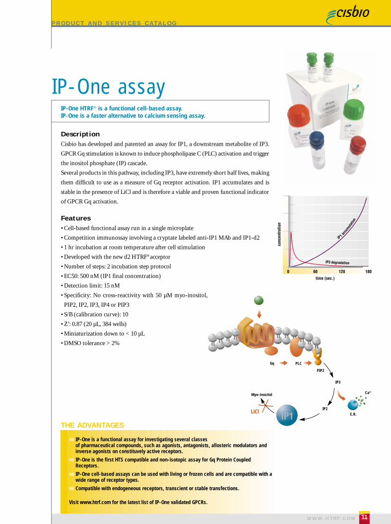
DescriptionThe IP-One ELISA assay has been designed to monitor the activation of Phospholipase
C (PLC) coupled receptors. Among them, the Gq coupled GPCRs represent the most
important family of receptors which can activate the ß subtype of the PLC family.
Other receptor types, like protein tyrosine kinase receptors, antigen or immunoglobulin
receptors, or collagen receptors, are known to activate another PLC subtype, PLC-γ .
Features• Cell-based functional assay
• Monoclonal antibody based
• Highly sensitive (detection limit: 10 nM)
• Simple protocol
• EC50: 110 nM
• No cross-reactivity with 50 µM myo-inositol, PIP2, IP2, IP3, IP4 or PIP3
THE ADVANTAGES
The fundamental assay for IP1 quantification and assessment of Gq coupledGPCR activation.
IP-One EL ISA
IP1-HRP
goat anti-mouse
lgGanti IP1 MAb
1. Plate cells2. Cell stimulation
3. Cell lysis
washOD 450 nm with
optional correction
transfer
4. IP1-HRP + IP1 MAb(3 hour incubation)
cell culture plate ELISA plate ELISA plate
5. TMB (20 to 30 min incubation)6. Stop solution
Cell stimulation step Detection with IP-One ELISA reagents
Cell stimulation step1. Plate cells in appropriate cell culture plate (overnight incubation)
2. Stimulate cells with ligand of choice or the drug of interest (1 hour incubation)
3. Lyse cells (30 min incubation)
Detection with IP-One ELISA reagents4. Transfer cell lysate to the ELISA plate supplied with the kit, add IP1-HRP conjugate and anti-IP1 MAb (3 hour incubation)
5. Add HRP substrate TMB following plate wash
6. Stop the reaction after 20 to 30 minutes and read optical density at 450nm
IP1
Ordering information
DesignationIP-One ElisaIP-One Elisa
Size96 wells
5x96 wells
Cat#72IP1PEA72IP1PED
Lyophilized4°C
These HTRF® kit components can be ordered separately
(except for Cryptate and XL665 conjugates).
IP-One stimulation buffer 5X (8ml)
DescriptionLyophilized4°C
62IP1FDC
Cat#
S E L E C T E D B I B L I O G R A P H Y
P A G E
15W W W . H T R F . C O M
The perfect tool to select and validate GPCR functionality during cell engineeringprocesses.
IP-One ELISA is a functional and sensitive assay developed to follow Inositol 1phosphate accumulation following Phospholipase C coupled receptors’ activation.
High pharmacological relevance, ideal for lead optimization phases.
P A G E
14
P R O D U C T A N D S E R V I C E S C A T A L O G
IP-One EL ISA
Assay principleIP-One ELISA is a competitive immunoassay which uses IP1-HRP, IP1 and an anti-IP1 monoclonal antibody. The protocol
consists of two incubation steps following cell stimulation.
HRP substrate TMB (3, 3’, 5, 5’-Tetramethylbenzidine) is added following a final wash step.
The HRP reaction is stopped and the optical density (OD) read at 450 nm.
Thomsen, W., Frazer, J. & Unett, D. Functional assays for screening GPCR targets.Curr Opin Biotechnol. 2005;16,655-665.Gibbins, J.M. Platelet adhesion signalling and the regulation of thrombus formation. J Cell Sci. 2004;117,3415-3425
Rhee, S.G. Regulation of phosphoinositide-specific phospholipase C. Annu RevBiochem. 2001;70,281-312.Berridge, M.J. Inositol trisphosphate and calcium signalling. Nature. 1993;361,315-325.
P R O D U C T A N D S E R V I C E S C A T A L O G
DescriptionThe IP-One ELISA assay has been designed to monitor the activation of Phospholipase
C (PLC) coupled receptors. Among them, the Gq coupled GPCRs represent the most
important family of receptors which can activate the ß subtype of the PLC family.
Other receptor types, like protein tyrosine kinase receptors, antigen or immunoglobulin
receptors, or collagen receptors, are known to activate another PLC subtype, PLC-γ .
Features• Cell-based functional assay
• Monoclonal antibody based
• Highly sensitive (detection limit: 10 nM)
• Simple protocol
• EC50: 110 nM
• No cross-reactivity with 50 µM myo-inositol, PIP2, IP2, IP3, IP4 or PIP3
THE ADVANTAGES
The fundamental assay for IP1 quantification and assessment of Gq coupledGPCR activation.
IP-One EL ISA
IP1-HRP
goat anti-mouse
lgGanti IP1 MAb
1. Plate cells2. Cell stimulation
3. Cell lysis
washOD 450 nm with
optional correction
transfer
4. IP1-HRP + IP1 MAb(3 hour incubation)
cell culture plate ELISA plate ELISA plate
5. TMB (20 to 30 min incubation)6. Stop solution
Cell stimulation step Detection with IP-One ELISA reagents
Cell stimulation step1. Plate cells in appropriate cell culture plate (overnight incubation)
2. Stimulate cells with ligand of choice or the drug of interest (1 hour incubation)
3. Lyse cells (30 min incubation)
Detection with IP-One ELISA reagents4. Transfer cell lysate to the ELISA plate supplied with the kit, add IP1-HRP conjugate and anti-IP1 MAb (3 hour incubation)
5. Add HRP substrate TMB following plate wash
6. Stop the reaction after 20 to 30 minutes and read optical density at 450nm
IP1
Ordering information
DesignationIP-One ElisaIP-One Elisa
Size96 wells
5x96 wells
Cat#72IP1PEA72IP1PED
Lyophilized4°C
These HTRF® kit components can be ordered separately
(except for Cryptate and XL665 conjugates).
IP-One stimulation buffer 5X (8ml)
DescriptionLyophilized4°C
62IP1FDC
Cat#
P A G E
17W W W . H T R F . C O M
Monitoring Gs or Gi coupled receptors for high throughput agonist/antagonist screening incell-based assays on living or frozen cells.
Related applications:• Phosphodiesterase (PDE) follow-up by measuring the degradation of cAMP into AMP.
• Adenylate cyclase activity.
• Measuring GPCR activity in membrane preparations with cAMP HiRange.P A G E
16
These HTRF® kit components can be ordered separately
(except for Cryptate and d2 conjugates).
P R O D U C T A N D S E R V I C E S C A T A L O G
cAMP assays
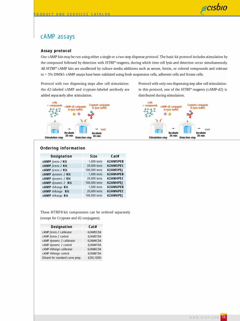
Assay protocolOur cAMP kits may be run using either a single or a two-step dispense protocol. The basic kit protocol includes stimulation by
the compound followed by detection with HTRF® reagents, during which time cell lysis and detection occur simultaneously.
All HTRF® cAMP kits are unaffected by culture media additions such as serum, biotin, or colored compounds and tolerant
to > 5% DMSO. cAMP assays have been validated using fresh suspension cells, adherent cells and frozen cells.
THE ADVANTAGES
P R O D U C T A N D S E R V I C E S C A T A L O G
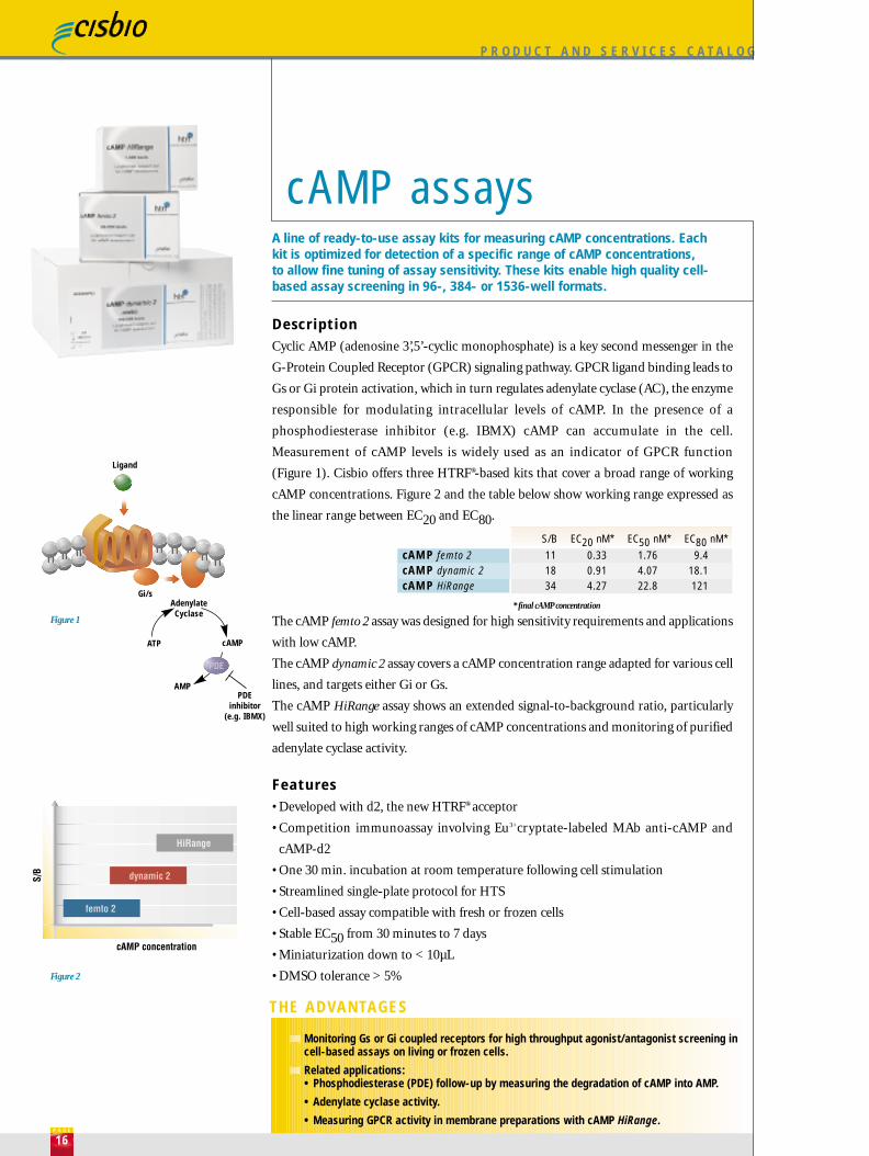
DescriptionCyclic AMP (adenosine 3’,5’-cyclic monophosphate) is a key second messenger in the
G-Protein Coupled Receptor (GPCR) signaling pathway. GPCR ligand binding leads to
Gs or Gi protein activation, which in turn regulates adenylate cyclase (AC), the enzyme
responsible for modulating intracellular levels of cAMP. In the presence of a
phosphodiesterase inhibitor (e.g. IBMX) cAMP can accumulate in the cell.
Measurement of cAMP levels is widely used as an indicator of GPCR function
(Figure 1). Cisbio offers three HTRF®-based kits that cover a broad range of working
cAMP concentrations. Figure 2 and the table below show working range expressed as
the linear range between EC20 and EC80.
The cAMP femto 2 assay was designed for high sensitivity requirements and applications
with low cAMP.
The cAMP dynamic 2 assay covers a cAMP concentration range adapted for various cell
lines, and targets either Gi or Gs.
The cAMP HiRange assay shows an extended signal-to-background ratio, particularly
well suited to high working ranges of cAMP concentrations and monitoring of purified
adenylate cyclase activity.
Features• Developed with d2, the new HTRF® acceptor
• Competition immunoassay involving Eu3+cryptate-labeled MAb anti-cAMP and
cAMP-d2
• One 30 min. incubation at room temperature following cell stimulation
• Streamlined single-plate protocol for HTS
• Cell-based assay compatible with fresh or frozen cells
• Stable EC50 from 30 minutes to 7 days
• Miniaturization down to < 10µL
• DMSO tolerance > 5%
A line of ready-to-use assay kits for measuring cAMP concentrations. Eachkit is optimized for detection of a specific range of cAMP concentrations, to allow fine tuning of assay sensitivity. These kits enable high quality cell-based assay screening in 96-, 384- or 1536-well formats.
cAMP assays
Gi/sAdenylateCyclase
ATP cAMP
AMPPDE
inhibitor(e.g. IBMX)
Ligand
Incubate30 min
Incubate30 minStimulation step Detection step
read
Protocol with two dispensing steps after cell stimulation:
the d2-labeled cAMP and cryptate-labeled antibody are
added separately after stimulation.
Protocol with only one dispensing step after cell stimulation:
in this protocol, one of the HTRF® reagents (cAMP-d2) is
distributed during stimulation.
cells+ compounds cAMP-d2 conjugate
in lysis buffer
Cryptate conjugatein lysis buffer
cells+ compounds cAMP-d2 conjugate
in lysis buffer
Cryptate conjugatein lysis buffer
Incubate30 min
Incubate30 min
Stimulation step Detection step
read
Ordering information
DesignationcAMP femto 2 KitcAMP femto 2 Kit cAMP femto 2 Kit cAMP dynamic 2 Kit cAMP dynamic 2 Kit cAMP dynamic 2 KitcAMP HiRange KitcAMP HiRange KitcAMP HiRange Kit
Size1,000 tests
20,000 tests100,000 tests
1,000 tests20,000 tests
100,000 tests1,000 tests
20,000 tests100,000 tests
Cat#62AM5PEB62AM5PEC62AM5PEJ62AM4PEB62AM4PEC62AM4PEJ62AM6PEB62AM6PEC62AM6PEJ
cAMP femto 2 calibratorcAMP femto 2 controlcAMP dynamic 2 calibratorcAMP dynamic 2 controlcAMP HiRange calibratorcAMP HiRange controlDiluent for standard curve prep.
Designation62AM5CDA62AM5TDA62AM4CDA62AM4TDA62AM6CDA62AM6TDA62DL1DDD
Cat#
Figure 1
Figure 2
111834
0.330.914.27
1.764.0722.8
9.418.1121
S/B EC20 nM* EC50 nM* EC80 nM*
cAMP femto 2cAMP dynamic 2cAMP HiRange
* final cAMP concentration
P A G E
17W W W . H T R F . C O M
Monitoring Gs or Gi coupled receptors for high throughput agonist/antagonist screening incell-based assays on living or frozen cells.
Related applications:• Phosphodiesterase (PDE) follow-up by measuring the degradation of cAMP into AMP.
• Adenylate cyclase activity.
• Measuring GPCR activity in membrane preparations with cAMP HiRange.P A G E
16
These HTRF® kit components can be ordered separately
(except for Cryptate and d2 conjugates).
P R O D U C T A N D S E R V I C E S C A T A L O G
cAMP assays
Assay protocolOur cAMP kits may be run using either a single or a two-step dispense protocol. The basic kit protocol includes stimulation by
the compound followed by detection with HTRF® reagents, during which time cell lysis and detection occur simultaneously.
All HTRF® cAMP kits are unaffected by culture media additions such as serum, biotin, or colored compounds and tolerant
to > 5% DMSO. cAMP assays have been validated using fresh suspension cells, adherent cells and frozen cells.
THE ADVANTAGES
P R O D U C T A N D S E R V I C E S C A T A L O G
DescriptionCyclic AMP (adenosine 3’,5’-cyclic monophosphate) is a key second messenger in the
G-Protein Coupled Receptor (GPCR) signaling pathway. GPCR ligand binding leads to
Gs or Gi protein activation, which in turn regulates adenylate cyclase (AC), the enzyme
responsible for modulating intracellular levels of cAMP. In the presence of a
phosphodiesterase inhibitor (e.g. IBMX) cAMP can accumulate in the cell.
Measurement of cAMP levels is widely used as an indicator of GPCR function
(Figure 1). Cisbio offers three HTRF®-based kits that cover a broad range of working
cAMP concentrations. Figure 2 and the table below show working range expressed as
the linear range between EC20 and EC80.
The cAMP femto 2 assay was designed for high sensitivity requirements and applications
with low cAMP.
The cAMP dynamic 2 assay covers a cAMP concentration range adapted for various cell
lines, and targets either Gi or Gs.
The cAMP HiRange assay shows an extended signal-to-background ratio, particularly
well suited to high working ranges of cAMP concentrations and monitoring of purified
adenylate cyclase activity.
Features• Developed with d2, the new HTRF® acceptor
• Competition immunoassay involving Eu3+cryptate-labeled MAb anti-cAMP and
cAMP-d2
• One 30 min. incubation at room temperature following cell stimulation
• Streamlined single-plate protocol for HTS
• Cell-based assay compatible with fresh or frozen cells
• Stable EC50 from 30 minutes to 7 days
• Miniaturization down to < 10µL
• DMSO tolerance > 5%
A line of ready-to-use assay kits for measuring cAMP concentrations. Eachkit is optimized for detection of a specific range of cAMP concentrations, to allow fine tuning of assay sensitivity. These kits enable high quality cell-based assay screening in 96-, 384- or 1536-well formats.
cAMP assays
Gi/sAdenylateCyclase
ATP cAMP
AMPPDE
inhibitor(e.g. IBMX)
Ligand
Incubate30 min
Incubate30 minStimulation step Detection step
read
Protocol with two dispensing steps after cell stimulation:
the d2-labeled cAMP and cryptate-labeled antibody are
added separately after stimulation.
Protocol with only one dispensing step after cell stimulation:
in this protocol, one of the HTRF® reagents (cAMP-d2) is
distributed during stimulation.
cells+ compounds cAMP-d2 conjugate
in lysis buffer
Cryptate conjugatein lysis buffer
cells+ compounds cAMP-d2 conjugate
in lysis buffer
Cryptate conjugatein lysis buffer
Incubate30 min
Incubate30 min
Stimulation step Detection step
read
Ordering information
DesignationcAMP femto 2 KitcAMP femto 2 Kit cAMP femto 2 Kit cAMP dynamic 2 Kit cAMP dynamic 2 Kit cAMP dynamic 2 KitcAMP HiRange KitcAMP HiRange KitcAMP HiRange Kit
Size1,000 tests
20,000 tests100,000 tests
1,000 tests20,000 tests
100,000 tests1,000 tests
20,000 tests100,000 tests
Cat#62AM5PEB62AM5PEC62AM5PEJ62AM4PEB62AM4PEC62AM4PEJ62AM6PEB62AM6PEC62AM6PEJ
cAMP femto 2 calibratorcAMP femto 2 controlcAMP dynamic 2 calibratorcAMP dynamic 2 controlcAMP HiRange calibratorcAMP HiRange controlDiluent for standard curve prep.
Designation62AM5CDA62AM5TDA62AM4CDA62AM4TDA62AM6CDA62AM6TDA62DL1DDD
Cat#
Figure 1
Figure 2
111834
0.330.914.27
1.764.0722.8
9.418.1121
S/B EC20 nM* EC50 nM* EC80 nM*
cAMP femto 2cAMP dynamic 2cAMP HiRange
* final cAMP concentration

Cisbio created the cAMP membrane kit specifically to address the use of cellular membranesin GPCR activation and adenylate cyclase activity screening. The cAMP membrane assay ishighly robust and produces quality results while eliminating the need for a continuous supplyof living cells.
P R O D U C T A N D S E R V I C E S C A T A L O G
DescriptionThe localization of the adenylate cyclase enzyme within membranes allows the use of
cellular membrane preparations rather than live cells when measuring Gi and Gs-
coupled GPCR activation.
Cisbio’s cAMP membrane assay exhibits a high signal-to-background ratio (S/B=12)
and EC50 of 6.6 nM in 20 µL final assay volume (132 fmol/well). This level of assay
sensitivity thus requires the use of only a small quantity of membrane per well.
Features• Membrane-based assay
• Immunoassay competition between cAMP-XL665 and cAMP with a monoclonal anti-
cAMP-cryptate antibody conjugate
• Number of steps: two incubation step protocols (see assay principle)
• Incubation: 90-120 min. at room temperature
• Small quantity of membrane required per assay
(0.125 µg to 1.5 µg depending on receptor type)
• Amenable to automation
• S/B > 10
• EC50 = 6.6 nM
THE ADVANTAGES
cAMP membrane assayA cAMP detection assay to monitor Gi/s activation and adenylate cyclasescreening optimized for use with cellular membranes.
P A G E
19W W W . H T R F . C O M
Gi/sAdenylateCyclase
ATP cAMP
Ligand
P R O D U C T A N D S E R V I C E S C A T A L O G
cAMP assays
S E L E C T E D B I B L I O G R A P H Y
Amoravain M, Lyotard S, Jaga D, Lebreton ML, Servent F, Bömer U, Bersdorf C,Seguin P, Sulocha S, Tardieu JL, Trinquet E. Introduction of a new HTRF® acceptor,d2. Development of a complete GPCR platform for a Gs, Gi and Gq screening. SBS11th Annual Conference. September 2005, Geneva (CH).
Cenni B, Drexler C, Gaveriaux S, Achard S, Martinez S, Sulocha S, Préaudat M.HTRF® cyclic AMP assay: new optimized cell-based assay for better investigation ofGi coupled receptors. Smart assays for screening, IIR congress. 2003, Zurich(CH).
Gabriel D, Vernier M, Pfeifer MJ, Dasen B, Tenaillon L, Bouhelal R. High throughputscreening technologies for direct cyclic AMP measurement. Assay Drug Dev Technol.2003; 2: 291-303.
Bonnet F, Mensat P, Achard S, Martinez S, Sulocha S, Tokuda C, Durroux T, SeguinP, Degorce F. HTRF® cyclic AMP assay: new optimized cell-based solutions for GPCRscreening. IBC’s 7th Annual Conference on G-Protein Coupled Receptors. 2002, SanDiego (USA).
Hamon J, Martin B, Meunier E, Casamitjana O, Ouagued M, Roman F. Validationof the HTRF® technology for high throughput quantication of cAMP productionmediated by activation of GPCR – Comparison with flashplate. SBS 7th AnnualConference. September 2001, Baltimore (USA).
Degorce F, Achard S, Préaudat M, Cougouluègne F, Durroux T, Bazin H, Aono Y,Kawamoto K, Dohi K, Takemoto H, Seguin P, Mathis G. A new HTRF® assay for thedirect assessment of cyclic AMP in high throughput screening. SBS 6th AnnualConference. September 2000, Vancouver (CAN).
P A G E
18
T h i s p o s t e r c a n b e d o w n l o a d e d f r o m w w w. h t r f . c o m
A S S A Y I N A C T I O N
Thawed CHO-K1 cells that stably expressed the H3 receptor (Gi) were exposed to increasing
concentrations of Methylhistamine in the presence of 30 µM of forskolin (EC80). Assays were
performed in 20 µL in Greiner 384-well plates. The plates were read at several intervals following
cell stimulation, from 1 hour to 7 days.
The IC50 values calculated with the three kits are remarkably stable even after 7 days, which is
a significant advantage during screening campaigns.From Amoravain M. et al. SBS 11th Annual conference. September 2005, Geneva (CH).
IC50 nM
cAMP femto 2
cAMP dynamic 2
cAMP HiRange
1 hour
3.45
7.01
4.42
Overnight
2.89
8.88
4.75
7 days
2.86
9.54
3.94
Introduction of a new HTRF® acceptor: d2. Development of a complete GPCR platform for Gs, Gi and Gq screening.

P R O D U C T A N D S E R V I C E S C A T A L O G
cAMP membrane assay
Assay principleTwo dispensing protocols are available for use depending on automation constraints. Both protocols include:
Stimulation: membrane and compounds are dispensed followed by incubation (30 to 60 minutes at room temperature).
Stop: the stop reagent supplied with the kit is added.
Detection: addition of HTRF® detection reagents (1 hour at room temperature).
S E L E C T E D B I B L I O G R A P H YAmoravain M, Mensat P, Loison C, Bernard J, Morandi D, Alaoui N, Sulocha S, JeanA. Lyotard S, Degorce F, Méoni P. HTRF® cyclic AMP membrane assay: a new kitspecifically designed for the measurement of GPCR activity in membranepreparations. SBS 10th Annual Conference. September 2004, Orlando (USA).
P A G E
20
T h i s p o s t e r c a n b e d o w n l o a d e d f r o m w w w. h t r f . c o m
These HTRF® kit components can be ordered separately
(except for Cryptate and XL665 conjugates).
Ordering information
DesignationcAMP membrane Kit
Size1,000 tests
20,000 tests
Cat#62AM3PEB62AM3PEC*
cAMP membrane calibrator cAMP membrane control Diluent for standard curve prep.
Designation
Lyophilized4°C
62AM3CDA62AM3TDA62DL1DDD
Cat#
compounds membranes
incubation10 min at RT
incubation1h at RT
cAMP-XL665in lysis buffer
anti cAMP-cryptatein lysis buffer
read
incubation30 min
Stimulation step Detection step
STOPreagent
* on request
Kinase so lu t ionsKinase too lboxHTRF® KinEASE™ ki tsKinase assay development
Oncology-re la ted so lu t ionsUbiqui t in assayHeparanase assay
Kinaseand oncology
relatedproducts
P R O D U C T A N D S E R V I C E S C A T A L O G
P A G E
21W W W . H T R F . C O M
Top Related