


Languages
Pages
Legal


Essentials of Human Anatomy & Physiology
Copyright © 2003 Pearson Education, Inc. publishing as Benjamin Cummings
Slides 13.1 – 13.30
Seventh Edition
Elaine N. Marieb
Chapter 13
The Respiratory System
Lecture Slides in PowerPoint by Jerry L. Cook

Organs of the Respiratory system
Slide 13.1 Copyright © 2003 Pearson Education, Inc. publishing as Benjamin Cummings
________
Pharynx
Larynx
________
Bronchi
_______- ________
Figure 13.1

Function of the Respiratory System
Slide 13.2 Copyright © 2003 Pearson Education, Inc. publishing as Benjamin Cummings
Oversees ______ _____________between the blood and external environment
Exchange of gasses takes place within the lungs in the _______________
Passageways to the lungs _______, ______________, and ________________ the incoming air
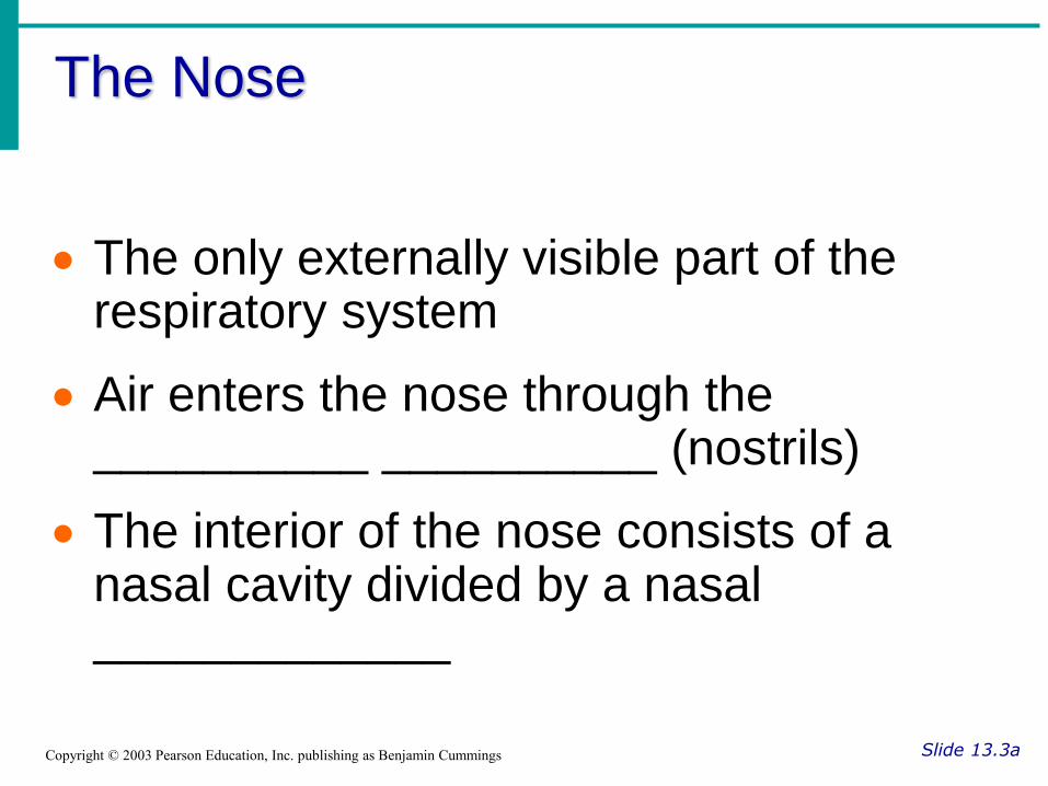
The Nose
Slide 13.3a Copyright © 2003 Pearson Education, Inc. publishing as Benjamin Cummings
The only externally visible part of the respiratory system
Air enters the nose through the __________ __________ (nostrils)
The interior of the nose consists of a nasal cavity divided by a nasal _____________

Slide 13.3b Copyright © 2003 Pearson Education, Inc. publishing as Benjamin Cummings
Figure 13.2
Upper Respiratory Tract

Anatomy of the Nasal Cavity
Slide 13.4a Copyright © 2003 Pearson Education, Inc. publishing as Benjamin Cummings
_______________ _________are located in the mucosa on the superior surface
The rest of the cavity is lined with respiratory mucosa
Moistens air
Traps incoming foreign particles

Anatomy of the Nasal Cavity
Slide 13.4b Copyright © 2003 Pearson Education, Inc. publishing as Benjamin Cummings
Lateral walls have projections called ________________
Increases surface area
Increases air turbulence within the nasal cavity
The nasal cavity is separated from the oral cavity by the palate
Anterior hard ___________ (bone)
Posterior ____________ palate (muscle)

Paranasal ______________
Slide 13.5a Copyright © 2003 Pearson Education, Inc. publishing as Benjamin Cummings
______________ within bones surrounding the nasal cavity
Frontal bone
Sphenoid bone
Ethmoid bone
Maxillary bone

Paranasal Sinuses
Slide 13.5b Copyright © 2003 Pearson Education, Inc. publishing as Benjamin Cummings
____________ of the sinuses
____________ the skull
Act as _____________ chambers for speech
Produce _________________ that drains into the nasal cavity

__________________ (Throat)
Slide 13.6 Copyright © 2003 Pearson Education, Inc. publishing as Benjamin Cummings
Muscular passage from nasal cavity to larynx
Three regions of the pharynx
__________________ – superior region behind nasal cavity
Oropharynx – middle region behind mouth
Laryngopharynx – inferior region attached to larynx
The oropharynx and laryngopharynx are ______________passageways for air and food

Structures of the Pharynx
Slide 13.7 Copyright © 2003 Pearson Education, Inc. publishing as Benjamin Cummings
___________________ tubes enter the nasopharynx
_______________ of the pharynx
Pharyngeal tonsil (adenoids) in the nasopharynx
Palatine tonsils in the oropharynx
Lingual tonsils at the base of the tongue

_________________ (Voice Box)
Slide 13.8 Copyright © 2003 Pearson Education, Inc. publishing as Benjamin Cummings
Routes air and food into proper channels
Plays a role in speech
Made of eight rigid hyaline _________________and a spoon-shaped flap of elastic cartilage (____________________)

Structures of the Larynx
Slide 13.9a Copyright © 2003 Pearson Education, Inc. publishing as Benjamin Cummings
______________ cartilage
Largest hyaline cartilage
Protrudes anteriorly (_________ _________)
_________________
Superior opening of the larynx
Routes ____________ to the larynx and ___________toward the trachea

Structures of the Larynx
Slide 13.9b Copyright © 2003 Pearson Education, Inc. publishing as Benjamin Cummings
__________ __________(vocal folds)
Vibrate with expelled air to create sound (speech)
Glottis – opening between vocal cords

______________ (Windpipe)
Slide 13.10 Copyright © 2003 Pearson Education, Inc. publishing as Benjamin Cummings
Connects _________ with ______________
Lined with ciliated mucosa
Beat continuously in the opposite direction of incoming air
Expel mucus loaded with dust and other debris __________ from lungs
Walls are reinforced with C-shaped hyaline cartilage

Primary _____________
Slide 13.11 Copyright © 2003 Pearson Education, Inc. publishing as Benjamin Cummings
Formed by division of the trachea
Enters the lung at the hilus (medial depression)
Right bronchus is wider, shorter, and straighter than left
Bronchi _______________ into smaller and smaller branches
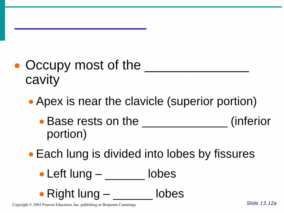
_______________
Slide 13.12a Copyright © 2003 Pearson Education, Inc. publishing as Benjamin Cummings
Occupy most of the ______________ cavity
Apex is near the clavicle (superior portion)
Base rests on the _____________ (inferior portion)
Each lung is divided into lobes by fissures
Left lung – ______ lobes
Right lung – ______ lobes

Lungs
Slide 13.12b Copyright © 2003 Pearson Education, Inc. publishing as Benjamin Cummings
Figure 13.4b

Coverings of the Lungs
Slide 13.13 Copyright © 2003 Pearson Education, Inc. publishing as Benjamin Cummings
______________ (visceral) pleura covers the lung surface
______________ pleura lines the walls of the thoracic cavity
____________ __________fills the area between layers of pleura to allow gliding

Respiratory Tree Divisions
Slide 13.14 Copyright © 2003 Pearson Education, Inc. publishing as Benjamin Cummings
____________ bronchi
Secondary bronchi
Tertiary bronchi
________________
Terminal bronchioli

Bronchioles
Slide 13.15a Copyright © 2003 Pearson Education, Inc. publishing as Benjamin Cummings
Figure 13.5a
____________ branches of the bronchi

Bronchioles
Slide 13.15b Copyright © 2003 Pearson Education, Inc. publishing as Benjamin Cummings
Figure 13.5a
All but the smallest branches have reinforcing cartilage

Bronchioles
Slide 13.15c Copyright © 2003 Pearson Education, Inc. publishing as Benjamin Cummings
Terminal bronchioles end in _____________
Figure 13.5a

Respiratory Zone
Slide 13.16 Copyright © 2003 Pearson Education, Inc. publishing as Benjamin Cummings
Structures
Respiratory bronchioli
Alveolar duct
Alveoli
Site of gas exchange

Alveoli
Slide 13.17 Copyright © 2003 Pearson Education, Inc. publishing as Benjamin Cummings
Structure of alveoli
Alveolar duct
Alveolar sac
Alveolus
Gas exchange takes place within the ____________in the respiratory membrane

Respiratory Membrane
(Air-Blood Barrier)
Slide 13.18a Copyright © 2003 Pearson Education, Inc. publishing as Benjamin Cummings
Thin squamous epithelial layer lining
alveolar walls
Pulmonary capillaries cover external
surfaces of alveoli

Respiratory Membrane
(Air-Blood Barrier)
Slide 13.18b Copyright © 2003 Pearson Education, Inc. publishing as Benjamin Cummings
Figure 13.6

Gas Exchange
Slide 13.19 Copyright © 2003 Pearson Education, Inc. publishing as Benjamin Cummings
Gas crosses the respiratory membrane by ______________
Oxygen ______________ the blood
__________ ____________enters the alveoli
Macrophages add protection
Surfactant coats gas-exposed alveolar surfaces

Events of _________________
Slide 13.20a Copyright © 2003 Pearson Education, Inc. publishing as Benjamin Cummings
____________ ______________– moving air in and out of the lungs
External respiration – gas exchange between __________ _____________and alveoli

Events of Respiration
Slide 13.20b Copyright © 2003 Pearson Education, Inc. publishing as Benjamin Cummings
Respiratory gas transport – transport of oxygen and carbon dioxide via the bloodstream
_______________ ____________– gas exchange ___________ blood and ______________ cells in systemic capillaries

Mechanics of Breathing
(___________ ______________)
Slide 13.21a Copyright © 2003 Pearson Education, Inc. publishing as Benjamin Cummings
Completely ________________ process
Depends on _________ ____________in the thoracic cavity
Volume changes lead to _____________changes, which lead to the flow of gases to equalize pressure

Mechanics of Breathing
(Pulmonary Ventilation)
Slide 13.21b Copyright © 2003 Pearson Education, Inc. publishing as Benjamin Cummings
Two phases
__________ – flow of air into lung
____________ – air leaving lung

Inspiration
Slide 13.22a Copyright © 2003 Pearson Education, Inc. publishing as Benjamin Cummings
Diaphragm and intercostal muscles _________________
The size of the thoracic cavity ______________
External air is _______ ______the lungs due to an increase in intrapulmonary volume

Inspiration
Slide 13.22b Copyright © 2003 Pearson Education, Inc. publishing as Benjamin Cummings
Figure 13.7a

Exhalation
Slide 13.23a Copyright © 2003 Pearson Education, Inc. publishing as Benjamin Cummings
Largely a ______________ process which depends on natural lung elasticity
As muscles __________, air is pushed _________ of the lungs
Forced expiration can occur mostly by contracting internal intercostal muscles to depress the rib cage

Exhalation
Slide 13.23b Copyright © 2003 Pearson Education, Inc. publishing as Benjamin Cummings
Figure 13.7b

Pressure Differences in the
Thoracic Cavity
Slide 13.24 Copyright © 2003 Pearson Education, Inc. publishing as Benjamin Cummings
Normal pressure within the pleural space is always negative (intrapleural pressure)
Differences in lung and pleural space pressures keep lungs from collapsing

Nonrespiratory Air Movements
Slide 13.25 Copyright © 2003 Pearson Education, Inc. publishing as Benjamin Cummings
Can be caused by reflexes or voluntary actions
Examples
___________ __________– clears lungs of debris
Laughing
___________
Yawn
____________

Respiratory Volumes and Capacities
Slide 13.26 Copyright © 2003 Pearson Education, Inc. publishing as Benjamin Cummings
Normal breathing moves about __________ ml of air with each breath (________ ___________[TV])
Many factors that affect respiratory capacity
A person’s ______
___________
__________
___________ ____________
Residual volume of air – after exhalation, about __________ ml of air remains in the lungs

Respiratory Volumes and Capacities
Slide 13.27a Copyright © 2003 Pearson Education, Inc. publishing as Benjamin Cummings
_________ ____________ ______(IRV)
Amount of air that can be taken in ___________ over the tidal volume
Usually between 2100 and 3200 ml
_____________ reserve volume (ERV)
Amount of air that can be __________ exhaled
Approximately 1200 ml

Respiratory Volumes and Capacities
Slide 13.27b Copyright © 2003 Pearson Education, Inc. publishing as Benjamin Cummings
________ ___________
Air remaining in lung after expiration
About 1200 ml

Respiratory Volumes and Capacities
Slide 13.28 Copyright © 2003 Pearson Education, Inc. publishing as Benjamin Cummings
__________ ____________
The total amount of _____________ air
Vital capacity = TV + IRV + ERV
Dead space volume
Air that remains in conducting zone and ___________ reaches alveoli
About 150 ml

Respiratory Volumes and Capacities
Slide 13.29 Copyright © 2003 Pearson Education, Inc. publishing as Benjamin Cummings
Functional volume
Air that actually reaches the respiratory zone
Usually about ____________ ml
Respiratory capacities are measured with a ______________

Respiratory Capacities
Slide 13.30 Copyright © 2003 Pearson Education, Inc. publishing as Benjamin Cummings
Figure 13.9
Top Related