


Languages
Pages
Legal


Blood, Vol. 55, No. 4 (April), 1980 629
The Molecular Mechanism of the Inherited Phosphofructokinase
Deficiency Associated With Hemolysis and Myopathy
By Shobhana Vora, Laurence Corash, W. King Engel, Susan Durham, Carol Seaman, and Sergio Piomelli
Normal human erythrocyte phosphofructokinase (ATP: o-fructose-6. P-i -phosphotransferase. EC 2.7.1 .1 1 ; PFK) has
recently been shown to consist of a heterogeneous mixture of five tetrameric isozymes: M4. M3L. M2L2. ML3, and L4 (M.
muscle type; L. liver type). In the light of these findings. we have investigated the molecular basis of the inherited
erythrocyte PFK deficiency associated with myopathy and hemolysis (Tarui disease). The propositus. a 31 -yr-old male,
suffered from muscle weakness and myoglobinuria on exertion. He showed mild erythrocytosis despite laboratory
evidence of hemolysis. In his erythrocytes a metabolic crossover point was found at the level of PFK; 2,3-diphosphoglycer-
ate (2,3-DPG) was also significantly reduced. The PFK from the patient’s erythrocytes consisted exclusively of the L4
isozyme. and there was a complete absence of the other four. The leukocyte and platelet PFKs from the patient showed
normal activities, chromatographic profiles, and precipitation with anti-M4 antibody. These studies provide direct evidence
that in Tarui disease the M-type subunits are absent; but the liver- and platelet-type subunits of PFK are unaffected. The
paradox of mild erythrocytosis despite hemolysis reflects the decreased production of 2,3-DPG.
I N THE ERYTHROCYTES, inherited deficiencies
have been described for almost all the glycolytic
enzymes. Since the glycolytic pathway is universally
present, an enzymatic defect of the erythrocytes
should reflect a generalized defect in all the tissues.
Although most glycolytic defects of the erythrocytes
are associated with a hemolytic syndrome, concomi-
tant dysfunction of other organ systems is not always
observed. Neurologic syndromes occur with triose-
P-isomerase, phosphoglycerate kinase, and aldolase
deficiencies,’ while the deficiency of phosphofructoki-
nase (ATP: D-fructose-6,P- I -phosphotransferase,
EC.2.7.l.1 1; PFK) is often associated with severe
generalized muscle dysfunction. An inherited defi-
ciency of PFK (complete lack in the muscles, but
partial deficiency in the erythrocytes) was first
reported by Tarui et al. in 1965.2 As a moderate
deposition of glycogen occurred in the muscles, Brown
and Brown designated this syndrome as glycogen stor-
age disease VII.’ Since the original description, eight
additional cases of inherited PFK deficiency have been
reported.� Of these, only one manifested the same
clinical features of Tarui disease, i.e., myopathy and
hemolysis.’ The other cases exhibited either only
myopathy or hemolysis or no clinical symptoms at
all.5’6’8�’2 A relative deficiency of erythrocyte PFK has
been described in normal newborns”4 that has been
shown to be due to increased in vivo lability of the
I 5, I 6
Like other allosteric enzymes, PFK is an oligomeric
protein, the smallest active oligomer being a tetram-
er.’7 The existence of two distinct subunits in human
erythrocyte PFK (one of which is identical to the sole
subunit present in muscle PFK) had been suggested
initially by the immunologic studies of the residual
erythrocyte PFK from the patients with Tarui
disease”8 and demonstrated more recently by SDS-
polyacrylamide gel electrophoresis of normal erythro-
cyte PFK.’9’2#{176}Recently, we have elucidated the molec-
ular structure of normal human erythrocyte PFK. We
have shown it to consist of a heterogeneous mixture of
five isozymes resulting from all the possible combina-
tions of the M (muscle-type) and L (liver-type) sub-
units to form various tetramers, i.e., M4, M,L, M,L2,
ML,, and L4.2’
In this article, we report an additional patient with
Tarui disease and observations on his residual erythro-
cyte PFK, in the light of the recent knowledge. A
preliminary report of these studies has been
presented.22
CASE REPORT
The propositus, an apparently healthy and bright 31-yr-old male,
was referred to the Medical Neurology Branch of the National
Institute of Neurological and Communicative Disorders and Stroke
in 1974. He complained of easy fatiguability, muscle weakness, and
myoglobinuria following vigorous exercise since his early childhood.
On one occasion myoglobinuria led to acute renal failure, which
resolved uneventfully. His physical examination was essentially
normal except for slight scleral icterus (total bilirubin: 2.4 mg/dl;
direct: 0.3 mg/dl), secondary to a well compensated hemolytic
process (hemoglobin 14.7 g/dl; reticulocytes, 6.3%; 5’Cr survival,
12.5 days; haptoglobin, 12.0 mg/dl). There was modest macrocyto-
sis (mean cell volume [MCV]: 100 cu is). Other normal studies
included autohemolysis, direct and indirect Coombs tests, hemoglo-
bin A2 and F levels, hemoglobin electrophoresis, renal and liver
function tests, intravenous pyelogram (IVP), chest X-ray, electro-
From the Division ofPediatric Hematology-Oncology. Columbia
University College of Physicians and Surgeons, New York, N.Y.
and the Hematology Service, Clinical Pathology Department. Clin-
ical Center, National Institute of Health and the Medical Neurol-
ogy Branch, National Institute of Neurological and Communica-
live Disorders and Stroke, Bethesda, Md.
Supported in part by NIH Research Grants AM-26793-Ol and
AM-26437-O1.
Submitted August 16, 1979; accepted December 12. 1979.
Address reprint requests to Shobhana Vora, M.D.. Department
of Pediatrics. Columbia University, College of Physicians and
Surgeons. 70/ West 168th Street, New York, N. Y. 10032.
© I 980 by Grune & Stratton, Inc.
0006-4971/80/5504-0015$0I.00/0
For personal use only.on April 3, 2019. by guest www.bloodjournal.orgFrom

630 VORA ET AL.
�Range of measurements, see text.
cardiogram (EKG), serum GOT, glutamate pyruvate transaminase
(GPT), lactate dehydrogenase (LDH), aldolase, and creatine phos-
phokinase (CPK). Nerve conduction and electromyographic studies
were completely normal. During several ischemic exercise tests, the
patient failed to increase the levels ofvenous lactate and pyruvate. A
diagnosis of muscle PFK deficiency was then established by direct
biochemical analysis of the patient’s muscle biopsy specimen. The
simultaneously measured erythrocyte PFK was found to be approxi-
mately 50% of normal. These findings suggested that his defect was
the same as in the families reported by Tarui et al.2 and Layzer et
al.7
The patient is of Jewish ancestry and his parents are nonconsan-
guineous. The erythrocyte PFK in both the parents was also found to
be half-normal. However, both of them and a female sibling (not
studied) are completely asymptomatic.
Further biochemical and immunologic studies carried out on the
patient in I 978 form the basis of this report.
MATERIALS AND METHODS
Adenosine 5’-mono-, di-, and triphosphates. nicotinamide adenine
dinucleotide/phosphate (NAD/NADP) and their reduced forms,
hexose mono- and diphosphates, dithiothreitol, glycylglycine, and
the sodium salt of�3-glycerol-P were purchased from Sigma Chemi-
cal Co., St. Louis, Mo. Aldolase, cr-glycerol-P-dehydrogenase,
triose-P-isomerase, hexokinase, glucose-6-phosphate dehydrogenase
(G6PD), LDH, and other enzymes were purchased from Boehringer
Mannheim. Enzyme-grade ammonium sulfate was obtained from
Schwarz/ Mann. Phosphoenolypyruvate and phosphoglycerates
were obtained from Calbiochem. Freund’s complete and incomplete
adjuvants came from Difco. DEAE-Cellulose (DE-52) was obtained
from Whatman, DEAE-Sephadex A-25 from Pharmacia, Agarose
from Colab Labs, and Nonidet (NP-40) from Particle Data Inc. All
other chemicals were of reagent grade.
Cell suspensions were prepared from blood samples stored at 4#{176}C
immediately after collection. Simultaneously collected blood
samples from normal individuals served as controls. The removal of
leukocytes and platelets was achieved by filtration of whole blood on
a-cellulose columns.23 PFK assays of the erythrocytes were done
within 8 hr of collection, whereas the rest of the investigations were
done within the next 4 days. Leukocytes were prepared by dextran
sedimentation technique24 and platelet suspensions according to the
method of Corash et al.25 The leukocytes and platelet concentrations
in the final preparations were determined using an electronic coun-
ter (Particle Data Inc.). To assay glycolytic intermediates, whole
blood was immediately added to an equal volume of cold perchloric
acid and the supernatant was used. Routine hematologic and
biochemical determinations were done using standard techniques.
Phosphofructokinase assays were performed as described by
Beutler.26 One unit of enzyme was defined as that amount of enzyme
that converts I �mole of F6P to F-l,6-P/g of hemoglobin (Hb) or
1010 cells/mm at 37#{176}C.For erythrocyte activity, hemolysates were
prepared just prior to the assay by hypotonic lysis in S mM TES
buffer (pH 7.4) containing I mM EDTA and 1 mM fl-mercapto-
ethanol. Leukocytes and platelets were suspended in 10 mM
K2HPO4 buffer, pH 8.0 containing 0.2 mM EDTA, 0.2 mM AMP
and 0.7 mM dithiothreitol and disrupted by two freeze-thaws and
sonication, respectively. The resulting suspensions were centrifuged
at I 2,000 g for I 0 mm and the supernatants were assayed.
Ischemic exercise test was performed according to the method of
McArdle.2’
Muscle specimens were obtained by biopsy of the left quadriceps.
Another biopsy was obtained from the left forearm muscles after an
ischemic exercise test. PFK was assayed in the supernatant of the
muscle homogenate prepared according to the method described by
Tarui et al.2 Phosphorylase, acid maltase, and neutral maltase were
assayed according to the methods described by Layzer et al.7
Immunologic studies consisted of double diffusion and enzyme
precipitation studies and were performed using an anti-human
muscle PFK rabbit antiserum as previously described.2’
Partial purifications of erythrocyte and liver PFKs were carried
out according to the methods of Hennessey et al.28 and Brock,29
respectively. Muscle PFK was purified according to Kemp et al.3#{176}
Metabolic studies included glycolytic intermediates, glucose
consumption, and lactate production, as well as PK, HK, and G6PD
activities, and were determined according to Beutler.26
Chromatographic separation of PFK isozymes was obtained on
DEAE-Sephadex A-25, equilibrated with 0.1 M Tris-P04 buffer
(pH 8) containing 0.2 mM EDTA, 0.2 mM AMP, and I mM
dithiothreitol. A concave gradient of NaCI was used for elution.
Details of this technique have been previously described.2’
Enzyme stability studies were performed to assess the in vivo
stability of the enzyme; the biological half-life (0/2) of the erythro-
cyte PFK was determined according to the method of Corash et al.3’
In vitro stability was determined by allowing hemolysates of similar
enzyme concentrations to stand at various temperatures with pen-
odic assays over the next 24 hr.
Protein determinations in the solutions were performed according
to Lowry et al.32 using bovine serum albumin as a standard.
RESULTS
Hematologic and enzyme studies. Table 1 sum-
marizes the representative hematologic values and
erythrocyte, leukocyte, and platelet enzyme activities
of the patient and his parents. From September 1974
through March 1977, the propositus demonstrated
normal hemoglobin values (14.7-16 g/dl) with hema-
tocrits ranging between 45% and 47%. At the time of
the study in August 1978, he exhibited a hemoglobin
level of 18.0 g/dl with a hematocrit of 53%. He
showed a persistent reticulocytosis of approximately
7% (range 49�o-1 1%). The hemoglobin levels and reti-
Table 1 . Hematol ogic Values and Enzyme Act ivity in the Patient an d his Parents
Hemoglobin
(g/dl)
Hematocrit
(%)
Reticulocytes
1%)
ErythrocyteLeukocyte
PFKt
Platelet
PFKtPFK HK PK G6PD
Propositus 14.7-18� 53 7.0 7.22 0.95 7.9 21.8 341 12.6
Father 15.5 44 0.9 7.80 0.76 5.8 15.9 284 14.3
Mother 13.1 39 1.5 6.35 0.85 7.4 15.2 365 13.9
Control 15.0 45 1.0 14.35 0.51 7.3 13.3 310 12.0
9n zmole/min/g hemoglobin at 37#{176}C.
tIn zmole/min/10’#{176} cells at 37#{176}C.
For personal use only.on April 3, 2019. by guest www.bloodjournal.orgFrom
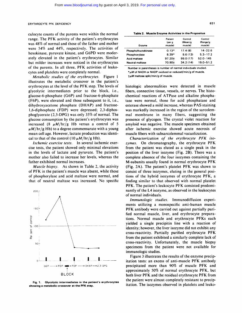
Table 2. Muscle Enzyme Activities in the Propositus
Number in parentheses is number of normal individuals studied.
MM of NADH or NADP oxidized or reduced/min/g of muscle.
tMM maltose split/min/g of muscle.
200 r
� 50
0
0
0
a)0�
50
BLOCK
ERYTHROCYTE PFK DEFICIENCY 631
Fig. 1 . Glycolytic intermediates in the patient’s erythrocytesshowing a metabolic crossover at the PFK step.
culocyte counts of the parents were within the normal
range. The PFK activity of the patient’s erythrocytes
was 48% of normal and those of the father and mother
were 54% and 44%, respectively. The activities of
hexokinase, pyruvate kinase, and G6PD were moder-
ately elevated in the patient’s erythrocytes. Similar
but milder increases were noticed in the erythrocytes
of the parents. In all three, PFK activities of leuko-
cytes and platelets were completely normal.
Metabolic studies of the erythrocytes. Figure 1
illustrates the metabolic crossover in the patient’s
erythrocytes at the level of the PFK step. The levels of
glycolytic intermediates prior to the block, i.e.,
glucose-6-phosphate (G6P) and fructose-6-phosphate
(F6P), were elevated and those subsequent to it, i.e.,
dihydroxyacetone phosphate (DHAP) and fructose-
1,6-diphosphate ( FDP) were depressed; 2,3-diphos-
phoglycerate (2,3-DPG) was only 35% of normal. The
glucose consumption by the patient’s erythrocytes was
increased (8 �zM/hr/g Hb versus a control of 5
.tM/hr/g Hb) to a degree commensurate with a young
mean cell age. However, lactate production was identi-
cal to that ofthe control (10 �tM/hr/g Hb).
Ischemic exercise tests. In several ischemic exer-
cise tests, the patient showed only minimal elevations
in the levels of lactate and pyruvate. The patient’s
mother also failed to increase her levels, whereas the
father exhibited normal increases.
Muscle biopsy. As shown in Table 2, the activity
of PFK in the patient’s muscle was absent, while those
of phosphorylase and acid maltase were normal, and
that of neutral maltase was increased. No specific
Enzyme
Patient
(per g
muscle)
Control
(Mean/gmuscle)
Control
(Range/gmuscle)
Phosphofructokinase 0.13 17.4 (8) 14-22.6
Phosphorylase 8.39’ 9.6(13) 5.3-17.2
Acidmaltase 97.20t 99.0(17) 52.0-145
Neutralmaltase 70.90t 34.2(14) 19.0-51.2
histologic abnormalities were detected in muscle
fibers, connective tissue, vessels, or nerves. The histo-
chemical reactions of ATPase and alkaline phospha-
tase were normal, those for acid phosphatase and
esterase showed a mild increase, whereas PAS staining
was markedly increased in the region of the sarcolem-
mal membrane in many fibers, suggesting the
presence of glycogen. The crystal violet reaction for
amyloid was negative. The muscle specimen obtained
after ischemic exercise showed acute necrosis of
muscle fibers with subsarcolemmal vacuolization.
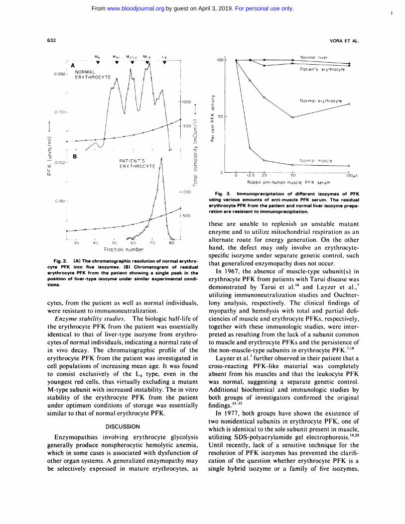
Characterization of the erythrocyte PFK iso-
zymes. On chromatography, the erythrocyte PFK
from the patient was eluted as a single peak in the
position of the liver isozyme (Fig. 2B). There was a
complete absence of the four isozymes containing the
M-subunits usually found in normal erythrocyte PFK
(Fig. 2A). The patient’s platelet PFK was shown to
consist of three isozymes, eluting in the general posi-
tions of the hybrid isozymes of erythrocyte PFK, a
finding similar to that observed with normal platelet
PFK. The patient’s leukocyte PFK consisted predomi-
nantly of the L4 isozyme, as observed in the leukocytes
of normal individuals.
Immunologic studies. Immunodi ffusion experi-
ments utilizing a monospecific anti-human muscle
PFK antibody were carried out against partially pun-
fled normal muscle, liver, and erythrocyte prepara-
tions. Normal muscle and erythrocyte PFKs each
yielded a single precipitin line with a reaction of
identity; however, the liver isozyme did not exhibit any
cross-reactivity. Partially purified erythrocyte PFK
from the patient exhibited a similarly complete lack of
cross-reactivity. Unfortunately, the muscle biopsy
specimens from the patient were not available for
immunologic studies.
Figure 3 illustrates the results of the enzyme precip-
itation tests: an excess of anti-muscle PFK antibody
precipitated more than 90% of muscle PFK and
approximately 50% of normal erythrocyte PFK, but
both liver PFK and the residual erythrocyte PFK from
the patient were almost completely resistant to precip-
itation. The isozymes observed in platelets and leuko-
For personal use only.on April 3, 2019. by guest www.bloodjournal.orgFrom
M4 M3L M2L2 ML3 L4
OOO2� NORMAL
0 flOl -
Normot liver
E
C
LL
0�
0
U-
0.
a)0.
B0 00?
-�
PATIENTS
ERYTHROCYTE
>�
0
0
ELfl0
0
0F-
0 OO� -
I 000
25 25
NormjI muscle
50
Rabbit anti-human muscle PFK serum
l00/)t
i . A Q’#{176}� - - I --30 40 50 60 70 80
FractIon number
632 VORA ET AL.
Fig. 2. (A) The chromatographic resolution of normal erythro-cyte PFK into five isozymes. (B) Chromatogram of residualerythrocyte PFK from the patient showing a single peak in theposition of liver-type isozyme under similar experimental condi-
tions.
cytes, from the patient as well as normal individuals,
were resistant to immunoneutralization.
Enzyme stability studies. The biologic half-life of
the erythrocyte PFK from the patient was essentially
identical to that of liver-type isozyme from erythro-
cytes of normal individuals, indicating a normal rate of
in vivo decay. The chromatographic profile of the
erythrocyte PFK from the patient was investigated in
cell populations of increasing mean age. It was found
to consist exclusively of the L4 type, even in the
youngest red cells, thus virtually excluding a mutant
M-type subunit with increased instability. The in vitro
stability of the erythrocyte PFK from the patient
under optimum conditions of storage was essentially
similar to that of normal erythrocyte PFK.
DISCUSSION
Enzymopathies involving erythrocyte glycolysis
generally produce nonspherocytic hemolytic anemia,
which in some cases is associated with dysfunction of
other organ systems. A generalized enzymopathy may
be selectively expressed in mature erythrocytes, as
Fig. 3. Immunoprecipitation of different isozymes of PFKusing various amounts of anti-muscle PFK serum. The residualerythrocyte PFK from the patient and normal liver isozyme prepa-ration are resistant to immunoprecipitation.
these are unable to replenish an unstable mutant
enzyme and to utilize mitochondrial respiration as an
alternate route for energy generation. On the other
hand, the defect may only involve an erythrocyte-
specific isozyme under separate genetic control, such
that generalized enzymopathy does not occur.
In 1967, the absence of muscle-type subunit(s) in
erythrocyte PFK from patients with Tarui disease was
demonstrated by Tarui et al.’8 and Layzer et al.,7
utilizing immunoneutralization studies and Ouchter-
lony analysis, respectively. The clinical findings of
myopathy and hemolysis with total and partial defi-
ciencies of muscle and erythrocyte PFKs, respectively,
together with these immunologic studies, were inter-
preted as resulting from the lack of a subunit common
to muscle and erythrocyte PFKs and the persistence of
the non-muscle-type subunits in erythrocyte PFK.7”8
Layzer et al.7 further observed in their patient that a
cross-reacting PFK-like material was completely
absent from his muscles and that the leukocyte PFK
was normal, suggesting a separate genetic control.
Additional biochemical and immunologic studies by
both groups of investigators confirmed the original
findings.3335
In 1977, both groups have shown the existence of
two nonidentical subunits in erythrocyte PFK, one of
which is identical to the sole subunit present in muscle,
utilizing SDS-polyacrylamide gel 1920
Until recently, lack of a sensitive technique for the
resolution of PFK isozymes has prevented the clarifi-
cation of the question whether erythrocyte PFK is a
single hybrid isozyme or a family of five isozymes,
For personal use only.on April 3, 2019. by guest www.bloodjournal.orgFrom
ERYTHROCYTE PFK DEFICIENCY 633
composed of both muscle and non-muscle types of
subunits. Using a high-resolution chromatographic
technique, we have recently demonstrated that
erythrocyte PFK is indeed a mixture of five isozymes
resulting from all the possible combinations of two
distinct subunits, M (muscle-type) and L (liver-type),
to form various tetramers. These findings have been
corroborated by the fact that an identical set of five
isozymes is produced when purified muscle and liver
PFKs are hybridized in vitro.2’ The similarity between
the non-muscle subunit of erythrocyte PFK and the
subunit from liver PFK is supported by the identical
chromatographic elution patterns of the respective
homotetramers, nonreactivity with anti-M4 antibody,
and by the fact that the kinetic properties of erythro-
cyte PFK are intermediate between those of muscle
and liver isozymes.2’ The recent observation by Kahn
et al. that normal erythrocyte PFK is also precipitated
by anti-liver PFK antibody further corroborates our
interpretation that the non-muscle subunit of erythro-
cyte PFK is of liver-type.36
The patient reported in this study exhibits clinical
and biochemical characteristics essentially identical to
those reported by Tarui et al.2 and Layzer et al.;7 the
findings in his parents are also identical to those
already reported (partial deficiency of erythrocyte
PFK and normal hematologic values), except that his
mother showed an abnormal response to ischemic
exercise test. The results of the muscle biopsy are also
in agreement with those reported by other workers.8’9
The observation that in our patient acute necrosis of
muscle fibers with subsarcolemmal vacuolization
occurred after ischemic exercise provides an explana-
tion of the mechanism of the exertional myopathy, at
times resulting in myoglobinuria.
Our metabolic studies indicate that the deficiency in
the erythrocytes, although partial, nevertheless results
in a significant block in glycolysis. A metabolic cross-
over was found at the level of the PFK reaction. The
increased glucose consumption without a concomitant
rise in lactate production attests to the block and the
probable shunting of glycolytic flux via the hexose
monophosphate shunt. These metabolic alterations
most probably account for the significant shortening
of the erythrocyte lifespan. The block in glycolysis also
accounts for the observed marked decrease in the
2,3-DPG level in the patient’s erythrocytes. This
results in increased oxygen affinity, which in turn
produces a compensatory erythrocytosis. The combi-
nation of these factors appears to be responsible for the
paradoxical finding of shortened erythrocyte lifespan,
accompanied by erythrocytosis, instead of anemia.
The stability of the L4 from patient’s erythrocytes
was assessed, since hemolysis was present despite 50%
residual PFK activity. Both in vivo t’/2 and in vitro
stability on storage were normal, indicating that the
cause of hemolysis must be sought elsewhere. It is
known that under existing intraerythrocytic condi-
tions, the activity of PFK is scaled down to 0.5% of its
potential capacity and is commensurate with the
observed rate of erythrocyte glycolysis.37 It is possible
that even a 50% reduction in erythrocyte PFK
becomes critically rate-limiting for glycolysis and
energy generation, which results in premature cell
death. Alternatively, the turnover number of liver-type
isozyme or its apparent Km and K could be of such
magnitude that glycolysis in the erythrocyte is severely
impaired despite adequate in vitro enzymatic activity.
Our chromatographic studies provide direct molec-
ular evidence that the PFK deficiency results from the
absence of the M-type subunits as originally proposed
by Tarui et al.’8 and Layzer et al.7 The chromato-
graphic profiles of PFKs from the normal and defi-
cient erythrocytes were strikingly different. The
normal PFK was resolved into five peaks, whereas that
from the patient was eluted as a solitary peak in the
same position as the final peak of the normal enzyme.
Immunologically, the residual PFK from our patient
failed to react with anti-muscle PFK antibody, as
previously demonstrated by Tanui et al.’8 and Layzer
et al.7 We have additionally shown that purified
human liver PFK, which chromatographically is
eluted in the same position as the patient’s erythrocyte
PFK, also fails to react with the anti-muscle PFK
antibody.
The presence of an unstable M-subunit instead of its
total absence in our patient was virtually excluded by
the failure to detect any M-containing isozymes in the
reticulocytes and youngest erythrocytes separated by
density gradient centrifugation. Thus, the most likely
reason for undetectable M-subunits in our patient
would seem to be absent or extremely reduced synthe-
sis.
The existence of multiple isozymes of PFK in differ-
ent organs had been suggested initially by Layzer et
al.38’39 on the basis of the differing immunologic reac-
tivity of PFK from various tissues to anti-muscle PFK
antibody. Based on immunologic studies utilizing
three different antisera, Kahn et al.’�#{176}have recently
suggested the existence of three types of PFK sub-
units, i.e., muscle, liver, and fibroblast type. Using
molecular hybridization, chromatographic and immu-
nologic techniques, we have recently shown that in
leukocytes L4 predominates and that in platelets there
are three isozymes (P4. P3L, P2L2) consisting of a
unique platelet-type or P-subunit (probably the same
For personal use only.on April 3, 2019. by guest www.bloodjournal.orgFrom
634 VORA ET AL.
Table 3. Phosphofructok inase Deficie ncy: Clinical and Bioc hemical Aspects
Enzyme Activity
Author
1% of normal)
Myopathy Hemolysis
Reticulocyte
1%)Muscle RBC
Tarui et al.2 (3 sibs) 1 -3 29-48 + + 4-6
Layzeretal.7 2 50 + + 4
Voraetal.22 1 48 + + 4-11
Serratnice et al.8 0 1 7 + ? #{149} ND
Tobinetal.9 6 ND + ? ND
Waterbury et al.’#{176} ND 62 0 + 9
Miwaetal.” ND 8 0 + 7-14
Odaetal. NDt 0 + 7-11
Boulard et al.’2 ND 28 0 0 1
Kahnetal.#{176} 100 61 0 0 ND
ND, not done.
Hemolysis not adequately ruled out.
tMuscle involvement indicated by increased plasma PK level of muscle-type isozyme.�Reported as “decreased.”
as Kahn’s fibroblast type) and liver-type or L sub-
unit.4’ These findings are further corroborated by the
observations that in our patient, leukocyte and platelet
PFKs were found to be normal, both chromatographi-
cally and immunologically.
So far, I 2 cases of inherited PFK deficiency occur-
ring in 10 unrelated families (including the present
report) have been described. Table 3 lists the clinical
and biochemical profiles of these cases. In the light of
the present findings, one can speculate on the molecu-
lar basis of PFK deficiency in these cases. The cases
reported by Serratnice et al.8 and Tobin et al.9 both
showed a total lack of muscle PFK and partial reduc-
tion oferythrocyte PFK. The hemolysis was (probably
unjustifiably) presumed to be absent only because the
patients were found not to be anemic; however, labora-
tory evidence of hemolysis (i.e., reticulocyte count or
erythrocyte survival) was not sought. It appears most
likely that these cases were essentially similar to those
mentioned above, i.e., homozygous deficient for M
subunits. In contrast, the patients reported by Water-
bury et al.’#{176}and Miwa et al.” presented with hemoly-
sis but no myopathic symptoms and had normal
lactate production after exercise. Their molecular
defect could be due either to the absence or the
extreme instability of L-type subunits or to the insta-
bility of the M-subunits. The case described by Oda et
al.6 showed partial reduction in erythrocyte PFK with
a compensated hemolytic syndrome. Despite the
absence of myopathic symptoms, involvement of the
muscle was implicated because of an increase in the
plasma level of pyruvate kinase of the muscle type. In
this case, the molecular defect could be due to an
unstable M-type PFK subunit, which is compensated
in the muscle cells because of active protein synthesis.
Boulard et al.’2 described a healthy individual, with
partial reduction of erythrocyte PFK, discovered
during a large-scale survey; he could be a heterozygote
for the deficiency of M- or L-subunit. Kahn et al.5
reported another individual with normal muscle func-
tion and PFK level, despite a half-normal erythrocyte
PFK level; the muscle PFK was unstable and had fast
electrophoretic mobility. These findings were inter-
preted as due to the presence of a mutant M-subunit.
From the foregoing discussion, it is obvious that a
variety of clinical syndromes associated with erythro-
cyte PFK deficiency may stem from different defects
of either of the two subunits. Some syndromes may
even result from the inheritance of a combination of
different genetic lesions. Our studies provide direct
and conclusive evidence that the most common form of
PFK deficiency (myopathy with compensated hemoly-
sis-Tarui disease) stems from the exclusive defi-
ciency of the M-type subunit of PFK. The precise
characterization of the other forms of this enzymatic
defect will demand detailed studies along similar
lines.
ACKNOWLEDGMENT
The authors are extremely grateful to Stephen Vasta for his help
in the preparation of the manuscript.
REFERENCES
I. Valentine WN, Tanaka KR: Pyruvate kinase and other
enzyme deficiency hereditary hemolytic anemia, in Stanbury JB,
Wyngaarden JB, Fredenickson DS (eds): Metabolic Basis of lnher-
ited Diseases. New York, McGraw-Hill, 1978, p 1410
2. Tarui 5, Okuno G, Ikura Y, Tanaka T, Suda M, Nishikawa
M: Phosphofructokinase deficiency in skeletal muscle: A new type
ofglycogenosis. Biochem Biophys Res Commun (9:517, 1965
3. Brown BI, Brown DH: Glycogen-storage diseases: types I, Ill,
For personal use only.on April 3, 2019. by guest www.bloodjournal.orgFrom

ERYTHROCYTE PFK DEFICIENCY 635
IV, V, VII and unclassified glycogenoses, in Dickens F, Randle, PJ,
Whelan WJ (eds): Carbohydrate Metabolism and Its Disorders, vol
2. New York, Academic, 1968, p 123
4. Piomelli 5, Corash L: Hereditary hemolytic anemia due to
enzyme defects of glycolysis, in Harris H, Hirschhorn K (eds):
Advances in Human Genetics, vol 6. New York, Plenum, 1976, p
I65
5. Kahn A, Etiemble J, Meienhofer MC, Bovin P: Erythrocyte
phosphofructokinase deficiency associated with an unstable variant
ofmuscle phosphofructokinase. Clin Chim Acta 61:415, 1975
6. Oda 5, Oda E, Tanaka KR: Erythrocyte phosphofructokinase
(PFK) deficiency: Characterization and metabolic studies. Clin Res
25: 344A, 1977 (abstr)
7, Layzer RB, Rowland LP, Ranney HM: Muscle phosphofruc-
tokinasedeficiency. Arch Neurol 17:512, 1967
8. Serratnice G, Monges A, Roux H: Forme myopathique du
deficit en phosphofructokinase. Rev Neurol 1 20:271 , 1969
9. Tobin WE, Huijing F, Porro RS, Salzman RT: Muscle phos-
phofructokinasedeficiency. Arch Neurol 28:128, 1973
10. Waterbury L, Frankel EP: Hereditary nonspherocytic hemo-
lysis with erythrocyte phosphofructokinase deficiency. Blood
39:445, 1972
1 1 . Miwa S, Sato T, Murao H: A new type of phosphofructoki-
nase deficiency: Hereditary non-spherocytic hemolytic anemia.
Acta Hematol Jpn 35:1 13, 1972
12. Boulard MR. Meienhofer MC, Bois M, Reviron M, Najean
Y: Red cell phosphofructokinase deficiency. N EngI J Med 291:978,
I 974
I 3. Gross RT, Schroeder BAR, Brounstein SA: Energy metabo-
lism in the erythrocytes of premature infants compared to full-term
newborn infants and adults. Blood 2 1:755, 1963
14. Oski FA: Red cell metabolism in the newborn infant. V.
Glycolytic intermediates and glycolytic enzymes. Pediatrics 44:84,
1969
15. Travis SF, Garvin JH: In vivo lability of red cell phospho-
fructokinase in term infants: The possible molecular basis of the
relative phosphofructokinase deficiency in neonatal red cells. Pedi-
atrRes 11:1159, 1977
16. Vora 5, Piomelli 5: A fetal isozyme of phosphofructokinase
in newborn erythrocytes. Pediatr Res I I :483, 1977
17. Bloxham DP, Lardy HA: Phosphofructokinase, in Boyer PD
(ed): The Enzymes, vol 8A. New York, Academic, 1973, p 239
18. Tarui 5: Phosphofructokinase deficiency. Proceedings of the
17th General Assembly of the Japan Medical Congress 3:51 2, 1967
19. Karadsheh NS, Uyeda K: Studies on structure of human
erythrocyte phosphofructokinase. J Biol Chem 252:35 15, 1977
20. Kaur J, Layzer RB: Nonidentical subunits of human erythro-
cyte phosphofructokinase. Biochem Genet I 5: 1 133, 1977
21. Vora 5, Seaman C, Durham S. Piomelli S: Isozymes of
human phosphofructokinase: Identification and subunit structural
characterization of a new system. Proc NatI Acad Sci USA 77:62,
I 980
22. Vora 5, Corash L, Piomelli 5: The molecular mechanism of
inherited red cell phosphofructokinase deficiency associated with
hemolysis and myopathy (Tarui’s disease). Blood 52 (Suppl l):105,
1978 (abstr)
23. Beutler E, West C, Blume KG: The removal of leukocytes
and platelets from whole blood. J Lab Clin Med 88:328, 1976
24. Clark RA, Kimball HR: Defective granulocyte chemotaxis in
the Chediak-Higashi syndrome. J Clin Invest 50:2645, 1971
25. Corash L, Shafer B, Perlow M: Heterogeneity of human
whole blood platelet subpopulations. II. Use of sub-human primate
model to analyze the relationship between density and platelet age.
Blood 52:726, 1978
26. Beutler E: Red Cell Metabolism. A Manual of Biochemical
Methods (ed 2). New York, Grune & Stratton, 1975
27. McArdle B: Myopathy due to a defect in muscle glycogen
breakdown.ClinSci 10:13, 195l
28. Hennessey MA, Waltersdorph AM: Erythrocyte metabo-
lism. VI. Separation of erythrocyte enzymes from hemoglobin. J
Clin lnvest4l:l257, 1962
29. Brock DJH: Purification and properties of sheep liver phos-
phofructokinase. Biochem J I 1 3:235, 1969
30. Kemp RG: Methods Enzymol 42:7 1-77, 1975
31. Corash LM, Piomelli 5, Chen HC, Seaman C, Gross E:
Separation of erythrocytes according to age on a simplified density
gradient. J Lab Clin Med 84: 147. 1974
32. Lowry OH, Rosebrough NJ, Farr AL, Randall RJ: Protein
measurement with the folin phenol reagent. J Biol Chem 193:265,
1951
33, Tarui S, Kono N, Nasu T, Nishikawa M: Enzymatic basis
for the existence of myopathy and hemolytic disease in inherited
muscle phosphofructokinase deficiency. Biochem Biophys Res
Commun 34:77, 1969
34. Layzer RB, Rowland LP, Bank WJ: Physical and kinetic
properties of human phosphofructokinase from skeletal muscle and
erythrocytes. J Biol Chem 244:3823, 1969
35. Layzer RB, Rasmussen J: The molecular basis of muscle
phosphofructokinase deficiency. Arch Neurol 31 :41 1, 1974
36. Meienhofer M, Lagrange J, Cottreau D, Lenoir G, Dreyfus
J, Kahn A: Phosphofructokinase in human blood cells. Blood
53:389, 1979
37. Rapoport 5: The regulation of glycolysis in mammalian
erythrocytes, in Campbell PN, Greville GD (eds): Essays in
Biochemistry, vol 4. New York, Academic, 1968, p 69
38. Layzer RB, Conway MM: Multiple isozymes of human
phosphofructokinase. Biochem Biophys Res Commun 40:1259,
I970
39. Layzer RB: Enzyme genetics in muscle phosphofructokinase
deficiency, in Proceedings of the Second International Congress on
MuscleDiseases, 1971, p62
40. Kahn A, Meienhofer M, Cottreau D, Lagrange J, Dreyfus J:
Phosphofructokinase (PFK) isozymes in man. I. Studies of adult
human tissues. Human Genet 48:93, 1979
41 . Vora S, Durham 5, Piomelli S: Isozymes of phosphofructoki-
nase in human blood cells: Molecular and genetic evidence for a
multigenic system. Blood 54 (Suppl I ):35a, I 979 (abstr)
For personal use only.on April 3, 2019. by guest www.bloodjournal.orgFrom

1980 55: 629-635
S Vora, L Corash, WK Engel, S Durham, C Seaman and S Piomelli associated with hemolysis and myopathyThe molecular mechanism of the inherited phosphofructokinase deficiency
http://www.bloodjournal.org/content/55/4/629.full.htmlUpdated information and services can be found at:
Articles on similar topics can be found in the following Blood collections
http://www.bloodjournal.org/site/misc/rights.xhtml#repub_requestsInformation about reproducing this article in parts or in its entirety may be found online at:
http://www.bloodjournal.org/site/misc/rights.xhtml#reprintsInformation about ordering reprints may be found online at:
http://www.bloodjournal.org/site/subscriptions/index.xhtmlInformation about subscriptions and ASH membership may be found online at:
Copyright 2011 by The American Society of Hematology; all rights reserved.Hematology, 2021 L St, NW, Suite 900, Washington DC 20036.Blood (print ISSN 0006-4971, online ISSN 1528-0020), is published weekly by the American Society of
For personal use only.on April 3, 2019. by guest www.bloodjournal.orgFrom
Top Related