


Languages
Pages
Legal


The Larynx
Table of Contents • Functions • Anatomy • Subdivisions • Cartilages • Vocal Cords • Muscles • Nerves • Vessels • Special Cases

Functions
• To produce voice • To protect the airway,
especially during swallowing • To keep the airway patent
Functions
Anatomy
Subdivisions
Cartilages Vocal Cords
Muscles
Nerves Vessels
Special Cases

Anatomy • The larynx is located at the point
where the respiratory and digestive tracts separate.
• The entrance to the larynx, or laryngeal inlet, is in the anterior wall of the laryngopharynx.
• Internally, the wall of the larynx is modified to form the vocal cords.
Functions
Anatomy
Subdivisions
Cartilages Vocal Cords
Muscles
Nerves Vessels
Special Cases

Anatomy • The larynx lies in the
mid-line of the neck, deep to the strap muscles and partly covered by the thyroid gland.
• At roughly the C vertebral level, the larynx is continuous with the trachea.
Functions
Anatomy
Subdivisions
Cartilages
Vocal Cords
Muscles
Nerves
Vessels
Special Cases

Subdivisions • Vertically, the larynx is
divided into 3 regions: • 1. Supraglottis –
Includes the epiglottis, aryepiglottic folds, false vocal folds, arytenoids, and ventricle
• 2. Glottis – true vocal folds
• 3. Subglottis –below the true vocal folds to the inferior border of the cricoid cartilage
Functions
Anatomy
Subdivisions
Cartilages
Vocal Cords
Muscles
Nerves
Vessels
Special Cases
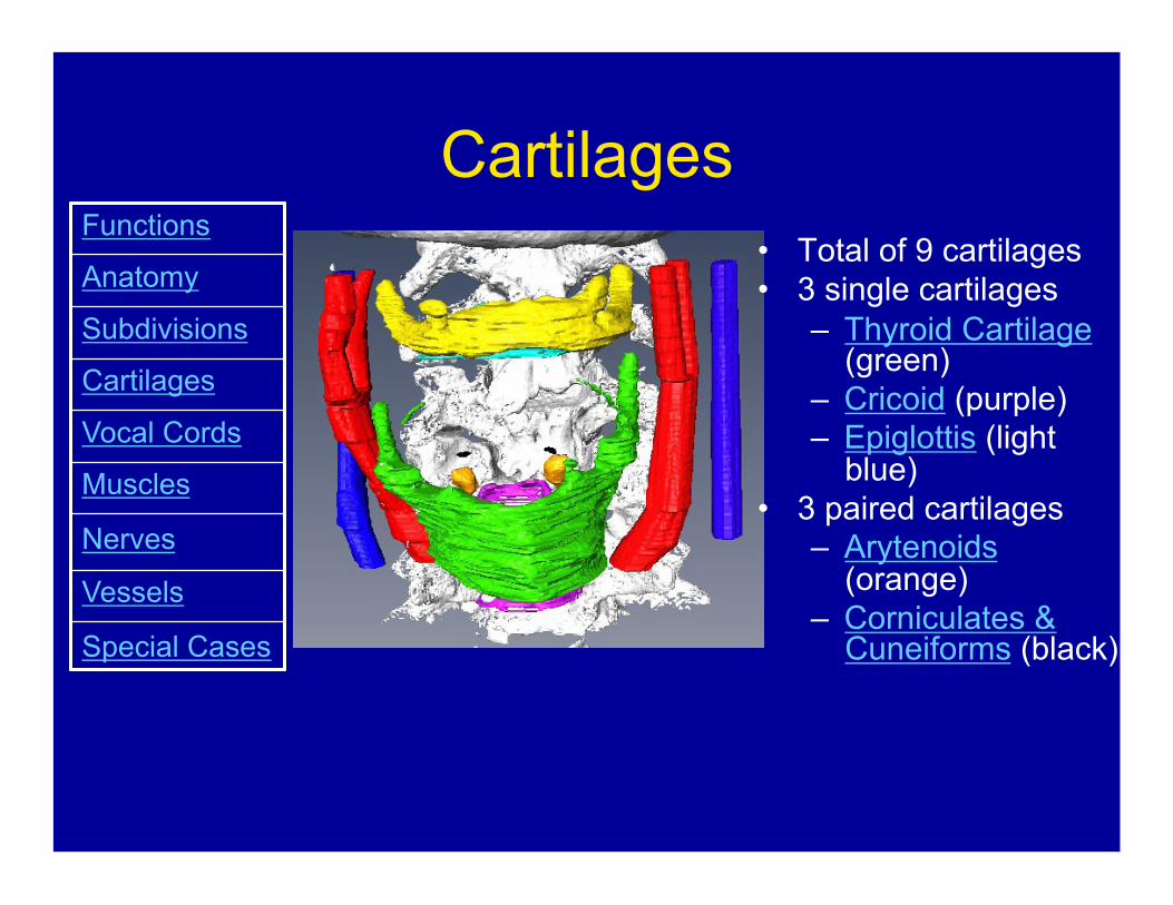
Cartilages • Total of 9 cartilages • 3 single cartilages
– Thyroid Cartilage (green)
– Cricoid (purple) – Epiglottis (light
blue) • 3 paired cartilages
– Arytenoids (orange)
– Corniculates & Cuneiforms (black)
Functions
Anatomy
Subdivisions
Cartilages
Vocal Cords
Muscles
Nerves
Vessels
Special Cases

Cartilages
• The hyoid bone (yellow) and the cartilages are collectively referred to as the visceral skeleton of the neck.
Functions
Anatomy
Subdivisions
Cartilages
Vocal Cords
Muscles
Nerves
Vessels
Special Cases

Thyroid Cartilage • Thyroid = shield-like • The largest cartilage • The two laminae
meet anteriorly at the superior thyroid notch (Adam's apple).
• The inferior horns articulate with the cricoid cartilage at the cricothyroid joints.
• The thyroid cartilage suspended from the hyoid bone by the thyrohyoid membrane.
<- Cartilages
Functions
Anatomy
Subdivisions
Cartilages
Vocal Cords
Muscles
Nerves
Vessels
Special Cases

Cricoid • Cricoid = ring-
shaped • The only
complete ring of cartilage in the respiratory tract.
• Cricoid pressure is applied to the esophagus during intubation to prevent gastric contents from refluxing into the airway.
<- Cartilages
Functions
Anatomy
Subdivisions
Cartilages
Vocal Cords
Muscles
Nerves
Vessels
Special Cases

Cricoid • Anteriorly - a
narrow arch. • Posteriorly -
enlarges to form the lamina.
• The cricoid lamina articulates with the inferior horns of the thyroid cartilage at the crico-thyroid joints.
<- Cartilages
Functions
Anatomy
Subdivisions
Cartilages
Vocal Cords
Muscles
Nerves
Vessels
Special Cases

Epiglottis • Guards the entrance
of the larynx. It folds posteriorly over the opening of the larynx during swallowing.
• Leaf-shaped, flexible, elastic cartilage.
• It attaches to the back of the thyroid cartilage via the thyroepiglottic ligament.
• Unlike the other cartilages, the epiglottis remains unossified.
<- Cartilages
Functions
Anatomy
Subdivisions
Cartilages
Vocal Cords
Muscles
Nerves
Vessels
Special Cases

Arytenoids • Paired and
pyramidal in shape.
• The base rests on the upper surface of the cricoid and forms the crico-arytenoid joint.
<- Cartilages
Functions
Anatomy
Subdivisions
Cartilages
Vocal Cords
Muscles
Nerves
Vessels
Special Cases

Arytenoids
• Postero-laterally - muscular process
• Anteriorly - vocal process provides attachment for the vocal cords.
• Superiorly - apex
<- Cartilages
Functions
Anatomy
Subdivisions
Cartilages
Vocal Cords
Muscles
Nerves
Vessels
Special Cases
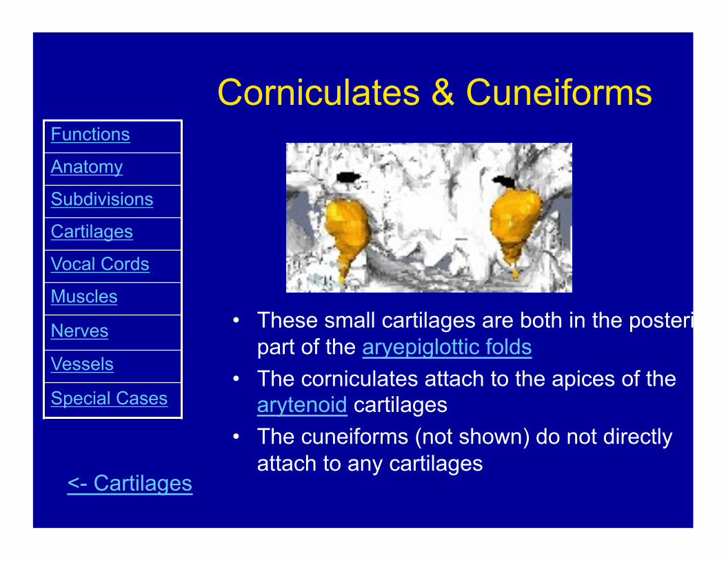
Corniculates & Cuneiforms
• These small cartilages are both in the posterior part of the aryepiglottic folds
• The corniculates attach to the apices of the arytenoid cartilages
• The cuneiforms (not shown) do not directly attach to any cartilages
<- Cartilages
Functions
Anatomy
Subdivisions
Cartilages
Vocal Cords
Muscles
Nerves
Vessels
Special Cases

Vocal Cords
Epiglottis
Arytenoids
True Vocal Cords
False Vocal Cords
Glottis
Ventricle Aryepiglottic Fold
Functions
Anatomy
Subdivisions
Cartilages
Vocal Cords
Muscles
Nerves
Vessels
Special Cases

True Vocal Cords • Vocal folds (true vocal cords) control
sound production (tone). Each vocal fold includes: – Vocal ligament – elastic tissue that
is the thickened medial free edge of the lateral cricothyroid ligament (conus elasticus)
– Vocalis muscle – fibres that form the most medial part of the thyroarytenoid muscle
<- Photo
Functions
Anatomy
Subdivisions
Cartilages
Vocal Cords
Muscles
Nerves
Vessels
Special Cases

False Vocal Cords • Vestibular folds (false vocal cords)
extend between the thyroid and arytenoids. – Have little to no part in voice
production – Serve a protective function
• Vestibular folds are the mucous membrane covering the lower border of the quadrangular membrane
<- Photo
Functions
Anatomy
Subdivisions
Cartilages
Vocal Cords
Muscles
Nerves
Vessels
Special Cases

Ventricle
• Between the true vocal cords and false vocal cords, on each side, is a lateral depression, lined by mucous membrane, known as the ventricle of the larynx.
<- Photo
Functions
Anatomy
Subdivisions
Cartilages
Vocal Cords
Muscles
Nerves
Vessels
Special Cases
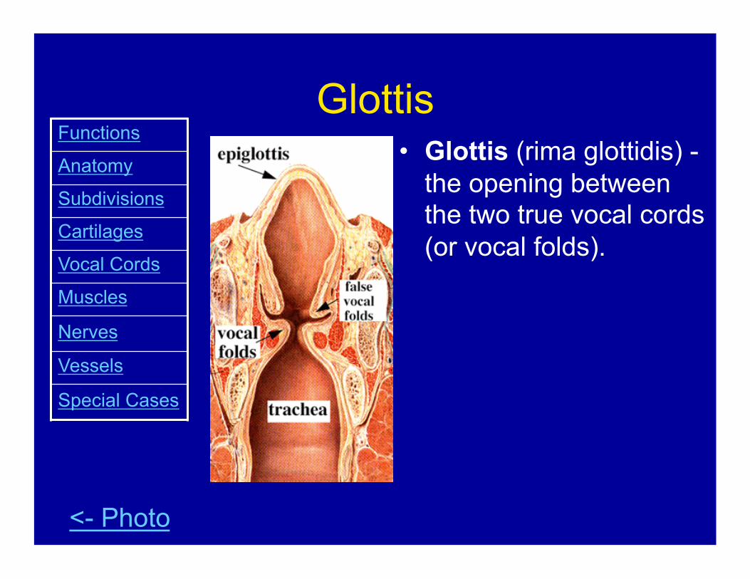
Glottis • Glottis (rima glottidis) -
the opening between the two true vocal cords (or vocal folds).
<- Photo
Functions
Anatomy
Subdivisions
Cartilages
Vocal Cords
Muscles
Nerves
Vessels
Special Cases

Larynx – Sagittal View
Epiglottis
Aryepiglottic Fold
Arytenoids
True Vocal Cords
Ventricle
False Vocal Cords
Thyroid Cartilage
Cricoid
Conus Elasticus
Quadrangular Membrane
Functions
Anatomy
Subdivisions Cartilages
Vocal Cords
Muscles
Nerves
Vessels
Special Cases

Larynx – Posterior View
Epiglottis
Aryepiglottic Fold
Arytenoids
Cricoid Thyroid Cartilage
Quadrangular Membrane
Hyoid Bone Thyrohyoid Membrane
Thyroepiglottic Ligament
<- Photo
Functions Anatomy Subdivisions Cartilages
Vocal Cords
Muscles
Nerves
Vessels
Special Cases

Quandrangular Membrane (In more detail)
• The quadrangular membrane is a sheet of fibrous connective tissue that extends from the arytenoids to the epiglottis.
• The upper border, covered by mucous membrane, is the aryepiglottic fold.
• The lower border is the vestibular ligament.
• The latter, together with its covering of mucous membrane, is the vestibular fold, or false vocal chord.
<- Diagram
Functions Anatomy Subdivisions Cartilages Vocal Cords Muscles
Nerves Vessels
Special Cases

Conus Elasticus (In more detail) • The conus elasticus attaches to
the upper surface of the cricoid arch.
• Its upper border is the vocal ligament which extends between the vocal process of the arytenoid cartilage and the thyroid lamina.
• The vocal ligament, covered with mucous membrane, is the vocal fold or true vocal chord.
• The membrane between the thyroid cartilage and cricoid is the cricothyroid membrane.
<- Diagram
Functions Anatomy Subdivisions Cartilages Vocal Cords Muscles
Nerves Vessels
Special Cases

Vocal Cords
Abducted Adducted
Functions Anatomy Subdivisions Cartilages Vocal Cords Muscles Nerves Vessels Special Cases

Vocal Cord Abduction & Adduction
• The vocal cords are abducted during breathing
• The vocal cords are tightly adducted in straining efforts and before a cough or sneeze.
• Voice production is the result of the escape of small amounts of air between the adducted vocal cords.
<- Photos
Functions Anatomy Subdivisions Cartilages Vocal Cords Muscles Nerves Vessels Special Cases

Phonation Physiology
• Power source – Lungs & Diaphragm • Pitch & quality – Larynx • Articulation – Lips and Tongue
<- Photos
Functions Anatomy Subdivisions Cartilages Vocal Cords Muscles Nerves Vessels Special Cases

Muscles • The muscles of the larynx are classified
as extrinsic or intrinsic • Extrinsic laryngeal muscles
– Move the larynx as a whole – Depress or elevate the hyoid bone & larynx – Infrahyoid strap muscles (omohyoid, sternohyoid,
sternothyroid, thyrohyoid)– depressors – Palato-pharyngeus & stylopharyngeus muscles –
elevators
• Intrinsic laryngeal muscles – Move parts of the larynx – Control the length/tension and movements of the
vocal folds and may help in the closure of the laryngeal inlet
Functions
Anatomy
Subdivisions
Cartilages
Vocal Cords
Muscles
Nerves
Vessels
Special Cases

Extrinsic Muscles
Functions
Anatomy
Subdivisions
Cartilages
Vocal Cords
Muscles
Nerves
Vessels
Special Cases
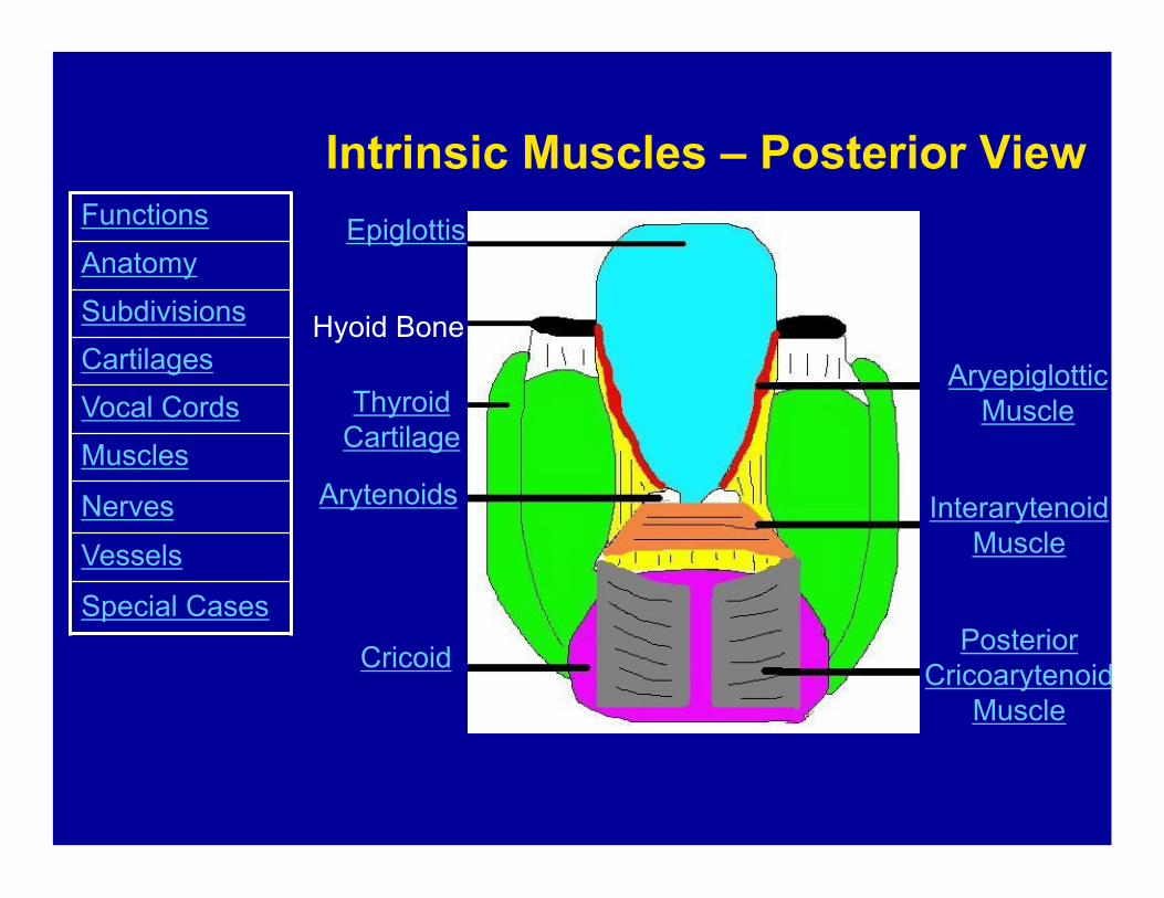
Intrinsic Muscles – Posterior View Epiglottis
Arytenoids
Cricoid
Thyroid Cartilage
Hyoid Bone
Posterior Cricoarytenoid
Muscle
Aryepiglottic Muscle
Interarytenoid Muscle
Functions Anatomy Subdivisions Cartilages Vocal Cords Muscles
Nerves
Vessels
Special Cases

Intrinsic Muscles Lateral View from Inside
Arytenoids
Thyroid Cartilage
Cricoid
Aryepiglottic Fold
Quadrangular Membrane
Thyroepiglottic Muscle
Thyroarytenoid Muscle
Lateral Cricoarytenoid Muscle
Functions Anatomy Subdivisions Cartilages Vocal Cords Muscles
Nerves Vessels
Special Cases
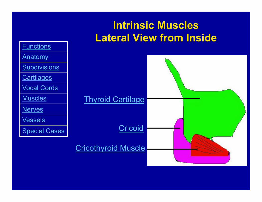
Intrinsic Muscles Lateral View from Inside
Thyroid Cartilage
Cricoid
Cricothyroid Muscle
Functions Anatomy Subdivisions Cartilages Vocal Cords Muscles
Nerves Vessels
Special Cases

Posterior Cricoarytenoid Muscle • From the posterior surface of
the lamina of the cricoid, its fibres converge to insert into the muscular process of the arytenoid.
• The two posterior cricoarytenoid muscles abduct the vocal chords by both rotating and separating the two arytenoid cartilages. <- Diagram
Functions Anatomy Subdivisions Cartilages Vocal Cords Muscles Nerves Vessels Special Cases

Interarytenoid muscle
• Consists of transverse and oblique fibres which pass between the two arytenoid cartilages.
• They adduct the vocal chords by drawing the two arytenoid cartilages together.
<- Diagram
Functions Anatomy Subdivisions Cartilages Vocal Cords Muscles Nerves Vessels Special Cases
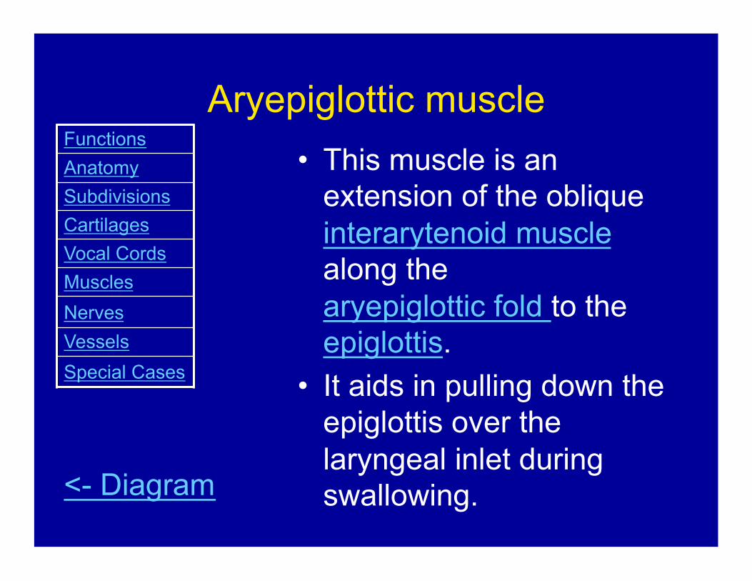
Aryepiglottic muscle • This muscle is an
extension of the oblique interarytenoid muscle along the aryepiglottic fold to the epiglottis.
• It aids in pulling down the epiglottis over the laryngeal inlet during swallowing. <- Diagram
Functions Anatomy Subdivisions Cartilages Vocal Cords Muscles Nerves Vessels Special Cases

Lateral Cricoarytenoid Muscle • Originates from the upper
margin of the cricoid arch and inserts into the muscular process of the arytenoid.
• It adducts the vocal chord by rotating the arytenoid cartilage, so that the vocal process swings towards the mid-line. <- Diagram
Functions Anatomy Subdivisions Cartilages Vocal Cords Muscles Nerves Vessels Special Cases

Cricothyroid Muscle
• Passes from the arch of the cricoid to the inferior margin and inferior horn of the thyroid cartilage.
• It acts on the cricothyroid joint, causing an increase in the length, and/or tension of the vocal chords.
• This movement is opposed by the thyroarytenoid muscle.
<- Diagram
Functions Anatomy Subdivisions Cartilages Vocal Cords Muscles Nerves Vessels Special Cases
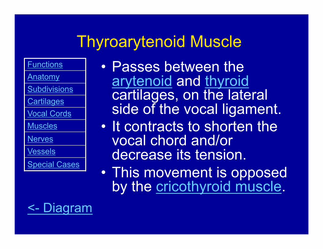
Thyroarytenoid Muscle • Passes between the
arytenoid and thyroid cartilages, on the lateral side of the vocal ligament.
• It contracts to shorten the vocal chord and/or decrease its tension.
• This movement is opposed by the cricothyroid muscle.
<- Diagram
Functions Anatomy Subdivisions Cartilages Vocal Cords Muscles Nerves Vessels Special Cases
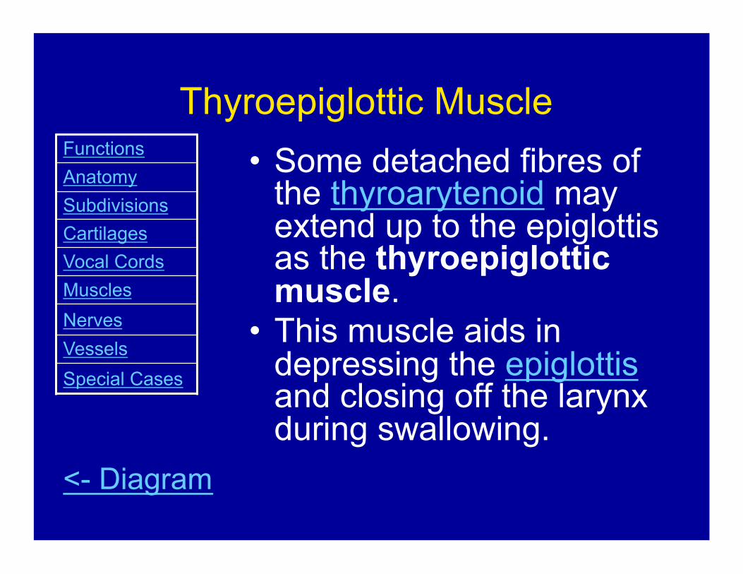
• Some detached fibres of the thyroarytenoid may extend up to the epiglottis as the thyroepiglottic muscle.
• This muscle aids in depressing the epiglottis and closing off the larynx during swallowing.
Thyroepiglottic Muscle
<- Diagram
Functions Anatomy Subdivisions Cartilages Vocal Cords Muscles Nerves Vessels Special Cases
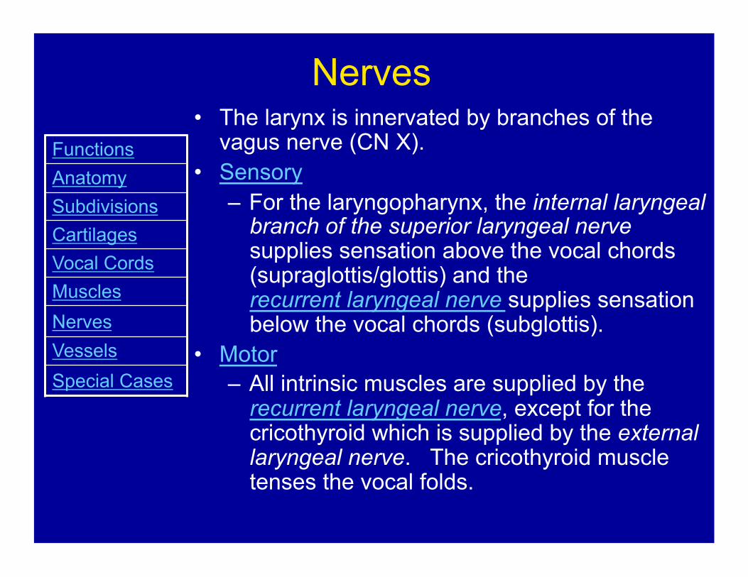
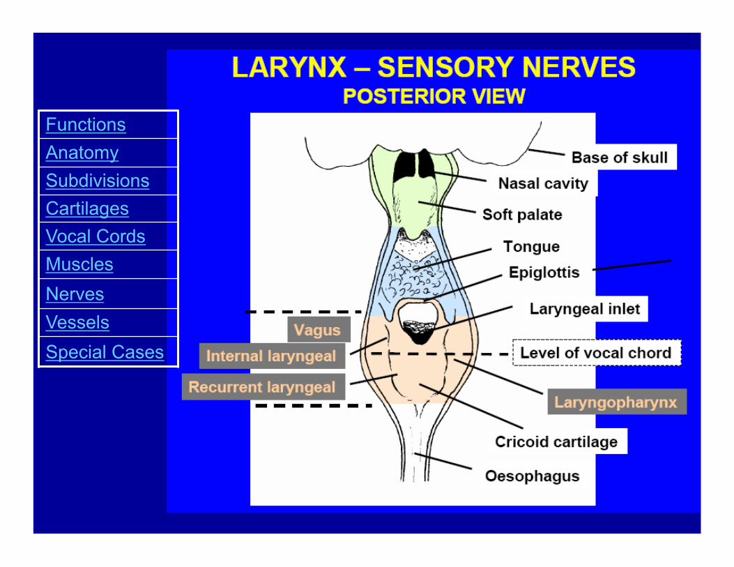
Nerves • The larynx is innervated by branches of the
vagus nerve (CN X). • Sensory
– For the laryngopharynx, the internal laryngeal branch of the superior laryngeal nerve supplies sensation above the vocal chords (supraglottis/glottis) and the recurrent laryngeal nerve supplies sensation below the vocal chords (subglottis).
• Motor – All intrinsic muscles are supplied by the
recurrent laryngeal nerve, except for the cricothyroid which is supplied by the external laryngeal nerve. The cricothyroid muscle tenses the vocal folds.
Functions Anatomy Subdivisions Cartilages Vocal Cords Muscles Nerves Vessels Special Cases
Functions Anatomy Subdivisions Cartilages Vocal Cords Muscles Nerves Vessels Special Cases

Recurrent Laryngeal
Nerve
• LEFT – Loops around
aortic arch • RIGHT
– Loops around subclavian artery
Functions Anatomy Subdivisions Cartilages Vocal Cords Muscles Nerves Vessels Special Cases

Vessels • Arteries
– Superior and inferior laryngeal arteries (from the superior and inferior thyroid arteries) accompany the internal and recurrent laryngeal nerves, respectively.
• Vein – Venous drainage is by
corresponding veins.
Functions Anatomy Subdivisions Cartilages Vocal Cords Muscles Nerves Vessels Special Cases

Special Cases
• Child’s Larynx • Laryngeal Carcinoma • Tracheostomy • Epiglottitis • Cricothyrotomy • Spasmodic Dysphonia
Functions Anatomy Subdivisions Cartilages Vocal Cords Muscles Nerves Vessels Special Cases

Child’s Larynx There are several
differences between an adult and child’s larynx and airway:
• More anterior and cephalad larynx
• Short trachea and neck. Beware of right mainstem bronchus intubation
• Proportionally larger head and tongue
Functions Anatomy Subdivisions Cartilages Vocal Cords Muscles Nerves Vessels Special Cases
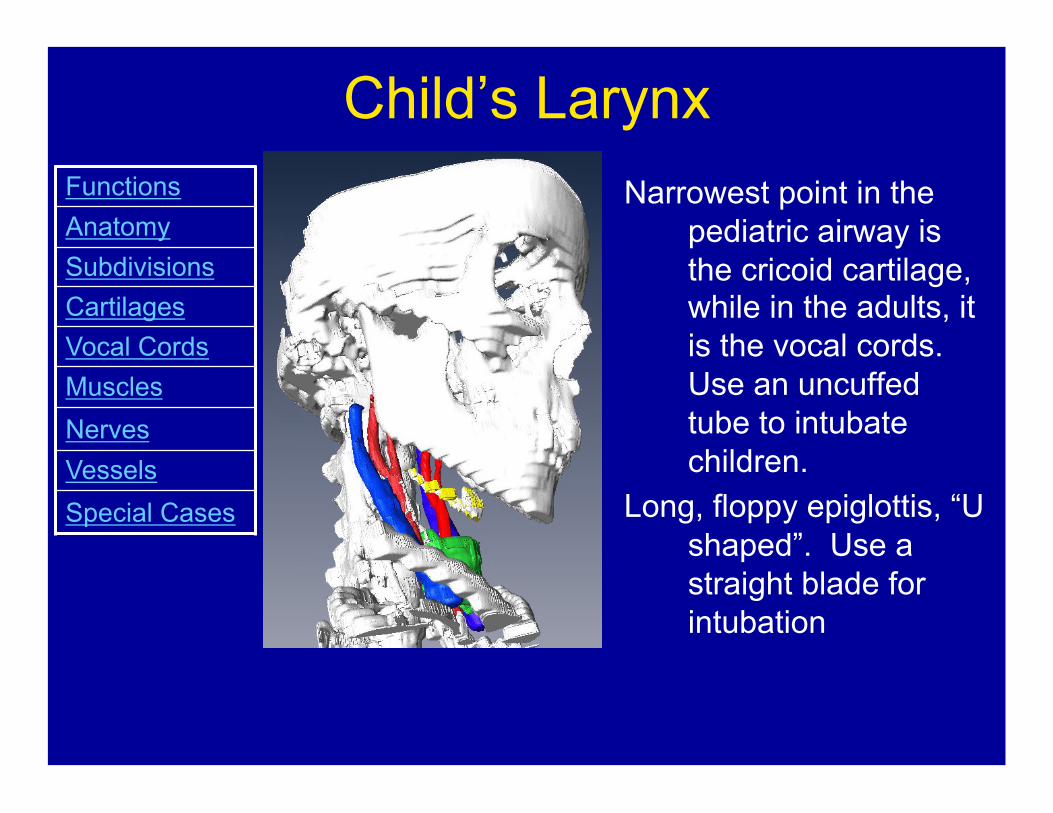
Child’s Larynx Narrowest point in the
pediatric airway is the cricoid cartilage, while in the adults, it is the vocal cords. Use an uncuffed tube to intubate children.
Long, floppy epiglottis, “U shaped”. Use a straight blade for intubation
Functions Anatomy Subdivisions Cartilages Vocal Cords Muscles Nerves Vessels Special Cases
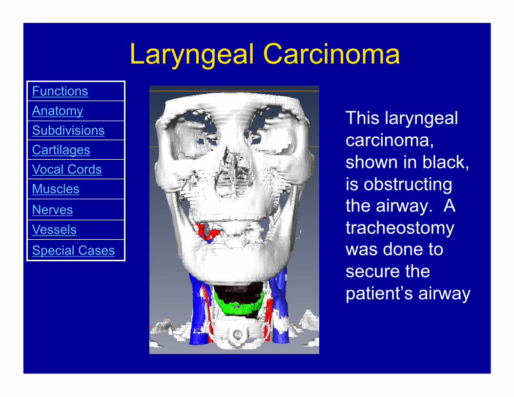
Laryngeal Carcinoma
This laryngeal carcinoma, shown in black, is obstructing the airway. A tracheostomy was done to secure the patient’s airway
Functions Anatomy Subdivisions Cartilages Vocal Cords Muscles Nerves Vessels Special Cases

Laryngeal Carcinoma • The most common head & neck cancer • 5% of all malignancies diagnosed
annually • 20,000 new cases in US annually • Mean age: 60-62 years • Predisposing factors: smoking & alcohol • Most are squamous cell carcinomas
(>95%)
Functions Anatomy Subdivisions Cartilages Vocal Cords Muscles Nerves Vessels Special Cases

Laryngeal Carcinoma • Presenting symptoms: hoarseness,
dysphagia, odynophagia, sore throat, referred otalgia, globus sensation, weight loss, and neck mass.
• Laryngeal tumors arise in the glottis (67%), supraglottis (31%), and subglottis (2%)
• Early cancers (T1/T2) are treated with single-modality treatment (surgery or radiation), while late cancers (T3/T4) are treated with multimodality treatment (surgery with postoperative radiation or organ preserving therapy)
Functions Anatomy Subdivisions Cartilages Vocal Cords Muscles Nerves Vessels Special Cases

Tracheostomy
A tracheostomy is an opening is made into the anterior wall of the trachea to establish an airway
Functions Anatomy Subdivisions Cartilages Vocal Cords Muscles Nerves Vessels Special Cases

Indications for Tracheostomy • Upper airway obstruction
– Due to burns or corrosive injury, laryngeal dysfunction, foreign bodies, infections, inflammatory conditions, neoplasms, OSA
• Inability of patient to manage secretions – Due to aspiration or excessive
bronchopulmonary secretions • Prolonged intubation • Facilitation of ventilation support • Inability to intubate • Adjunct to major head & neck surgery • Adjunct to management of major head
& neck trauma
Functions Anatomy Subdivisions Cartilages Vocal Cords Muscles Nerves Vessels Special Cases

Tracheostomy Functions Anatomy Subdivisions Cartilages Vocal Cords Muscles Nerves Vessels Special Cases
Tracheostomy
Airway Obstruction
http://www.nlm.nih.gov/medlineplus/ency/presentations/100043_4.htm

Epiglottitis • Acute inflammation of the
epiglottis and the supraglottic structures surrounding it
• Can be a severe, life-threatening disease of the upper airway
• Haemophilus influenzae was the predominant organism. – Hib vaccine has decreased the
incidence of epiglottitis • Age: 1-6 years old
Functions Anatomy Subdivisions Cartilages Vocal Cords Muscles Nerves Vessels Special Cases
Epiglottitis • Clinical triad of the 3 Ds:
– Drooling – Dysphagia – respiratory Distress
• Rapid onset of fever and sore throat
• Patient anxious and toxic looking
Functions Anatomy Subdivisions Cartilages Vocal Cords Muscles Nerves Vessels Special Cases

Epiglottitis • “Hot potato" muffled voice
– supraglottic • May have inspiratory stridor
– Inspiratory – supraglottic or glottic – Biphasic – subglottic – Expiratory – distal tracheobronchial tree
• “Sniffing position" with their nose pointed superiorly, head forward, sitting erect to maintain an adequate airway.
• Cough is rare
Functions Anatomy Subdivisions Cartilages Vocal Cords Muscles Nerves Vessels Special Cases

Epiglottitis
• Secure the AIRWAY first before any tests – ENT/Anesthesia consults – May need to intubate in the OR
• Do not agitate the child in any way – e.g. iv, tests
• Administer humidified oxygen • iv Antibiotics to cover causative
agents X 7-10 d
Functions Anatomy Subdivisions Cartilages Vocal Cords Muscles Nerves Vessels Special Cases
Epiglottitis Functions Anatomy Subdivisions Cartilages Vocal Cords Muscles Nerves Vessels Special Cases
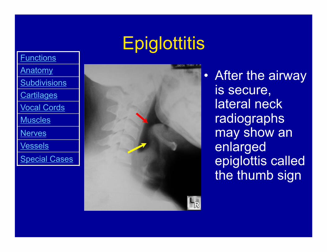
• After the airway is secure, lateral neck radiographs may show an enlarged epiglottis called the thumb sign
Cricothyrotomy • Emergency incision through the
skin and cricothyroid membrane to secure a patient's airway during certain emergency situations
• Easier and faster than a tracheostomy
• Only used when oral or nasal intubation is not possible
• A cricothyrotomy is a temporary airway, while a tracheostomy is a definitive airway
Functions Anatomy Subdivisions Cartilages Vocal Cords Muscles Nerves Vessels Special Cases

Cricothyrotomy Functions Anatomy Subdivisions Cartilages Vocal Cords Muscles Nerves Vessels Special Cases
http://www.fotosearch.com/comp/LIF/LIF141/NU304004.jpg
Adductor Spasmodic Dysphonia
• Sudden involuntary muscle movements or spasms cause the vocal cords to slam together and stiffen.
• Speech may be choppy and sound similar to stuttering. The voice is commonly described as strained or strangled and full of effort.
Functions Anatomy Subdivisions Cartilages Vocal Cords Muscles Nerves Vessels Special Cases
Abductor Spasmodic Dysphonia
• Sudden involuntary muscle movements or spasms cause the vocal folds to open.
• The vocal folds can not vibrate when they are open.
• The open position of the vocal folds also allows air to escape from the lungs during speech.
• The voices sounds weak, quiet and breathy or whispery.
Functions Anatomy Subdivisions Cartilages Vocal Cords Muscles Nerves Vessels Special Cases
Spasmodic Dysphonia • With both abductor and adductor
spasmodic dysphonia, the spasms are often absent during activities such as laughing or singing
• Stress often makes the spasms worse • There is no cure • The most effective treatment for
reducing symptoms is injections of very small amounts of botulinum toxin (Botox) directly into the affected muscles of the larynx
Functions Anatomy Subdivisions Cartilages Vocal Cords Muscles Nerves Vessels Special Cases

References • Essential Clinical Anatomy • ENT Secrets • Wikipedia® - the Free encyclopedia http://en.wikipedia.org/wiki/Larynx http://en.wikipedia.org/wiki/
Spasmodic_dysphonia#Adductor_spasmodic_dysphonia • Dr. Peter Haase’s Laryngeal Anatomy lecture notes for first
year medicine, UWO • Dr. Kevin Fung’s Hoarseness lecture notes for first year
medicine, UWO • Amira 4.1 • Netters • http://www.nlm.nih.gov/medlineplus/ency/presentations/
100043_4.htm - trach photo • http://www.fotosearch.com/comp/LIF/LIF141/NU304004.jpg -
cricothyrotomy photo
Top Related