


Languages
Pages
Legal


The Imaging Facility Newsletter
Inside this issue:
Featured
Microscope 2
Zeiss 710 Update 3
Celebrate
Biomedical Art 4
IF Updates 5
IF Survey Results 6
Recent Publications 7
Contact Us 10
I am a 2nd year Masters student in Dr. Pistilli’s lab in the Exercise
Physiology Department. We study the effects of cancer cachexia on
skeletal muscle wasting and function. We recently classified a murine
-derived mammary tumor cell line as having the ability to form
tumors in vivo and induce cachexia within 4 weeks post injection.
We monitored tumor status weekly using both optical and
ultrasound imaging, and we performed physiological assessment of
muscle function.
Large-stitch images of immunostained muscle cross sections were
acquired using the Nikon TE2000 Sweptfield microscope. These
images were then analyzed using the NIS Elements General Analysis
program to automatically determine muscle fiber area. We showed
severe fatigue at the 4 week time point for our mice, highlighting
both skeletal muscle wasting and dysfunction. Building upon these
results, we repeated these experiments in our transgenic model of
IL-15 skeletal muscle overexpression where we observed
preservation in function and fiber area. We are now continuing to
characterize the effectiveness of IL-15 in the attenuation of cancer
cachexia.
Volume 5, Issue 2
August 2016
Featured Researcher: Joseph Bohlen
Large-stitch image of a mouse muscle cross section (left). Muscles were labeled with laminin
and DAPI, and using the Nikon Sweptfield microscope, 25 images at 20X were captured and
stitched to show the entire section. Since the individual images were obtained with a 20X
objective lens, zooming in to an area of the muscle does not result in loss of resolution.
IVIS optical imaging (left) was used to non-invasively monitor tumor growth over time. A
series of 2D ultrasound images (middle) were collected and reconstructed into a 3D structure
(right) for accurate tumor volume measurements.

Page 2
Lightning-Fast
Brightfield
Scanning
3D Virtual
Slides
Multi-Channel Fluorescent
Slides
New Equipment in the Imaging Facility!
6 Position
Slide Holder
for High
Throughput
The Microscope Imaging Facility has recently installed a new Olympus VS120 Slide Scanner
to replace the old Olympus Histology
Microscope.
This system can image entire tissue sections,
either in brightfield or fluorescence.
This system was purchased with funding from the INBRE
grant 5P20GM103434 and the WVU HSC Office of
Research & Graduate Education. Please remember to
acknowledge this support in your publications.
Olympus VS120
Slide Scanner
The system is equipped with 5 objectives for
2x - 40x magnification.
This scanner is super-duper fast and easy to
use.
Please contact Mandy Ammer for a demo or
to setup training.
Page 3
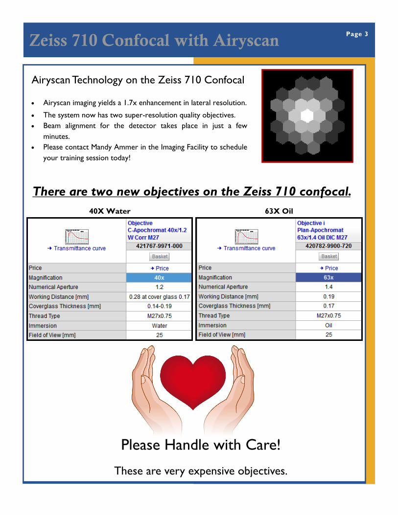
Zeiss 710 Confocal with Airyscan
There are two new objectives on the Zeiss 710 confocal.
40X Water 63X Oil
Airyscan Technology on the Zeiss 710 Confocal
Airyscan imaging yields a 1.7x enhancement in lateral resolution.
The system now has two super-resolution quality objectives.
Beam alignment for the detector takes place in just a few
minutes.
Please contact Mandy Ammer in the Imaging Facility to schedule
your training session today!
Please Handle with Care!
These are very expensive objectives.

Celebrate Biomedical Art Page 4
The National Institutes of Health Life: Magnified exhibition has turned
life into art. This gift, displayed in the Pylons area and library at the
Health Sciences Center, will enlighten and educate for years to come.
Join the celebration of this exhibit at a reception in the Pylons lobby
Wednesday, August 24, 2016, 3:30 - 5:30 p.m.
In addition, the NIH has contacted us looking for more breath-taking
images to add to their collection. If you have images you’d be willing
to share, please let us know and we’ll help pass them along.
The IF would like to wish Emily Ellis the best of luck in her new
position in the laboratory of Dr. Timothy Eubank.
We will miss you!
newsletter

Page 5
New Server For Storing Your Awesome Images The Imaging Facility Users now have space on a brand
new server that will provide tons of room for data
storage. All of your data in your AMIF/MIF User Folders
was successfully transferred to the new location, and it
is ready to use.
You should now have a shortcut named "Imaging Folders" on your desktop that will
take you to the new server location; you should also see this location mapped to your
P: drive. If you don't see these links, try logging out and back in and/or restarting your
computer. If that doesn't work, let us know and we'll figure out how to make those
connections.
\\hs-mbrcc\mbrcc\RESLAB\Imaging (for PCs)
smb://hs-mbrcc/mbrcc/reslab/imaging/ (for Macs)
Service Rate/Hr
AMIF IVIS Lumina II $30.00
AMIF Vevo 2100 $38.50
AMIF XenX $20.00
AMIF SkyScan 1272 $16.50
AMIF Procedures Room $5.50
Assistance/Procedures/Training $30.00
The Core Resources Management Committee (CRMC) has recently completed the
annual review of the Imaging Facilities, and the following rate schedule was approved
effective October 1, 2016.
Imaging Facility Rates Starting Oct 1
Service Rate/Hr
Assistance/Training $30.00
MIF Nikon Live Cell $15.00
MIF Nikon Sweptfield (<4 hours) $15.00
MIF Nikon Sweptfield (> 4 hours) $10.00
MIF NLOM 2-Photon $15.00
MIF Olympus MVX10 $0.00
MIF Olympus Slide Scanner $10.00
MIF Zeiss 710 Confocal (<4 hours) $25.00
MIF Zeiss 710 Confocal (>4 hours) $18.50
MIF Zeiss Fluorescent $15.00
MIF Zeiss PALM Laser Microdissection $15.00
MIF Zeiss Violet Confocal $30.00
Workstations $5.00
Microscope Imaging Facility Animal Models & Imaging Facility

Imaging Facility Surveys Page 6
Thanks to everyone who has taken our Imaging Facility Surveys over
the past year! Your feedback is really important for us to learn what
we’re doing well and what we need to improve to best support your
research.
Here’s a
summary of
what you
told us.
Your feedback is also critical in identifying new equipment needs.
Here are a couple of requests and an update on what we’re doing...
*Two-photon microscope with live animal imaging capabilities is needed.
The imaging facility is working with Nikon to bring in a two-photon
microscope for a demo, hopefully later this fall. Please let us know
ASAP if you’d like to participate. We will need to get ACUC protocols
submitted soon in order to make the most of this demonstration.
*Super resolution imaging system is needed.
We hosted a demo of the Nikon A1R confocal / N-SIM super-
resolution imaging system this spring. With your help, we had a very
successful demo and were able to submit an instrumentation grant to
fund the purchase of this system. Our application will be reviewed in
November - we’ll keep you posted.
For training and assistance from the AMIF, please
keep in mind that Sarah’s regular schedule is Monday,
Tuesday and Thursday 7:30 a.m. - 5:30 p.m. The
calendar can fill up quickly, so please schedule in
advance to make sure we can accommodate your
imaging needs.
Survey Results (Scale of 1 to 5) AMIF MIF
Availability of facility & equipment 4.7 4.5
Quality of results/data 4.4 4.5
Availability of staff 4.7 4.7
Knowledge and expertise of staff 4.3 4.8
Quality of consultation 4.6 4.8
Cost of service 4.0 3.8
Overall experience 4.7 4.7 Take the
AMIF
Survey
Take the
MIF
Survey
https://goo.gl/forms/
BMzvGPCw7JLKUzXW2
https://goo.gl/
forms/4CLrBVbanZpDNYCj

Page 7
Early detection of cardiac dysfunction in the type 1 diabetic heart using speckle-
tracking based strain imaging. Shepherd DL, Nichols CE, Croston TL, McLaughlin SL,
Petrone AB, Lewis SE, Thapa D, Long DM, Dick GM, Hollander JM. J Mol Cell Cardiol.
2016 Jan;90:74-83. PMID: 26654913
Cardiac and mitochondrial dysfunction following acute pulmonary exposure to moun-
taintop removal mining particulate matter. Nichols CE, Shepherd DL, Knuckles TL,
Thapa D, Stricker JC, Stapleton PA, Minarchick VC, Erdely A, Zeidler-Erdely PC, Alway
SE, Nurkiewicz TR, Hollander JM. Am J Physiol Heart Circ Physiol. 2015 Dec 15;309
(12):H2017-30. PMID: 26497962
Deficiency of isoprenylcysteine carboxyl methyltransferase (ICMT) leads to progres-
sive loss of photoreceptor function. Christiansen JR, Pendse ND, Kolandaivelu S, Ber-
go MO, Young SG, Ramamurthy V. J Neurosci. 2016 May 4;36(18):5107-14. PMID:
27147662
Grainyhead-like 2 inhibits the coactivator p300, suppressing tubulogenesis and the epi-
thelial-mesenchymal transition. Pifer PM, Farris JC, Thomas AL, Stoilov P, Denvir J,
Smith DM, Frisch SM. Mol Biol Cell. 2016 Aug 1;27(15):2479-92. PMID: 27251061
Recent Publications
Shepherd et al. Nichols et al.
Christiansen et al. Pifer et al.
Page 8
Please let us know
when you publish a manuscript with da-
ta from the MIF or AMIF so that we may acknowledge
your achievement in
our newsletter!
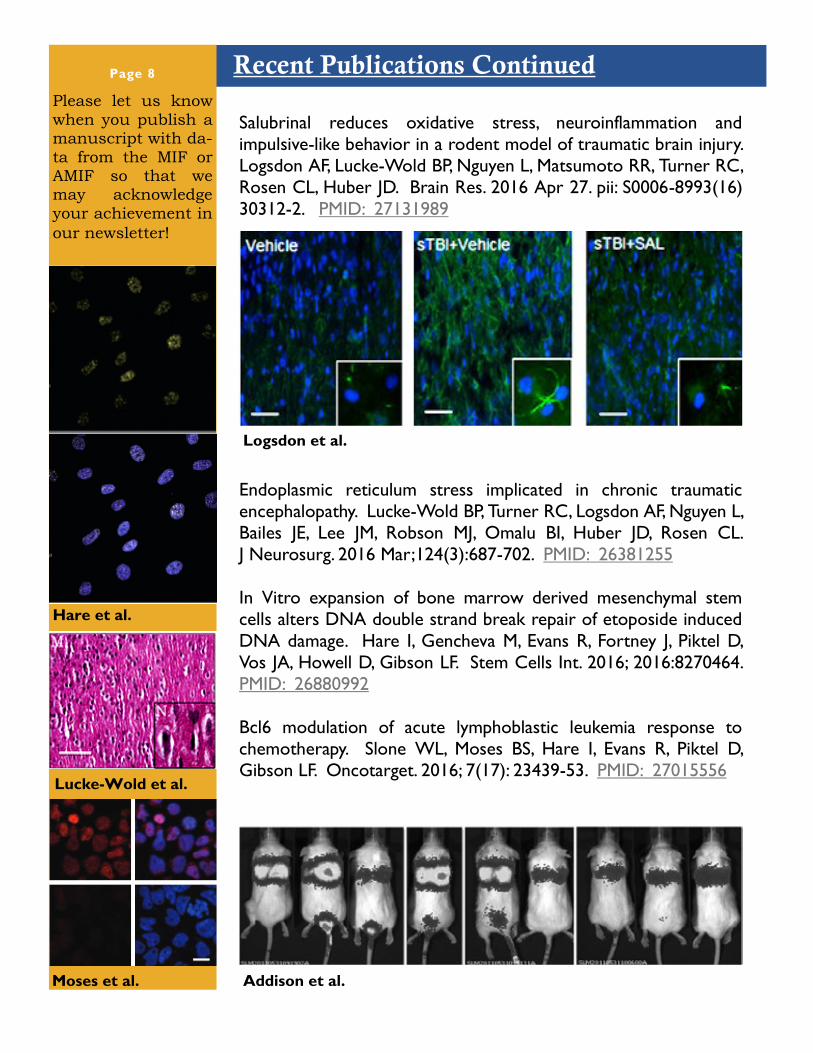
Salubrinal reduces oxidative stress, neuroinflammation and
impulsive-like behavior in a rodent model of traumatic brain injury.
Logsdon AF, Lucke-Wold BP, Nguyen L, Matsumoto RR, Turner RC,
Rosen CL, Huber JD. Brain Res. 2016 Apr 27. pii: S0006-8993(16)
30312-2. PMID: 27131989
Endoplasmic reticulum stress implicated in chronic traumatic
encephalopathy. Lucke-Wold BP, Turner RC, Logsdon AF, Nguyen L,
Bailes JE, Lee JM, Robson MJ, Omalu BI, Huber JD, Rosen CL.
J Neurosurg. 2016 Mar;124(3):687-702. PMID: 26381255
In Vitro expansion of bone marrow derived mesenchymal stem
cells alters DNA double strand break repair of etoposide induced
DNA damage. Hare I, Gencheva M, Evans R, Fortney J, Piktel D,
Vos JA, Howell D, Gibson LF. Stem Cells Int. 2016; 2016:8270464.
PMID: 26880992
Bcl6 modulation of acute lymphoblastic leukemia response to
chemotherapy. Slone WL, Moses BS, Hare I, Evans R, Piktel D,
Gibson LF. Oncotarget. 2016; 7(17): 23439-53. PMID: 27015556
Recent Publications Continued
Logsdon et al.
Moses et al.
Lucke-Wold et al.
Addison et al.
Hare et al.

Recent Publications Continued Page 9
Please Remember to Acknowledge Us!
KAP1 promotes proliferation and metastatic progression of breast cancer cells. Addison
JB, Koontz C, Fugett JH, Creighton CJ, Chen D, Farrugia MK, Padon RR, Voronkova MA,
McLaughlin SL, Livengood RH, Lin CC, Ruppert JM, Pugacheva EN, Ivanov AV. Cancer Res.
2015; 75(2): 344-55. PMID: 25421577.
Grainyhead-like 2 reverses the metabolic changes induced by
the oncogenic epithelial-mesenchymal transition: effects on
anoikis. Farris JC, Pifer PM, Zheng L, Gottlieb E, Denvir J,
Frisch SM. Mol Cancer Res. 2016; 2016; 27(15): 2479-92..
PMID: 27084311
Bone marrow microenvironment niche regulates miR-221/222
in acute lymphoblastic leukemia. Moses BS, Evans R, Slone WL,
Piktel D, Martinez I, Craig MD, Gibson LF. Mol Cancer Res.
2016 Jun 29. PMID: 27358112
In vivo assessment of coronary flow and cardiac function after
bolus adenosine injection in adenosine receptor knockout
mice. Teng B, Tilley SL, Ledent C, Mustafa SJ. Physiol Rep. 2016
Jun;4(11). PMID:27302991
AMIF: “Small animal imaging and image analysis were performed in the West Virginia University
Animal Models & Imaging Facility, which has been supported by the WVU Cancer Institute and
NIH grants P20 RR016440, P30 GM103488 and S10 RR026378.”
MIF: “Imaging experiments and image analysis were performed in the West Virginia University
Microscope Imaging Facility, which has been supported by the WVU Cancer Institute and NIH
grants P20 RR016440, P30 GM103488 and P20 GM103434.”
Farris et al. Slone et al.
Teng et al.

1 Medical Center Drive
Erma Byrd BMRC
West Virginia University
Morgantown WV 26506
Karen Martin
Phone: 304-293-6965
Fax: 304-293-4667
The purpose of this newsletter is to inform researchers about the AMIF and MIF.
We want all investigators, graduate students and staff to be knowledgeable about
the equipment and resources that are available. The staff are always glad to
discuss upcoming studies with investigators to best utilize the core resources
available. To learn more about our facilities, please check out our websites (to
the left) or contact us to speak directly with AMIF or MIF staff.
wvucancer.org/AMIF
Contact Us!
wvucancer.org/MIF
Sarah McLaughlin
Phone: 304-293-0518
Fax: 304-293-4667
Mandy Ammer
Phone: 304-293-0942
Fax: 304-293-4667
Page 10 Humor to encourage your science
Picture Perfect Image Analysis using
Nikon NIS Elements Tuesday, September 27 at
1:00 PM
Erma Byrd 201
Top Related