


Languages
Pages
Legal


TAVR: A Fresh Look at Aortic Stenosis
Thomas Smith, MD
May 5, 2012
Disclosures
• I have no specific conflicts of interest pertaining to this presentation.
• I will discuss valves not currently available/approved for use in the United States.

Outline
Aortic Stenosis
• Etiology/Pathophysiology
• Clinical course/symptoms
• Assessment – Echocardiography
Transcatheter Aortic Valve Replacement - Imaging • Echo and CT • Safety of valve placement
– Echocardiography (TEE) and CT • Procedural guidance
– Implant angle (CT) – Final valve size and complications (TEE)
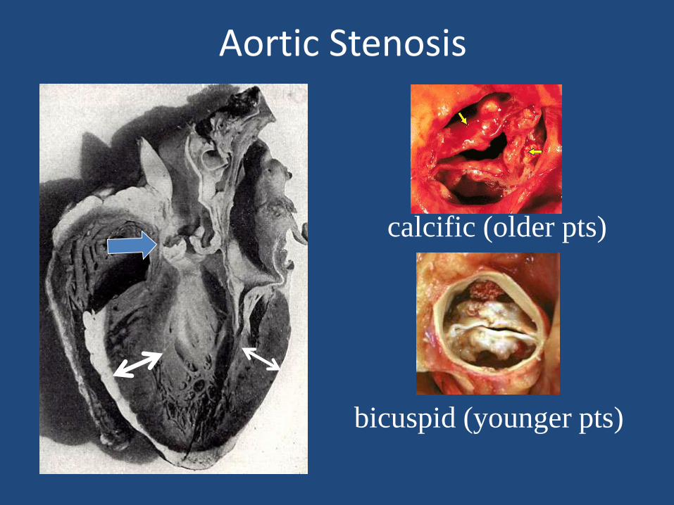
Aortic Stenosis
calcific (older pts)
bicuspid (younger pts)

Pathology
• Similar to atherosclerosis
– Cellular proliferation, inflammation, lipid and increased macrophages and T-lymphocytes
• Congenitally deformed valves
– Turbulent flow leads to disruption of endothelium and collagen -> calcium deposit
• Rheumatic AS
– inflammation leads to organization and fibrosis of the valve with fusion of the commissures
Aortic Stenosis
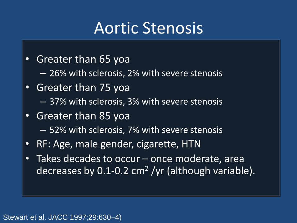
• Greater than 65 yoa – 26% with sclerosis, 2% with severe stenosis
• Greater than 75 yoa – 37% with sclerosis, 3% with severe stenosis
• Greater than 85 yoa – 52% with sclerosis, 7% with severe stenosis
• RF: Age, male gender, cigarette, HTN
• Takes decades to occur – once moderate, area decreases by 0.1-0.2 cm2 /yr (although variable).
Stewart et al. JACC 1997;29:630–4)
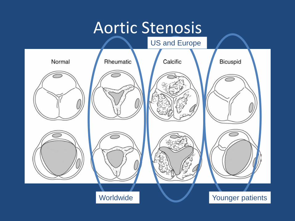
Aortic Stenosis
Worldwide
US and Europe
Younger patients
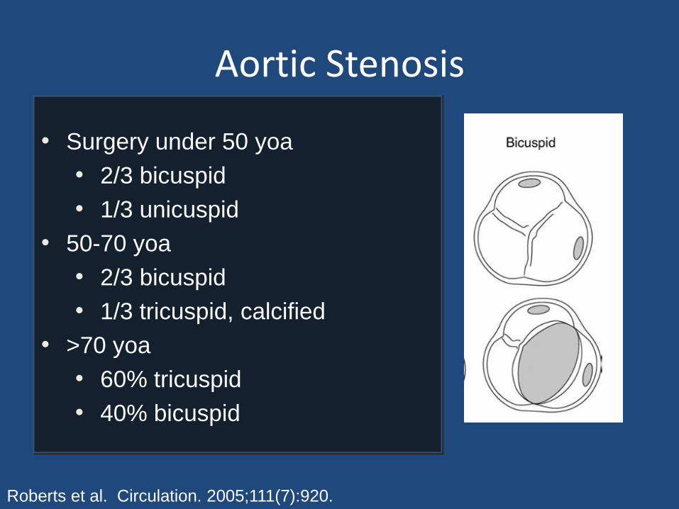
• Surgery under 50 yoa
• 2/3 bicuspid
• 1/3 unicuspid
• 50-70 yoa
• 2/3 bicuspid
• 1/3 tricuspid, calcified
• >70 yoa
• 60% tricuspid
• 40% bicuspid
Aortic Stenosis
Roberts et al. Circulation. 2005;111(7):920.
Pathophysiology
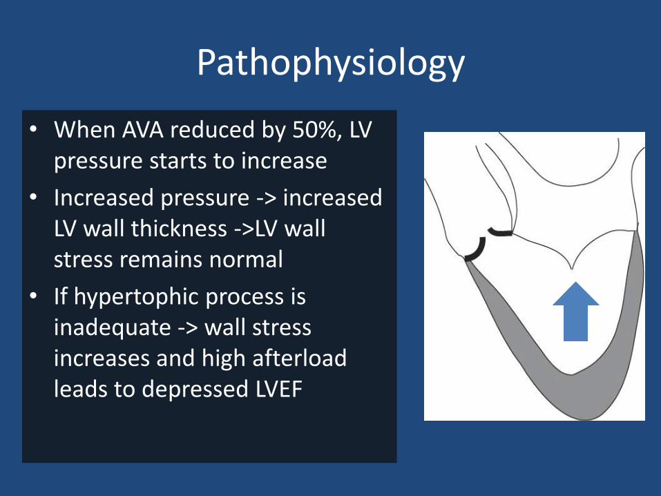
• When AVA reduced by 50%, LV pressure starts to increase
• Increased pressure -> increased LV wall thickness ->LV wall stress remains normal
• If hypertophic process is inadequate -> wall stress increases and high afterload leads to depressed LVEF
Pathophysiology
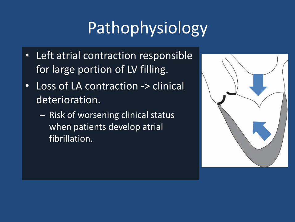
• Left atrial contraction responsible for large portion of LV filling.
• Loss of LA contraction -> clinical deterioration.
– Risk of worsening clinical status when patients develop atrial fibrillation.

Natural History
Ross and Braunwald. Circulation 1968;38(Suppl. V):61.
Angina
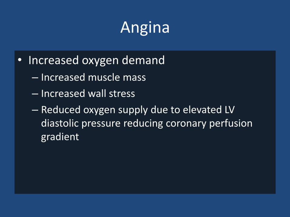
• Increased oxygen demand
– Increased muscle mass
– Increased wall stress
– Reduced oxygen supply due to elevated LV diastolic pressure reducing coronary perfusion gradient

Exertional syncope
• Exercise leads to vasodilation in peripheral muscles
• LV unable to augment output enough for exercise – unable to increase cardiac output
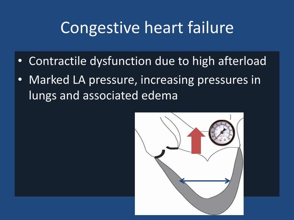
Congestive heart failure
• Contractile dysfunction due to high afterload
• Marked LA pressure, increasing pressures in lungs and associated edema
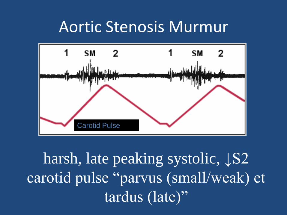
Aortic Stenosis Murmur
Carotid Pulse
harsh, late peaking systolic, ↓S2
carotid pulse “parvus (small/weak) et
tardus (late)”
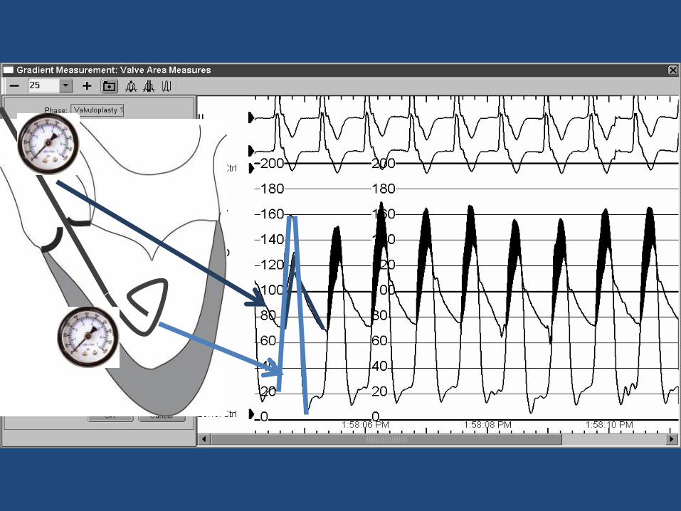

AV area = (A1V1)/V2
AV Area = (3.14 x1.1x1.1)x76/471.87 AV Area= 0.62 sq cm

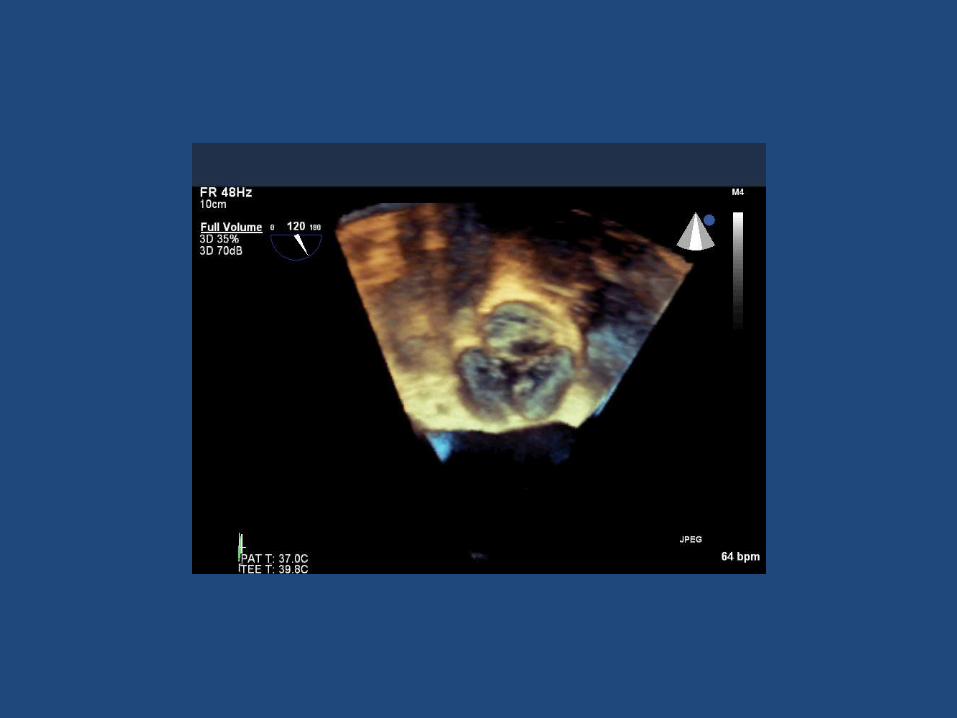
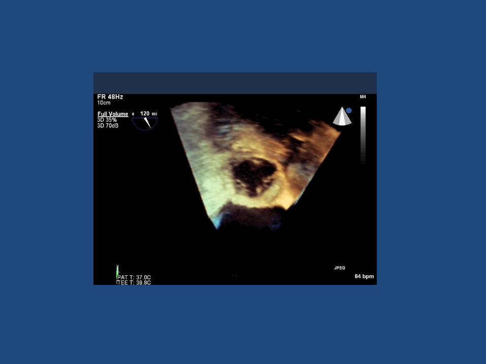
Echocardiography


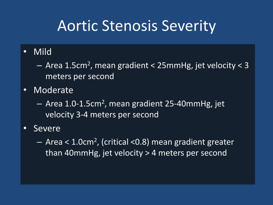
Aortic Stenosis Severity
• Mild
– Area 1.5cm2, mean gradient < 25mmHg, jet velocity < 3 meters per second
• Moderate
– Area 1.0-1.5cm2, mean gradient 25-40mmHg, jet velocity 3-4 meters per second
• Severe
– Area < 1.0cm2, (critical <0.8) mean gradient greater than 40mmHg, jet velocity > 4 meters per second

Medical Therapy for Severe AS
• No medication can alter natural history
• Maintain euvolemia, sinus rhythm
• Digitalis with decreased EF
• Caution with beta blockers, ACE inhibitors, nitroglycerin or any agents that decrease CO or decrease systemic vascular resistance (SVR)

Surgery for AS Gold Standard
• Indicated for critical AS, or for severe AS if patient has any symptoms attributable to AS
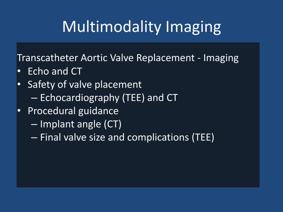
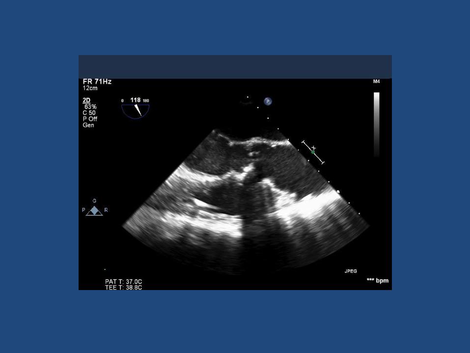
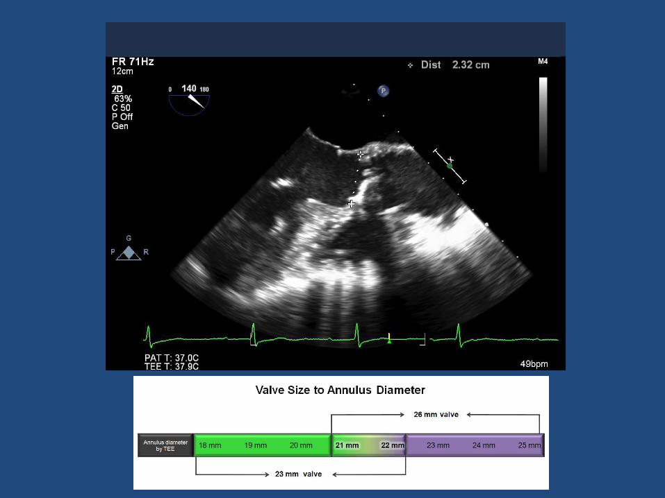
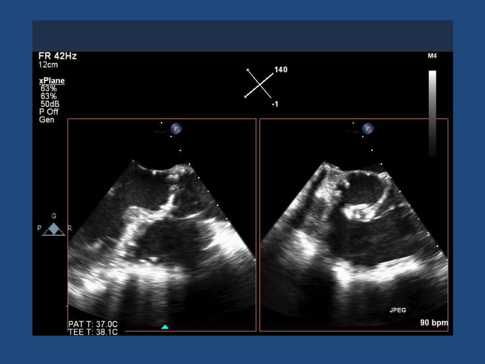
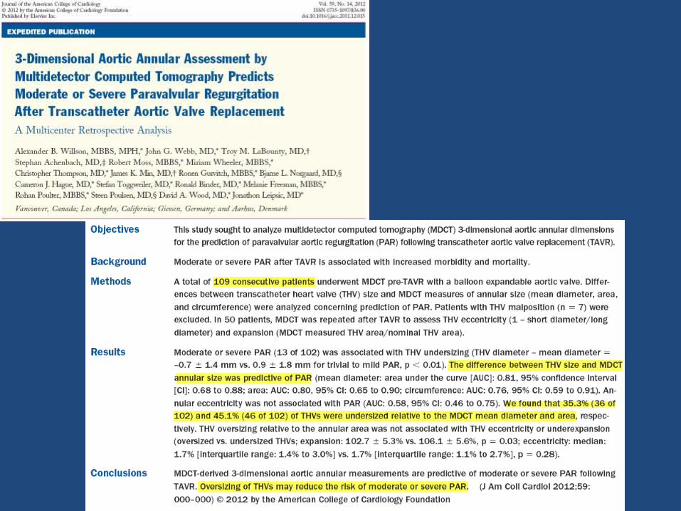
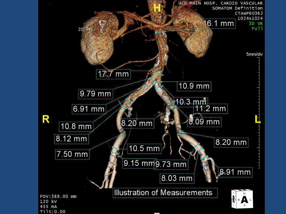
Multimodality Imaging
Transcatheter Aortic Valve Replacement - Imaging • Echo and CT • Safety of valve placement
– Echocardiography (TEE) and CT • Procedural guidance
– Implant angle (CT) – Final valve size and complications (TEE)


SAFETY OF IMPLANTATION CT
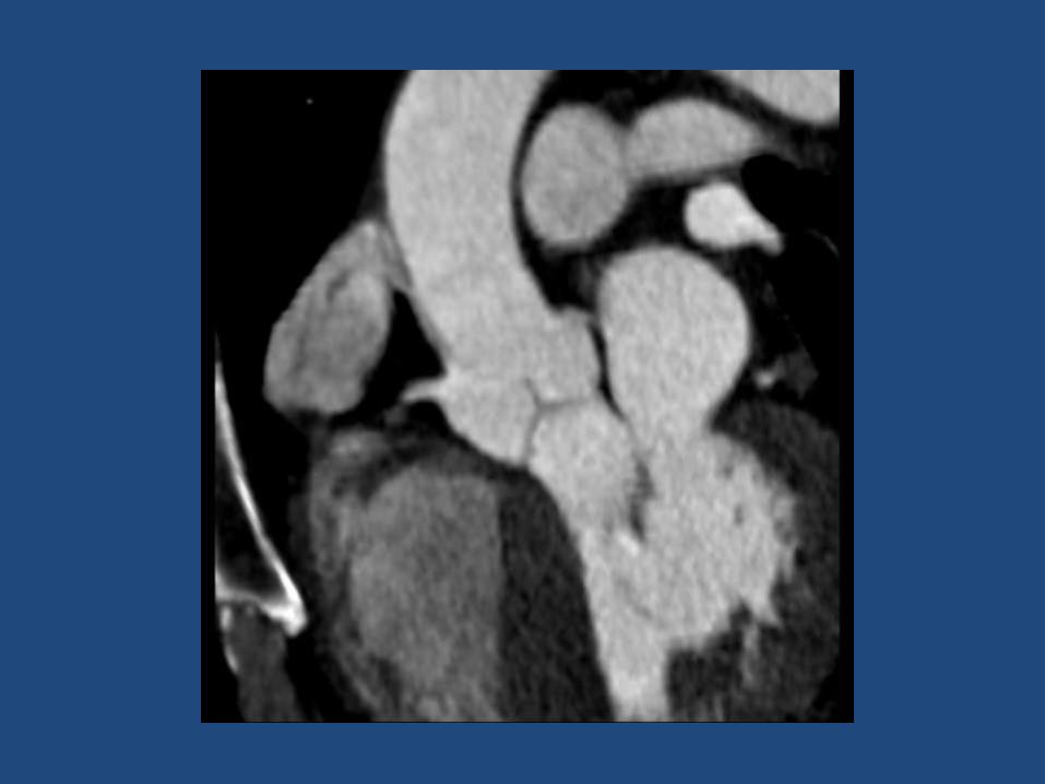
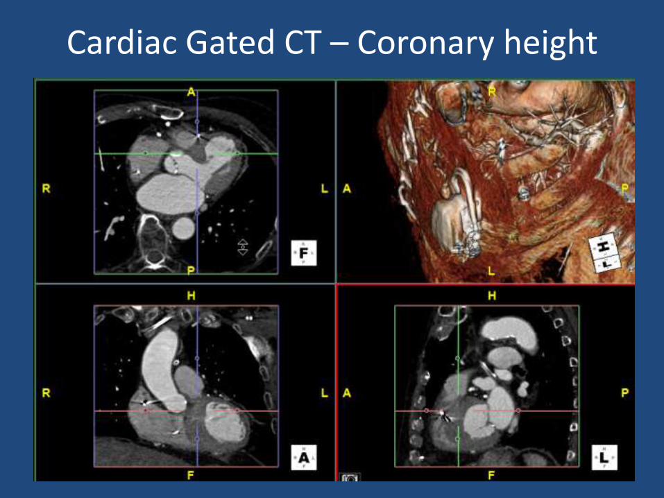
Cardiac Gated CT – Coronary height

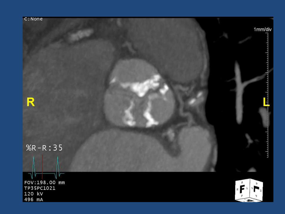


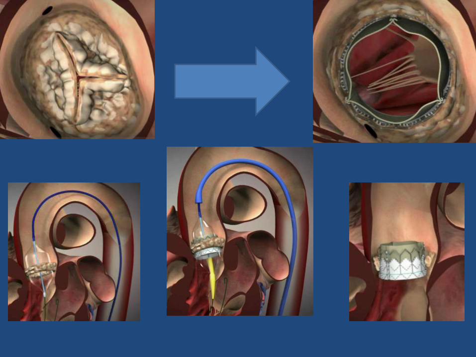
PROCEDURAL

Deployment angle lining up all the cusps


LAO 15, CAUDAL 20
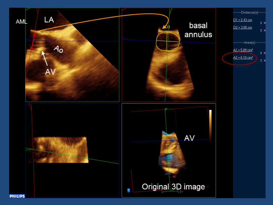
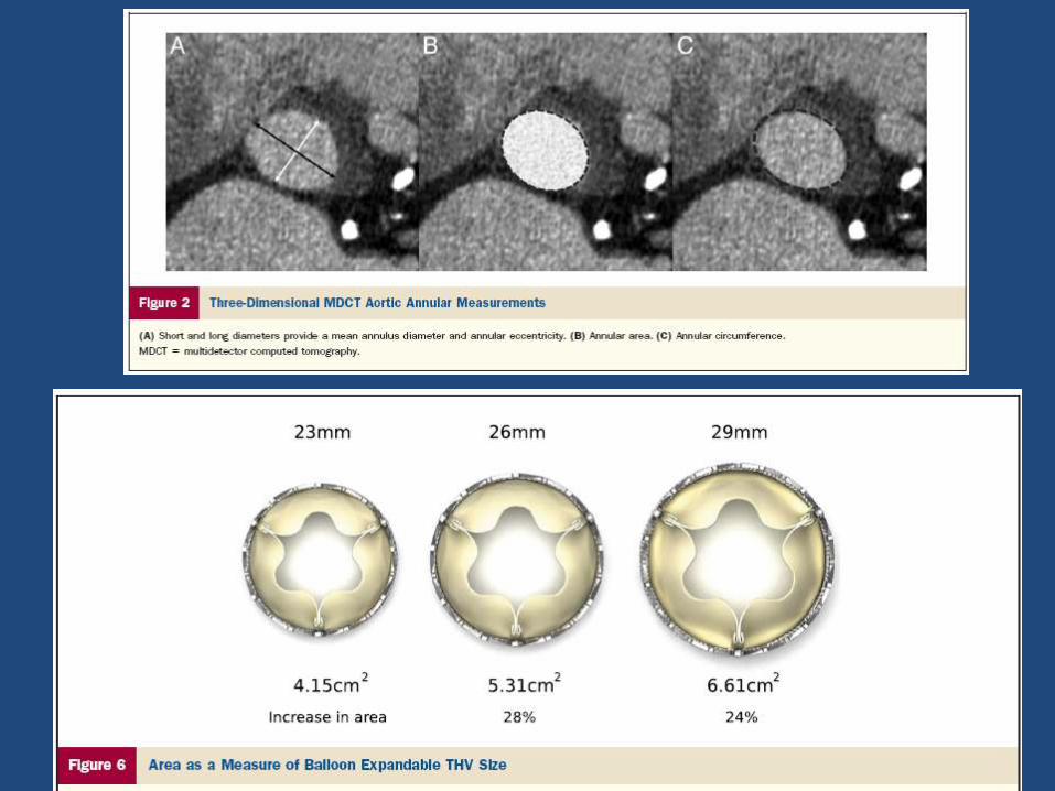
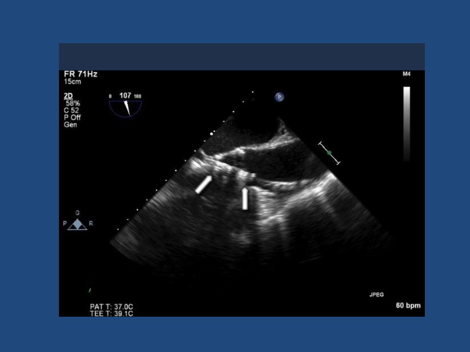
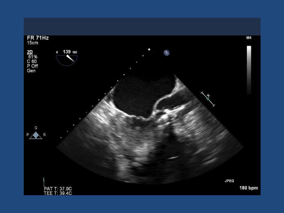
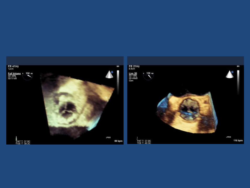
Transesophageal Echo
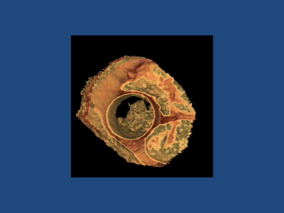
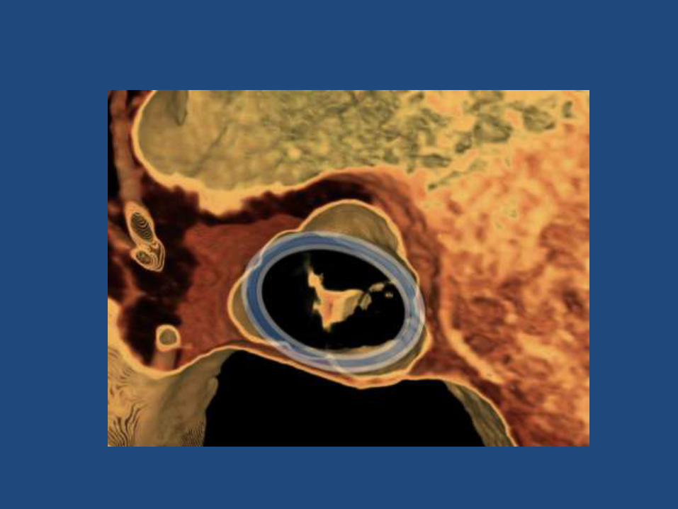
• Annulus assessment prior to opening seal on valve.
• Baseline TEE (valves, function, ?effusion, working views).
• Valve placement (secondary)
• Ongoing assessment for complications.


Recognition of complications
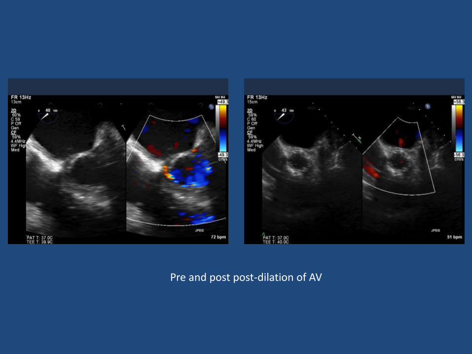
Pre and post post-dilation of AV
Conclusions/Summary
• Aortic stenosis is quite common in our aging population.
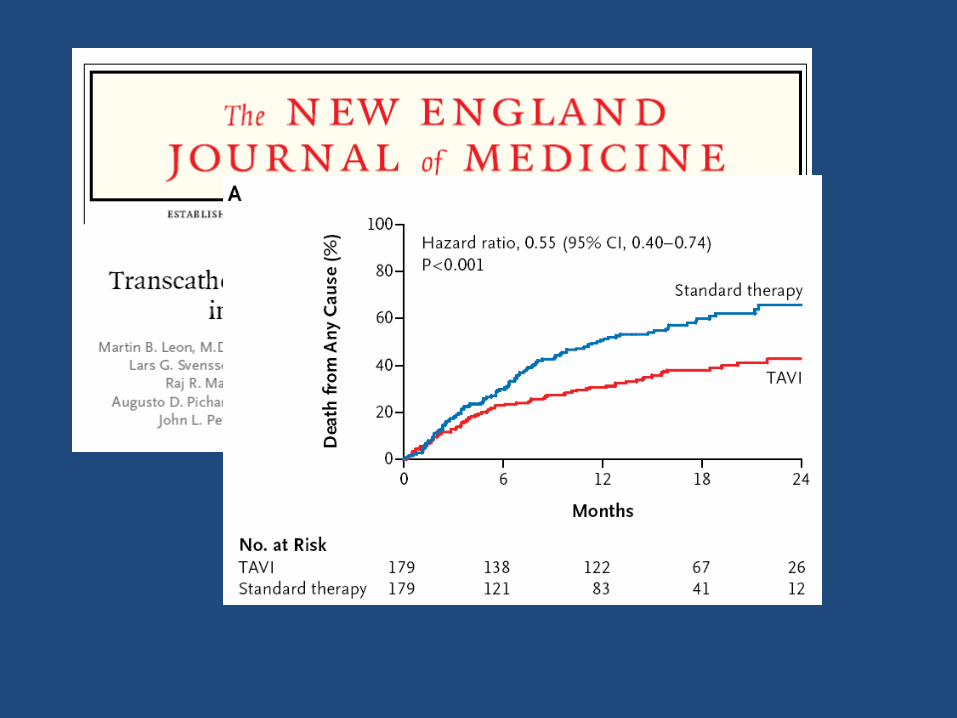
• Percutaneous aortic valve therapies provide patients who are not operative candidates the benefit of valve replacement.
• Echocardiography and Cardiac CT are complementary modalities in assessment of patients undergoing TAVR.

Thank you
• Questions?