


Languages
Pages
Legal

1037
Synthesis of new pyrrole–pyridine-based ligandsusing an in situ Suzuki coupling method
Matthias Böttger1, Björn Wiegmann1, Steffen Schaumburg1,Peter G. Jones2, Wolfgang Kowalsky1 and Hans-Hermann Johannes*1
Full Research Paper Open Access
Address:1Labor für Elektrooptik am Institut für Hochfrequenztechnik,Technische Universität Braunschweig, Bienroder Weg 94, 38106Braunschweig, Germany and 2Institut für Anorganische undAnalytische Chemie, Technische Universität Braunschweig,Hagenring 30, 38106 Braunschweig, Germany
Email:Hans-Hermann Johannes* - [email protected]
* Corresponding author
Keywords:boronic acids; complex chemistry; in situ generation; pyrrole-pyridine;Suzuki coupling
Beilstein J. Org. Chem. 2012, 8, 1037–1047.doi:10.3762/bjoc.8.116
Received: 04 April 2012Accepted: 18 June 2012Published: 09 July 2012
Associate Editor: I. Marek
© 2012 Böttger et al; licensee Beilstein-Institut.License and terms: see end of document.
AbstractThe compounds 6-(pyrrol-2-yl)-2,2‘-bipyridine, 2-(pyrrol-2-yl)-1,10-phenanthroline and 2-(2-(N-methylbenz[d,e]imidazole)-6-
(pyrrol-2-yl)-pyridine were synthesized by using an in situ generated boronic acid for the Suzuki coupling. Crystals of the products
could be grown and exhibited interesting structures by X-ray analysis, one of them showing a chain-like network with the adjacent
molecules linked to each other via intermolecular N–H…N hydrogen bonds.
1037
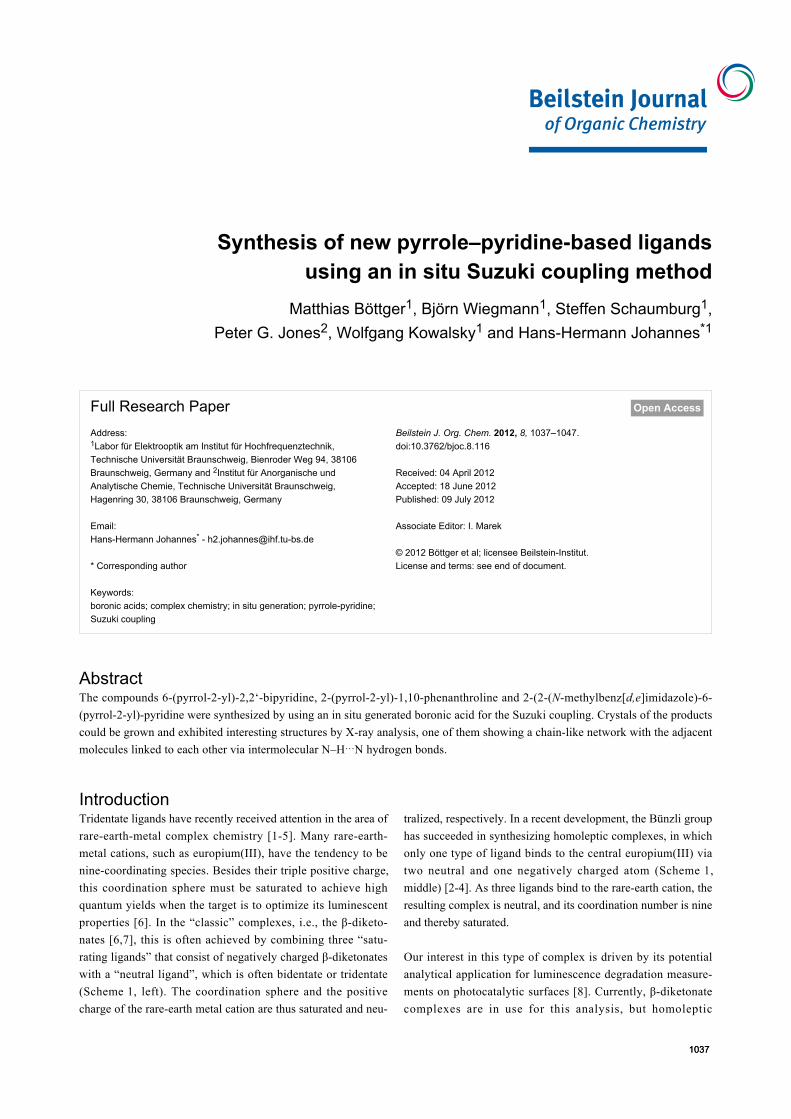
IntroductionTridentate ligands have recently received attention in the area of
rare-earth-metal complex chemistry [1-5]. Many rare-earth-
metal cations, such as europium(III), have the tendency to be
nine-coordinating species. Besides their triple positive charge,
this coordination sphere must be saturated to achieve high
quantum yields when the target is to optimize its luminescent
properties [6]. In the “classic” complexes, i.e., the β-diketo-
nates [6,7], this is often achieved by combining three “satu-
rating ligands” that consist of negatively charged β-diketonates
with a “neutral ligand”, which is often bidentate or tridentate
(Scheme 1, left). The coordination sphere and the positive
charge of the rare-earth metal cation are thus saturated and neu-
tralized, respectively. In a recent development, the Bünzli group
has succeeded in synthesizing homoleptic complexes, in which
only one type of ligand binds to the central europium(III) via
two neutral and one negatively charged atom (Scheme 1,
middle) [2-4]. As three ligands bind to the rare-earth cation, the
resulting complex is neutral, and its coordination number is nine
and thereby saturated.
Our interest in this type of complex is driven by its potential
analytical application for luminescence degradation measure-
ments on photocatalytic surfaces [8]. Currently, β-diketonate
complexes are in use for this analysis, but homoleptic
Beilstein J. Org. Chem. 2012, 8, 1037–1047.
1038
Scheme 1: β-diketonate complexes (left), homoleptic complexes (middle) and planned homoleptic complexes of europium(III) (right).
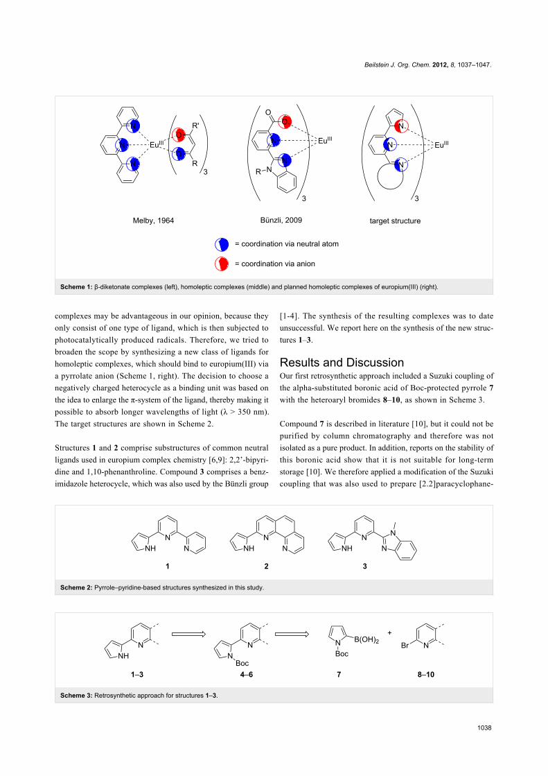
Scheme 2: Pyrrole–pyridine-based structures synthesized in this study.
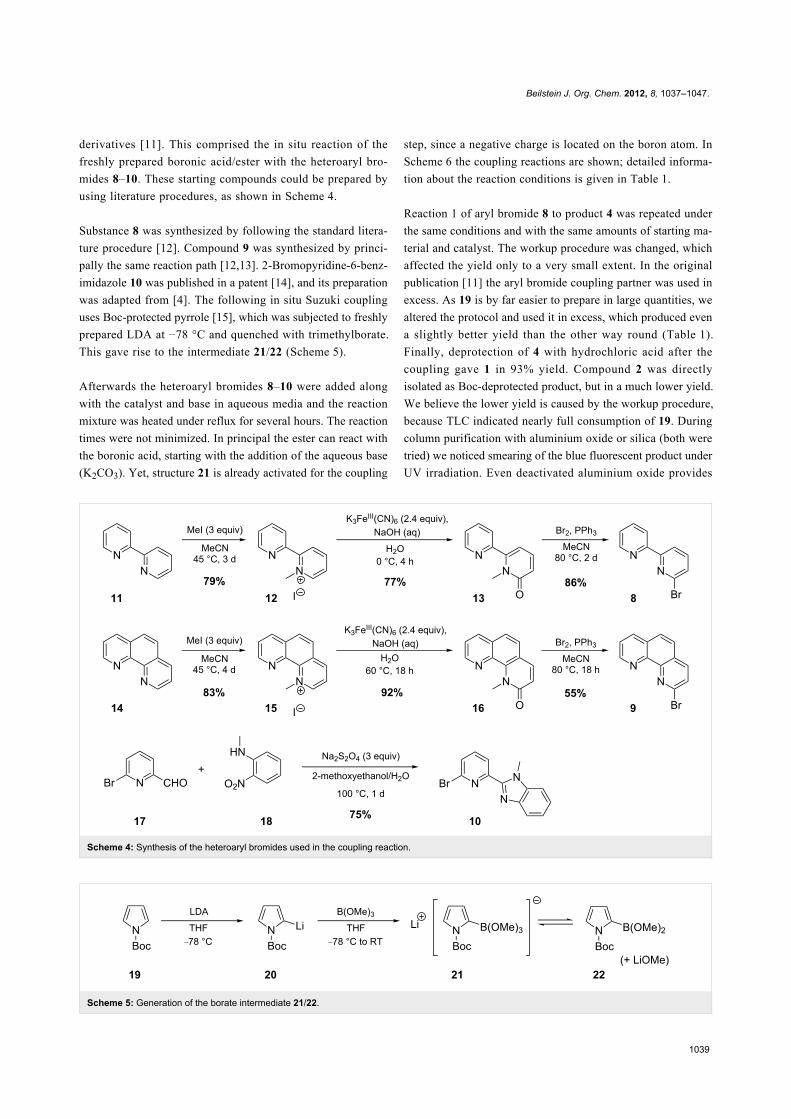
Scheme 3: Retrosynthetic approach for structures 1–3.
complexes may be advantageous in our opinion, because they
only consist of one type of ligand, which is then subjected to
photocatalytically produced radicals. Therefore, we tried to
broaden the scope by synthesizing a new class of ligands for
homoleptic complexes, which should bind to europium(III) via
a pyrrolate anion (Scheme 1, right). The decision to choose a
negatively charged heterocycle as a binding unit was based on
the idea to enlarge the π-system of the ligand, thereby making it
possible to absorb longer wavelengths of light (λ > 350 nm).
The target structures are shown in Scheme 2.
Structures 1 and 2 comprise substructures of common neutral
ligands used in europium complex chemistry [6,9]: 2,2’-bipyri-
dine and 1,10-phenanthroline. Compound 3 comprises a benz-
imidazole heterocycle, which was also used by the Bünzli group
[1-4]. The synthesis of the resulting complexes was to date
unsuccessful. We report here on the synthesis of the new struc-
tures 1–3.
Results and DiscussionOur first retrosynthetic approach included a Suzuki coupling of
the alpha-substituted boronic acid of Boc-protected pyrrole 7
with the heteroaryl bromides 8–10, as shown in Scheme 3.
Compound 7 is described in literature [10], but it could not be
purified by column chromatography and therefore was not
isolated as a pure product. In addition, reports on the stability of
this boronic acid show that it is not suitable for long-term
storage [10]. We therefore applied a modification of the Suzuki
coupling that was also used to prepare [2.2]paracyclophane-
Beilstein J. Org. Chem. 2012, 8, 1037–1047.
1039
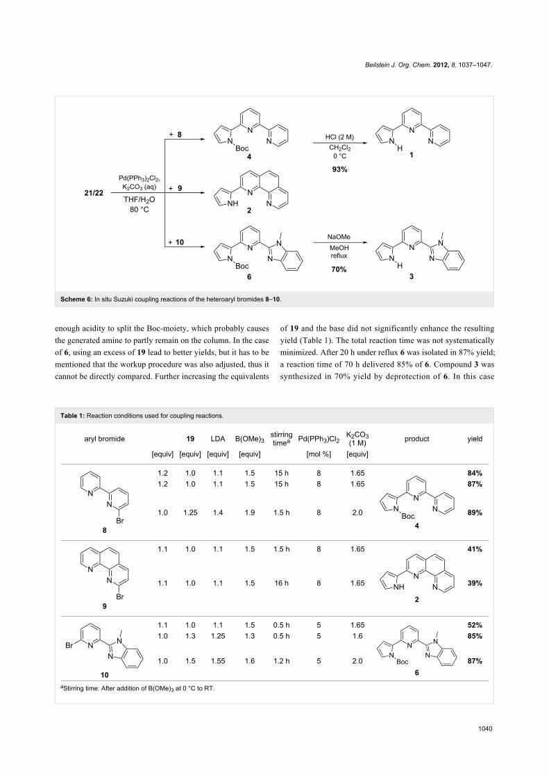
Scheme 4: Synthesis of the heteroaryl bromides used in the coupling reaction.
Scheme 5: Generation of the borate intermediate 21/22.
derivatives [11]. This comprised the in situ reaction of the
freshly prepared boronic acid/ester with the heteroaryl bro-
mides 8–10. These starting compounds could be prepared by
using literature procedures, as shown in Scheme 4.
Substance 8 was synthesized by following the standard litera-
ture procedure [12]. Compound 9 was synthesized by princi-
pally the same reaction path [12,13]. 2-Bromopyridine-6-benz-
imidazole 10 was published in a patent [14], and its preparation
was adapted from [4]. The following in situ Suzuki coupling
uses Boc-protected pyrrole [15], which was subjected to freshly
prepared LDA at −78 °C and quenched with trimethylborate.
This gave rise to the intermediate 21/22 (Scheme 5).
Afterwards the heteroaryl bromides 8–10 were added along
with the catalyst and base in aqueous media and the reaction
mixture was heated under reflux for several hours. The reaction
times were not minimized. In principal the ester can react with
the boronic acid, starting with the addition of the aqueous base
(K2CO3). Yet, structure 21 is already activated for the coupling
step, since a negative charge is located on the boron atom. In
Scheme 6 the coupling reactions are shown; detailed informa-
tion about the reaction conditions is given in Table 1.
Reaction 1 of aryl bromide 8 to product 4 was repeated under
the same conditions and with the same amounts of starting ma-
terial and catalyst. The workup procedure was changed, which
affected the yield only to a very small extent. In the original
publication [11] the aryl bromide coupling partner was used in
excess. As 19 is by far easier to prepare in large quantities, we
altered the protocol and used it in excess, which produced even
a slightly better yield than the other way round (Table 1).
Finally, deprotection of 4 with hydrochloric acid after the
coupling gave 1 in 93% yield. Compound 2 was directly
isolated as Boc-deprotected product, but in a much lower yield.
We believe the lower yield is caused by the workup procedure,
because TLC indicated nearly full consumption of 19. During
column purification with aluminium oxide or silica (both were
tried) we noticed smearing of the blue fluorescent product under
UV irradiation. Even deactivated aluminium oxide provides
Beilstein J. Org. Chem. 2012, 8, 1037–1047.
1040
Scheme 6: In situ Suzuki coupling reactions of the heteroaryl bromides 8–10.
Table 1: Reaction conditions used for coupling reactions.
aryl bromide 19 LDA B(OMe)3stirringtimea Pd(PPh3)Cl2
K2CO3(1 M) product yield
[equiv] [equiv] [equiv] [equiv] [mol %] [equiv]
8
1.2 1.0 1.1 1.5 15 h 8 1.65
4
84%1.2 1.0 1.1 1.5 15 h 8 1.65 87%
1.0 1.25 1.4 1.9 1.5 h 8 2.0 89%
9
1.1 1.0 1.1 1.5 1.5 h 8 1.65
2
41%
1.1 1.0 1.1 1.5 16 h 8 1.65 39%
10
1.1 1.0 1.1 1.5 0.5 h 5 1.65
6
52%1.0 1.3 1.25 1.3 0.5 h 5 1.6 85%
1.0 1.5 1.55 1.6 1.2 h 5 2.0 87%
aStirring time: After addition of B(OMe)3 at 0 °C to RT.
enough acidity to split the Boc-moiety, which probably causes
the generated amine to partly remain on the column. In the case
of 6, using an excess of 19 lead to better yields, but it has to be
mentioned that the workup procedure was also adjusted, thus it
cannot be directly compared. Further increasing the equivalents
of 19 and the base did not significantly enhance the resulting
yield (Table 1). The total reaction time was not systematically
minimized. After 20 h under reflux 6 was isolated in 87% yield;
a reaction time of 70 h delivered 85% of 6. Compound 3 was
synthesized in 70% yield by deprotection of 6. In this case
Beilstein J. Org. Chem. 2012, 8, 1037–1047.
1041
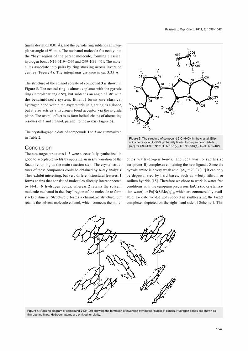
Figure 2: Packing diagram of compound 1, viewed parallel to the y-axis in the range y ≈ 1/4. Hydrogen bonds are indicated by dashed lines.Hydrogen atoms not involved in the hydrogen bonds are omitted for clarity. Hydrogen bond details (Å,°) for N17–H17…N1: H…N 0.88(2), N…N2.861(2), N–H…N 158(2), operator 1/2 + x, 1 1/2 − y, z.
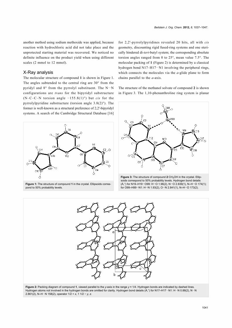
Figure 1: The structure of compound 1 in the crystal. Ellipsoids corres-pond to 50% probability levels.
another method using sodium methoxide was applied, because
reaction with hydrochloric acid did not take place and the
unprotected starting material was recovered. We noticed no
definite influence on the product yield when using different
scales (2 mmol to 12 mmol).
X-Ray analysisThe molecular structure of compound 1 is shown in Figure 1.
The angles subtended to the central ring are 30° from the
pyridyl and 8° from the pyrrolyl substituent. The N…N
configurations are trans for the bipyridyl substructure
(N–C–C–N torsion angle −155.8(1)°) but cis for the
pyrrolylpyridine substructure (torsion angle 3.8(2)°). The
former is well-known as a structural preference of 2,2'-bipyridyl
systems. A search of the Cambridge Structural Database [16]
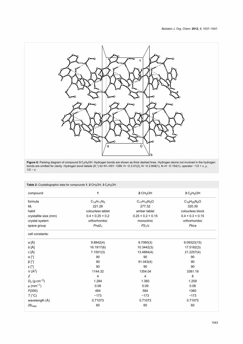
Figure 3: The structure of compound 2·CH3OH in the crystal. Ellip-soids correspond to 50% probability levels. Hydrogen bond details(Å,°) for N19–H19…O99: H…O 1.96(2), N…O 2.839(1), N–H…O 174(1);for O99–H99…N1: H…N 1.93(2), O…N 2.841(1), N–H…O 173(2).
for 2,2'-pyrrolylpyridines revealed 20 hits, all with cis
geometry, discounting rigid fused-ring systems and one steri-
cally hindered di-tert-butyl system; the corresponding absolute
torsion angles ranged from 0 to 25°, mean value 7.5°. The
molecular packing of 1 (Figure 2) is determined by a classical
hydrogen bond N17–H17…N1 involving the peripheral rings,
which connects the molecules via the a-glide plane to form
chains parallel to the a-axis.
The structure of the methanol solvate of compound 2 is shown
in Figure 3. The 1,10-phenanthroline ring system is planar
Beilstein J. Org. Chem. 2012, 8, 1037–1047.
1042
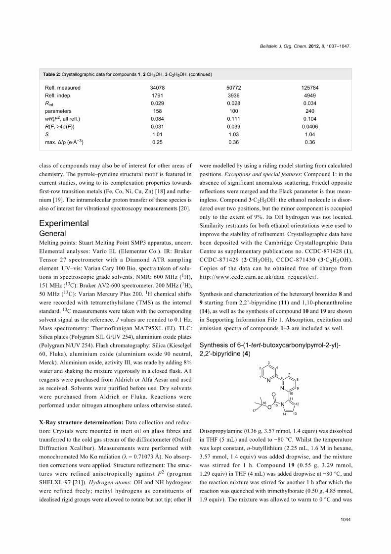
Figure 4: Packing diagram of compound 2·CH3OH showing the formation of inversion-symmetric "stacked" dimers. Hydrogen bonds are shown asthin dashed lines. Hydrogen atoms are omitted for clarity.
(mean deviation 0.01 Å), and the pyrrole ring subtends an inter-
planar angle of 9° to it. The methanol molecule fits neatly into
the “bay” region of the parent molecule, forming classical
hydrogen bonds N19–H19…O99 and O99–H99…N1. The mole-
cules associate into pairs by ring stacking across inversion
centres (Figure 4). The interplanar distance is ca. 3.35 Å.
The structure of the ethanol solvate of compound 3 is shown in
Figure 5. The central ring is almost coplanar with the pyrrole
ring (interplanar angle 9°), but subtends an angle of 38° with
the benzimidazole system. Ethanol forms one classical
hydrogen bond within the asymmetric unit, acting as a donor,
but it also acts as a hydrogen bond acceptor via the a-glide
plane. The overall effect is to form helical chains of alternating
residues of 3 and ethanol, parallel to the a-axis (Figure 6).
The crystallographic data of compounds 1 to 3 are summarized
in Table 2.
ConclusionThe new target structures 1–3 were successfully synthesized in
good to acceptable yields by applying an in situ variation of the
Suzuki coupling as the main reaction step. The crystal struc-
tures of these compounds could be obtained by X-ray analysis.
They exhibit interesting, but very different structural features: 1
forms chains that consist of molecules directly interconnected
by N–H…N hydrogen bonds, whereas 2 retains the solvent
molecule methanol in the “bay” region of the molecule to form
stacked dimers. Structure 3 forms a chain-like structure, but
retains the solvent molecule ethanol, which connects the mole-
Figure 5: The structure of compound 3·C2H5OH in the crystal. Ellip-soids correspond to 50% probability levels. Hydrogen bond details(Å,°) for O99–H99…N17: H…N 1.91(2), O…N 2.813(1), O–H…N 174(2).
cules via hydrogen bonds. The idea was to synthesize
europium(III) complexes containing the new ligands. Since the
pyrrole amine is a very weak acid (pKa = 23.0) [17] it can only
be deprotonated by hard bases, such as n-butyllithium or
sodium hydride [18]. Therefore we chose to work in water-free
conditions with the europium precursors EuCl3 (no crystalliza-
tion water) or Eu[N(SiMe3)2]3, which are commercially avail-
able. To date we did not succeed in synthesizing the target
complexes depicted on the right-hand side of Scheme 1. This
Beilstein J. Org. Chem. 2012, 8, 1037–1047.
1043
Figure 6: Packing diagram of compound 3·C2H5OH. Hydrogen bonds are shown as thick dashed lines. Hydrogen atoms not involved in the hydrogenbonds are omitted for clarity. Hydrogen bond details (Å,°) for N1–H01…O99: H…O 2.01(2), N…O 2.848(1), N–H…O 154(1), operator −1/2 + x, y,1/2 − z.
Table 2: Crystallographic data for compounds 1, 2·CH3OH, 3·C2H5OH.
compound 1 2·CH3OH 3·C2H5OH
formula C14H11N3 C17H15N3O C19H20N4OMr 221.26 277.32 320.39habit colourless tablet amber tablet colourless blockcrystallite size (mm) 0.4 × 0.25 × 0.2 0.25 × 0.2 × 0.15 0.4 × 0.3 × 0.15crystal system orthorhombic monoclinic orthorhombicspace group Pna21 P21/c Pbca
cell constants:
a [Å] 9.8842(4) 9.7060(3) 9.09323(15)b [Å] 16.1917(6) 10.3442(3) 17.5182(3)c [Å] 7.1501(3) 13.4884(4) 21.2257(4)α [°] 90 90 90β [°] 90 91.043(4) 90γ [°] 90 90 90V (Å3) 1144.32 1354.04 3381.19Z 4 4 8Dx (g·cm−3) 1.284 1.360 1.259μ (mm−1) 0.08 0.09 0.08F(000) 464 584 1360T (°C) −173 −173 −173wavelength (Å) 0.71073 0.71073 0.710732θmax 60 60 60
Beilstein J. Org. Chem. 2012, 8, 1037–1047.
1044
Table 2: Crystallographic data for compounds 1, 2·CH3OH, 3·C2H5OH. (continued)
Refl. measured 34078 50772 125784Refl. indep. 1791 3936 4949Rint 0.029 0.028 0.034parameters 158 100 240wR(F2, all refl.) 0.084 0.111 0.104R(F, >4σ(F)) 0.031 0.039 0.0406S 1.01 1.03 1.04max. Δ/ρ (e·Å−3) 0.25 0.36 0.36
class of compounds may also be of interest for other areas of
chemistry. The pyrrole–pyridine structural motif is featured in
current studies, owing to its complexation properties towards
first-row transition metals (Fe, Co, Ni, Cu, Zn) [18] and ruthe-
nium [19]. The intramolecular proton transfer of these species is
also of interest for vibrational spectroscopy measurements [20].
ExperimentalGeneralMelting points: Stuart Melting Point SMP3 apparatus, uncorr.
Elemental analyses: Vario EL (Elementar Co.). IR: Bruker
Tensor 27 spectrometer with a Diamond ATR sampling
element. UV–vis: Varian Cary 100 Bio, spectra taken of solu-
tions in spectroscopic grade solvents. NMR: 600 MHz (1H),
151 MHz (13C): Bruker AV2-600 spectrometer. 200 MHz (1H),
50 MHz (13C): Varian Mercury Plus 200. 1H chemical shifts
were recorded with tetramethylsilane (TMS) as the internal
standard. 13C measurements were taken with the corresponding
solvent signal as the reference. J values are rounded to 0.1 Hz.
Mass spectrometry: Thermofinnigan MAT95XL (EI). TLC:
Silica plates (Polygram SIL G/UV 254), aluminium oxide plates
(Polygram N/UV 254). Flash chromatography: Silica (Kieselgel
60, Fluka), aluminium oxide (aluminium oxide 90 neutral,
Merck). Aluminium oxide, activity III, was made by adding 8%
water and shaking the mixture vigorously in a closed flask. All
reagents were purchased from Aldrich or Alfa Aesar and used
as received. Solvents were purified before use. Dry solvents
were purchased from Aldrich or Fluka. Reactions were
performed under nitrogen atmosphere unless otherwise stated.
X-Ray structure determination: Data collection and reduc-
tion: Crystals were mounted in inert oil on glass fibres and
transferred to the cold gas stream of the diffractometer (Oxford
Diffraction Xcalibur). Measurements were performed with
monochromated Mo Kα radiation (λ = 0.71073 Å). No absorp-
tion corrections were applied. Structure refinement: The struc-
tures were refined anisotropically against F2 (program
SHELXL-97 [21]). Hydrogen atoms: OH and NH hydrogens
were refined freely; methyl hydrogens as constituents of
idealised rigid groups were allowed to rotate but not tip; other H
were modelled by using a riding model starting from calculated
positions. Exceptions and special features: Compound 1: in the
absence of significant anomalous scattering, Friedel opposite
reflections were merged and the Flack parameter is thus mean-
ingless. Compound 3·C2H5OH: the ethanol molecule is disor-
dered over two positions, but the minor component is occupied
only to the extent of 9%. Its OH hydrogen was not located.
Similarity restraints for both ethanol orientations were used to
improve the stability of refinement. Crystallographic data have
been deposited with the Cambridge Crystallographic Data
Centre as supplementary publications no. CCDC-871428 (1),
CCDC-871429 (2·CH3OH), CCDC-871430 (3·C2H5OH).
Copies of the data can be obtained free of charge from
http://www.ccdc.cam.ac.uk/data_request/cif.
Synthesis and characterization of the heteroaryl bromides 8 and
9 starting from 2,2’-bipyridine (11) and 1,10-phenanthroline
(14), as well as the synthesis of compound 10 and 19 are shown
in Supporting Information File 1. Absorption, excitation and
emission spectra of compounds 1–3 are included as well.
Synthesis of 6-(1-tert-butoxycarbonylpyrrol-2-yl)-2,2’-bipyridine (4)
Diisopropylamine (0.36 g, 3.57 mmol, 1.4 equiv) was dissolved
in THF (5 mL) and cooled to −80 °C. Whilst the temperature
was kept constant, n-butyllithium (2.25 mL, 1.6 M in hexane,
3.57 mmol, 1.4 equiv) was added dropwise, and the mixture
was stirred for 1 h. Compound 19 (0.55 g, 3.29 mmol,
1.29 equiv) in THF (4 mL) was added dropwise at −80 °C, and
the reaction mixture was stirred for another 1 h after which the
reaction was quenched with trimethylborate (0.50 g, 4.85 mmol,
1.9 equiv). The mixture was allowed to warm to 0 °C and was
Beilstein J. Org. Chem. 2012, 8, 1037–1047.
1045
stirred for 1.5 h. At room temperature 8 (0.6 g, 2.55 mmol) and
Pd(PPh3)2Cl2 (144 mg, 0.2 mmol, 8 mol %) were added. The
reaction mixture was heated under reflux and aq K2CO3
(5.1 mL, 1 M, 5.1 mmol, 2 equiv) was added meanwhile. It was
kept at that temperature for 2 h, then cooled to room tempera-
ture and diluted with diethyl ether. The organic phase was
washed with sat. aq NaCl and the aqueous phase was extracted
two times with diethyl ether. The combined organic extracts
were then dried over MgSO4, filtered and the solvent was
removed. The raw product was preadsorbed onto silica. Column
chromatography (SiO2, hexane/ethyl acetate 3:1, Rf = 0.4) gave
4 as a yellow resin (0.73 g, 2.27 mmol, 89%). 1H NMR
(600 MHz, CDCl3) δ 8.67 (ddd, 3JH,H= 4.8 Hz, 4JH,H = 1.8 Hz,5JH,H = 0.9 Hz, 1H, 1-H), 8.46 (pseudo-dt, 3JH,H = 8.0 Hz, J =
1.1 Hz, 1H, 4-H), 8.34 (dd, 3JH,H= 7.9 Hz, 4JH,H = 1.0 Hz, 1H,
7-H), 7.81 (dd, 3JH,H = 7.9 Hz, 3JH,H = 7.7 Hz, 1H, 8-H), 7.76
(ddd, 3JH,H = 7.9 Hz, 3JH,H = 7.6 Hz, 4JH,H = 1.8 Hz, 1H, 3-H),
7.43 (dd, 3JH,H= 7.7 Hz, 4JH,H = 1.0 Hz, 1H, 9-H), 7.41 (dd,3JH,H= 3.3 Hz, 4JH,H = 1.8 Hz, 1H, 14-H), 7.28 (ddd, 3JH,H=
7.2 Hz, 3JH,H = 4.6 Hz, 4JH,H = 1.1 Hz, 1H, 2-H), 6.48 (dd,3JH,H= 3.3 Hz, 4JH,H = 1.8 Hz, 1H, 12-H), 6.27 (dd, 3JH,H =
3.3 Hz, 3JH,H = 3.3 Hz, 1H, 13-H), 1.28 (s, 9H, 17-H) ppm; 13C
NMR (151 MHz, CDCl3) δ 156.0 (s, C-5), 155.0 (s, C-6), 152.1
(s, C-10), 149.4 (s, C-15), 149.0 (d, C-1), 136.7 (d, C-8), 136.7
(d, C-3), 134.3 (s, C-11), 123.6 (d, C-2), 123.6 (d, C-14), 123.1
(d, C-9), 121.3 (d, C-4), 118.8 (d, C-7), 115.6 (d, C-12), 110.5
(d, C-13), 83.5 (s, C-16), 27.4 (q, C-17) ppm; EIMS (70 eV)
m/z (% relative intensity): M+● 321 (5), [M − Boc]+● 222/221/
220 (14/100/9); anal. calcd for C19H19N3O2: C 71.01, H 5.96,
N 13.08; found: C 70.95, H 6.43, N 12.96.
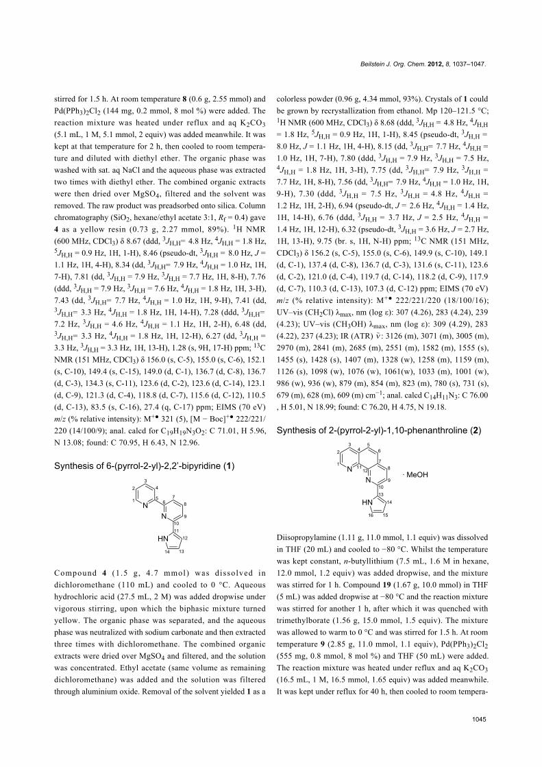
Synthesis of 6-(pyrrol-2-yl)-2,2’-bipyridine (1)
Compound 4 (1 .5 g , 4 .7 mmol) was dissolved in
dichloromethane (110 mL) and cooled to 0 °C. Aqueous
hydrochloric acid (27.5 mL, 2 M) was added dropwise under
vigorous stirring, upon which the biphasic mixture turned
yellow. The organic phase was separated, and the aqueous
phase was neutralized with sodium carbonate and then extracted
three times with dichloromethane. The combined organic
extracts were dried over MgSO4 and filtered, and the solution
was concentrated. Ethyl acetate (same volume as remaining
dichloromethane) was added and the solution was filtered
through aluminium oxide. Removal of the solvent yielded 1 as a
colorless powder (0.96 g, 4.34 mmol, 93%). Crystals of 1 could
be grown by recrystallization from ethanol. Mp 120–121.5 °C;1H NMR (600 MHz, CDCl3) δ 8.68 (ddd, 3JH,H = 4.8 Hz, 4JH,H
= 1.8 Hz, 5JH,H = 0.9 Hz, 1H, 1-H), 8.45 (pseudo-dt, 3JH,H =
8.0 Hz, J = 1.1 Hz, 1H, 4-H), 8.15 (dd, 3JH,H= 7.7 Hz, 4JH,H =
1.0 Hz, 1H, 7-H), 7.80 (ddd, 3JH,H = 7.9 Hz, 3JH,H = 7.5 Hz,4JH,H = 1.8 Hz, 1H, 3-H), 7.75 (dd, 3JH,H= 7.9 Hz, 3JH,H =
7.7 Hz, 1H, 8-H), 7.56 (dd, 3JH,H= 7.9 Hz, 4JH,H = 1.0 Hz, 1H,
9-H), 7.30 (ddd, 3JH,H = 7.5 Hz, 3JH,H = 4.8 Hz, 4JH,H =
1.2 Hz, 1H, 2-H), 6.94 (pseudo-dt, J = 2.6 Hz, 4JH,H = 1.4 Hz,
1H, 14-H), 6.76 (ddd, 3JH,H = 3.7 Hz, J = 2.5 Hz, 4JH,H =
1.4 Hz, 1H, 12-H), 6.32 (pseudo-dt, 3JH,H = 3.6 Hz, J = 2.7 Hz,
1H, 13-H), 9.75 (br. s, 1H, N-H) ppm; 13C NMR (151 MHz,
CDCl3) δ 156.2 (s, C-5), 155.0 (s, C-6), 149.9 (s, C-10), 149.1
(d, C-1), 137.4 (d, C-8), 136.7 (d, C-3), 131.6 (s, C-11), 123.6
(d, C-2), 121.0 (d, C-4), 119.7 (d, C-14), 118.2 (d, C-9), 117.9
(d, C-7), 110.3 (d, C-13), 107.3 (d, C-12) ppm; EIMS (70 eV)
m/z (% relative intensity): M+● 222/221/220 (18/100/16);
UV–vis (CH2Cl) λmax, nm (log ε): 307 (4.26), 283 (4.24), 239
(4.23); UV–vis (CH3OH) λmax, nm (log ε): 309 (4.29), 283
(4.22), 237 (4.23); IR (ATR) : 3126 (m), 3071 (m), 3005 (m),
2970 (m), 2841 (m), 2685 (m), 2551 (m), 1582 (m), 1555 (s),
1455 (s), 1428 (s), 1407 (m), 1328 (w), 1258 (m), 1159 (m),
1126 (s), 1098 (w), 1076 (w), 1061(w), 1033 (m), 1001 (w),
986 (w), 936 (w), 879 (m), 854 (m), 823 (m), 780 (s), 731 (s),
679 (m), 628 (m), 609 (m) cm−1; anal. calcd C14H11N3: C 76.00
, H 5.01, N 18.99; found: C 76.20, H 4.75, N 19.18.
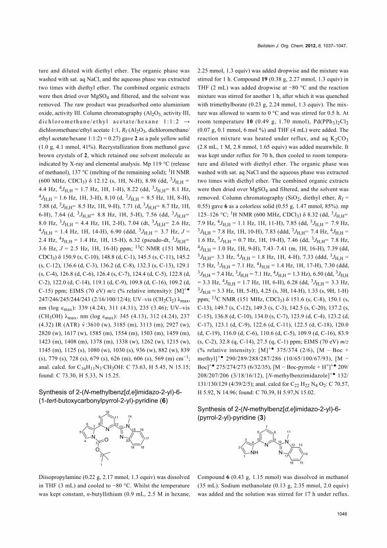
Synthesis of 2-(pyrrol-2-yl)-1,10-phenanthroline (2)
Diisopropylamine (1.11 g, 11.0 mmol, 1.1 equiv) was dissolved
in THF (20 mL) and cooled to −80 °C. Whilst the temperature
was kept constant, n-butyllithium (7.5 mL, 1.6 M in hexane,
12.0 mmol, 1.2 equiv) was added dropwise, and the mixture
was stirred for 1 h. Compound 19 (1.67 g, 10.0 mmol) in THF
(5 mL) was added dropwise at −80 °C and the reaction mixture
was stirred for another 1 h, after which it was quenched with
trimethylborate (1.56 g, 15.0 mmol, 1.5 equiv). The mixture
was allowed to warm to 0 °C and was stirred for 1.5 h. At room
temperature 9 (2.85 g, 11.0 mmol, 1.1 equiv), Pd(PPh3)2Cl2
(555 mg, 0.8 mmol, 8 mol %) and THF (50 mL) were added.
The reaction mixture was heated under reflux and aq K2CO3
(16.5 mL, 1 M, 16.5 mmol, 1.65 equiv) was added meanwhile.
It was kept under reflux for 40 h, then cooled to room tempera-
Beilstein J. Org. Chem. 2012, 8, 1037–1047.
1046
ture and diluted with diethyl ether. The organic phase was
washed with sat. aq NaCl, and the aqueous phase was extracted
two times with diethyl ether. The combined organic extracts
were then dried over MgSO4 and filtered, and the solvent was
removed. The raw product was preadsorbed onto aluminium
oxide, activity III. Column chromatography (Al2O3, activity III,
d i c h l o r o m e t h a n e / e t h y l a c e t a t e / h e x a n e 1 : 1 : 2 →
dichloromethane/ethyl acetate 1:1, Rf (Al2O3, dichloromethane/
ethyl acetate/hexane 1:1:2) = 0.27) gave 2 as a pale yellow solid
(1.0 g, 4.1 mmol, 41%). Recrystallization from methanol gave
brown crystals of 2, which retained one solvent molecule as
indicated by X-ray and elemental analysis. Mp 119 °C (release
of methanol), 137 °C (melting of the remaining solid); 1H NMR
(600 MHz, CDCl3) δ 12.12 (s, 1H, N-H), 8.98 (dd, 3JH,H =
4.4 Hz, 4JH,H = 1.7 Hz, 1H, 1-H), 8.22 (dd, 3JH,H= 8.1 Hz,4JH,H = 1.6 Hz, 1H, 3-H), 8.10 (d, 3JH,H = 8.5 Hz, 1H, 8-H),
7.88 (d, 3JH,H= 8.5 Hz, 1H, 9-H), 7.71 (d, 3JH,H= 8.7 Hz, 1H,
6-H), 7.64 (d, 3JH,H= 8.8 Hz, 1H, 5-H), 7.56 (dd, 3JH,H=
8.0 Hz, 3JH,H = 4.4 Hz, 1H, 2-H), 7.04 (dt, 3JH,H= 2.6 Hz,4JH,H = 1.4 Hz, 1H, 14-H), 6.90 (ddd, 3JH,H = 3.7 Hz, J =
2.4 Hz, 4JH,H = 1.4 Hz, 1H, 15-H), 6.32 (pseudo-dt, 3JH,H=
3.6 Hz, J = 2.5 Hz, 1H, 16-H) ppm; 13C NMR (151 MHz,
CDCl3) δ 150.9 (s, C-10), 148.8 (d, C-1), 145.5 (s, C-11), 145.2
(s, C-12), 136.6 (d, C-3), 136.2 (d, C-8), 132.3 (s, C-13), 129.1
(s, C-4), 126.8 (d, C-6), 126.4 (s, C-7), 124.4 (d, C-5), 122.8 (d,
C-2), 122.0 (d, C-14), 119.1 (d, C-9), 109.8 (d, C-16), 109.2 (d,
C-15) ppm; EIMS (70 eV) m/z (% relative intensity): [M]+●
247/246/245/244/243 (2/16/100/12/4); UV–vis (CH2Cl2) λmax,
nm (log εmax): 339 (4.24), 311 (4.31), 235 (3.46); UV–vis
(CH3OH) λmax, nm (log εmax): 345 (4.13), 312 (4.24), 237
(4.32) IR (ATR) :3610 (w), 3185 (m), 3113 (m), 2927 (w),
2820 (w), 1617 (w), 1585 (m), 1554 (m), 1503 (m), 1459 (m),
1423 (m), 1408 (m), 1378 (m), 1338 (w), 1262 (w), 1215 (w),
1145 (m), 1125 (s), 1080 (w), 1030 (s), 936 (w), 882 (w), 839
(s), 779 (s), 728 (s), 679 (s), 626 (m), 606 (s), 569 (m) cm−1;
anal. calcd. for C16H11N3·CH3OH: C 73.63, H 5.45, N 15.15;
found: C 73.30, H 5.33, N 15.25.
Synthesis of 2-(N-methylbenz[d,e]imidazo-2-yl)-6-(1-tert-butoxycarbonylpyrrol-2-yl)-pyridine (6)
Diisopropylamine (0.22 g, 2.17 mmol, 1.3 equiv) was dissolved
in THF (3 mL) and cooled to −80 °C. Whilst the temperature
was kept constant, n-butyllithium (0.9 mL, 2.5 M in hexane,
2.25 mmol, 1.3 equiv) was added dropwise and the mixture was
stirred for 1 h. Compound 19 (0.38 g, 2.27 mmol, 1.3 equiv) in
THF (2 mL) was added dropwise at −80 °C and the reaction
mixture was stirred for another 1 h, after which it was quenched
with trimethylborate (0.23 g, 2.24 mmol, 1.3 equiv). The mix-
ture was allowed to warm to 0 °C and was stirred for 0.5 h. At
room temperature 10 (0.49 g, 1.70 mmol), Pd(PPh3)2Cl2
(0.07 g, 0.1 mmol, 6 mol %) and THF (4 mL) were added. The
reaction mixture was heated under reflux, and aq K2CO3
(2.8 mL, 1 M, 2.8 mmol, 1.65 equiv) was added meanwhile. It
was kept under reflux for 70 h, then cooled to room tempera-
ture and diluted with diethyl ether. The organic phase was
washed with sat. aq NaCl and the aqueous phase was extracted
two times with diethyl ether. The combined organic extracts
were then dried over MgSO4 and filtered, and the solvent was
removed. Column chromatography (SiO2, diethyl ether, Rf =
0.55) gave 6 as a colorless solid (0.55 g, 1.47 mmol, 85%). mp
125–126 °C; 1H NMR (600 MHz, CDCl3) δ 8.32 (dd, 3JH,H=
7.9 Hz, 4JH,H = 1.1 Hz, 1H, 11-H), 7.85 (dd, 3JH,H = 7.9 Hz,3JH,H = 7.8 Hz, 1H, 10-H), 7.83 (ddd, 3JH,H= 7.4 Hz, 4JH,H =
1.6 Hz, 5JH,H = 0.7 Hz, 1H, 19-H), 7.46 (dd, 3JH,H= 7.8 Hz,4JH,H = 1.0 Hz, 1H, 9-H), 7.43–7.41 (m, 1H, 16-H), 7.39 (dd,3JH,H= 3.3 Hz, 4JH,H = 1.8 Hz, 1H, 4-H), 7.33 (ddd, 3JH,H =
7.5 Hz, 3JH,H = 7.1 Hz, 4JH,H = 1.4 Hz, 1H, 17-H), 7.30 (ddd,3JH,H = 7.4 Hz, 3JH,H = 7.1 Hz, 4JH,H = 1.3 Hz), 6.50 (dd, 3JH,H
= 3.3 Hz, 4JH,H = 1.7 Hz, 1H, 6-H), 6.28 (dd, 3JH,H = 3.3 Hz,3JH,H = 3.3 Hz, 1H, 5-H), 4.25 (s, 3H, 14-H), 1.33 (s, 9H, 1-H)
ppm; 13C NMR (151 MHz, CDCl3) δ 151.6 (s, C-8), 150.1 (s,
C-13), 149.7 (s, C-12), 149.3 (s, C-3), 142.5 (s, C-20), 137.2 (s,
C-15), 136.8 (d, C-10), 134.0 (s, C-7), 123.9 (d, C-4), 123.2 (d,
C-17), 123.1 (d, C-9), 122.6 (d, C-11), 122.5 (d, C-18), 120.0
(d, C-19), 116.0 (d, C-6), 110.6 (d, C-5), 109.9 (d, C-16), 83.9
(s, C-2), 32.8 (q, C-14), 27.5 (q, C-1) ppm; EIMS (70 eV) m/z
(% relative intensity): [M]+● 375/374 (2/6), [M – Boc +
methyl]+● 290/289/288/287/286 (10/65/100/67/93), [M −
Boc]+● 275/274/273 (6/32/35), [M − Boc-pyrrole + H+]+● 209/
208/207/206 (3/18/16/12), [N-methylbenzimidazole]+● 132/
131/130/129 (4/39/2/5); anal. calcd for C22 H22 N4 O2: C 70.57,
H 5.92, N 14.96; found: C 70.39, H 5.97,N 15.02.
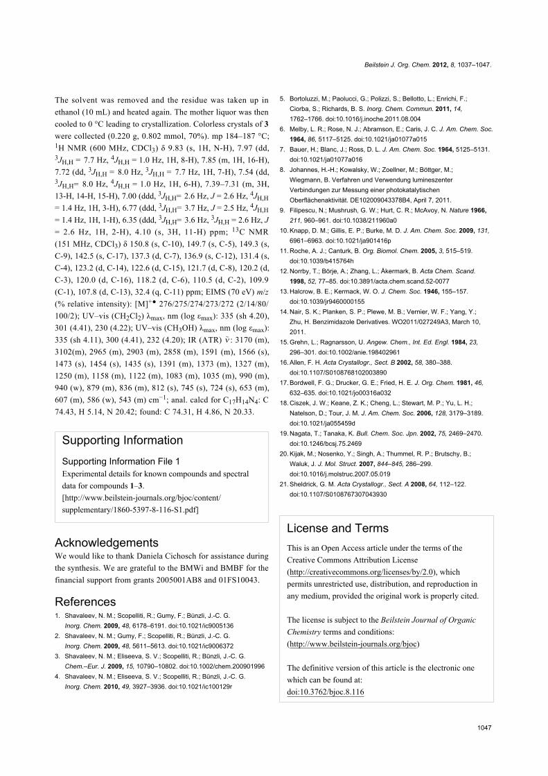
Synthesis of 2-(N-methylbenz[d,e]imidazo-2-yl)-6-(pyrrol-2-yl)-pyridine (3)
Compound 6 (0.43 g, 1.15 mmol) was dissolved in methanol
(35 mL). Sodium methanolate (0.13 g, 2.35 mmol, 2.0 equiv)
was added and the solution was stirred for 17 h under reflux.
Beilstein J. Org. Chem. 2012, 8, 1037–1047.
1047
The solvent was removed and the residue was taken up in
ethanol (10 mL) and heated again. The mother liquor was then
cooled to 0 °C leading to crystallization. Colorless crystals of 3
were collected (0.220 g, 0.802 mmol, 70%). mp 184–187 °C;1H NMR (600 MHz, CDCl3) δ 9.83 (s, 1H, N-H), 7.97 (dd,3JH,H = 7.7 Hz, 4JH,H = 1.0 Hz, 1H, 8-H), 7.85 (m, 1H, 16-H),
7.72 (dd, 3JH,H = 8.0 Hz, 3JH,H = 7.7 Hz, 1H, 7-H), 7.54 (dd,3JH,H= 8.0 Hz, 4JH,H = 1.0 Hz, 1H, 6-H), 7.39–7.31 (m, 3H,
13-H, 14-H, 15-H), 7.00 (ddd, 3JH,H= 2.6 Hz, J = 2.6 Hz, 4JH,H
= 1.4 Hz, 1H, 3-H), 6.77 (ddd, 3JH,H= 3.7 Hz, J = 2.5 Hz, 4JH,H
= 1.4 Hz, 1H, 1-H), 6.35 (ddd, 3JH,H= 3.6 Hz, 3JH,H = 2.6 Hz, J
= 2.6 Hz, 1H, 2-H), 4.10 (s, 3H, 11-H) ppm; 13C NMR
(151 MHz, CDCl3) δ 150.8 (s, C-10), 149.7 (s, C-5), 149.3 (s,
C-9), 142.5 (s, C-17), 137.3 (d, C-7), 136.9 (s, C-12), 131.4 (s,
C-4), 123.2 (d, C-14), 122.6 (d, C-15), 121.7 (d, C-8), 120.2 (d,
C-3), 120.0 (d, C-16), 118.2 (d, C-6), 110.5 (d, C-2), 109.9
(C-1), 107.8 (d, C-13), 32.4 (q, C-11) ppm; EIMS (70 eV) m/z
(% relative intensity): [M]+● 276/275/274/273/272 (2/14/80/
100/2); UV–vis (CH2Cl2) λmax, nm (log εmax): 335 (sh 4.20),
301 (4.41), 230 (4.22); UV–vis (CH3OH) λmax, nm (log εmax):
335 (sh 4.11), 300 (4.41), 232 (4.20); IR (ATR) : 3170 (m),
3102(m), 2965 (m), 2903 (m), 2858 (m), 1591 (m), 1566 (s),
1473 (s), 1454 (s), 1435 (s), 1391 (m), 1373 (m), 1327 (m),
1250 (m), 1158 (m), 1122 (m), 1083 (m), 1035 (m), 990 (m),
940 (w), 879 (m), 836 (m), 812 (s), 745 (s), 724 (s), 653 (m),
607 (m), 586 (w), 543 (m) cm−1; anal. calcd for C17H14N4: C
74.43, H 5.14, N 20.42; found: C 74.31, H 4.86, N 20.33.
Supporting InformationSupporting Information File 1Experimental details for known compounds and spectral
data for compounds 1–3.
[http://www.beilstein-journals.org/bjoc/content/
supplementary/1860-5397-8-116-S1.pdf]
AcknowledgementsWe would like to thank Daniela Cichosch for assistance during
the synthesis. We are grateful to the BMWi and BMBF for the
financial support from grants 2005001AB8 and 01FS10043.
References1. Shavaleev, N. M.; Scopelliti, R.; Gumy, F.; Bünzli, J.-C. G.
Inorg. Chem. 2009, 48, 6178–6191. doi:10.1021/ic90051362. Shavaleev, N. M.; Gumy, F.; Scopelliti, R.; Bünzli, J.-C. G.
Inorg. Chem. 2009, 48, 5611–5613. doi:10.1021/ic90063723. Shavaleev, N. M.; Eliseeva, S. V.; Scopelliti, R.; Bünzli, J.-C. G.
Chem.–Eur. J. 2009, 15, 10790–10802. doi:10.1002/chem.2009019964. Shavaleev, N. M.; Eliseeva, S. V.; Scopelliti, R.; Bünzli, J.-C. G.
Inorg. Chem. 2010, 49, 3927–3936. doi:10.1021/ic100129r
5. Bortoluzzi, M.; Paolucci, G.; Polizzi, S.; Bellotto, L.; Enrichi, F.;Ciorba, S.; Richards, B. S. Inorg. Chem. Commun. 2011, 14,1762–1766. doi:10.1016/j.inoche.2011.08.004
6. Melby, L. R.; Rose, N. J.; Abramson, E.; Caris, J. C. J. Am. Chem. Soc.1964, 86, 5117–5125. doi:10.1021/ja01077a015
7. Bauer, H.; Blanc, J.; Ross, D. L. J. Am. Chem. Soc. 1964, 5125–5131.doi:10.1021/ja01077a016
8. Johannes, H.-H.; Kowalsky, W.; Zoellner, M.; Böttger, M.;Wiegmann, B. Verfahren und Verwendung lumineszenterVerbindungen zur Messung einer photokatalytischenOberflächenaktivität. DE102009043378B4, April 7, 2011.
9. Filipescu, N.; Mushrush, G. W.; Hurt, C. R.; McAvoy, N. Nature 1966,211, 960–961. doi:10.1038/211960a0
10. Knapp, D. M.; Gillis, E. P.; Burke, M. D. J. Am. Chem. Soc. 2009, 131,6961–6963. doi:10.1021/ja901416p
11. Roche, A. J.; Canturk, B. Org. Biomol. Chem. 2005, 3, 515–519.doi:10.1039/b415764h
12. Norrby, T.; Börje, A.; Zhang, L.; Åkermark, B. Acta Chem. Scand.1998, 52, 77–85. doi:10.3891/acta.chem.scand.52-0077
13. Halcrow, B. E.; Kermack, W. O. J. Chem. Soc. 1946, 155–157.doi:10.1039/jr9460000155
14. Nair, S. K.; Planken, S. P.; Plewe, M. B.; Vernier, W. F.; Yang, Y.;Zhu, H. Benzimidazole Derivatives. WO2011/027249A3, March 10,2011.
15. Grehn, L.; Ragnarsson, U. Angew. Chem., Int. Ed. Engl. 1984, 23,296–301. doi:10.1002/anie.198402961
16. Allen, F. H. Acta Crystallogr., Sect. B 2002, 58, 380–388.doi:10.1107/S0108768102003890
17. Bordwell, F. G.; Drucker, G. E.; Fried, H. E. J. Org. Chem. 1981, 46,632–635. doi:10.1021/jo00316a032
18. Ciszek, J. W.; Keane, Z. K.; Cheng, L.; Stewart, M. P.; Yu, L. H.;Natelson, D.; Tour, J. M. J. Am. Chem. Soc. 2006, 128, 3179–3189.doi:10.1021/ja055459d
19. Nagata, T.; Tanaka, K. Bull. Chem. Soc. Jpn. 2002, 75, 2469–2470.doi:10.1246/bcsj.75.2469
20. Kijak, M.; Nosenko, Y.; Singh, A.; Thummel, R. P.; Brutschy, B.;Waluk, J. J. Mol. Struct. 2007, 844–845, 286–299.doi:10.1016/j.molstruc.2007.05.019
21. Sheldrick, G. M. Acta Crystallogr., Sect. A 2008, 64, 112–122.doi:10.1107/S0108767307043930
License and TermsThis is an Open Access article under the terms of the
Creative Commons Attribution License
(http://creativecommons.org/licenses/by/2.0), which
permits unrestricted use, distribution, and reproduction in
any medium, provided the original work is properly cited.
The license is subject to the Beilstein Journal of Organic
Chemistry terms and conditions:
(http://www.beilstein-journals.org/bjoc)
The definitive version of this article is the electronic one
which can be found at:
doi:10.3762/bjoc.8.116
Top Related