


Languages
Pages
Legal


Sports Medicine Problems in Primary Care
Eric J. Anish, MD, FACP, FACSM
Associate Professor of Medicine and Orthopaedic Surgery
University of Pittsburgh School of Medicine
Head Team Physician, Duquesne University1

Speaker Disclosures
None
2

Introduction
• Sports and exercise-related musculoskeletal injuries are common in primary care
• Patients with such injuries commonly present first to their PCP or are referred from urgent care or the ER
• Sports medicine is an integral component of primary care practice
3

Educational Objectives
• Recognize and treat 3 common sports medicine “syndromes” that are frequently seen in primary care:
➢Shoulder Impingement Syndrome
➢Femoroacetabular Impingement Syndrome
➢Patellofemoral Pain Syndrome
4
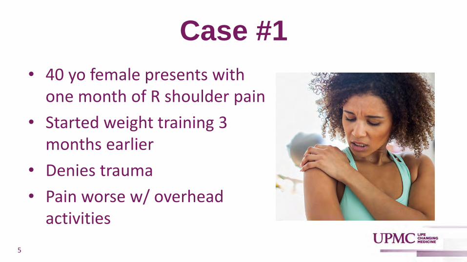
Case #1
• 40 yo female presents with one month of R shoulder pain
• Started weight training 3 months earlier
• Denies trauma
• Pain worse w/ overhead activities
5
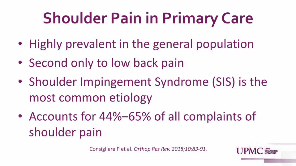
Shoulder Pain in Primary Care
• Highly prevalent in the general population
• Second only to low back pain
• Shoulder Impingement Syndrome (SIS) is the most common etiology
• Accounts for 44%–65% of all complaints of shoulder pain
Consigliere P et al. Orthop Res Rev. 2018;10:83-91.

What is Shoulder Impingement Syndrome?
• NOT injury to one specific structure!
• Combination of shoulder symptoms, exam findings, and radiological signs attributable to compression of structures around the glenohumeral joint that occurs with shoulder elevation
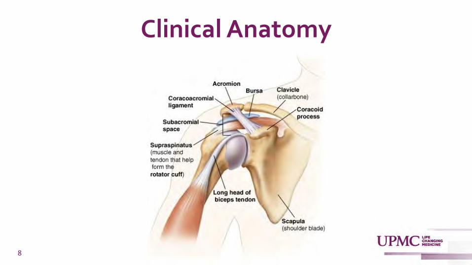
Clinical Anatomy
8

Shoulder Impingement Syndrome• Stage 1:
➢Edema and hemorrhage of the bursa and cuff
• Stage 2:
➢Cuff fibrosis and tendonitis
➢Thickened, fibrotic bursa
• Stage 3:
➢Partial or complete cuff tears
9Neer CS 2nd. Clin Orthop Relat Res. 1983;173:70-77.

Etiology – Primary Impingement
• Structural changes that mechanically narrow the subacromial space
➢Bony narrowing on the cranial side (outlet impingement)
➢Increase in the volume of the subacromial soft tissues on the caudal side (non-outlet impingement)
10
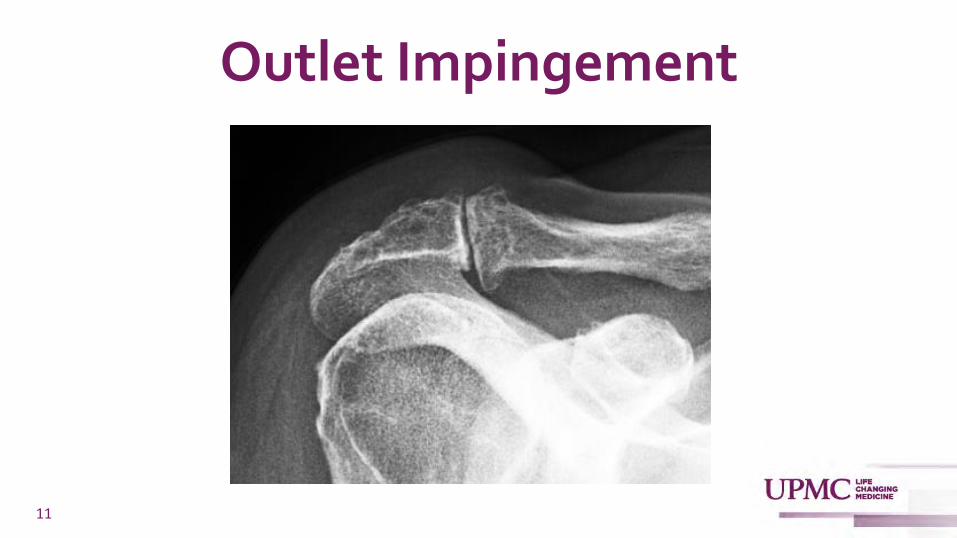
Outlet Impingement
11

Non-Outlet Impingement
12

Etiology – Secondary Impingement
• Results from a functional disturbance of centering the humeral head
13
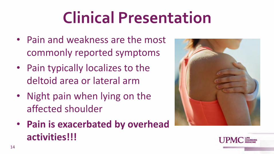
Clinical Presentation• Pain and weakness are the most
commonly reported symptoms
• Pain typically localizes to the deltoid area or lateral arm
• Night pain when lying on the affected shoulder
• Pain is exacerbated by overhead activities!!!
14
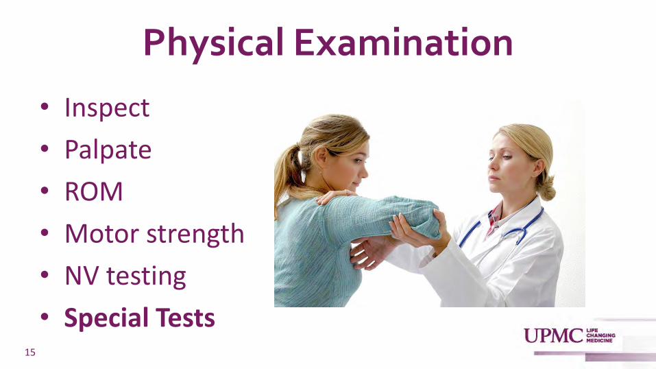
Physical Examination
• Inspect
• Palpate
• ROM
• Motor strength
• NV testing
• Special Tests15
Physical Examination
• Multiple prospective, observational studies report that physical exam techniques for the shoulder are sensitive for the presence of SIS
• Caveat: Can’t reliably distinguish among specific causes of pain and dysfunction
16
Hanchard NC et al. Cochrane Database Syst Rev. 2013;2013:CD007427. Lange T et al. British Journal of Sports Medicine 2017;51:511-518.

Hawkins Impingement Test
17
Caliş M et al. Ann Rheum Dis. 2000;59:44-47.MacDonald PB et al. J Shoulder Elbow Surg. 2000;9:299-301.
Sensitivity 88-92% / Specificity 25-42%

Neer Impingement Test
18
Sensitivity 83-89% / Specificity 30-50%
Caliş M et al. Ann Rheum Dis. 2000;59:44-47.MacDonald PB et al. J Shoulder Elbow Surg. 2000;9:299-301.
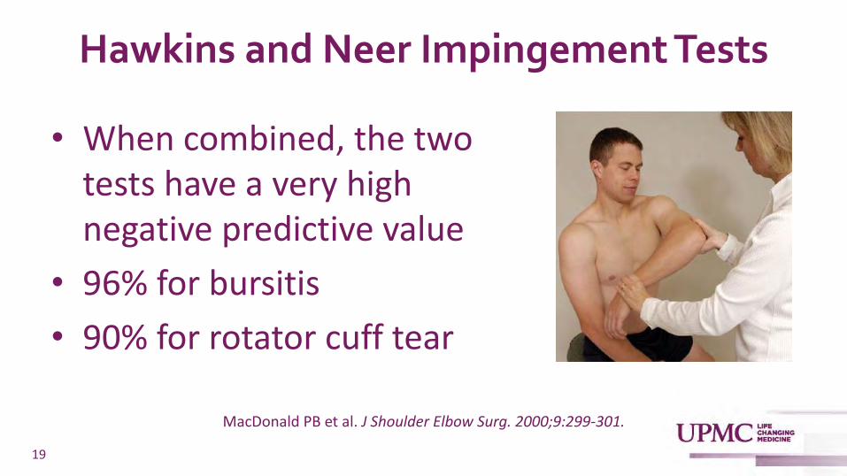
Hawkins and Neer Impingement Tests
• When combined, the two tests have a very high negative predictive value
• 96% for bursitis
• 90% for rotator cuff tear
19
MacDonald PB et al. J Shoulder Elbow Surg. 2000;9:299-301.

Diagnostic Imaging
• Radiographs are unnecessary for the initial evaluation of shoulder impingement
• Appropriate if no response to conservative treatment
20

Radiographs
21
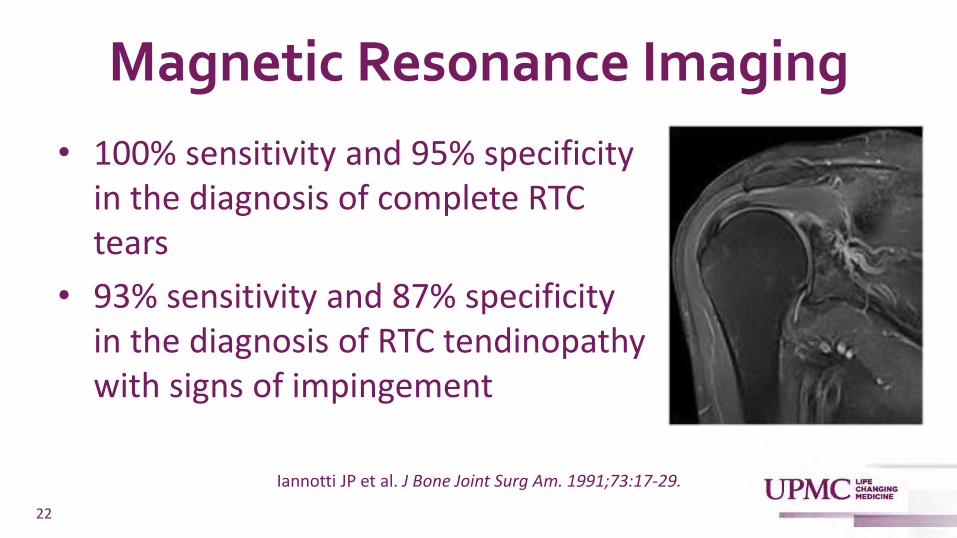
Magnetic Resonance Imaging
• 100% sensitivity and 95% specificity in the diagnosis of complete RTC tears
• 93% sensitivity and 87% specificity in the diagnosis of RTC tendinopathy with signs of impingement
22
Iannotti JP et al. J Bone Joint Surg Am. 1991;73:17-29.
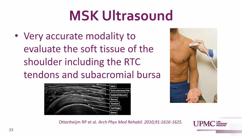
MSK Ultrasound
• Very accurate modality to evaluate the soft tissue of the shoulder including the RTC tendons and subacromial bursa
23
Ottenheijm RP et al. Arch Phys Med Rehabil. 2010;91:1616-1625.
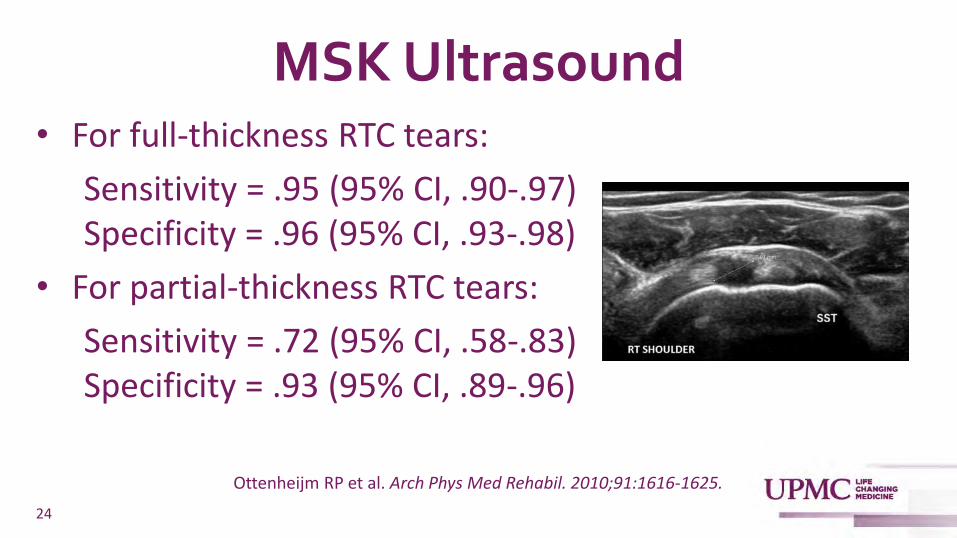
MSK Ultrasound• For full-thickness RTC tears:
Sensitivity = .95 (95% CI, .90-.97) Specificity = .96 (95% CI, .93-.98)
• For partial-thickness RTC tears:
Sensitivity = .72 (95% CI, .58-.83) Specificity = .93 (95% CI, .89-.96)
24
Ottenheijm RP et al. Arch Phys Med Rehabil. 2010;91:1616-1625.

MSK Ultrasound
• For Subacromial bursitis: Sensitivity = .79 (95% CI, .56-.94) Specificity = .95 (95% CI, .70-1.00)
• For RTC tendinopathy: Sensitivity = .80 (95% CI, .41-1.00) Specificity = .94 (95% CI, .75-1.00)
25
Ottenheijm RP et al. Arch Phys Med Rehabil. 2010;91:1616-1625.

Management
• Overall quality of evidence supporting various treatment modalities is relatively weak
• Most significant intervention -rehabilitation exercises
• Avoidance of aggravating activities
26
Steuri R et al. Br J Sports Med. 2017;51:1340-1347.
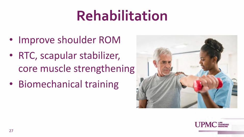
Rehabilitation
• Improve shoulder ROM
• RTC, scapular stabilizer, core muscle strengthening
• Biomechanical training
27

Rehabilitation
28

Rehabilitation• Specific exercise program was assoc. w/ significantly
more patients in the formal PT group reporting successful outcome
• Defined as large improvement or recovered in the patients’ global assessment of change
• 69% (35/51) v 24% (11/46); OR 7.6, 3.1 to 18.9; P<0.001
29Holmgren T et al. BMJ. 2012;344:e787.

Rehabilitation
• Significantly lower proportion of patients in the specific exercise group subsequently chose to undergo surgery: 20% (10/51) v 63% (29/46); OR 7.7, 3.1 to 19.4; P<0.001)
30
Holmgren T et al. BMJ. 2012;344:e787.

Other Treatments
• Electrical stimulation
• Phonopheresis / iontopheresis
• Therapeutic ultrasound
• Low-level laser therapy
• Acupuncture
• ESWL
31
Steuri R et al. Br J Sports Med. 2017;51:1340-1347.

Corticosteroid Injection• Small, randomized trials have
demonstrated short-term benefit from subacromial glucocorticoid injection
• May allow pts to engage in PT more effectively
• Should not be used as sole therapy
32
Gaujoux-Viala C et al. Ann Rheum Dis. 2009;68:1843-1849.

Conservative Therapy for SIS
• Conservative management of SIS resolves the problem in 70%–90% of patients
• In the absence of major structural damage, conservative multi-modal treatment for 3–6 months is the initial therapy of choice
33
Consigliere P et al. Orthop Res Rev. 2018;10:83-91.

Surgical Intervention
34
Arthroscopic subacromial decompression and bursectomy
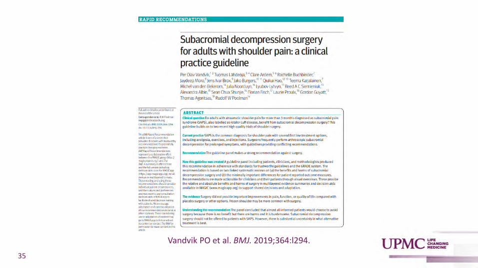
35
Vandvik PO et al. BMJ. 2019;364:l294.

Surgical Intervention
• 2 systematic reviews and a meta-analysis of data from 7 RCTs that involved 1000 participants with SIS (mean age, 49; median symptom duration, 2 years)
• Surgery provided no improvements in pain, function, or QOL compared w/ placebo surgery or other options
36
Vandvik PO et al. BMJ. 2019;364:l294.

Subacromial Decompression• 2 trials that involved 331 patients, researchers
compared decompressive surgery with placebo surgery
• Decompression provided no benefit for pain, function, health-related QOL, patient-perceived effect, or return to work at any time point from 6 months to 5 years
37
Vandvik PO et al. BMJ. 2019;364:l294.

Subacromial Decompression• 5 non-blinded trials, researchers compared exercise
therapy alone with decompressive surgery plus postoperative exercise therapy
• Compared with exercise therapy alone, surgery provided no benefit for pain, function, health-related QOL, patient-perceived effect, or return to work
38
Vandvik PO et al. BMJ. 2019;364:l294.

Subacromial Decompression
• Extrapolating from data in the 2 placebo-controlled trials, decompressive surgery would be associated with 12 additional frozen shoulders per 1000 patients
39
Vandvik PO et al. BMJ. 2019;364:l294.

40

41

Controversy Regarding Subacromial Decompression Studies
• Heterogeneous patient population
• Short-term follow-up
• Small sample size
• Group crossovers
• Surgeon experience
• Failure to standardize the surgical technique
42

Surgical Intervention
• Arthroscopic subacromial decompression is overused
• Need to be cautious when referring patients for this procedure
• Current evidence is not strong enough to say it should never be performed
43
Case #1• Modified her activity
• Performed 6 weeks of PT w/ some improvement
• X-rays showed mild AC joint arthritis
• Subacromial corticosteroid injection was performed
• Continued PT for an additional 4 weeks
• Completely pain-free and back to performing all overhead activities
44
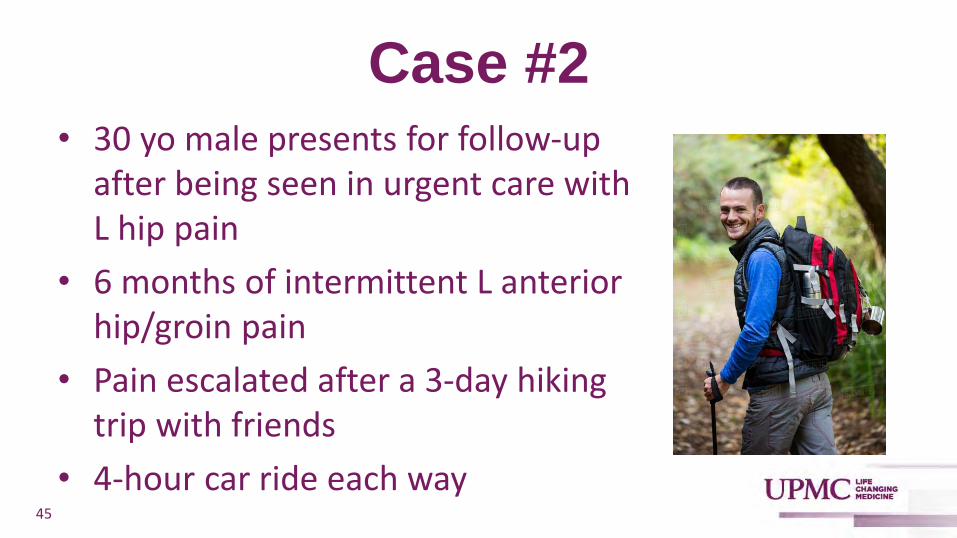
Case #2• 30 yo male presents for follow-up
after being seen in urgent care with L hip pain
• 6 months of intermittent L anterior hip/groin pain
• Pain escalated after a 3-day hiking trip with friends
• 4-hour car ride each way45

Case #2• X-rays performed at urgent care
indicate that pt has a left hip “Cam Deformity”
• Radiologist comment: “Correlate clinically for femoroacetabular impingement”
46

Femoroacetabular Impingement (FAI)
• Describes pathological contact between the femoral head-neck junction and the acetabular rim during a functional range of hip movement

Femoroacetabular Impingement (FAI)
• Abutment of femoral head-neck junction against the acetabular rim occurs as a result of:
1. Aspherical femoral head (cam morphology)
2. Over-coverage of the femoral head (pincer morphology)
3. Combination of the two (mixed morphology)

Types of FAI Morphology
49
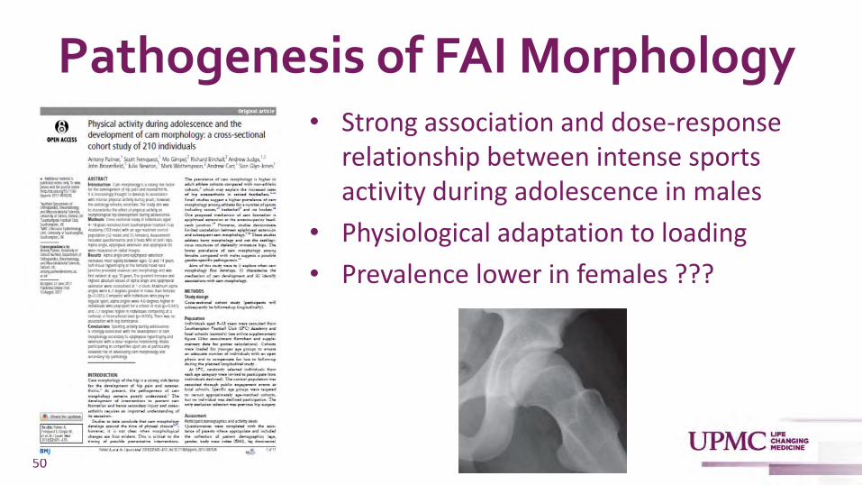
Pathogenesis of FAI Morphology• Strong association and dose-response
relationship between intense sports activity during adolescence in males
• Physiological adaptation to loading
• Prevalence lower in females ???
50

Pathogenesis of FAI Morphology
• Genetic contribution of FAI has been evaluated
• Pollard et al. observed a relative risk ≥ 2 for having a cam or pincer deformity in siblings of patients with cam or pincer-type FAI
51Pollard TC et al. J Bone Joint Surg Br. 2010;92:209-216

FAI vs. FAIS
• Only a small proportion of individuals with FAI morphology develop symptoms
• Femoroacetabular Impingement Syndrome (FAIS)* is defined as a triad of symptoms, clinical signs, and imaging findings
*The Warwick Agreement on femoroacetabular impingement syndrome
(FAI syndrome): an international consensus statement.
52
Griffin DR et al. Br J Sports Med. 2016;50:1169-1176.

Clinical Presentation of FAIS
• Primary sx is movement- or position-related groin pain
• Typically insidious in onset
• Often ignored or dismissed as a “groin strain”
• Most notable after sitting w/ hip flexed to 90 degrees for a prolonged period of time
• Decreases when rising to a standing position
• As sx progress, physical activity is affected53

54
Clinical Presentation of FAIS
• Clicking
• Catching
• Locking
• Stiffness
• Restricted motion
• Giving wayThe “C” Sign
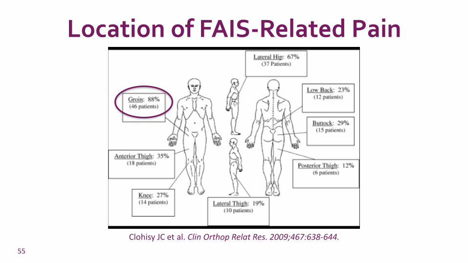
Location of FAIS-Related Pain
55
Clohisy JC et al. Clin Orthop Relat Res. 2009;467:638-644.

Physical Examination of FAIS
• Gait pattern
• Soft tissue palpation
• Hip ROM
• LE muscle strength
• Special Tests
56

Special Tests
• There is no single diagnostic sign for FAIS
• Most commonly used is the FADIR or anterior impingement test
57

FADIR Test
58Reiman MP et al. Br J Sports Med 2015;49:811
Sens: 94-99% / Spec: 5-9%

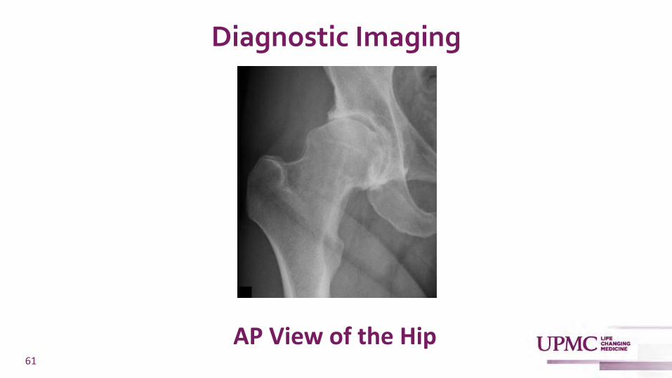
Diagnostic Imaging
• AP Pelvis radiograph (weight-bearing)
• AP view of the symptomatic hip
• Lateral view of the symptomatic hip:
➢ Frog-leg Lateral View – CAM deformity at the anterior aspect of the femoral head-neck junction
➢Modified Dunn Lateral View – CAM deformity at the anterolateral region of the femoral head-neck junction
59

Diagnostic Imaging
60
Diagnostic Imaging
61
AP View of the Hip

Diagnostic ImagingFrog-Leg Lateral
• Knee joint is flexed 30-40° in a supine position, while the hip is externally rotated by 45°
62
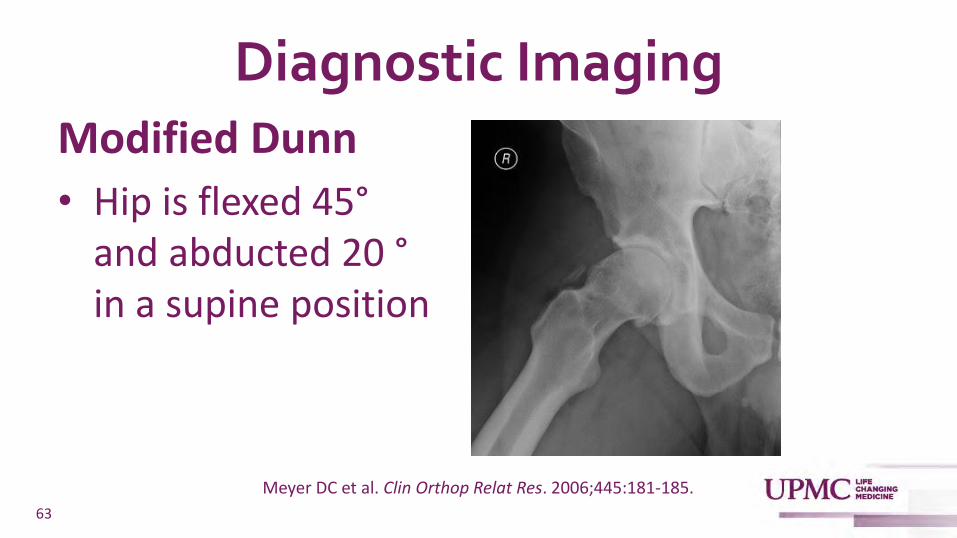
Diagnostic ImagingModified Dunn
• Hip is flexed 45°and abducted 20 °in a supine position
63
Meyer DC et al. Clin Orthop Relat Res. 2006;445:181-185.

Advanced Imaging?
• Not necessary in cases where hx and exam are consistent w/ FAIS and plain x-rays reveal cam and/or pincer morphology
• Reasonable if clinical suspicion persists and plain x-rays are not definitive
• MRI is the preferred study
64
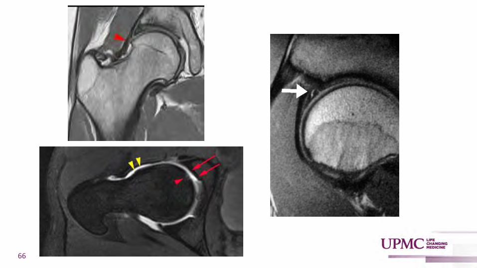
Role of Hip MRI
• Allows for evaluation of hip morphology in 3 dimensions
• Delivers high-resolution images of soft tissue structures
• May provide additional information about associated pathology such as tendinopathy, labral pathology, and chondrosis
65
66
67
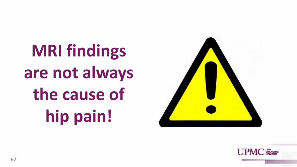
MRI findings are not always
the cause of hip pain!
68
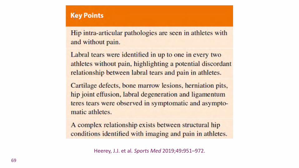
69
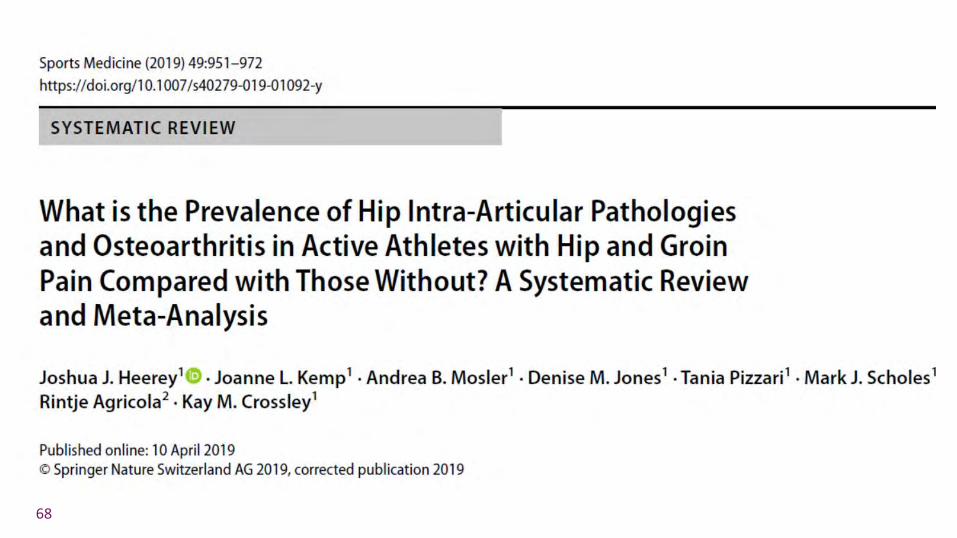
Heerey, J.J. et al. Sports Med 2019;49:951–972.

70
Frank JM et al. Arthroscopy. 2015;31:1199-1204.

Case #2• X-ray performed at urgent care
indicate that pt has a left hip “CAM Deformity”
• Radiologist comment: “correlate clinically for femoroacetabular impingement”
71

Case #2• X-ray performed at urgent care
indicate that pt has a left hip “CAM Deformity”
• Radiologist comment: “correlate clinically for femoroacetabular impingement syndrome”
72
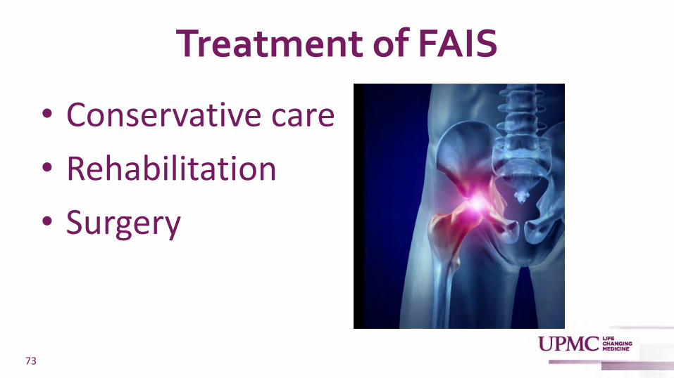
Treatment of FAIS
• Conservative care
• Rehabilitation
• Surgery
73

Conservative Care
• Education
• Lifestyle / Activity Modification
• Analgesics
• Watchful waiting
74
Griffin DR et al. Br J of Sports Med 2016;50:1169-1176.

Rehabilitation
• Muscle imbalances and biomechanical factors contribute to pain in FAIS
• Provides the foundational basis for physiotherapy as a treatment
75
Hoit G. et al. Am J Sports Med 2020;48:2042–2050.

Rehabilitation
• Hip stability
• Neuromuscular control
• Strength
• ROM
• Movement patterns
76

Rehabilitation
• Systematic review of 5 randomized trials involving 128 patients
• Supervised physiotherapy programs focusing on active strengthening and core strengthening are more effective than unsupervised, passive, and non-core focused programs
77Hoit G. et al. Am J of Sports Med 2020;48:2042–2050.
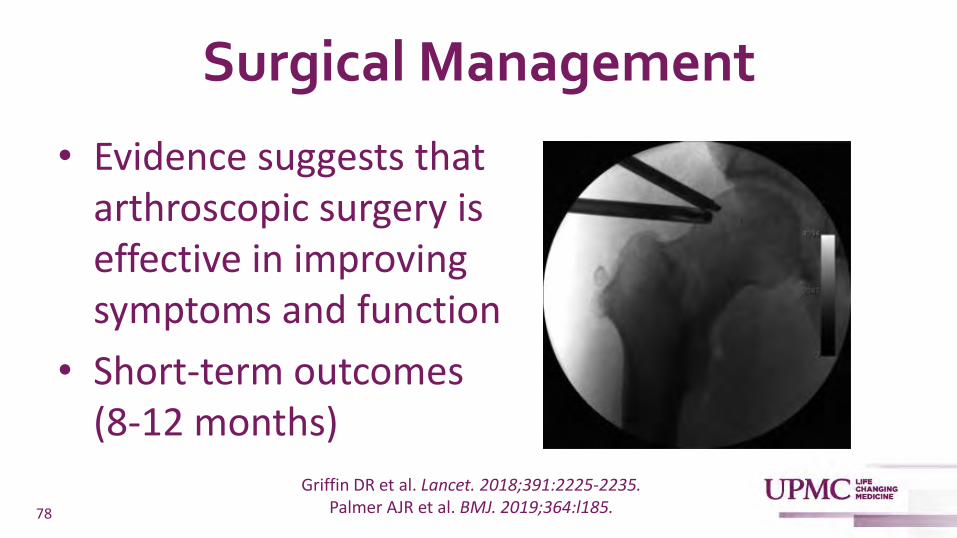
Surgical Management
• Evidence suggests that arthroscopic surgery is effective in improving symptoms and function
• Short-term outcomes (8-12 months)
78
Griffin DR et al. Lancet. 2018;391:2225-2235. Palmer AJR et al. BMJ. 2019;364:l185.

Surgical Management
• Consists of procedures to excise the impinging bone and address injury to the adjacent labrum and/or cartilage
• Arthroscopic osteotomy, chondroplasty, labral debridement or repair
79

Indications for FAIS Surgery???
• Literature has shown there is inconsistency regarding surgical indications
• Failure to respond to non-operative management for 3-6 months
80
Griffin DR et al. Lancet. 2018;391:2225-2235. Maupin JJ et al. Orthop Res Rev. 2019;11:99-108.

Contraindications for FAIS Surgery
• Absolute: joint space narrowing <2mm
• Relative:
➢Hip OA
➢Advanced age
➢Ligamentous laxity
➢Morbid obesity
81Maupin JJ et al. Orthop Res Rev. 2019;11:99-108

Case #2
• Prescribed oral NSAIDS
• Counseled about activity modification
• PT referral
• No improvement after 12 weeks
• MRI revealed FAI morphology with an associated labral tear
• Referred for surgical intervention82

Case #3
• 35 yo female presents with 1 month of worsening anterior knee pain
• Denies any trauma
• Increased her running mileage a few weeks before the pain began
83

Case #3
• Pain going up and down stairs and with prolonged sitting
• No improvement with rest, ice, NSAIDS
• Training partner thinks she has “Runner’s Knee”
• Is she right?
84

Patellofemoral Pain Syndrome (PFPS)
• Runner’s knee
• Anterior knee pain syndrome
• Chondromalacia patellae
85

Patellofemoral Pain Syndrome (PFPS)
• One of the most common causes of knee pain
• Annual prevalence for patellofemoral pain in the general population was reported as 22.7%, and adolescents as 28.9%
86
Smith BE et al. PLoS One. 2018;13:e0190892.

Patellofemoral Pain Syndrome (PFPS)
• 2016 consensus statement defines PFPS as pain occurring around or behind the patella ANDaggravated by at least one activity that loads the patella during weight-bearing on a flexed knee
87
Crossley KM et al. Br J Sports Med. 2016;50:839-843.

Patellofemoral Joint Anatomy
88

Patellar Maltracking
89
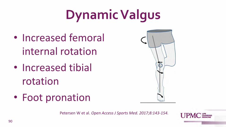
Dynamic Valgus
• Increased femoral internal rotation
• Increased tibial rotation
• Foot pronation
90
Petersen W et al. Open Access J Sports Med. 2017;8:143-154.

Excessive Patellofemoral Joint Loading
91

92

Pathogenesis of PFPS
93
Petersen W et al. Open Access J Sports Med. 2017;8:143-154.

Diagnosis of PFPS - History
• Cardinal feature is pain around or behind the patella that intensifies when the knee is flexed during weight-bearing activities
• Pain or stiffness exacerbated by prolonged sitting with the knee flexed
94

Diagnosis of PFPS - Exam
• NO effusion, erythema, warmth!!!
• Medial or lateral patellar facet tenderness Sens: 92% / Spec: 65%
• Pain during squatting Sens: 91% / 50%
• Patellar tilt test Sens: 43% / Spec: 92%
95Gaitonde DY et al. Am Fam Physician. 2019;99:88-94.
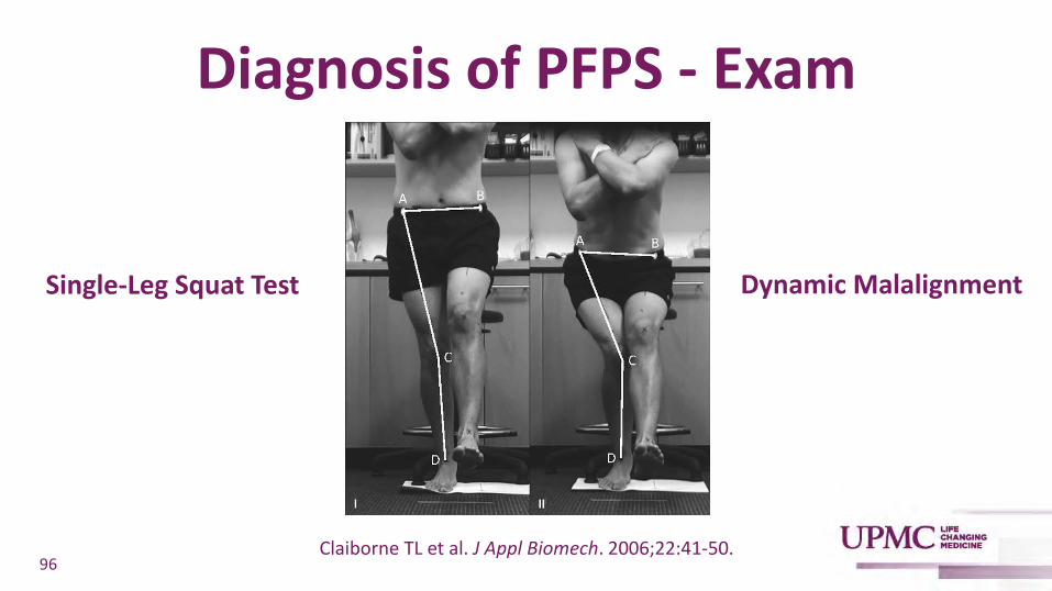
Diagnosis of PFPS - Exam
Single-Leg Squat Test
96Claiborne TL et al. J Appl Biomech. 2006;22:41-50.
Dynamic Malalignment

Imaging
• Unnecessary for initial management in the absence of a history of trauma, overt instability, effusion, prior surgery, or pain at rest with knee extended
• X-rays are appropriate if no improvement after 1-2 months of conservative treatment
97
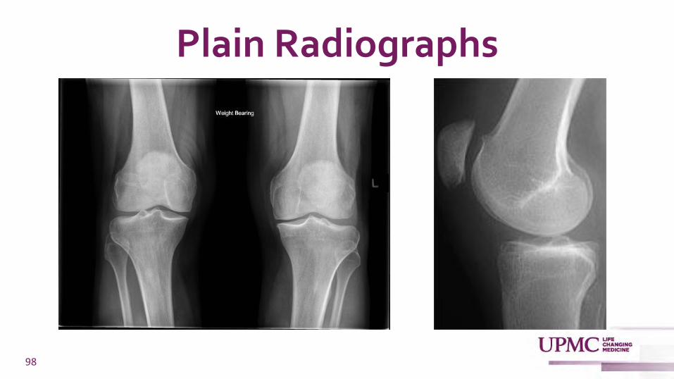
Plain Radiographs
98
Plain Radiographs
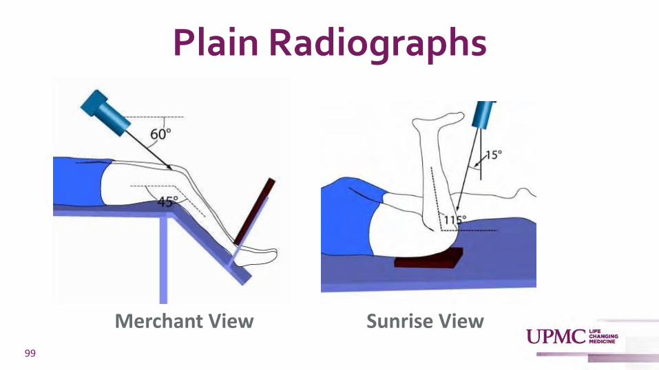
99
Merchant View Sunrise View

Plain Radiographs
100

Advanced Imaging
Role of MRI
• None in the initial evaluation of PFPS
• Reasonable if no response to ongoing conservative treatment
• Can detect articular cartilage injuries, OCDs, and injuries to the patellar retinaculum, patellofemoral ligament, and patellar / quad tendons
101

Treatment of PFPS
• Relative rest / activity modification
• Only limited evidence for the effectiveness of NSAIDs for short term pain reduction
• High-quality studies showing pain reduction with longer courses of NSAIDS are lacking
102
Heintjes E et al. Cochrane Database Syst Rev. 2004;(3):CD003470.

Treatment of PFPS
• Physical therapy is the cornerstone of treatment!!!
• Consistent evidence that exercise therapy for PFPS can result in clinically important reduction in pain, improvement in functional ability, and enhance long‐term recovery
103
van der Heijden RA et al. Cochrane Database of Systematic Reviews 2015, Issue 1. Art. No.: CD010387.
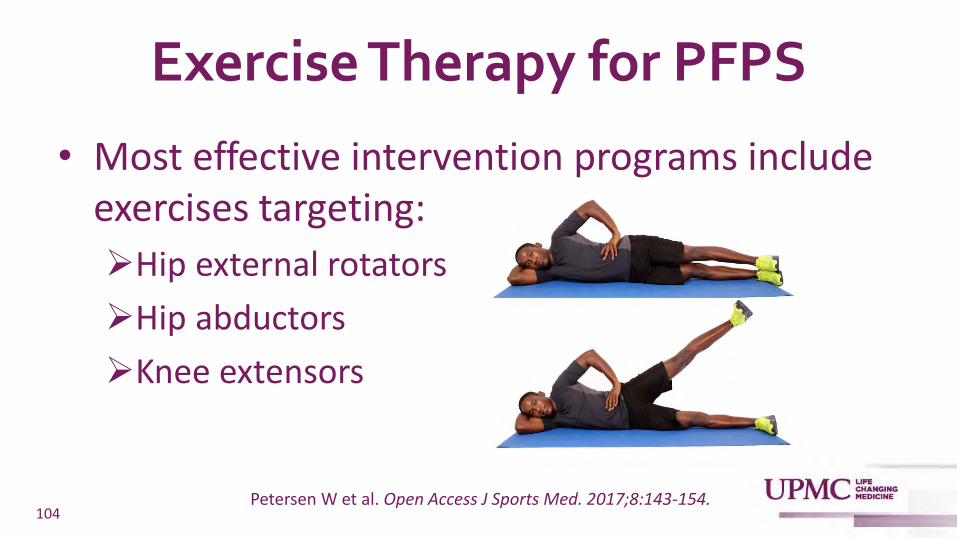
Exercise Therapy for PFPS
• Most effective intervention programs include exercises targeting:
➢Hip external rotators
➢Hip abductors
➢Knee extensors
104Petersen W et al. Open Access J Sports Med. 2017;8:143-154.

Exercise Therapy for PFPS
• Trunk stabilization
• Core strengthening
• Active stretching
105

Adjunctive Therapy for PFPS
• Foot orthoses
• Patellar taping
• Patellar bracing
106
May be of benefit when used in combination with physical therapy
Collins NJ et al. Br J Sports Med. 2018;52:1170-1178.Logan CA et al. Sports Health. 2017;9:456-461.
Willy RW et al. J Orthop Sports Phys Ther. 2019;49:CPG1-CPG95.

Surgical Intervention
• Need to have failed exhaustive conservative therapy (24 months)
• Consider only if there is evidence of lateral patellar compression or patellar instability
107
Fulkerson JP. Am J Sports Med. 2002;30:447-456. Bolgla LA et al. J Athl Train. 2018;53:820-836.

Surgical Intervention
• Failure of non-operative therapy does not imply successful surgical treatment
• A significant number of patients do not improve or are made worse by surgery for PFPS
108
Fulkerson JP. Am J Sports Med. 2002;30:447-456.

Case #3• Advised to refrain from running
• Allowed to bike and swim for fitness
• Referred for PT
• Had recurrent pain when she attempted to return to running after 6 weeks of PT
109

Case #3• Prescribed an orthotic
• Continued with aggressive rehabilitation
• Able to successfully return to running 12 weeks after starting PT
110

Summary
• Sports and exercise-related musculoskeletal injuries are common in primary care
• Shoulder Impingement, Femoroacetabular Impingement, and Patellofemoral Pain are 3 “syndromes” that are frequently seen in a primary care setting
111
Summary
• A good history and physical exam utilizing a few “special tests” will often allow primary care physicians to make these diagnoses
• Imaging should be used judiciously and studies such as MRI are typically needed only if patients aren’t responding to conservative treatment
112
Summary
• For SIS, FAIS, and PFPS, activity modification and physiotherapy remain the foundation of treatment
• Most patients will respond to conservative treatment
• Surgical outcomes for FAIS are extremely good
• Surgery for SIS remains controversial
• Surgery is rarely indicated for PFPS
113

Thank You!
114
Top Related