


Languages
Pages
Legal


C. Prothmann and K.-P. Zauner, Semibiotic Persistence, Journal of the British Interplanetary Society (JBIS), 2014, Volume 67, Number 7-9, pp. 314-321 (http://www.jbis.org.uk/paper.php?p=2014.67.314)
1
Semibiotic Persistence
C. Prothmann (1) and K.-P. Zauner (2)
(1)
Massachusetts Institute of Technology
77 Massachusetts Avenue
Cambridge, Massachusetts 02139, USA
(2)
Electronics and Computer Science
University of Southampton
Highfield, Southampton, SO17 1PQ, UK

C. Prothmann and K.-P. Zauner, Semibiotic Persistence, Journal of the British Interplanetary Society (JBIS), 2014, Volume 67, Number 7-9, pp. 314-321 (http://www.jbis.org.uk/paper.php?p=2014.67.314)
2
Abstract
From observation, we find four different strategies to successfully enable structures to persist
over extended periods of time. If functionally relevant features are very large compared to the
changes that can be effectuated by entropy, the functional structure itself has a high enough
probability to erode only slowly over time. If the functionally relevant features are protected from
environmental influence by sacrificial layers that absorb the impinging of the environment,
deterioration can be avoided or slowed. Loss of functionality can be delayed, even for complex
systems, by keeping alternate options for all required components available. Biological systems
also apply information processing to actively counter the impact of entropy. The latter strategy
increases the overall persistence of living systems and enables them to maintain a highly
complex functional organisation during their lifetime and over generations. In contrast to the
other strategies, information processing has only low material overhead. While at present
engineered technology is far from achieving the self-repair of evolved systems, the semibiotic
combination of biological components with conventionally engineered systems may open a path
to long-term persistence of functional devices in harsh environments. We review nature’s
strategies for persistence, and consider early steps taken in the laboratory to import such
capabilities into engineered architectures.
Keywords
Bio-hybrid architectures, information processing, resistance, resilience
C. Prothmann and K.-P. Zauner, Semibiotic Persistence, Journal of the British Interplanetary Society (JBIS), 2014, Volume 67, Number 7-9, pp. 314-321 (http://www.jbis.org.uk/paper.php?p=2014.67.314)
3
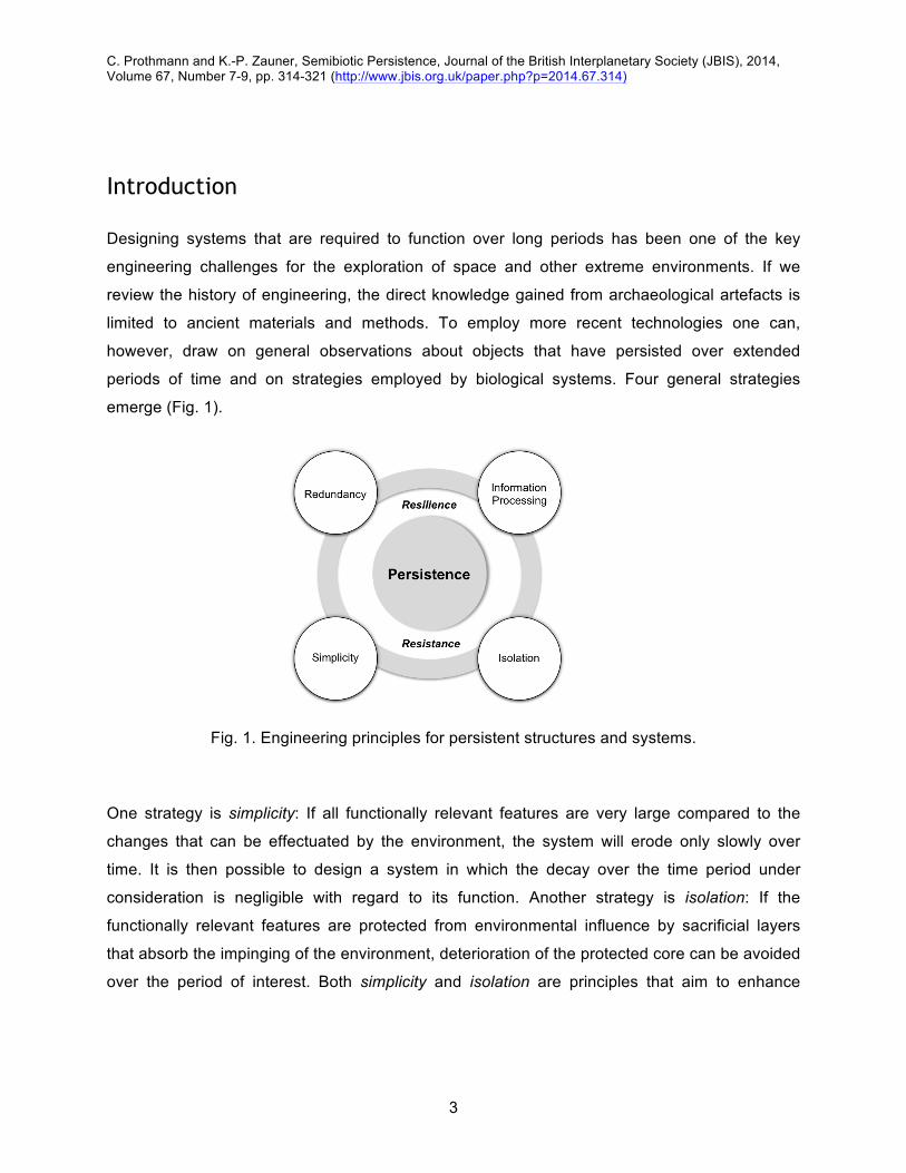
Introduction
Designing systems that are required to function over long periods has been one of the key
engineering challenges for the exploration of space and other extreme environments. If we
review the history of engineering, the direct knowledge gained from archaeological artefacts is
limited to ancient materials and methods. To employ more recent technologies one can,
however, draw on general observations about objects that have persisted over extended
periods of time and on strategies employed by biological systems. Four general strategies
emerge (Fig. 1).
Fig. 1. Engineering principles for persistent structures and systems.
One strategy is simplicity: If all functionally relevant features are very large compared to the
changes that can be effectuated by the environment, the system will erode only slowly over
time. It is then possible to design a system in which the decay over the time period under
consideration is negligible with regard to its function. Another strategy is isolation: If the
functionally relevant features are protected from environmental influence by sacrificial layers
that absorb the impinging of the environment, deterioration of the protected core can be avoided
over the period of interest. Both simplicity and isolation are principles that aim to enhance

C. Prothmann and K.-P. Zauner, Semibiotic Persistence, Journal of the British Interplanetary Society (JBIS), 2014, Volume 67, Number 7-9, pp. 314-321 (http://www.jbis.org.uk/paper.php?p=2014.67.314)
4
system resistance. The Temple Mount platform in Jerusalem and the Egyptian Pyramids could
serve as examples of, respectively, simplicity and isolation. Arguably, the Chinese Wall requires
more active maintenance and would be closer to resilience in Fig. 1, if manual repair is viewed
as an integral part of the architecture. Redundancy of critical system modules has been applied
to many modern engineering challenges, including nuclear reactors and space exploration
devices, and is particularly useful if there is only a limited possibility of repair during operation
[1–3]. Simplicity, isolation and redundancy are engineering principles, which have been
successfully battling with system disturbances, however, only to a certain point. Historical
examples of resistance engineering clearly demonstrate that these structures decay and that
deterioration can only be counteracted by active restoration from outside of the system.
Furthermore, structures and systems with a high level of resistance exhibit a very low overall
functional complexity. Maintaining highly complex functional systems requires other strategies to
ensure functional integrity. Although redundancy is certainly one option to address this
challenge, an exclusive reliance on redundancy is an inefficient solution.
All three principles discussed so far are not well suited for space-travel due to their inefficient
use of resources and material. Long-term functionality can be also achieved by actively
countering deterioration through using information processing. The system-inherent capability of
repairing complex functional systems has only been observed in biological systems that use
information processing to actively counter the impact of entropy [4–7]. Evolution has optimized
biological systems to resist, respond, and recover from environmental disturbances [7, 8].
Organisms exemplify a range of solutions, combining structural resistance, redundancy, and the
ability to actively self-repair, to achieve persistence and maintain functionality [7]. Over the past
six decades, design principles derived from biological systems have found their way into
engineering, material sciences and architecture. Biomimetics [9] has become a growing field
that focuses on studying mechanisms, processes and structures of biological systems with the
goal of imitating solutions optimized over evolutionary time scales [10]. Biomimicry-inspired
applications include biomaterials [10, 11], biomechanics and robotics [12], and architecture [13,
14], as well as algorithms for machine learning [15] and optimization [16]. Practical experience
with these application domains has, however, demonstrated that biomimetic approaches - while

C. Prothmann and K.-P. Zauner, Semibiotic Persistence, Journal of the British Interplanetary Society (JBIS), 2014, Volume 67, Number 7-9, pp. 314-321 (http://www.jbis.org.uk/paper.php?p=2014.67.314)
5
in some areas competitive with existing engineering methods - in many instances do not live up
to the expectations generated by the performance of organisms. Obstacles that stand in the way
of more successful biomimetic architectures arise from two sources. Firstly, the limitations in
fabricating specialised materials, such as nature produces in the form of customised
macromolecules. And, secondly, from the limited integration density of manufacturing processes
compared to the developmental processes available to nature. A case in point is the integration
of electronic circuits that has seen exponential advancement over the past half century. Even
after its astonishing progress, a connection density comparable to that achieved by natural
neuronal networks - where the average number of inputs received by a single neuron in the
mouse cortex is around 8000 [17] - is not in sight.
It appears unlikely that in the near term the gap between synthetic and natural materials and
functional systems will significantly narrow. As a consequence, it is attractive to make use of the
materials and systems available in nature by integrating them with conventionally engineered
components and devices into hybrid biosystems [18, 19]. Such semibiotic architectures
incorporate biological components into a technical setting to transfer some of the desirable
qualities of biosystems to the resulting architecture [20]. In-situ replenishment of complex
components that have degraded, dynamic reallocation of material and energy resources, and
self-repair following damage are typical examples of what can be achieved in semibiotic
architectures - but what is currently out of reach for conventional engineering.
The developments at the interface between engineering and biology are summarized in Fig. 2.
Biology, on the one hand, supplies ideas for solutions to engineering problems, giving rise to the
field of biomimetic engineering. In this field, the concepts inspired by biology are implemented
with conventional fabrication methods and materials. Both, methods and materials fall far short
of what nature has available. For example, growth processes and self-assembly allow for
biological systems with high interconnection density, and macromolecules with customized
functionality enable high integration density in organisms. In contrast, the limitations of the
materials and fabrication methods available in engineering hamper biomimetic engineering.
However, the interfacing between conventionally engineered systems and biomimetically
engineered systems is typically not difficult. Biology, on the other hand, acts as a source of
components for hybrid devices developed in the field of semibiotic engineering. Here the key

C. Prothmann and K.-P. Zauner, Semibiotic Persistence, Journal of the British Interplanetary Society (JBIS), 2014, Volume 67, Number 7-9, pp. 314-321 (http://www.jbis.org.uk/paper.php?p=2014.67.314)
6
challenges are the lack of direct control over the autonomously acting biological subsystem and
the development of suitable interfaces between biological and conventionally engineered
subsystems.
At present, most of the interface development falls on the side of the conventionally engineered
subsystems. However, initial steps to reengineer organisms to optimise the interface from the
biological side have also been taken [21]. The engineered adaptation of organisms specifically
for integration with conventional architectures is becoming increasingly feasible as the field of
synthetic biology progresses, and future semibiotic architectures are likely to comprise
engineered biosystems rather than components extracted from nature. Before we consider the
contribution that information processing can make to the persistence of semibiotic systems, it
will be instructive to consider examples of resilience in biological systems.
Fig. 2. Engineering approaches.

C. Prothmann and K.-P. Zauner, Semibiotic Persistence, Journal of the British Interplanetary Society (JBIS), 2014, Volume 67, Number 7-9, pp. 314-321 (http://www.jbis.org.uk/paper.php?p=2014.67.314)
7
Persistence Strategies of Biological Systems
Biological systems have developed strategies and mechanisms of persistence - resistance and
resilience - on a molecular, cellular and organismic level. All of these strategies differ in their
metabolic cost, which has led to the evolution of different mechanisms or combinations of
mechanisms employed by individual species [7]. Information processing, which is at the core of
all life, enables biological systems to maintain their state by mechanisms such as self-repair on
all levels of organization [4, 6, 7, 22]. Cells of single and multi-cellular organisms are constantly
exposed to exogenous (environmental) as well as endogenous challenges to their structural,
functional and informational integrity. Damaging agents, which impair DNA and protein function,
include metabolic by-products (such as free radicals), chemicals, and high-energy radiation [23].
DNA, which is the repository of genetic information, is under constant self-repair by a range of
mechanisms present in prokaryotic and eukaryotic life forms. DNA repair, which involves
multiple and overlapping processes (partial redundancy), relies on proteins that can be
inactivated by chemicals or oxidative radicals formed by radiation [24]. Organisms with high
radiation tolerance have a highly developed antioxidant system, which actively protects proteins
from oxidative damage, efficient DNA repair mechanisms, and processes to recognize and
degrade damaged proteins [25]. Components of these processes are further induced after
radiation exposure to increase cellular protection and allow for fast recovery from damage [26].
Bacteria such as Deinococcus radiodurans, some fungi and animals, including some rotifers
and tardigrades, show an extraordinary tolerance to radiation [26–30]. These organisms also
demonstrate exceptional robustness towards other extreme environmental conditions and are
referred to as extremotolerant or extremophiles, capable of surviving high radiation, desiccation,
low temperatures, high pressure, and even vacuum. Tardigrades, which are animals below one
millimetre in size and considered phylogenetically related to arthropods [31], are able to survive
exposure to the vacuum of space and even solar radiation [32]. Studies carried out with other
prokaryotic and eukaryotic organisms indicated that some bacterial spores, some microbial
communities, and some forms of lichens have the ability to survive prolonged exposure to space
conditions [33, 34].

C. Prothmann and K.-P. Zauner, Semibiotic Persistence, Journal of the British Interplanetary Society (JBIS), 2014, Volume 67, Number 7-9, pp. 314-321 (http://www.jbis.org.uk/paper.php?p=2014.67.314)
8
Many extremophiles are capable of coping with highly challenging environmental conditions in a
dormant as well as in a metabolic active state [35]. These life forms possess mechanisms to
actively respond to extreme physical environments allowing them to transition into a protective
dormant state, such as anhydrobiosis, which is characterized by a very low metabolic rate. The
transition back into a metabolically more active state is carefully orchestrated to enable the
repair of sustained damage, e.g. DNA repair, before the cells become fully metabolically active.
Depending on the environmental extreme, dormant cells are not completely metabolically
inactive. Studies of the metabolic rate of organisms in permafrost indicate that microorganisms
can retain a very low metabolic rate, which could enable them to actively counteract degradation
and repair damage [36, 37]. Extremophiles have evolved an extraordinary ability to limit the
impact of environmental disturbances and to actively repair molecular damage at low and active
metabolic rate. Several organisms have taken it even a step further and thrive in highly
radioactive environments such as nuclear waste or power plants. Deinococcus radiodurans
exhibits an unmatched level of resilience against multiple physically extreme environments,
including high levels of radiation, which seems to be based on having up to 10 copies of its
genome (redundancy) [38], highly developed mechanisms for actively protecting proteins from
radiation-induced oxidation [39], and DNA repair (information processing) [40].
Melanised fungi, which have been reported in places such as the Chernobyl nuclear reactor, not
only survive when exposed to ionizing radiation, they have also been described to show
increased growth and are therefore referred to as radiotrophic fungi. The molecular mechanisms
that promote increased cell growth are still being elucidated, though data indicates that some
melanised fungi are able to effectively protect themselves from ionizing radiation (isolation) and
repair radiation-induced damage (information processing) while enhancing their growth rate [28,
41, 42]. Some authors hypothesise that these fungi are able to utilize radiation as an additional
energy source [41].
Biological systems have developed astounding molecular and cellular mechanisms to ensure
survival in harsh environments. Although organisms exhibit a certain level of redundancy and
employ protective measures, e.g. melanin in case of the radiotrophic fungi (isolation), to
increase their environmental resistance, self-repair of cellular functions (information processing)

C. Prothmann and K.-P. Zauner, Semibiotic Persistence, Journal of the British Interplanetary Society (JBIS), 2014, Volume 67, Number 7-9, pp. 314-321 (http://www.jbis.org.uk/paper.php?p=2014.67.314)
9
is a key mechanism for achieving resilience.
Slime moulds are organisms that undergo a complex life cycle including a single-cellular/mono-
nucleated, a unicellular/multi-nucleated (plasmodium), and a multi-cellular differentiation stage
ending in a fruiting body carrying spores [43]. The plasmodium of Physarum polycephalum can
reach a size measured in square meters and is basically one very large cell with millions of
nuclei [44, 45]. The mechanical fragmentation of the plasmodium leads to smaller
microplasmodia, which are capable of either fusing back to a macroplasmodia or individually
undergoing the full lifecycle of Physarum polycephalum (redundancy) [46]. True multicellular
organisms with complex bodies have also developed mechanisms to maintain and restore their
overall structural integrity and function. Complex organisms establish their morphological
features, including different cell types, tissues and organs, as a result of a development process
[47]. Injuries to tissues and body structure trigger response mechanisms that contain the
damage and aim to compensate for the loss of structure and function. Repair may lead to some
structural but only limited functional recovery, unless compensatory growth is possible.
Regeneration is a self-repair process that leads to the reconstitution of most, if not all, of the lost
structures and functions. Plants and many animal species display regenerative processes [48].
Animal regeneration has been most extensively studied in invertebrates such as hydra and in
vertebrates such as various amphibians. Stem cells that are capable of differentiating into
various tissues are key to regeneration and have been identified in animals ranging from
primitive sponges to mammals [48].
Sponges represent an interesting example for investigating the relationships between structural
resistance, organizational complexity, regenerative self-repair, and overall persistence.
Interestingly, sponges, which were in the past mainly harvested for their skeletons, have
become of interest for biomimetic engineering due to composite-like biomaterials of their
skeletons, which might guide the development of novel biomimetic materials [49]. Recently, the
structural design of sponges also served as an inspiration for innovative architecture of
skyscrapers [50]. Sponges are sessile, mostly filter-feeding aquatic organisms that represent
one of the earliest multicellular forms of animals [51]. The body plan of sponges consists of an
outer and an inner layer of cells with the mesohyl that functions as an endoskeleton in between.

C. Prothmann and K.-P. Zauner, Semibiotic Persistence, Journal of the British Interplanetary Society (JBIS), 2014, Volume 67, Number 7-9, pp. 314-321 (http://www.jbis.org.uk/paper.php?p=2014.67.314)
10
Instead of tissues in the classical sense, sponges have a cellular grade organization consisting
of specialised and unspecialised cells supported by an endoskeleton with mineral and organic
components. Sponges grow in a variety of shapes, ranging in size from less than a centimetre
to giants measured in meters [52]. Pores and a canal system - through which water is actively
circulated by the movement of the flagella of the choanocytes to ensure the supply of oxygen
and food - characterize the basic body plan of sponges (Fig. 3). The morphology of sponge
species can differ and range from cup shape, branching, or tubular to globular and encrusting
forms [53, 54].
Fig. 3. Basic sponge anatomy with cross section, adapted from [54].
Although sponges have no nervous system or muscle tissue, they are capable of coordination
and reacting to environmental changes. Sponges can control their feeding circulation by
mechanisms that involve electrical impulses in response to sediments in their filtration system
[55]. Further, they have been reported to undergo contractions to remove waste from their body
system, which shows that sponges have some sensory capacity and the ability to coordinate
movements [56]. Despite the fact that sponges are considered sessile, except for their larvae,
some sponge species have demonstrated the ability to move or crawl short distances [57, 58].
Marine sponges contain complex microbial communities and interact with microorganisms such
as bacteria and fungi on multiple levels [59]. One of the well-studied symbiotic relationships is

C. Prothmann and K.-P. Zauner, Semibiotic Persistence, Journal of the British Interplanetary Society (JBIS), 2014, Volume 67, Number 7-9, pp. 314-321 (http://www.jbis.org.uk/paper.php?p=2014.67.314)
11
the metabolic interaction with photosynthetic cyanobacteria [60]. Microorganisms can contribute
more than 35% of sponge biomass [61] and are suggested to assist in a variety of host
functions, including nutrition, chemical host defence [62], and antifouling response [63] - and
might also contribute to the structural rigidity of some sponges [64]. Different species of
sponges vary in the morphology and composition of their skeletal structure, making them more
or less prone to damage from environmental disturbances (isolation) [65]. The structural
simplicity of sponges is exemplified by the lack of tissue, a limited number of cell types, and a
highly flexible cellular level of organization. Sponges have extraordinary reconstitutive abilities
as shown by the formation of functional sponges from dissociated sponge cells (redundancy
and information processing) [66]. The regenerative faculty of sponges is attributed to the
presumably totipotent archaeocytes and possibly choanocytes in some sponge species [67].
Molecular markers indicate that archaeocytes resemble stem cells, which would explain the
exceptional ability of sponges to reconstitute and recover from injuries [68]. Sponge species
differ in their strategies for improving their overall persistence by applying a species-distinctive
balance between their ability to resist damage and the ability to recover from damage [65]. This
has been documented in a large study that compared the extent of hurricane damage sustained
by sponges and their ability to recover after 5 weeks [65]. The study indicates that sponges with
strong resistance based on sturdy skeletons were on average better protected against damage.
However, when these sponges sustained significant damage, they had a more limited ability to
recover. As a consequence, more sponges with sturdy skeletons perished after the storm, which
led the authors to conclude that there is an inverse relationship and trade-off between the ability
to resist damage and the ability to recover from damage. The giant barrel sponges of the
species Xestospongia muta, which shows a balanced mix between damage resistance and
recovery, are assumed to be the longest-lived animals known, with a life-span exceeding more
than 2,000 years [65, 69].
Information processing lies at the core of living systems and enables the self-sustainability of life
by responding to internal and external changes on a molecular, cellular, organismal and even
ecological level to ensure propagation and persistence [4-6, 22, 70]. Evolutionary selection has
optimized biological systems to develop and employ diverse mechanisms for persistence,
including self-repair up to the point of full regeneration. Although most organisms come with a

C. Prothmann and K.-P. Zauner, Semibiotic Persistence, Journal of the British Interplanetary Society (JBIS), 2014, Volume 67, Number 7-9, pp. 314-321 (http://www.jbis.org.uk/paper.php?p=2014.67.314)
12
similar basic set of persistence mechanisms, which are critical for the sustainability and
perpetuation of life, they can exhibit varying degrees of resistance and resilience approaches to
improve overall persistence [7]. The ability of self-repair, however, remains the central trait for
achieving long-term sustainability and persistence, even in extreme environments. Next we will
turn to the question of how resilience, and especially the information processing aspect of it, can
guide the design of persistent semibiotic systems for the exploration of extreme environments
such as space.
Towards Semibiotic Persistence
The long-term survival strategies described in the previous section are so far not replicable in
engineered systems. Semibiotic engineering, however, provides a pathway for importing
capabilities of biological systems, such as self-repair, into subsystems of purpose-built
architectures. To clarify the mechanism of biological persistence, it will be useful to take an
abstract perspective on the role of self-repair in biological systems.
If disturbances from the environment impinge on a simple functional system, it will degrade over
time (Fig. 4). Entropy from the environment drives changes in the system. Because the system
in this case is assumed to be simple, many of these changes will not move the system out of the
subset of states in which it remains functional. Over time, the system will reach states in which it
is only marginally functional and from which further impact from the environment can push it into
a set of states of degraded functionality. For simple systems, the latter set is typically large and
it will on average take significant time until the accumulation of damage leads to the destruction
of the system. Entropy acts on the system without direction and consequently the transition
probabilities among the sets of states - and accordingly the lifetime of the system - are
determined by the relative size of the sets of states that correspond to a functional, degraded,
and destroyed system. The dominant transitions for a simple system are illustrated in Fig. 4.
Any transition backwards is exceedingly unlikely, because the set of functional states is much
smaller than the set of degraded states, and this in turn is far smaller than the set of states
commensurate with the system being destroyed. The vast size of the latter is symbolised by its

C. Prothmann and K.-P. Zauner, Semibiotic Persistence, Journal of the British Interplanetary Society (JBIS), 2014, Volume 67, Number 7-9, pp. 314-321 (http://www.jbis.org.uk/paper.php?p=2014.67.314)
13
dashed boundary in the figure. Transition pathways with negligible probability, such as the
backwards transitions and the direct transition from functional states to destroyed, are not
shown in this and the following transition graphs.
Fig. 4. Simple System
If we consider, in contrast to the simple system, the situation for a complex non-biological
system (Fig. 5), we find that more detailed requirements for the organisation and functionality of
the system restrict the set of possible system configurations, which are functional. Similarly, the
interdependencies in a complex system restrict the set of states that exhibit degraded
functionality rather than no functionality. As a result, the entropy from the environment impinging
on the system is likely to degrade the system or may even destroy it directly if critical
components or links are affected. The more sophisticated requirements for a complex system
also result in a much more rapid transition from a degraded state to the full destruction of the
system. The complex system is brittle in the sense that it deteriorates more rapidly along the
path indicated by thick arrows - a consequence of the relatively small sets of states that are
compatible with the requirements of the system. It is possible to reduce the transition
probabilities along this path to some degree by introducing redundancy, a measure that in effect
increases the sets of functional and of degraded states. In contemplating the complex system
(Fig. 5), we have in essence equated complexity with a small number of functional and

C. Prothmann and K.-P. Zauner, Semibiotic Persistence, Journal of the British Interplanetary Society (JBIS), 2014, Volume 67, Number 7-9, pp. 314-321 (http://www.jbis.org.uk/paper.php?p=2014.67.314)
14
degraded states. With the transition probability among these state sets being a direct
consequence of their relative size, it may appear at first sight as if shielding from the
environment is the only path to achieve a long lifetime for a complex system. However, the
transition probability towards destruction is only determined directly by the relative size of the
state sets if the transitions among states occur at random. That this does not necessarily have
to be the case is exploited by organisms. All forms of life are highly complex systems but
survive the impinging of environmental entropy over extended periods and do so by going
beyond shielding or isolation. Crucial to this ability is the use of information processing in order
to direct transitions from degraded states to functional states.
Fig. 5. Complex System
This scenario is depicted in Fig. 6. Biological systems are always complex systems and
therefore the underlying situation is as in Fig. 5. The specialised requirements for a living state
make state changes resulting in degraded functionality and further decay to destruction very

C. Prothmann and K.-P. Zauner, Semibiotic Persistence, Journal of the British Interplanetary Society (JBIS), 2014, Volume 67, Number 7-9, pp. 314-321 (http://www.jbis.org.uk/paper.php?p=2014.67.314)
15
likely. But there is a new path that takes degraded systems back to a functional state.
Fig. 6. Biological System.
If the transition probability along this new path would be determined by the relative size of the
state sets, this path would not exist. Instead, it is actively driven in the unlikely reverse direction.
The driving force could be free-energy minimization, as in the case of molecular self-assembly,
or provided through other energy sources. The effect of the new transition path is a reduced
probability for a degraded system to be destroyed and a reduced time for the system to be in a
degraded state. This requires the expenditure of energy to work against entropy and information
processing to determine the direction of the preferred transitions. If the deviation from normal
operation is slight, the active biasing of the transitions back to the fully functional state can be
viewed as dynamic control or stabilisation. If it is severe, it will give rise to self-repair. In either
case, information processing is key to preventing the complex organisation that underlies all life
forms from deteriorating rapidly. As a consequence, life as a robust phenomenon cannot exist
without information processing. This information processing is starkly different from conventional

C. Prothmann and K.-P. Zauner, Semibiotic Persistence, Journal of the British Interplanetary Society (JBIS), 2014, Volume 67, Number 7-9, pp. 314-321 (http://www.jbis.org.uk/paper.php?p=2014.67.314)
16
computing and tightly coupled to the physical properties of the underlying material substrate that
implements it. The course of computation is determined by free-energy minimisation rather than
by enforcing - from a materials perspective, arbitrary - mathematical constraints. Whereas a
computer follows a carefully arranged sequence of state transitions, natural information
processing can be better pictured as a process of crystallisation. The consequence is twofold.
Firstly, nature’s information processing is very energy efficient, because it does not need to be
driven through a prescribed sequence of state changes. Secondly, in the present context even
more importantly, it is more robust as it does not require the careful preparation of a starting
state, because of the availability of a very large set of potential starting states, all of which are
lead by energy minimisation to the solution [71].
Prototypical Semibiotic Systems
At present, it is not possible to reproduce the fine-grained, robust, and energy efficient
information processing that enables organisms to maintain their alive state with man-made
technology. However, a number of laboratory prototypes have shown that one method of
maintaining highly complex structures in engineered architectures is the integration of living
cells as functional components into the system. By doing so, it is possible to import the self-
repair capability of the living cell at least for the most complex part of the architecture. For
example, a cell can be used as a living biosensor that - in contrast to a conventionally
engineered device - can replenish its receptors upon contamination or degradation and thus
stay functional over a long period of time [21, 72]. Other prototypes have used living cells as
mechanical actuators. Prokaryotic [73, 74] and eukaryotic cells [75] have been employed to
pump fluids in microfluidic set-ups.
Early steps to interface living cells as information processors have also been taken. A number of
groups have interfaced neurons on chips (cf., e.g. [76]), but it is also possible to connect to
more robust cells. The plasmodia of Physarum polycephalum discussed above can be
integrated into electronic circuits and interfaced either optically [20] or with impedance

C. Prothmann and K.-P. Zauner, Semibiotic Persistence, Journal of the British Interplanetary Society (JBIS), 2014, Volume 67, Number 7-9, pp. 314-321 (http://www.jbis.org.uk/paper.php?p=2014.67.314)
17
measurements [77]. An interesting aspect of integrating plasmodia enclosed in microfluidic
chips with electronic circuits (Fig. 7) is that they can be stored for several months in a dormant
(sclerotia) state and activated within 1.5 h upon water contact. Plasmodia and sclerotia have a
very large number of redundant copies of their genetic information and can therefore withstand
significant damage between periods of self-repair. It is conceivable that in the future such
devices could be employed to withstand periods of high radiation without requirement for heavy
shielding, by relying on the combination of the genetic redundancy provided by millions of nuclei
in combination with self-repair. These prototypes point towards the development of bio-hybrid
devices with components that are alive. Such future living devices are attractive in the context
of space technology, because of their potential to enable self-repair and self-reconfiguration, as
well as efficiency with regard to both material and energy [78].
Fig. 7. Physarum polycephalum on microfluidic chips for integration with electronic circuit.

C. Prothmann and K.-P. Zauner, Semibiotic Persistence, Journal of the British Interplanetary Society (JBIS), 2014, Volume 67, Number 7-9, pp. 314-321 (http://www.jbis.org.uk/paper.php?p=2014.67.314)
18
Conclusions Biological systems achieve persistence through a combination of resistance and resilience
mechanisms - however, with a greater reliance on the latter by exploiting both redundancy and
information processing. The combination of both is a necessity, since information and the ability
of information processing are required for directing self-repair and other persistence measures.
Organisms evolved and apply varying degrees of resistance and resilience mechanisms
depending on the extremity of environmental disturbances. While redundancy is commonly
applied for mission-critical components in engineered persistence, the low-level physical
information processing required for self-repair is not easily duplicated in conventionally
engineered systems. A potential solution to the challenge of building light, persistent devices of
high complexity may be provided by semibiotic architectures that defer the burden of
maintaining the most complex components to biological systems. Such a strategy can be
viewed as an intermediate solution that is closer to realisation than completely artificial systems
capable of self-repair. This solution has its own challenges, such as the risk of contamination
and in case of living components, the potential of evolving away from the functionality required
by the host architecture. Among the challenges of making semibiotic persistence a useful
technology is the requirement to integrate biological components with conventional engineering.
There is rapid progress in the development and miniaturization of suitable interfaces, but it will
also be necessary to develop design strategies capable of coping with autonomous components
that cannot be characterized by rigid specifications. In fact, the need to adapt the prescriptive
control of present engineering approaches to architectural designs that are more suitable for
living matter, may be the largest obstacle to overcome on the path to semibiotic persistence.

C. Prothmann and K.-P. Zauner, Semibiotic Persistence, Journal of the British Interplanetary Society (JBIS), 2014, Volume 67, Number 7-9, pp. 314-321 (http://www.jbis.org.uk/paper.php?p=2014.67.314)
19
References
[1] R.C. Suich and R.L. Patterson, “How Much Redundancy? Some Cost Considerations, Including Examples for Spacecraft Systems”, NASA Technical Memorandum 103197, NASA Lewis Research Center, 1990.
[2] W. Bjorndahl and M. Byers, “Redundancy implementations and consideration of related failures in spacecraft electronic systems”, in Proceedings of the 2011 IEEE Aerospace Conference, pp.1–12, 2011.
[3] J. Downer, “When Failure is an Option: Redundancy, reliability and regulation in complex technical systems”, Discussion Paper No. 53, Centre for Analysis of Risk and Regulation at the London School of Economics and Political Science, 2009.
[4] M. Conrad, “Adaptability: the significance of variability from molecule to ecosystem”, Plenum Press, 1983.
[5] S.I. Walker and P.C.W. Davies, “The algorithmic origins of life”, J. R. Soc. Interface, 10, 79, 20120869, 2013.
[6] K.D. Farnsworth, J. Nelson, and C. Gershenson, “Living is Information Processing: From Molecules to Global Systems”, Acta Biotheor., 61, pp.203–222, 2013.
[7] L.S. Yafremava, M. Wielgos, S. Thomas, A. Nasir, M. Wang, J.E. Mittenthal, and G. Caetano-Anollés, “A general framework of persistence strategies for biological systems helps explain domains of life”, Front. Genet., 4:16, 2013.
[8] C. Adami, C. Ofria, and T.C. Collier, “Evolution of biological complexity”, Proc. Natl. Acad. Sci. U. S. A., 97, pp.4463–4468, 2000.
[9] O.H. Schmitt, “Some interesting and useful biomimetic transforms”, Proc. 3rd Int. Biophysics Congress, pp.297, 1969.
[10] Y. Bar-Cohen, “Biomimetics - using nature to inspire human innovation”, Bioinspir. Biomim., 1, pp.1-12, 2006.
[11] C.P. Brown, F. Rosei, E. Traversa, and S. Licoccia, “Spider silk as a load bearing biomaterial: tailoring mechanical properties via structural modifications”, Nanoscale, 3, pp.870–876, 2011.
[12] L.D. Paulson, “Biomimetic robots”, Computer, 37, pp.48–53, 2004. [13] M.P. Zari, “Biomimetic approaches to architectural design for increased sustainability,”
retrieved via https://www.academia.edu/7249036/BIOMIMETIC_APPROACHES_TO_ARCHITECTURAL_DESIGN_FOR_INCREASED_SUSTAINABILITY. (Last Accessed 28 November 2014)
[14] J.S. Turner and R.C. Soar, “Beyond biomimicry: What termites can tell us about realizing the living building”, in Proceedings of the First International Conference on Industrialized, Intelligent Construction (I3CON), eds T. Hassan and J. Ye, Loughborough University, pp.221-237, 2008.
[15] F. Rosenblatt, “The perceptron: A probabilistic model for information storage and organization in the brain”, Psychol. Rev., 65, pp.386-408, 1958.
[16] G.E.P. Box, “Evolutionary Operation: A Method for Increasing Industrial Productivity”, J. R. Stat. Soc. Ser. C Appl. Stat., 6, pp.81–101, 1957.
[17] A. Schüz, “Neuroanatomy in a computational perspective”, in The Handbook of Brain Theory and Neural Networks, ed. M. A. Arbib, MIT Press, Cambridge, MA, pp.622-626,

C. Prothmann and K.-P. Zauner, Semibiotic Persistence, Journal of the British Interplanetary Society (JBIS), 2014, Volume 67, Number 7-9, pp. 314-321 (http://www.jbis.org.uk/paper.php?p=2014.67.314)
20
1995. [18] M. Ayre, “Biomimetics applied to space exploration”, in Design and Nature II: Comparing
Science and Engineering, eds M. W. Collins and C. A. Brebbia, WIT Press, pp.603-612, 2004.
[19] A. Adamatzky, “Physarum machines for space missions”, Acta Futura, 6, pp. 53–67, 2013. [20] S. Tsuda, K.P. Zauner, and Y.P. Gunji“, Robot control with biological cells”, Biosystems.,
87, pp.215–223, 2007. [21] D.E. Nivens, T.E. McKnight, S.A. Moser, S.J. Osbourn, M.L. Simpson, and G.S. Sayler,
“Bioluminescent bioreporter integrated circuits: potentially small, rugged and inexpensive whole-cell biosensors for remote environmental monitoring”, J. Appl. Microbiol., 96, pp.33-46, 2004.
[22] M. Conrad, “Mutation-absorption model of the enzyme”, Bull. Math. Biol. 41, pp.387-405, 1979.
[23] A. Ciccia and S.J. Elledge, “The DNA damage response: making it safe to play with knives”, Mol. Cell, 40, pp.179-204, 2010.
[24] M.S. Cooke, M.D. Evans, M. Dizdaroglu, and J. Lunec, “Oxidative DNA damage: mechanisms, mutation, and disease”, FASEB J., 17, pp.1195–1214, 2003.
[25] M.J. Daly, “Death by protein damage in irradiated cells”, DNA Repair 11, pp.12–21, 2011. [26] H. Luan, N. Meng, J. Fu, X. Chen, X. Xu, Q. Feng, H. Jiang, J. Dai, X. Yuan, et al.,
“Genome-wide transcriptome and antioxidant analyses on gamma-irradiated phases of deinococcus radiodurans R1”, PLoS One, 9, e85649, 2014.
[27] D.D. Horikawa, T. Sakashita, C. Katagiri, M. Watanabe, T. Kikawada, Y. Nakahara, N. Hamada, S. Wada, T. Funayama, et al., “Radiation tolerance in the tardigrade Milnesium tardigradum”, Int. J. Radiat. Biol., 82, pp.843–848, 2006.
[28] T.I. Tugay, M.V. Zheltonozhskaya, L.V. Sadovnikov, A.V. Tugay, and E.B. Farfán, “Effects of ionizing radiation on the antioxidant system of microscopic fungi with radioadaptive properties found in the Chernobyl exclusion zone”, Health Phys., 101, pp.375–382, 2011.
[29] J. Dighton, T. Tugay, and N. Zhdanova, “Fungi and ionizing radiation from radionuclides”, FEMS Microbiol. Lett., 281, pp.109–120, 2008.
[30] A. Krisko and M. Radman, “Biology of extreme radiation resistance: the way of Deinococcus radiodurans”, Cold Spring Harb. Perspect. Biol., 5, a012765, 2013.
[31] D.R. Nelson, “Current status of the tardigrada: evolution and ecology”, Integr. Comp. Biol., 42, pp.652–659, 2002.
[32] K.I. Jönsson, E. Rabbow, R.O. Schill, M. Harms-Ringdahl, and P. Rettberg, “Tardigrades survive exposure to space in low Earth orbit”, Curr. Biol., 18, R729–R731, 2008.
[33] G. Horneck, D.M. Klaus, and R.L. Mancinelli, “Space microbiology”, Microbiol. Mol. Biol. Rev., 74, pp.121–156, 2010.
[34] S. Onofri, R. de la Torre, J.P. de Vera, S. Ott, L. Zucconi, L. Selbmann, G. Scalzi, K.J. Venkateswaran, E. Rabbow, et al., “Survival of rock-colonizing organisms after 1.5 years in outer space”, Astrobiology, 12, pp.508–516, 2012.
[35] T.H. Waterman, “Evolutionary challenges of extreme environments (Part 2)”, J. Exp. Zool., 291, pp.130–168, 2001.
[36] K. L. F. Brinton, A. I. Tsapin, D. Gilichinsky, and G. D. McDonald, “Aspartic acid racemization and age-depth relationships for organic carbon in Siberian permafrost”, Astrobiology, 2, pp.77–82, 2002.

C. Prothmann and K.-P. Zauner, Semibiotic Persistence, Journal of the British Interplanetary Society (JBIS), 2014, Volume 67, Number 7-9, pp. 314-321 (http://www.jbis.org.uk/paper.php?p=2014.67.314)
21
[37] P.B. Price and T. Sowers, “Temperature dependence of metabolic rates for microbial growth, maintenance, and survival”, Proc. Natl. Acad. Sci. U. S. A., 101, pp.4631–4636, 2004.
[38] K.S. Makarova, L. Aravind, Y.I. Wolf, R.L. Tatusov, K.W. Minton, E.V. Koonin, and M.J. Daly, “Genome of the extremely radiation-resistant bacterium Deinococcus radiodurans viewed from the perspective of comparative genomics”, Microbiol. Mol. Biol. Rev., 65, pp.44-79, 2001.
[39] M.J. Daly, E.K. Gaidamakova, V.Y. Matrosova, A. Vasilenko, M. Zhai, R.D. Leapman, B. Lai, B. Ravel, S.M.W. Li, et al., “Protein oxidation implicated as the primary determinant of bacterial radioresistance”, PLoS Biol., 5, e92, 2007.
[40] M.M. Cox, J.L. Keck, and J.R. Battista, “Rising from the Ashes: DNA Repair in Deinococcus radiodurans”, PLoS Genet., 6, e1000815, 2010.
[41] E. Dadachova, R.A. Bryan, X. Huang, T. Moadel, A.D. Schweitzer, P. Aisen, J.D. Nosanchuk, and A. Casadevall, “Ionizing radiation changes the electronic properties of melanin and enhances the growth of melanized fungi”, PLoS One, 2, e457, 2007.
[42] K.L. Robertson, A. Mostaghim, C.A. Cuomo, C.M. Soto, N. Lebedev, R.F. Bailey, and Z. Wang, “Adaptation of the black yeast Wangiella dermatitidis to ionizing radiation: molecular and cellular mechanisms”, PLoS One, 7, e48674, 2012.
[43] S.L. Stephenson, ”Myxomycetes: A Handbook of Slime Molds”, Timber Press, 2000. [44] T.G. Burland, L. Solnica-Krezel, J. Bailey, D.B. Cunningham, and W.F. Dove, “Patterns of
inheritance, development and the mitotic cycle in the protist Physarum polycephalum”, Adv. Microb. Physiol., 35, pp.1-69, 1993.
[45] D.P. Häder, R. Hemmersbach, and M. Lebert, “Gravity and the Behavior of Unicellular Organisms”, Cambridge University Press, 2005.
[46] E. Guttes, S. Guttes, and H.P. Rusch, “Morphological observations on growth and differentiation of Physarum polycephalum grown in pure culture”, Dev. Biol., 3, pp.588-614, 1961.
[47] J. Bonner, “First Signals: The Evolution of Multicellular Development”, Princeton University Press, 2009.
[48] K.D. Birnbaum and A. Sánchez Alvarado, “Slicing across kingdoms: regeneration in plants and animals”, Cell, 132, pp.697-710, 2008.
[49] H. Ehrlich and H. Worch, “Sponges as natural composites: from biomimetic potential to development of new biomaterials,” in Porifera Research: Biodiversity, Innovation & Sustainability, ed. E. Hajdu, Museu Nacional, Rio de Janeiro, pp.217-223, 2007.
[50] C. Aiello (ed), “Evolo Skyscrapers 2”, Evolo Publishing Company, New York, NY, 2012. [51] W.E.G. Müller (ed), “ Sponges (Porifera)”, Springer, 2003. [52] R.W.M. Van Soest, N. Boury-Esnault, J. Vacelet, M. Dohrmann, D. Erpenbeck, N.J. De
Voogd, N. Santodomingo, B. Vanhoorne, M. Kelly, et al., “Global diversity of sponges (Porifera)”, PLoS One, 7, e35105, 2012.
[53] N. Boury-Esnault and K. Rützler, “Thesaurus of sponge morphology”, Smithsonian Institution Press, 55, 1997.
[54] E.E. Ruppert, R.S. Fox, and R.D. Barnes, “Invertebrate Zoology: A Functional Evolutionary Approach”, Chapter 5, 7th Edition, Brooks Cole Thomson, Belmont, CA, USA, 2004.
[55] G.J. Tompkins-MacDonald and S.P. Leys, “Glass sponges arrest pumping in response to sediment: implications for the physiology of the hexactinellid conduction system”, Mar. Biol.,

C. Prothmann and K.-P. Zauner, Semibiotic Persistence, Journal of the British Interplanetary Society (JBIS), 2014, Volume 67, Number 7-9, pp. 314-321 (http://www.jbis.org.uk/paper.php?p=2014.67.314)
22
154, pp.973-984, 2008. [56] D.A. Ludeman, N. Farrar, A. Riesgo, J. Paps, and S.P. Leys, “Evolutionary origins of
sensation in metazoans: functional evidence for a new sensory organ in sponges”, BMC Evol. Biol., 14:3, 2014.
[57] L. Fishelson, “Observations on the moving colonies of the genus Tethya (Demospongia, Porifera)”, Zoomorphology, 98, pp. 89-99, 1981.
[58] C. Bond and A.K. Harris, “Locomotion of sponges and its physical mechanism”, J. Exp. Zool., 246, pp.271-284, 1988.
[59] M.W. Taylor, R. Radax, D. Steger, and M. Wagner, “Sponge-associated microorganisms: evolution, ecology, and biotechnological potential”, Microbiol. Mol. Biol. Rev., 71, pp.295-347, 2007.
[60] Y.K. Lee, J.H. Lee, and H.K. Lee, “Microbial symbiosis in marine sponges”, J. Microbiol., 39, pp.254-264, 2001.
[61] J. Vacelet, “Étude en microscopie électronique de l’association entre bactéries et spongiaires du genre Verongia (Dictyoceratida)”, J. Microsc. Biol. Cell, 23, pp.271-288, 1975.
[62] N.S. Webster and M.W. Taylor, “Marine sponges and their microbial symbionts: love and other relationships”, Environ. Microbiol., 14, pp.335-346, 2012.
[63] R.N. Aguila-Ramírez, C. J. Hernández-Guerrero, B. González-Acosta, G. Id-Daoud, S. Hewitt, J. Pope, and C. Hellio, “Antifouling activity of symbiotic bacteria from sponge Aplysina gerardogreeni”, Int. Biodeterior. Biodegradation, 90, pp.64-70, 2014.
[64] C. R. Wilkinson, M. Nowak, B. Austin, and R.R. Colwell, “Specificity of bacterial symbionts in Mediterranean and Great Barrier Reef sponges”, Microb. Ecol., 7, pp.13-21,1981.
[65] J.L. Wulff, “Resistance vs recovery: morphological strategies of coral reef sponges”, Funct. Ecol., 20, pp.699-708, 2006.
[66] E.G. Müller and I. Müller, “Sponge cell aggregation”, Mol. Cell. Biochem., 29, pp.131-143, 1980.
[67] N. Funayama, “The stem cell system in demosponges: suggested involvement of two types of cells: archeocytes (active stem cells) and choanocytes (food-entrapping flagellated cells)”, Dev. Genes Evol., 223, pp.23-38, 2013.
[68] K. Okamoto, M. Nakatsukasa, A. Alié, Y. Masuda, K. Agata, and N. Funayama, “The active stem cell specific expression of sponge Musashi homolog EflMsiA suggests its involvement in maintaining the stem cell state”, Mech. Dev., 129, pp.24-37, 2012.
[69] S.E. McMurray, J.E. Blum, and J.R. Pawlik, “Redwood of the reef: growth and age of the giant barrel sponge Xestospongia muta in the Florida Keys”, Mar. Biol., 155, pp.159-171, 2008.
[70] H.R. Maturana and F.J. Varela, “Autopoiesis and Cognition: The Realization of the Living“, Boston Studies in the Philosophy of Science, 42, D. Reidel Publishing Company, 1980.
[71] K.P. Zauner, “From Prescriptive Programming of Solid-State Devices to Orchestrated Self-organisation of Informed Matter”, in Unconventional Programming Paradigms, 3566, eds J.P. Banâtre, P.Fradet, J.L. Giavitto, and O. Michel, Springer Berlin Heidelberg, pp.47-55, 2005.
[72] R. Vijayaraghavan, S.K. Islam, M. Zhang, S. Ripp, S. Caylor, N. D. Bull, S. Moser, S.C. Terry, B.J. Blalock, et al., “A bioreporter bioluminescent integrated circuit for very low-level chemical sensing in both gas and liquid environments”, Sens. Actuators B Chem. 123,

C. Prothmann and K.-P. Zauner, Semibiotic Persistence, Journal of the British Interplanetary Society (JBIS), 2014, Volume 67, Number 7-9, pp. 314-321 (http://www.jbis.org.uk/paper.php?p=2014.67.314)
23
pp.922-928, 2007. [73] M.J. Kim and K.S. Breuer, “Controlled Mixing in Microfluidic Systems Using Bacterial
Chemotaxis”, Anal. Chem., 79, pp.955-959, 2007. [74] M. J. Kim and K. S. Breuer, “Microfluidic pump powered by self-organizing bacteria,” Small,
4, pp.111-118, 2008. [75] Y. Tanaka, K. Sato, T. Shimizu, M. Yamato, T. Okano, and T. Kitamori, “A micro-spherical
heart pump powered by cultured cardiomyocytes”, Lab Chip, 7(2), pp.207-212, 2007. [76] M. Jenkner, B. Müller, and P. Fromherz, “Interfacing a silicon chip to pairs of snail neurons
connected by electrical synapses”, Biol. Cybern., 84, pp.239-249, 2001. [77] S. Tsuda, S. Artmann, and K.P. Zauner, “The Phi-Bot: A Robot Controlled by a Slime
Mould”, in Artificial Life Models in Hardware, Springer London, pp.213-232, 2009. [78] J. Gough, G. Jones, C. Lovell, P. Macey, H. Morgan, F. Revilla, R. Spanton, S. Tsuda, and
K.P. Zauner, “Integration of Cellular Biological Structures Into Robotic Systems”, Acta Futura, 3, pp.43-49, 2009.
Top Related