


Languages
Pages
Legal


Rev. Med. Chir. Soc. Med. Nat., Iaşi – 2015 – vol. 119, no. 2
DENTAL MEDICINE CASE REPORTS
572
SECOND MOLAR UPRIGHTING AFTER PREMATURE LOSS
OF MANDIBULAR FIRST PERMANENT MOLAR- CASE REPORT
E. R. Cernei1, R. B. Mavru2, Irina Nicoleta Zetu1*
University of Medicine and Pharmacy, “Grigore T. Popa”-Iaşi
Faculty of Dental Medicine
1. Department of Orthodontics
2. Department of Pedodontics
*Corresponding author. E-mail: [email protected]
SECOND MOLAR UPRIGHTING AFTER PREMATURE LOSS OF MANDIBULAR
FIRST PERMANENT MOLAR- CASE REPORT (Abstract): Introduction: Uprighting a
tipped molar by using an uprighting spring is a fundamental orthodontic treatment technique.
Following a weak anchorage done only by the anterior teeth the permanent lower second mo-
lar will rotate around its center of resistance, and besides the distalizing movement will oc-
cur its extrusion. Following the reaction, the mandibular anterior teeth will have a movement
of intrusion. All these elements will lead to anterior open bite. Correction of vertical prob-
lems has become easier with the advent of mini-implants. Case presentation: U.I patient
aged 24 years presenting for aesthetic and functional disorders. Clinical examination reveals
intraoral Class I molar malocclusion on the right sight, upper narrowing arch with a slight
overbite, and the both permanent first molars on the left side lost prematurely. The tooth 37
is inclined at 600 degrees to the plane of occlusion. To avoid front opening occlusion we
used an orthodontic miniscrew inserted between the canine and the first premolar on the
same side to achieve the second molar up righting. Lower second molar uprighting was
achieved without unwanted movements in anterior mandibular region and without molar ex-
trusion. Conclusions: Using mini implants in uprighting a tipped molar will achieve the de-
sired tooth movement and will reduce the number of unwanted side effects and eventually to
improve patient’s esthetics. Keywords: SECOND MOLAR UPRIGHTING, PREMATURE
LOSS, MANDIBULAR FIRST MOLAR, ORTHODONTIC MINISCREW
The first permanent molar has been re-
ported to be the most caries prone tooth in
the permanent dentition. More than 50% of
children over the age of 11 years have
some caries experience in this tooth (1, 2,
3). The early loss of permanent first molars
is a routine problem in the orthodontic
clinic and results in mesial inclination of
the second molars. Especially the early loss
of the lower first permanent molar should
be immediately addressed by prosthetic
replacement or orthodontic space closure as
it may lead to functional and anatomical
disturbances. The sequelae of events in-
clude distal migration of premolars, extru-
sion of the antagonist molar, premature
contacts in centric relation, occlusal inter-
ferences during lateral and protrusive
movements. In addition there is a difficulty
in prosthetic adaptation when the inclina-

Second molar uprighting after premature loss of mandibular first permanent molar - case report
573
tion is excessive. Mesial inclination favors
the appearance of vertical bone defects and
infra-osseous pockets in the mesial region
of molars (4, 5).
Molar uprighting into its correct posi-
tion leads to the normalization of function-
al and periodontal occlusion situation, ena-
bling the roots to be positioned perpendicu-
lar to the occlusal plane so that they are
better able to resist to occlusal forces, in
addition to facilitating the plane of inser-
tion of the prosthesis parallel to the long
axis of the tooth (6). Various conventional
orthodontic approaches were used to
achieve such a movement, but all have
been plagued to a greater or lesser extent
by the following issues: extrusion of the
molar to be up righted, unwanted move-
ment of the anchorage unit, the need for
bulky appliances, the frequent need of
accessories to strengthen anchorage, and
long treatment time (7).
With the advent of the use of minis-
crews as orthodontic anchorage, molar
uprighting procedures have become sim-
pler, with more predictable results, and
since there is no need to use other teeth as
anchorage, there is better control of unde-
sirable effects (6).
The aim of this study is to describe a
method for molar uprighting with the use
of a titanol spring supported on a messially
placed miniscrew.
CASE REPORT
Therefore we want to present a case that
presented in the clinic of Orthodontics and
dento-facial orthopedics in the Ambulatory
Pediatric Dentistry from Iasi, Romania. U.
I. patient aged 24 years presenting for func-
tional disorders. The extra-oral examination
(fig. 1) revealed an oval shape of the face, a
mesocephalic head shape, a mesoprosopic
face, and a mild convex profile. Facial
changes occur hyperdivergent profile and an
acute naso-labial angle. The patient showed
a good range of mandibular movements and
no temporomandibular-joint symptoms.
Fig. 1. Extraoral aspect-before treatment
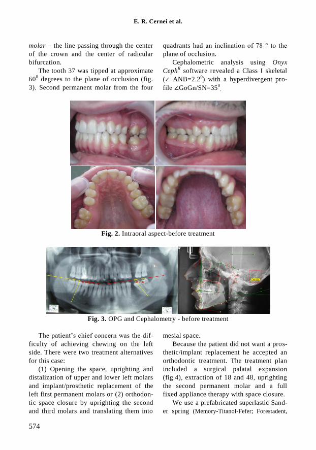
Clinical examination revealed intraoral
Class I molar malocclusion on the right
sight, upper narrowing arch with a slight
overbite, and the both permanent first mo-
lars on the left side lost prematurely. The
space for 26 and 36 decreased due to mesi-
al inclination and migration of the second
molars. Tooth 16 was covered by a metal-
ceramic crown (fig. 2).
Orthopantomogram (OPG) examination
revealed good root canal treatment on tooth
16. All wisdom teeth were present.
The OPG was processed by tracking the
following structures (fig. 3):
-the occlusion plane – line tangent to
the vestibular cusps tips of premolars and
the canine tip on the same side;
-the axis of lower second permanent
E. R. Cernei et al.
574
molar – the line passing through the center
of the crown and the center of radicular
bifurcation.
The tooth 37 was tipped at approximate
600 degrees to the plane of occlusion (fig.
3). Second permanent molar from the four
quadrants had an inclination of 78 ° to the
plane of occlusion.
Cephalometric analysis using Onyx
CephR software revealed a Class I skeletal
(∠ ANB=2.20) with a hyperdivergent pro-
file ∠GoGn/SN=350
.
Fig. 2. Intraoral aspect-before treatment
Fig. 3. OPG and Cephalometry - before treatment
The patient’s chief concern was the dif-
ficulty of achieving chewing on the left
side. There were two treatment alternatives
for this case:
(1) Opening the space, uprighting and
distalization of upper and lower left molars
and implant/prosthetic replacement of the
left first permanent molars or (2) orthodon-
tic space closure by uprighting the second
and third molars and translating them into
mesial space.
Because the patient did not want a pros-
thetic/implant replacement he accepted an
orthodontic treatment. The treatment plan
included a surgical palatal expansion
(fig.4), extraction of 18 and 48, uprighting
the second permanent molar and a full
fixed appliance therapy with space closure.
We use a prefabricated superlastic Sand-
er spring (Memory-Titanol-Fefer; Forestadent,

Second molar uprighting after premature loss of mandibular first permanent molar - case report
575
Bernard Förster GmBH, Pforzheim, Germany) to
achieve the second molar uprighting. Be-
cause the patient had a slightly hiper-
divergent profile (∠GoGn/SN=350) and to
avoid front opening occlusion we used a
titan orthodontic miniscrew (8 mm, Ortho-
Easy®Pin Forestadent®) inserted between
the canine and the first premolar on the
lower left side as for anterior anchorage
(fig. 5).
Fig. 4. After surgical palatal
expansion
Fig. 5. Achieving uprighting of 37: superlastic Sander spring and mini implant insertion

E. R. Cernei et al.
576
Fig. 6. Achieving uprighting
of 37: final situation
DISCUSSION
Permanent first molars are the first
permanent teeth to erupt into the oral cavi-
ty. They have been quoted as the most
caries-prone tooth in the permanent denti-
tion leading to their early loss, probably as
a result of their early exposure to the oral
environment (1, 2). Loss of a first perma-
nent molar should be immediately ad-
dressed by prosthetic replacement or ortho-
dontic space closure as it may lead to func-
tional and anatomical disturbances (4). The
second and third molars from that side will
incline and rotate, canine and premolars
will move distally into the molar space, and
the opposing first molar will extrude. Cor-
rection of these problems is the biggest
challenge to the orthodontist (5).
The ideal solution of six years molar
syndrome is protraction of the second and
the third molars and toothless gap closure
without biological sacrifice. The procedure
is not simple, as these strong roots and a
significant path that follows the apex, if we
take into account initial second molar in-
clination. In a second molar inclination of
300-40
0 correspond to 11 mm travel only
apical-uprighting recovery (8) (fig. 7).
The optimal molar uprighting technique
for most situations in adults should thus
apply an intrusive force to the molar
crown, within an inflammatory- free envi-
ronment. A safe and simple method to
achieve such conditions is to use the pre-
fabricated superlastic Sander spring
(Memory-Titanol-Fefer; Forestadent, Ber-

Second molar uprighting after premature loss of mandibular first permanent molar - case report
577
nard Förster GmBH, Pforzheim, Germany).
Although the prefabricated Sander spring is
a simple and effective method for mandibu-
lar molar up righting, the method can in-
volve unwanted movements in anterior
mandibular region (9) (fig. 8).
Lee et al. have promoted the use of
miniscrew as direct anchorage changing the
mini implant position in order to obtain two
different types of force. The miniscrew can
be positioned in the retromolar area, where
it acts as a fulcrum for the traction by pull-
ing distally to the second molar (10). This
position of the miniimplant can be used in
mild tipped molars cases with mesial incli-
nation of the crown, because it generates a
sufficient effective uprighting moment to
allow the complete straightening of the
molar. Instead in moderate to severe tipped
cases, the generated moment is too small to
produce a sufficient straightening of the
tooth (11). For this reason it is essential to
consider the possibility of placing the mini
implant mesially to the tipped tooth so as a
thrusting action force from mesial to distal
in generated (12). To avoid front opening
occlusion we used in this case an orthodon-
tic miniscrew inserted between the canine
and the first premolar on the same side to
achieve the second molar uprighting.
Achieving uprighting of 37 the slightly
hiperdivergent profile remained constant
(∠GoGn/SN=350).
Fig. 7. Closing gaps steps left by the
absence of a first lower molar
Fig. 8. Consequences of molar uprighting with poor anterior anchorage
and molar extrusion
CONCLUSIONS
In this article we used a miniimplant in
uprighting a tipped molar to achieve a de-
sired tooth movement and to reduce the
number of unwanted side effects and even-
tually to improve patient’s esthetics. It is
necessary a proper clinical, radiological, and
biomechanical evaluation and a thoughtful
selection of the best orthodontic devices to
grant successful treatment results.

E. R. Cernei et al.
578
ACKNOWLEDGMENTS
This work received financial support
through the "Program of Excellence in
multidisciplinary doctoral and postdoctoral
research in chronic diseases", contract no.
POSDRU / 159 / 1.5 / S / 133377 benefi-
ciary U.M.F "Gr. T. Popa" Iasi, financed
from the European Social Fund through the
Sectoral Operational Programme Human
Resources Development 2007-2013.
REFERENCES
1. J. L. Seddon . "Extraction of four first molars: a case for a general practitioner?" J Orthod. 2014;
31(2): 80-85.
2. Todd JE, Dodd T. Children`s dental health in the United Kingdom. Office of Population Censuses
and Surveys, London, 1983.
3. Rajashekhara B S, Keyur J M, Bhavna D, Poonacha K S. Management of early loss of first permanent
molar: A new technique. J Ind Soc Pedod Prev Dent. 2012; 30: 349-351.
4. Gill D, Lee R, Tredwin CJ. Treatment Planning for the Loss of Permanent First Molar. Dent Update
2001; 28: 304-308.
5. Capelluto E, Lauweryns. A Simple Technique for Molar Uprighting. J Clin Orthod. 1997; 31(2): 119-
125.
6. Ruellas ACO, Pithon MM, Santos RL. Miniscrew supported coil spring for molar uprighting: descrip-
tion. Dent Press J Orthod. 2013; 18(1): 45-49.
7. Derton N, Perini A, Mutinelli S, Gracco A. Mandibular molar uprighting using mini-implants: differ-
ent approaches for different clinical cases--two case reports. Orthod (Chic.) 2012; 13(1): 138-145.
8. Musilli M, Marsico M, Romanucci A, Grampone F. Molar uprighting with miniscrew: comparison
among different systems and relative biomechanical analysis. Prog Orthod. 2010; 11(2): 166-173.
9. Zachrisson BU, Bantleon HP.Optimal mechanics for mandibular molar uprighting. World J Orthod.
2005; 6(1): 80-87.
10. Lee KJ, Park YC, Hwang WS, Seong EH. Uprighting mandibular second molars with direct minis-
crew anchorage. J Clin Orthod. 2007; 41(10): 627-635.
11. Sohn BW, Choi JH, Jung SN, Lim KS. Uprighting mesially impacted second molars with miniscrew
anchorage. J Clin Orthod. 2007; 41(2): 94-97.
12. Muggiano F, Giannantoni I, Anastasi G, Giorgia C, Quaranta A. TADs versus traditional devices and
tecniques in lower second molar uprighting procedures. WebmedCentr Orthod. 2014;
5(1):WMC004494.
Top Related