


Languages
Pages
Legal


Rosacea

Maryam GhasemiDaneshjooye Pezeshki
Daneshgah Esfahan901111521DR Fatemi

epidemiology•relatively common disease •fair-skinned people
•rarer in dark-skinned people
•women are more often affected than men in earlier stages
•Men more rhynophima
•importance of sun-damaged skin

pathogenesis•precise etiology of rosacea remains a mystery •hypothesis :degenerative changes of the
perivascular, and possibly vascular, collagen and elastic tissues in inherently susceptible
individuals exposed to climatic factors .
•lead to small vessel dilatation resulting in flushing, telangiectases, and erythema.
• the dilated vessels become incompetent with perivascular leakage of potentially
inflammatory substances .
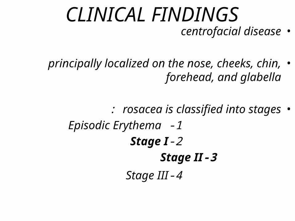
CLINICAL FINDINGS •centrofacial disease
•principally localized on the nose, cheeks, chin, forehead, and glabella
•rosacea is classified into stages: 1 -Episodic Erythema 2-Stage I
3-Stage II
4-Stage III
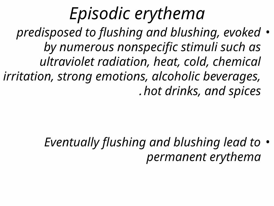
Episodic erythema•predisposed to flushing and blushing,
evoked by numerous nonspecific stimuli such as ultraviolet radiation, heat, cold,
chemical irritation, strong emotions, alcoholic beverages, hot drinks, and
spices.
•Eventually flushing and blushing lead to
permanent erythema
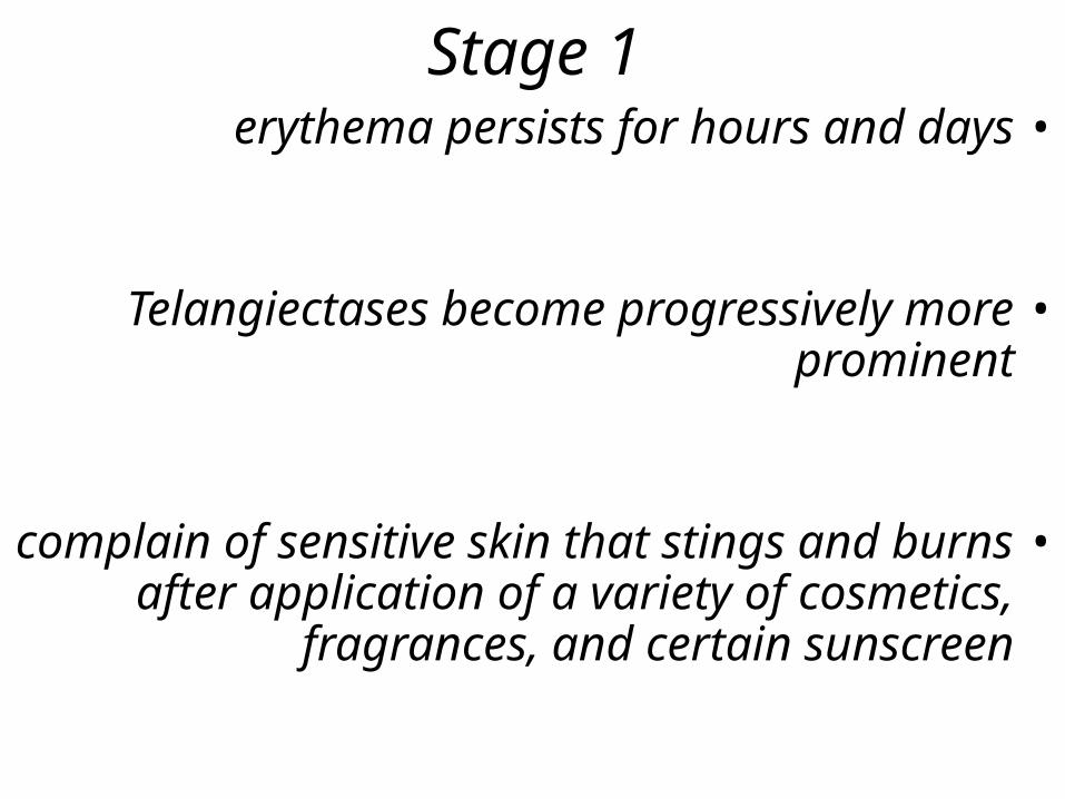
Stage 1•erythema persists for hours and days
•Telangiectases become progressively more prominent
•complain of sensitive skin that stings and burns after application of a variety of
cosmetics, fragrances, and certain sunscreen


Stage 2•inflammatory papules less than 0.5 to 1.0 mm
in size, with or without pustules •persist for weeks •Some papules show a small pustule at the
apex, justifying the term papulopustular.• lesions are always follicular in origin vellus and
sebaceous follicles are involved • deeper inflammatory lesions may heal with
scarring, but scars are small and tend to be shallow
•pores become more prominent •papulopustular attacks become increasingly
frequent


Stage 3•A small proportion of the patients•particularly on the cheeks and nose, less often
on the chin, forehead, or ears • large inflammatory nodules, furunculoid
infiltrations, and tissue hyperplasia.• Finally, the patient shows inflamed and
thickened edematous skin with large pores, resembling the surface of an orange ( peau
d'orange) .•features are caused by inflammatory
infiltration, connective tissue hypertrophy with masses of collagen deposition, diffuse
sebaceous gland hyperplasia, and overgrowth of individual sebaceous glands
•Ultimate deformity is rhinophyma


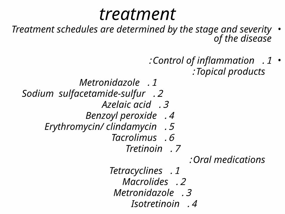
treatment•Treatment schedules are determined by the stage and
severity of the disease
•1 .Control of inflammation: Topical products:
1 .Metronidazole 2 .Sodium sulfacetamide-sulfur 3 .Azelaic acid 4 .Benzoyl peroxide 5 .Erythromycin/ clindamycin 6 .Tacrolimus 7 .Tretinoin
Oral medications: 1 .Tetracyclines 2 .Macrolides 3 .Metronidazole 4 .Isotretinoin

treatment•2 .Repair of structural damage:
1-Laser 2-Intense pulsed light 3-surgical techniques
(rhinophyma) •3 .Prevention of further damage:
1 -Sunscreens 2 -Cosmetics 3-Avoidanceof triggerfactors(flushing)

treatment•Box 13-6 Treatment of rosacea by subset•1 .All subsets:
Daily sunscreen Sun avoidance strategies Cosmetic coverage Avoidance of specific factors that trigger flushing Laser and intense pulsed light
•2 .Erythrotelangiectatic subset:
Morning:sodium sulfacetamide-sulfur cleanser followed by a moisturizing sunscreen and/or camouflaging cosmetic with sunscreen
Night: leave on metronidazole, azelaic acid or sodium sulfacetamide-sulfur product

treatment•3 .Papulopustular subset:
Morning:Topical metronidazole, azelaic acid, sodium sulfacetamide-sulfur or benzoyl peroxide-
antibiotic combination + suncreens
Nightly:sodium sulfacetamide-sulfur cleanser+ different one of the above topical products forAM usage
Oral antibiotics or isotretinoin depending on severity•4 .Glandular subset:
1-Benzoyl peroxide-antibiotic combination most effective, other topicals less so
2-Oral antibiotics or isotretinoin depending on severity
3-Surgical intervention as needed for phymatous changes