


Languages
Pages
Legal


Rhythmical Excitation of the Heart KALEB HOOD AND JIMMY JOHNSON

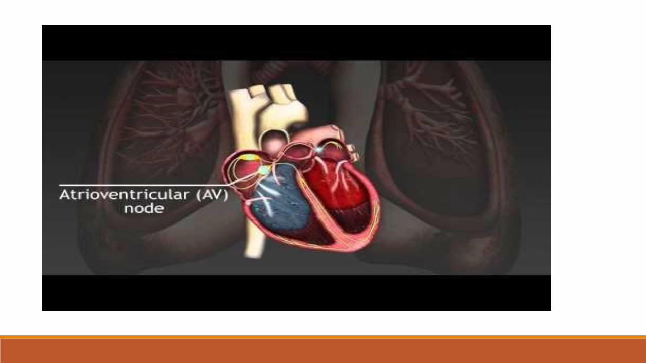
Special Excitory and Conductive System of the Heart
Sinus Node (or sinoatrial node or S-A): A small node with almost no contractile muscle, where signals originate.
A-V bundle: The conductive bridge between the atrial and ventricular lobes.
Purkinje Fibers: Transmit the signals to all parts of the ventricles.
Sinus Fibers: The sinus fibers have the ability to self-excite, and maintain the rhythm of the heart.

Mechanisms Fast Sodium Channels Slow Sodium Channels Pottassium Channels
Leakage of sodium ions and self-excitation
Reversion to normal


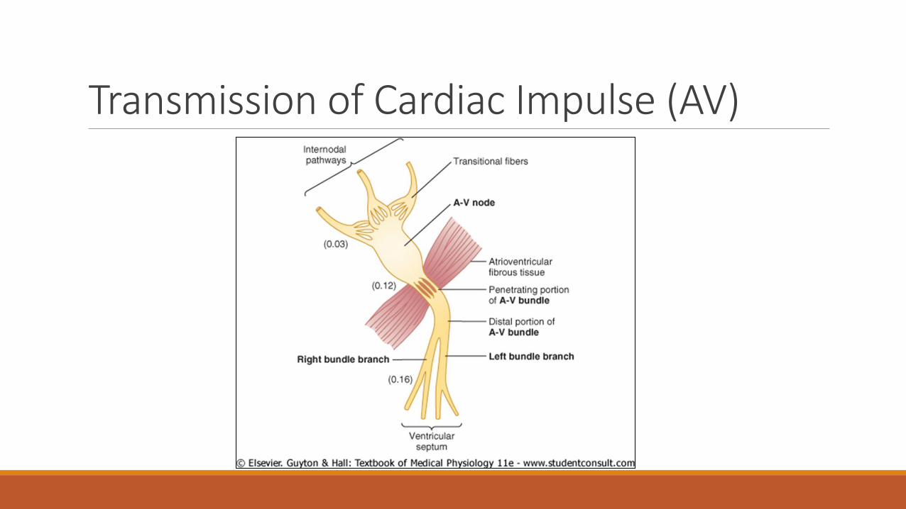
Transmission of Cardiac Impulse (AV)

Transmission of Cardiac Impulse (Purkinje)
Transmission is 6 times faster than in AV node
Action potentials can not travel backwards

Control of Excitation and Conduction ◦ Pacemaker of the heart (Sinus Node)
- cardiac impulse transmission does not always necessarily start in the Sinus node
- When start in the A-V nodal fibers and discharge at 40 to 60 times per minute or in the purkinje fibers
where they discharge between 15 to 40 times per minute
-regardless, cardiac transmissions mainly discharge from the sinus node
- 70-80 times per minute
- it’s discharge also excites the a-v nodal fibers and the purkinje fibers
- discharges the a-v nodal or purkinje fibers before they can self excite
- Sinus node controls the beat of the heart as the “pace maker” because it has the fastest rhythmical discharge

Abnormal Pacemaker ◦ “Ectopic” Pacemaker
- pacemaker anywhere other than the sinus node, causes abnormal contraction in heart and debility of the heart pumping
- this happens when another part of the heart such as the a-v node or purkinje fibers develop a rhythmic discharge faster than that of
the sinus node
- signal is blocked from the sinus node and pacemaker starts in the a-v node on the way to the ventricles
- atria continues to beat at normal rate of sinus node
- pacemaker develops in purkinje system for the ventricles to contract ventricle muscle
- purkinje “pacemaker” starts 5-20 seconds later because fibers are suppressed from previous sinus over-load
- during these 5-20 seconds the ventricle fails to pump blood and the person faints after 4-5 seconds
- this whole process is called Stokes-Adams syndrome and can lead to death if delay period is too long.
Purkinje System and synchronous contraction
◦ Purkinje system contractions happen only 0.03 to 0.06 apart
◦ - contractions are happening at almost the exact same time
◦ - any slowing in the purkinje fiber signaling will result in irregular contraction of the ventricle
◦ - this leads to many debilities of the heart and can decrease pumping effectiveness by 20-30 percent

Sympathetic and Parasympathetic Nerves
◦ Parasympathetic nerves (vagi)
◦ - mainly distributed to S-A and A-V nodes, and a small amount extend to the atria with even less to the ventricles
◦ Sympathetic nerves
◦ - conversely, distributed to all areas of the heart
Parasympathetic stimulation
◦ Can slow or even block cardiac rhythm
◦ - parasympathetic stimulation causes release of acetylcholine
- decreases rate of sinus node, and decreases excitability of the A-V junction fibers slowing contraction of ventricles
- can block complete transmission from atria to ventricles through A-V node
- ventricle stops beating for 5 to 20 seconds until purkinje fibers kick in
- this is called ventricular escape
- Acetylcholine release increases permeability of nerve endings and allows leakage of potassium causing “hyperpolarization”
- Because of this, membrane potential takes much longer to reach the threshold potential for excitation
- Because of this there are moderate delays or possible blocks the rate of rhythmicity of the nodal fibers.
Sympathetic stimulation
◦ Is responsible for the exact opposite effects of parasympathetic (vegal) stimulation
◦ - increases rate of nodal discharge
- Increases rate of conduction and level of excitability in all nervous fibers
- Increases force of contraction of all cardiac musculature (atrial/venticle)
- Basically increases overall function of the heart as much as two fold

Mechanisms of sympathetic stimulation
◦ Begins with the secretion of norepinephrine in sympathetic nerve endings
◦ - norepinephrine stimulates beta-1 andrenegic receptors
◦ - the exact effect is unclear but is believed to increase permeability of fiber membrane causing increase sodium-calcium levels
◦ - this causes a more positive resting potential increasing the threshold level for self excitation therefore increasing heart rate
◦ - A-V node and A-V bundles increase in sodium- calcium permeability makes action potential easier and therefore exciting each
conducting fiber faster and increasing conduction time from the atria to the ventricles
Top Related