


![[MDMA]MDMA Neurochemistry](https://static.fdocuments.us/doc/165x107/577dab601a28ab223f8c57f3/mdmamdma-neurochemistry.jpg)


Languages
Pages
Legal


Review of Neurochemistry
UNIVERSITY OF PNGSCHOOL OF MEDICINE AND HEALTH SCIENCES
DISCIPLINE OF BIOCHEMISTRY AND MOLECULAR BIOLOGYPBL MBBS YEAR III SEMINAR
VJ Temple
1
CEREBRAL METABOLISMWhat are some major uses of energy in nervous system?
• Neuron is an excitable cell: capable of changing membrane potential
• Excitation and Conduction are major functions of nervous tissues
• Reflected in unceasing electrical activity of cerebral tissue
• Electrical activity requires energy derived from metabolism
• Energy required for Active Transport of ions needed to Sustain and Restore Membrane Potentials discharged during Excitation and Conduction;
2

• Energy required for maintenance of blood-brain barrier;
• Energy required for biosynthesis of neurotransmitters and other components;
• Oxygen and Glucose are major substrates for energy production in cerebral tissue;
3
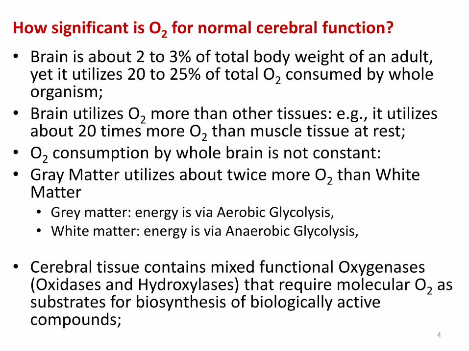
How significant is O2 for normal cerebral function?
• Brain is about 2 to 3% of total body weight of an adult, yet it utilizes 20 to 25% of total O2 consumed by whole organism;
• Brain utilizes O2 more than other tissues: e.g., it utilizes about 20 times more O2 than muscle tissue at rest;
• O2 consumption by whole brain is not constant:• Gray Matter utilizes about twice more O2 than White
Matter • Grey matter: energy is via Aerobic Glycolysis,• White matter: energy is via Anaerobic Glycolysis,
• Cerebral tissue contains mixed functional Oxygenases (Oxidases and Hydroxylases) that require molecular O2 as substrates for biosynthesis of biologically active compounds;
4

• Brain requires continuous supply of O2 because O2
stored in brain is limited compared to rate of utilization;
• During Ischemia, consciousness is loss within a few seconds, or time required for consuming O2 contained within brain and its blood content ;
• Reduced cerebral O2 uptake occurs under certain conditions that lead to depressed consciousness, eg:
• Hypoglycemia,
• Diabetic Coma,
• Cerebral Tumors,
• Uremia,
• Gross Liver damage leading to hepatic coma,
• Anesthetic used during surgery5
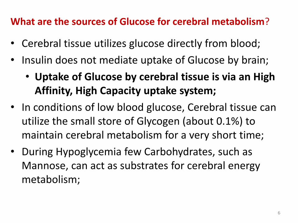
What are the sources of Glucose for cerebral metabolism?
• Cerebral tissue utilizes glucose directly from blood;
• Insulin does not mediate uptake of Glucose by brain;
• Uptake of Glucose by cerebral tissue is via an High Affinity, High Capacity uptake system;
• In conditions of low blood glucose, Cerebral tissue can utilize the small store of Glycogen (about 0.1%) to maintain cerebral metabolism for a very short time;
• During Hypoglycemia few Carbohydrates, such as Mannose, can act as substrates for cerebral energy metabolism;
6

• Maltose, Fructose, Galactose, Hexosephosphates, Lactate, Pyruvate and Glyceraldehyde cannot directly act as substrates for cerebral energy metabolism;
• They can act as substrate only after their conversion to glucose via Gluconeogenesis in the liver;
• These compounds act by raising blood glucose level;
• In absence of glucose brain can utilize Mannosedirectly and rapidly to restore or maintain normal metabolism;
7
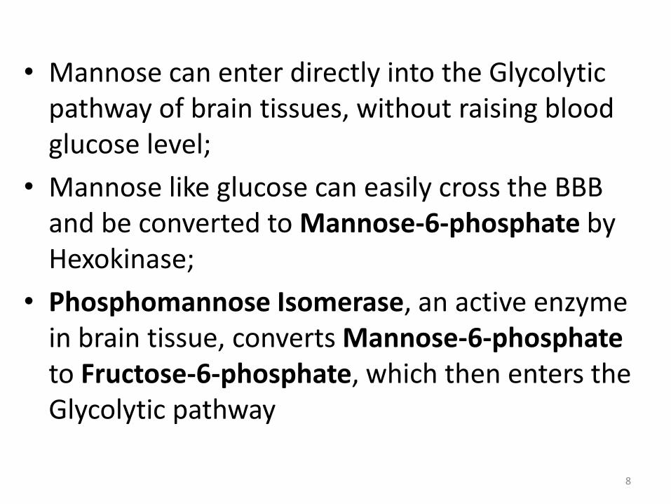
• Mannose can enter directly into the Glycolytic pathway of brain tissues, without raising blood glucose level;
• Mannose like glucose can easily cross the BBB and be converted to Mannose-6-phosphate by Hexokinase;
• Phosphomannose Isomerase, an active enzyme in brain tissue, converts Mannose-6-phosphateto Fructose-6-phosphate, which then enters the Glycolytic pathway
8
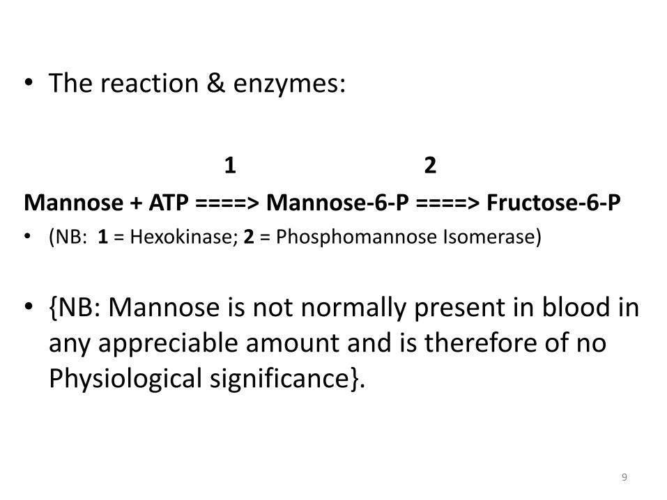
• The reaction & enzymes:
1 2
Mannose + ATP ====> Mannose-6-P ====> Fructose-6-P
• (NB: 1 = Hexokinase; 2 = Phosphomannose Isomerase)
• {NB: Mannose is not normally present in blood in any appreciable amount and is therefore of no Physiological significance}.
9

How is ammonia metabolized in cerebral tissue?
• Urea cycle is not the major pathway for removal of Ammonia in Cerebral tissue;
• Mitochondrial N-Acetyl-Glutamate activated Carbamoyl-Phosphate Synthetase that catalyzes the first reaction in Urea cycle, is low or absent in Cerebral tissue;
• Removal of Ammonia from Cerebral tissue involves two reactions:
10
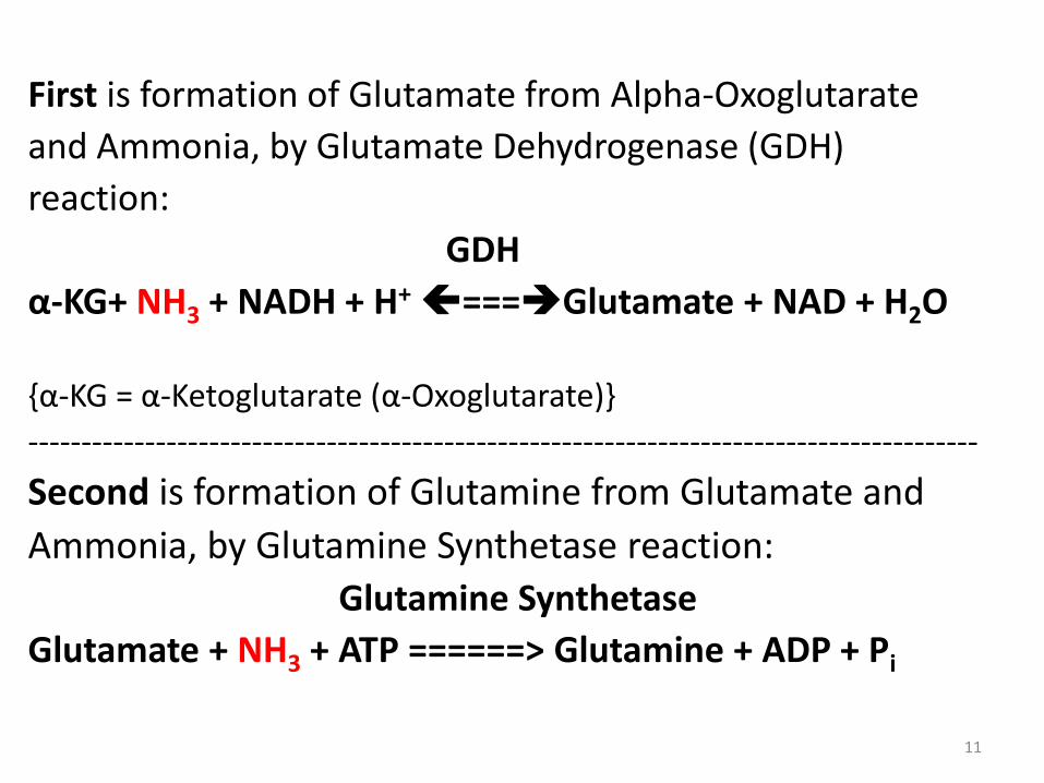
First is formation of Glutamate from Alpha-Oxoglutarate
and Ammonia, by Glutamate Dehydrogenase (GDH)
reaction:
GDH
α-KG+ NH3 + NADH + H+===Glutamate + NAD + H2O
{α-KG = α-Ketoglutarate (α-Oxoglutarate)}
-----------------------------------------------------------------------------------------
Second is formation of Glutamine from Glutamate and
Ammonia, by Glutamine Synthetase reaction:
Glutamine Synthetase
Glutamate + NH3 + ATP ======> Glutamine + ADP + Pi
11
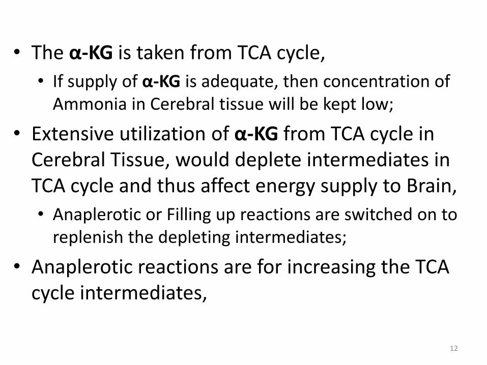
• The α-KG is taken from TCA cycle,
• If supply of α-KG is adequate, then concentration of Ammonia in Cerebral tissue will be kept low;
• Extensive utilization of α-KG from TCA cycle in Cerebral Tissue, would deplete intermediates in TCA cycle and thus affect energy supply to Brain,
• Anaplerotic or Filling up reactions are switched on to replenish the depleting intermediates;
• Anaplerotic reactions are for increasing the TCA cycle intermediates,
12

Give examples of the Anaplerotic reactions:
• Pyruvate Carboxylase reaction:
• catalyzes formation of Pyruvate from Oxaloacetate using ATP and Biotin;
• Transamination reactions:
• forms Alpha-Oxoglutarate, a TCA cycle intermediate
• Glutamate Dehydrogenase reaction:
• forms Alpha-Oxoglutarate
• Succinyl-CoA formation from Isoleucine, Valine, Methionine, and Threonine;
13
SOME FACTORS THAT CAN AFFECT CEREBRAL METABOLISM
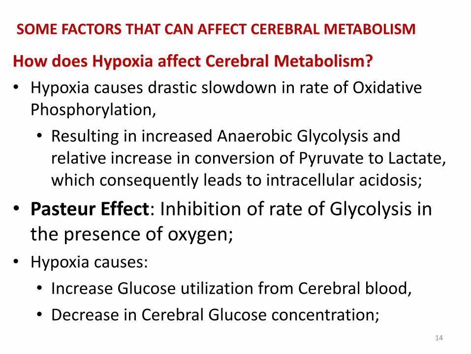
How does Hypoxia affect Cerebral Metabolism?
• Hypoxia causes drastic slowdown in rate of Oxidative Phosphorylation,
• Resulting in increased Anaerobic Glycolysis and relative increase in conversion of Pyruvate to Lactate, which consequently leads to intracellular acidosis;
• Pasteur Effect: Inhibition of rate of Glycolysis in the presence of oxygen;
• Hypoxia causes:
• Increase Glucose utilization from Cerebral blood,
• Decrease in Cerebral Glucose concentration;14

• Major effects of hypoxia on nervous system:
• Reduction in Rate of Conversion of Pyruvate to Acetyl-CoA with a resultant decrease in both biosynthesis of Acetylcholine and Activity of TCA cycle;
• In situations of low Acetyl-CoA availability the Cerebral tissue may use the available Acetyl-CoA for energy production so as to maintain membrane potentials in preference to its use in the biosynthesis of compounds;
15

How does Ischemia affect Cerebral Metabolism?
• Ischemia: Glucose and Oxygen supply to Cerebral tissue are deficient;
During Ischemia:
• Glucose and O2 supply are deficient;
• Cerebral Glucose level and Glycogen store are depleted;
• Coma can occur leading to cerebral tissue damage;
16

How does hypoglycemia affect cerebral metabolism?
• Hypoglycemia severely affect cerebral energy metabolism because, the brain uses glucose almost exclusively as substrate for energy metabolism;
• Nervous system does not depend on Insulin for uptake of Glucose from blood;
• During starvation Cerebral Tissue can use Ketone bodies (Beta-hydroxybutyrate, and Acetone) as substrate for energy metabolism;
• Ketone Bodies are usually very high in blood during starvation, thus they are able to cross the BBB without much restriction;
17

NEUROTRANSMISSION
• Neuron is an excitable cell:
• it is capable of generating and conducting electrical impulse by temporarily reversing its membrane potential;
• Synapse: contact between two neurons;
• Neuromuscular Junction: contact between neuron and muscle fiber;
18

What are the 2 modes of impulse transmission?
Electrical mode of transmission:
• Impulse transmission along axon – Membrane transmission:• Involves propagation of Action Potential along axonal
membrane;
Chemical mode of transmission:
• Impulse transmission across synapse – Synaptic transmission,
• Impulse transmission from neuron to muscle –Neuromuscular transmission, • Involves chemical process by group of compounds
called Chemical Neurotransmitters;19

Give brief description of neurotransmitter Acetylcholine
• Acetylcholine (Ach) is excitatory neurotransmitter
• Neurons that synthesize and release ACh are termed Cholinergic neurons;
• ACh is synthesized from Choline and Acetyl-CoA;
• Reaction catalyzed by Acetylcholine Transferase (Choline Acetyl-transferase);
• Removal of ACh from receptors at Postsynaptic membrane is by hydrolysis catalyzed by True Acetyl-Cholinesterase located at nerve endings;
20

• ACh receptors are Ligand-Gated Cation channels composed of four different polypeptide subunits arranged in the form [(2)()()()];
• Two main classes of ACh receptors:
• Muscarinic receptors,
• Nicotinic receptors;
• Nicotinic receptors are divided into those at:
• Neuromuscular junctions,
• Neuronal synapses;
21

Give brief description of Catecholamine Neurotransmitters
• Catecholamines: Norepinephrine, Epinephrine and Dopamine;
• Precursor for their biosynthesis are Phenylalanine or Tyrosine;
• Catecholamines are neurotransmitters for the sympathetic nervous system effect;
• Catecholamines bind to two different classes of receptors termed - and -Adrenergic receptors, which are classical serpentine receptors that couple to intracellular G-proteins
22

• Catecholamines are remove from receptors on post-synaptic membrane by “Active Reuptake”;
• Catecholamines are degraded by sequential actions of the enzymes:
• Catecholamine-O-Methyl-Transferase (COMT),
• Monoamine Oxidase (MAO),
• Aldehyde Dehydrogenase;
23
Briefly comment on neurotransmitter Gamma Amino Butyric Acid (GABA)
• Gamma-Amino-Butyrate, (4-Aminobutyrate or GABA) is an inhibitor neurotransmitter;
• Biosynthesis of GABA occurs via Decarboxylation of Glutamate catalyzed by Glutamate Decarboxylase (GAD);
• Neurons that secrete GABA are called GABAergic neurons;
•
24

• GABA exerts action by binding to 2 distinct receptors
• GABA-A,
• GABA-B;
• GABA-A receptors form Chloride ion channel:
• Binding of GABA to GABA-A receptors increases Chloride ion conductance of Presynaptic neurons;
• GABA-B receptors are coupled to Intracellular G-protein and act by increasing conductance of associated K+ channel;
25
State some characteristics of Neurotransmitter Receptors
• Neurotransmitter receptors are located on the surface of Postsynaptic neurons and also on some Presynaptic neurons;
• Receptors on Presynaptic neurons act to inhibit further release of neurotransmitter;
• Link between neurotransmitter and Intracellular signaling is by association with one of the following:
• G-proteins,
• Protein Kinase,
• Receptor itself in the form of a Ligand-gated ion channel (e.g, Acetylcholine receptor)
26

• Vast majority of neurotransmitter receptors are Serpentine receptors, because they exhibits a characteristic transmembrane structure that spans the cell membrane seven times;
• Neurotransmitter receptors are subjected to ligand-Induced Desensitization:
• they can become unresponsive upon prolonged exposure to their neurotransmitter;
27
What are the effects of Neurotransmitters on Post-synaptic receptors?
• Some Neurotransmitters (e.g. Acetylcholine, Glycine, Glutamate, GABA) have “Inherent” biological activity;
• Act directly to cause increase in conductance to certain ions by binding to “Ligand-Activated” ion channels at the post-synaptic membrane;
• Other Neurotransmitters (e.g. Norepinephrine, Dopamine and Serotonin) have no direct activity but act indirectly via Second Messenger systems to bring about Post-synaptic response;
28

• 2nd Messengers (e.g. cAMP, cGMP, ITP, PGs, Epoxides and Ca2+ ions) act in the Cytosol to activate target proteins, including protein kinases, which in turn act on Gated Ion Channels, to produce neurotransmission effect;
• Excitatory neurotransmitters bind to receptors to cause depolarization of Post-synaptic membrane;
• Inhibitory neurotransmitters cause Hyper-polarization by increasing Chloride ion conductance of Post-synaptic membrane, thus making it more difficult for the cell to become depolarized;
29

Some Toxins & Diseases that affect Neuromuscular Junction & Synaptic Transmission
• ACh release in the NMJ is inhibited by Botulinum Toxin causing flaccid paralysis;
• Glycine release in the CNS is inhibited by Tetanus Toxin causing spastic paralysis;
• Black widow spider toxin, Alpha-Latrotoxin, stimulates fusion and depletion of neurotransmitter vesicles;
• Plant poison, Physostigmine, Nerve Gases and Organo-phosphorus pesticides inhibit Acetyl-cholinesterase in Cholinergic neurons;
30

• Muscle ACh receptor is blocked by the South American arrow poison, Curare;
• Plant drug, Atropine, inhibits ACh receptors of the Autonomic nervous system (but not the NMJ)
• Strychnine binds to Glycine receptor protein and inhibits IPSPs in the spinal cord;
• Cocaine blocks recycling of Dopamine and Norepinephrine neurotransmitters in the brain causing excitatory effect;
31
Top Related