


Languages
Pages
Legal


HAL Id: hal-01729757https://hal.archives-ouvertes.fr/hal-01729757
Submitted on 13 Mar 2018
HAL is a multi-disciplinary open accessarchive for the deposit and dissemination of sci-entific research documents, whether they are pub-lished or not. The documents may come fromteaching and research institutions in France orabroad, or from public or private research centers.
L’archive ouverte pluridisciplinaire HAL, estdestinée au dépôt et à la diffusion de documentsscientifiques de niveau recherche, publiés ou non,émanant des établissements d’enseignement et derecherche français ou étrangers, des laboratoirespublics ou privés.
Distributed under a Creative Commons Attribution| 4.0 International License
Respective impact of implementation of preventionstrategies, colonization with multiresistant bacteria and
antimicrobial use on the risk of early- and late-onsetVAP: An analysis of the OUTCOMEREA network
Wafa Ibn Saied, Bertrand Souweine, Maite Garrouste-Orgeas, StephaneRuckly, Michael Darmon, Sebastien Bailly, Yves Cohen, Elie Azoulay, Carole
Schwebel, Aguila Radjou, et al.
To cite this version:Wafa Ibn Saied, Bertrand Souweine, Maite Garrouste-Orgeas, Stephane Ruckly, Michael Darmon, etal.. Respective impact of implementation of prevention strategies, colonization with multiresistantbacteria and antimicrobial use on the risk of early- and late-onset VAP: An analysis of the OUTCOM-EREA network. PLoS ONE, Public Library of Science, 2017, 12 (11), pp.e0187791. �10.1371/jour-nal.pone.0187791�. �hal-01729757�

RESEARCH ARTICLE
Respective impact of implementation of
prevention strategies, colonization with
multiresistant bacteria and antimicrobial use
on the risk of early- and late-onset VAP: An
analysis of the OUTCOMEREA network
Wafa Ibn Saied1,2, Bertrand Souweine3, Maite Garrouste-Orgeas4, Stephane Ruckly1,
Michael Darmon5, Sebastien Bailly1,6, Yves Cohen7, Elie Azoulay8, Carole Schwebel2,
Aguila Radjou9, Hatem Kallel10, Christophe Adrie11, Anne-Sylvie Dumenil12,
Laurent Argaud13, Guillaume Marcotte14, Samir Jamali15, Laurent Papazian16,
Dany Goldgran-Toledano17, Lila Bouadma9, Jean-Francois Timsit1,9*, for the
OUTCOMEREA study group¶
1 UMR 1137 - IAME Team 5 – DeSCID: Decision SCiences in Infectious Diseases, control and care, Inserm/
Paris Diderot University, Sorbonne Paris Cite, Paris, France, 2 Medical Intensive care unit, Grenoble
University Hospital, Grenoble 1 University, U823, La Tronche, France, 3 Medical Intensive Care Unit, Gabriel
Montpied University Hospital, Clermont-Ferrand, France, 4 Intensive Care Unit, Saint Joseph Hospital
Network, Paris, France, 5 Saint Etienne University Hospital, Medical Intensive Care Unit, Saint-Etienne,
France, 6 Grenoble Alpes University, U823, Rond-point de la Chantourne, La Tronche France, 7 AP-HP,
Avicenne Hospital, Intensive Care Unit, Paris and Medicine University, Paris 13 University, Bobigny, France,
8 Medical Intensive Care Unit, AP-HP, Saint Louis Hospital, Paris, France, 9 AP-HP, Bichat Hospital,
Medical and infectious diseases Intensive Care Unit, Paris Diderot university, Paris, France, 10 Medical
Surgical ICU, Centre hospitalier de Cayenne, Guyane, France, 11 Physiology department, Cochin University
Hospital, Sorbonne Cite, Paris, France, 12 AP-HP, Antoine Beclère University Hospital, Medical-surgical
Intensive Care Unit, Clamart, France, 13 Medical Intensive Care Unit, Lyon University Hospital, Lyon,
France, 14 Surgical ICU, Edouard Herriot University Hospital, Lyon, France, 15 Critical care Medicine Unit
Dourdan Hospital, Dourdan, France, 16 Respiratory and infectious diseases ICU, APHM Hopital Nord, Aix
Marseille University, Marseille, France, 17 Gonesse Hospital, Intensive Care Unit, Gonesse, France
¶ Membership of the OUTCOMEREA study group is provided in the Acknowledgments.
Abstract
Rationale
The impact of prevention strategies and risk factors for early-onset (EOP) versus late-onset
(LOP) ventilator-associated pneumonia (VAP) are still debated.
Objectives
To evaluate, in a multicenter cohort, the risk factors for EOP and LOP, as the evolution of
prevention strategies.
Methods
7,784 patients with mechanical ventilation (MV) for at least 48 hours were selected into the
multicenter prospective OUTCOMEREA database (1997–2016). VAP occurring between
PLOS ONE | https://doi.org/10.1371/journal.pone.0187791 November 29, 2017 1 / 16
a1111111111
a1111111111
a1111111111
a1111111111
a1111111111
OPENACCESS
Citation: Ibn Saied W, Souweine B, Garrouste-
Orgeas M, Ruckly S, Darmon M, Bailly S, et al.
(2017) Respective impact of implementation of
prevention strategies, colonization with
multiresistant bacteria and antimicrobial use on the
risk of early- and late-onset VAP: An analysis of the
OUTCOMEREA network. PLoS ONE 12(11):
e0187791. https://doi.org/10.1371/journal.
pone.0187791
Editor: Yu Ru Kou, National Yang-Ming University,
TAIWAN
Received: November 7, 2016
Accepted: October 26, 2017
Published: November 29, 2017
Copyright: © 2017 Ibn Saied et al. This is an open
access article distributed under the terms of the
Creative Commons Attribution License, which
permits unrestricted use, distribution, and
reproduction in any medium, provided the original
author and source are credited.
Data Availability Statement: All relevant data are
within the paper and its Supporting Information
files.
Funding: The study was entirely funded by the
OUTCOMEREA research network. The funder had
no role in study design, data collection and
analysis, decision to publish, or preparation of the
manuscript.
the 3rd and 6th day of MV defined EOP, while those occurring after defined LOPs. We used a
Fine and Gray subdistribution model to take the successful extubation into account as a
competing event.
Measurements and main results
Overall, 1,234 included patients developed VAP (EOP: 445 (36%); LOP: 789 (64%)). Male
gender was a risk factor for both EOP and LOP. Factors specifically associated with EOP
were admission for respiratory distress, previous colonization with multidrug-resistant Pseu-
domonas aeruginosa, chest tube and enteral feeding within the first 2 days of MV. Antimicro-
bials administrated within the first 2 days of MV were all protective of EOP. ICU admission
for COPD exacerbation or pneumonia were early risk factors for LOP, while imidazole
and vancomycin use within the first 2 days of MV were protective factors. Late risk factors
(between the 3rd and the 6th day of MV) were the intra-hospital transport, PAO2-FIO2<200
mmHg, vasopressor use, and known colonization with methicillin-resistant Staphylococcus
aureus. Among the antimicrobials administered between the 3rd and the 6th day, fluoroquin-
olones were the solely protective one.Contrarily to LOP, the risk of EOP decreased across
the study time periods, concomitantly with an increase in the compliance with bundle of pre-
vention measures.
Conclusion
VAP risk factors are mostly different according to the pneumonia time of onset, which should
lead to differentiated prevention strategies.
Introduction
Ventilator-acquired pneumonia (VAP) is defined as pneumonia arising after at least 48 hours
of mechanical ventilation (MV). VAP represents the leading type of nosocomial infection in
intensive care units (ICUs); its rate ranges from 8 to 25% among mechanically ventilated
patients [1]. VAP is associated with a significant increase in the mortality risk for ICU patients,
from 3 to 8% when using new statistical model for causal inference [2, 3]. It increases the
length of ICU stay by up to 9 days [1].
The major route for acquiring VAP is oropharyngeal colonization by the endogenous flora
or by pathogens acquired exogenously from the intensive care unit environment, especially the
hands of health-care workers or the contaminated respiratory equipment. The stomach repre-
sents a potential site of secondary colonization and reservoir of nosocomial Gram-negative
bacilli. On endotracheal-tube, the formation of biofilm may play a contributory role in sustain-
ing tracheal colonization, paving the way for VAP mostly caused by resistant organisms such
as methicillin-resistant staphylococcus aureus (MRSA) and non-fermentative gram-negative
bacilli (GNB). The immunoparalysis occurring after the initial phase of resuscitation may also
play a role in decreasing resistance to infections [4–6]
The changes in the pathogenesis of VAP over time have led to differentiate early-onset
pneumonia (EOP) from late-onset pneumonia (LOP) [7]. Time-points used for differentiating
them are slightly different according to studies, from >3 to�7 days of mechanical ventilation
[8–10].
Colonization with multiresistant bacteria and antimicrobial use on the risk of VAP
PLOS ONE | https://doi.org/10.1371/journal.pone.0187791 November 29, 2017 2 / 16
Competing interests: The authors have declared
that no competing interests exist.

According to the differences in the pathogenesis, different preventive measures might be
implemented. Indeed, the rapid decrease in the endogenous flora inoculum at the early phase
of ventilation by antimicrobial prophylaxis [11], selective digestive and oral decontamination,
subglottic aspiration [12, 13], semi-recumbent position [14], decreased the risk of EOP, but
have no or a moderate impact on LOP. When preventive bundles such as weaning trials, oro-
gastric tubes, semi-recumbent position, or oral care were applied, the decrease in VAP rates
was greatest for the period within 5 days after ICU admission and MV initiation, suggesting
that preventive measures were more effective when implemented early[6]. On the opposite, it
was shown that the constitution of a secondary reservoir in the stomach promotes infections
occurring later that might be partly prevented by the preservation of a low gastric pH [15].
The aim of our study was to assess specific risk factors for EOP and for LOP in a large mul-
ticenter database, and the impact of the progressive implementation of recommended preven-
tive strategies on the rates of onset of EOP and of LOP.
Methods
Study design and data source
We conducted a retrospective analysis using data prospectively entered into a multicenter
database from January 1997 to March 2016 (OUTCOMEREA database). The database is fed
by 22 different French ICUs, and contains data on admission features and diagnosis, daily dis-
ease severity, iatrogenic events, nosocomial infections, and vital status. ICU units participating
in the OUTCOMEREA database used a range of methods to include patients. Some ICUs
included all consecutive patients, while other units included patients admitted at predefined
periods of the year, and some other picked patients in certain beds that were randomly
selected, without considering patient’s characteristics such as the diseases at ICU admission
or their medical history [16]. All patients with MV lasting more than 48 hours were included
into the study. Patients were censored after 28 days of MV or death (Fig 1). Risk factors were
Fig 1. Flowchart.
https://doi.org/10.1371/journal.pone.0187791.g001
Colonization with multiresistant bacteria and antimicrobial use on the risk of VAP
PLOS ONE | https://doi.org/10.1371/journal.pone.0187791 November 29, 2017 3 / 16

evaluated for the first episode of EOP on the individuals under mechanical ventilation for
more than 2 days, and a similar analysis was performed for the first episode of LOP on the
patients who were still mechanically ventilated on the 7th day.
Ethical issues. This study was approved by our institutional review board (CECIC Cler-
mont-Ferrand—IRB n˚5891; Ref: 2007–16), which waived the need for signed informed con-
sent of the participants, in accordance with French legislation on non-interventional studies.
However, the patients and their next of kin were asked whether they were willing to participate
in the database, and none declined participation.
Population definitions. The diagnosis of VAP was suspected in patients who had
received at least 48 hours of MV and developed a new or persistent infiltrate on chest radiogra-
phy, associated with one of the following criteria: (1) purulent tracheal secretions, (2) fever
greater than or equal to 38.5˚C or hypothermia less than or equal to 36.5˚C, and (3) leukocyto-
sis greater than 109 cells/L or leukopenia less than 4.108 cells/L. The diagnosis of VAP was con-
firmed by a positive quantitative culture of a respiratory sample: bronchoalveolar lavage fluid
(significant threshold:� 104cfu/ml) or plugged telescopic catheter (significant threshold:�
103cfu/ml) or quantitative endotracheal aspirate (significant threshold,�105 cfu/ml). The
model was only based on confirmed VAPs with a positive result after bacteriological culture.
When VAP was suspected, samples were performed prior to any changes in the antibiotic
therapy.
We chose a cut-off point at day 7 of MV. EOP was defined as the occurrence of VAP
between the 3rd and the 6th day of MV. LOP was defined as the occurrence of VAP after the 6th
day of MV.
A new episode of VAP was defined either by: (1) the same causative microorganism after a
time interval of at least 5 days from the previous episode; (2) the acquisition of resistance of
the same pathogen; or (3) a different pathogen regardless the acquisition day. Positive samples
for fungi or viruses were not considered.
We also recorded data on previous carriage or colonization with MDR bacteria: multi-
drug-resistant P. aeruginosa (MDRPA), MRSA, extended-spectrum β-lactamase producing
Enterobacteriaceae (ESBLE), and A. baumannii. A patient was considered as colonized with
MDR bacteria if one of these 4 microorganisms was isolated from clinically-indicated samples,
or from samples drawn for systematic detection for MRSA and ESBLE, i.e., samples collected
at admission and weekly, from nasal and rectal swabs for MRSA and ESBLE, respectively. If
confirmed, patient was considered as carrier throughout the full study period.
Data collection. The following data were collected at admission, and daily if appropri-
ate: age; sex; comorbidities (assessed according to the Acute Physiology and Chronic Health
Evaluation II definitions); severity of illness at ICU admission and daily during the ICU stay
(assessed using the Simplified Acute Physiology Score II and the Logistic Organ Dysfunction
score); admission category (medical, surgery, or monitoring); admission diagnosis; whether
the patient was transferred from a hospital ward (defined as a stay in an acute-bed ward
lasting>24 h immediately before ICU admission); lengths of ICU stay and hospital stay; and
vital status at discharge from the ICU and from the hospital. Data on invasive procedures
(placement of an arterial or central venous catheter, and endotracheal intubation), treat-
ments of organ failures (catecholamine infusion, mechanical ventilation, renal replacement
therapy), colonization with ESBLE, MDRPA and MRSA, and antibiotic use were also cap-
tured daily.
Prevention bundle applied during each of the study periods: before 2001, between 2001 and
2006 and after 2006, was recorded using questionnaires filled by investigators (see description
in S1 Table). The compliance with prevention protocols was not monitored. We described
EOP and LOP tendency taking account of the modification of bundles over these 3 periods.
Colonization with multiresistant bacteria and antimicrobial use on the risk of VAP
PLOS ONE | https://doi.org/10.1371/journal.pone.0187791 November 29, 2017 4 / 16

Statistical analysis. Characteristics of patients and VAP episodes were described using
frequency and percentage for qualitative variables, median, and interquartile range (IQR) for
quantitative variables. Standard survival analyses are affected by the time of onset of the event
of interest. Patients who have not experienced the event at the end of follow-up were censored.
To determine the risk of an event occurrence at a certain time-point, a fundamental assump-
tion is that such censoring is not associated with an altered chance of the event occurring at
any given moment. In this study, the event of interest is the onset of pneumonia in patients
with MV and followed up until day 28 or until extubation for more than 48 hours. Indeed,
death and extubation for more than 48 hours is a competing event since, by definition, extuba-
tion precludes the observation of a ventilator-associated infection [17]. Risk factors of VAP
(EOP and LOP) occurrence were identified separately using Fine and Gray models stratified
by ICU. A multivariate model was built by including risk factors (at admission, the 2 first days
of MV for EOP, and between the third and the sixth day of MV for LOP) that met the 20% sig-
nificance threshold in univariate analysis. SAPS II at the beginning of mechanical ventilation
period was selected for the model, instead of organ failure scores and age. Then, a backward
selection process was applied until all remaining factors met the 5% threshold in the multivari-
ate context. For EOP as for LOP, some factors are specific to the patient, such as the male gen-
der; others were recorded at ICU admission, and others emerged later on, up to the 3rd to the
6th day after MV initiation for LOP. Sensitivity Analysis was also performed by varying MV
cut-off to five days for the definition of EOP and LOP without modification in the final model
(S2 Table). All models were stratified by centers and adjusted on time periods and SAPS II at
the beginning of mechanical ventilation period that correspond to the main changes in preven-
tion strategies. Results were given as subdistribution hazard ratios (sHR) and 95% confidence
intervals (CI). Using P values less than 0.05 were considered as significant. As it is an explor-
atory trial, p-values were further corrected for multiple comparisons using the Benjamini-
Hochberg procedure, as implemented in the MULTITEST procedure in SAS. Statistical analy-
sis was performed using SAS 9.4 (Cary, NC).
Results
Overall, 7,784 ICU patients with mechanically ventilation for more than 48 hours were
included (see CONSORT flowchart, Fig 1), including 1,234 (15.85%) patients who developed
VAP within the first 28 days of MV. Among them, 445 (36%) developed an EOP, and 789
(64%) developed a LOP. Characteristics at ICU admission, during the first 48 hours of MV
and between the third day of MV and the sixth day of MV for patients at risk for EOP and
LOP are described in Tables 1 and 2 respectively. The main causative pathogens according to
the type of VAP are described in Table 3.
Incidence of EOP, but not of LOP, decreased with the study time periods. Risk factors
selected separately for EOP and LOP at the final step of the multivariate models are reported
on Table 4. Male gender was the only risk factor common for EOP and LOP.
Factors that may lead to a decreased bronchial clearance or aspiration such as early enteral
feeding or presence of a chest tube were only associated with EOP. On the contrary, severe
organ dysfunction (low PaO2:FiO2 ratio) was associated with LOP.
Most antibacterial agents used within the first 48 hours of MV protected against EOP.The
early administration of Vancomycin and imidazole, and fluoroquinolones administered
between day 3 and 6 protected from LOP.
In term of previous carriage or colonization with multi-drug resistant pathogens, coloniza-
tion with MDRPA was a risk factor for EOP; whereas colonization with MRSA was a risk fac-
tor for LOP. When MRSA colonization was present on admission, it had a lower impact than
Colonization with multiresistant bacteria and antimicrobial use on the risk of VAP
PLOS ONE | https://doi.org/10.1371/journal.pone.0187791 November 29, 2017 5 / 16
MRSA colonization was acquired later on, between the 3rd and 6th day of mechanical ventila-
tion (15/34 vs 38/179, p<0.01).
Varying MV cut-off to five days for the definition of EOP and LOP had no impact on the
final model (S2 Table).
Discussion
Using commonly recognized definitions and quantitative bacterial samples, this study
included a large multicenter population at risk for VAP, and aimed at identifying and differen-
tiating risk factors associated with EOP and LOP, respectively. As one strategy for VAP pre-
vention might be to decrease the involved risk factors, identifying the risk factors involved in
VAP onset in each patient is critical. We used competing risk models in order to take account
of the informative censor due to extubation for more than 48 hours and death. Among the
mechanically-ventilated ICU patients included in our study, the incidence rate of VAP was
about 19%, similar to the values reported in the literature [5, 10]. In patients with VAP, we
observed rates of EOP and LOP of 40.2% and 59.8%, respectively, consistent with those
observed in other studies [2, 18–20]. The distribution between EOP and LOP depends on the
diagnostic method for pneumonia, and on the cut-off points used for the definition. We used a
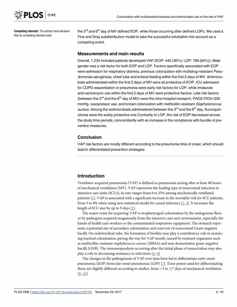
Table 1. Characteristics of study population at the ICU admission.
Variables at ICU admission Patients at risk for EOP (N = 7,784) Patients at risk for LOP (N = 3,878)
No EOP (N = 7,339) EOP (N = 445) P* No LOP (N = 3,089) LOP (N = 789) P**
Age (#miss = 5) 64.6 [52.1; 75.8] 64.1 [51.2; 72.8] 0.09 66.6 [54.6; 76.3] 65.1 [53.2; 75.7] 0.02
Male gender 4,564 (62.2) 312 (70.1) <.01 1,939 (62.8) 545 (69.1) <.01
Chronic diseases at ICU admission
Hepatic 528 (7.2) 22 (4.9) 0.07 197 (6.4) 57 (7.2) 0.39
Cardiovascular 1,049 (14.3) 67 (15.1) 0.66 491 (15.9) 110 (13.9) 0.18
Respiratory 1,182 (16.1) 78 (17.5) 0.43 587 (19) 165 (20.9) 0.23
Renal 355 (4.8) 30 (6.7) 0.07 147 (4.8) 29 (3.7) 0.19
Immunodeficiency 1,184 (16.1) 54 (12.1) 0.03 519 (16.8) 145 (18.4) 0.29
Diabetes 0.32
Complicated diabetes 333 (4.5) 19 (4.3) 0.71 144 (4.7) 39 (4.9)
Non complicated diabetes 810 (11) 44 (9.9) 351 (11.4) 75 (9.5)
No diabetes 6,196 (84.4) 382 (85.8) 2,594 (84) 675 (85.6)
Diagnosis category at ICU admission (#miss = 18) 0.03
Scheduled surgery 639 (8.7) 40 (9) 0.05 242 (7.8) 65 (8.2)
Emergency surgery 1,383 (18.8) 63 (14.2) 581 (18.8) 117 (14.8)
Medical 5,317 (72.4) 342 (76.9) 2,266 (73.4) 607 (76.9)
Diagnosis at ICU admission
Septic shock 1,486 (20.2) 46 (10.3) <.01 777 (25.2) 169 (21.4) 0.03
Other shock 1,088 (14.8) 69 (15.5) 0.70 444 (14.4) 15 (1.9) 0.56
Respiratory distress 2,172 (29.6) 157 (35.3) 0.01 1,090 (35.3) 252 (31.9) <.01
COPD exacerbation 412 (5.6) 26 (5.8) 0.84 213 (6.9) 73 (9.3) 0.05
Coma 1,759 (24) 131 (29.4) <.01 530 (17.2) 127 (16.1) 0.48
Pneumonia 1,507 (20.5) 105 (23.6) 0.12 773 (25) 252 (31.9) <.01
Drug overdose 384 (5.2) 23 (5.2) 0.95 52 (1.7) 13 (1.6) 0.94
P* = p value comparing EOP and No EOP patients /P** = p value comparing LOP and No LOP patients; EOP early onset pneumonia; LOP: late onset
pneumonia; MV: mechanical ventilation; COPD exacerbation = chronic obstructive pulmonary disease exacerbation; MV = mechanical ventilation; # miss:
number of missing data
https://doi.org/10.1371/journal.pone.0187791.t001
Colonization with multiresistant bacteria and antimicrobial use on the risk of VAP
PLOS ONE | https://doi.org/10.1371/journal.pone.0187791 November 29, 2017 6 / 16

Table 2. Characteristics of patients at risk for early and late onset pneumonia during their stay in ICU.
Characteristics Patients at risk for EOP (N = 7,784) Patients at risk for LOP (N = 3,878)
No EOP (N = 7,339) EOP (N = 445) P* No LOP (N = 3,089) LOP (N = 789) P**
Variables within the first 48 hours of MV
SAPSII score 48 [37; 59] 47 [37; 58] 50 [40; 60] 48 [39; 59]
�38 2,034 (27.7) 121 (27.2) 0.33 701 (22.7) 196 (24.8) 0.18
37–48 1,762 (24) 121 (27.2) 741 (24) 207 (26.2)
49–59 1,716 (23.4) 106 (23.8) 837 (27.1) 193 (24.5)
�60 1,827 (24.9) 97 (21.8) 810 (26.2) 193 (24.5)
Effective Glasgow† 4 [3; 8] 4 [3; 7] 4 [3; 8] 3 [3; 7]
� 5 3,693 (50.3) 237 (53.3) 0.02 1,559 (50.5) 453 (57.4) <.01
5>Glasgow<13 2,761 (37.6) 174 (39.1) 1,194 (38.7) 255 (32.3)
� 13 885 (12.1) 34 (7.6) 336 (10.9) 81 (10.3)
PAO2:FIO2, median (IQR) (# miss = 150)
<200mm Hg 3783 (51.5) 233 (52.4) 0.74 1766 (57.2) 515 (65.3) <.01
�200mm Hg 3556 (48.5) 212 (47.6) 1,364 (44.2) 274 (34.7)
Microbial colonization
ESBLE 186 (2.5) 11 (2.5) 0.94 98 (3.2) 22 (2.8) 0.58
MDR PA 76 (1) 9 (2) 0.05 38 (1.2) 12 (1.5) 0.52
Acinetobacter baumannii 216 (2.9) 12 (2.7) 0.76 113 (3.7) 27 (3.4) 0.75
MRSA 279 (3.8) 18 (4) 0.79 141 (4.6) 38 (4.8) 0.76
Treatments and procedures
Unplanned extubation 200 (2.7) 13 (2.9) 0.81 59 (1.9) 19 (2.4) 0.37
Chest tube 566 (7.7) 54 (12.1) <.01 263 (8.5) 84 (10.6) 0.06
Steroids 2,049 (27.9) 100 (22.5) 0.01 982 (31.8) 262 (33.2) 0.45
Paralytic agents 1,477 (20.1) 98 (22) 0.33 699 (22.6) 230 (29.2) 0.16
Proton Pump Inhibitors 4,526 (61.7) 250 (56.2) 0.02 1,940 (62.8) 471 (59.7) 0.11
Enteral feeding 2,320 (31.6) 177 (39.8) <.01 1,122 (36.3) 307 (38.9) 0.18
Intra-hospital transport 2,069 (28.2) 108 (24.3) 0.07 856 (27.7) 220 (27.9) 0.92
Patient isolation 1,288 (17.6) 64 (14.4) 0.09 574 (18.6) 151 (19.1) 0.72
Antibacterial agents administration
3rd and 4th generation cephalosporins 1,810 (24.7) 76 (17.1) <.01 901 (29.2) 246 (31.2) 0.33
ß-lactam/ ß-lactamase inhibitor 2,691 (36.7) 135 (30.3) <.01 1,107 (35.8) 295 (37.4) 0.09
Other penicillins 755 (10.3) 34 (7.6) 0.07 353 (11.4) 86 (10.9) 0.68
Penems 600 (8.2) 10 (2.2) <.01 323 (10.5) 64 (8.1) 0.05
Fluoroquinolones 1,205 (16.4) 44 (9.9) <.01 633 (20.5) 151 (19.1) 0.40
Aminoglycosides 1,784 (24.3) 51 (11.5) <.01 914 (29.6) 202 (25.6) 0.03
Imidazole 849 (11.6) 30 (6.7) <.01 448 (14.5) 86 (10.9) 0.71
Vancomycin 956 (13) 27 (6.1) <.01 519 (16.8) 94 (11.9) <.01
Other antibacterial agents 825 (11.2) 57 (12.8) 0.31 322 (10.4) 100 (12.7) 0.07
Variables between the 3 rd and the 6th days of MV
SAPSII score - - 46 [37; 56] 46 [39; 57] 0.51
�37 713 (23.1) 162 (20.5)
37–57 1,660 (53.7) 442 (56)
�57 716 (23.2) 185 (23.4)
Effective Glasgow† - - 6 [3; 10] 5 [3; 8] <.01
� 5 1,192 (38.6) 379 (48)
5>Glasgow<13 1484 (48) 319 (40.4)
� 13 413 (13.4) 91 (11.5)
(Continued )
Colonization with multiresistant bacteria and antimicrobial use on the risk of VAP
PLOS ONE | https://doi.org/10.1371/journal.pone.0187791 November 29, 2017 7 / 16
cut-off point of� 7 days of MV, and the diagnosis of pneumonia was confirmed by either
proximal or distal techniques with quantitative cultures of collected specimens. Varying the
MV cut-off from seven to five days for the definition of EOP and LOP keeps sHR roughly
unchanged in our study (S2 Table).
We observed a decrease in EOP but not in LOP over the three study time periods, concomi-
tantly with an increase in the compliance with bundles of prevention measures in the partici-
pating centers. It might be a chronological coincidence; however, it is in line with a previous
study that suggested that a bundle of preventive measures was more effective when imple-
mented early after MV initiation[6].
Differences in the pathogenesis of each VAP types contribute to explain the differences in
responsible pathogens. In the EOP, the lung is thought to be invaded by endogenous flora
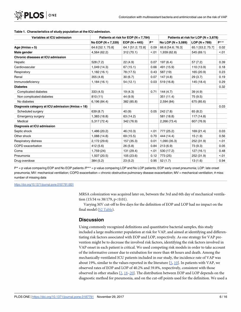
Table 2. (Continued)
Characteristics Patients at risk for EOP (N = 7,784) Patients at risk for LOP (N = 3,878)
No EOP (N = 7,339) EOP (N = 445) P* No LOP (N = 3,089) LOP (N = 789) P**
PAO2:FIO2, median (IQR) - -
<200mm Hg 1,499 (48.5) 492 (62.4) <.01
�200mm Hg 1,590 (51.5) 297 (37.6)
Microbial colonization - -
ESBLE 119 (3.9) 31 (3.9) 0.92
MDR PA 34 (1.1) 16 (2) 0.04
Acinetobacter baumannii 142 (4.6) 37 (4.7) 0.91
MRSA 123 (4) 44 (5.6) 0.05
Treatments and procedures - -
Unplanned extubation 140 (4.5) 40 (5.1) 0.52
Chest tube 311 (10.1) 107 (13.6) <.01
Steroids 1,132 (36.6) 309 (39.2) 0.19
Paralytic agents 429 (13.9) 179 (22.7) <.01
Proton Pump Inhibitors 2,121 (68.7) 531 (67.3) 0.46
Enteral feeding 921 (29.8) 202 (25.6) 0.02
Intra-hospital transport 610 (19.7) 189 (24) <.01
Patient isolation 610 (19.7) 166 (21) 0.42
Antibacterial agents administration - -
3rd and 4th generation cephalosporins 989 (32) 265 (33.6) 0.40
ß-lactam/ ß-lactamase inhibitor 1,149 (37.2) 303 (38.4) 0.53
Other penicillins 533 (17.3) 146 (18.5) 0.41
Penems 411 (13.3) 85 (10.8) 0.06
Fluoroquinolones 641 (20.8) 140 (17.7) 0.06
Aminoglycosides 805 (26.1) 193 (24.5) 0.36
Imidazole 542 (17.5) 110 (13.9) 0.02
Vancomycin 549 (17.8) 127 (16.1) 0.27
Other antibacterial agents 559 (18.1) 182 (23.1) <.01
Early-onset pneumonia 85 (9.7) 290 (8.8) 0.29
P** = p value comparing LOP and No LOP patients; ICU: intensive care unit; EOP early onset pneumonia; LOP: late onset pneumonia; MV: mechanical
ventilation; SAPSII score = Simplified Acute Physiology Score within the first 48h after ICU admission;† GLASGOW score = Glasgow coma scale within the first 48h after ICU admission—scored even in patients receiving sedation—represents the level of
awakening of patients; MDRPA = multi-drug-resistant Pseudomonas aeruginosa; MRSA = Methicillin-resistant Staphylococcus aureus; ESBL-PE:
extended-spectrum βlactamase producing Enterobacteriaceae; # miss: number of missing data
https://doi.org/10.1371/journal.pone.0187791.t002
Colonization with multiresistant bacteria and antimicrobial use on the risk of VAP
PLOS ONE | https://doi.org/10.1371/journal.pone.0187791 November 29, 2017 8 / 16

resulting from direct inoculation during emergency intubation or endotracheal tube manipu-
lation, whereas pathogens involved in LOP originate from flora modified by hospitalization
and antimicrobial treatment [5, 21]. The pattern of resistance highly depends on the level of
resistance of the country or hospital. In countries with high rate of MDR pathogens, the differ-
ence between rates of MDR pathogens in LOP and EOP was not significant [22, 23]. Coloniza-
tion with MDR bacteria represents a risk factor for the development of VAP [24]. In our study,
colonization with MDR P. aeruginosa within the first 48 hours of MV was associated with a
twice higher occurrence of EOP compared to that observed in non-colonized patients. It may
reflect the fact that antibiotic used in the first days of mechanical ventilation were most often
not active against MDR P. aeruginosa, and may induce colonization pressure. The colonization
with MRSA, especially when it was acquired between the 3rd and 6th day of mechanical ventila-
tion, was an important risk factor of LOP, with an incidence increased by 50% [21, 25]. Other
studies demonstrated that nasal colonization with S. aureus, including MRSA, is a risk factor
for the development of VAP [18]. The mechanisms by which previous colonization increased
the risk of LOP is partly related to the protective role of previous antimicrobial therapy active
on other microorganisms than MRSA. It could also be related to the decrease of prevention
and of control of VAP, already shown to be associated with isolation [26]. The impact of the
quantitative importance of colonization and the location of colonization was not available in
our database and would require further studies.
Table 3. Causative pathogens according to the type of ventilator-associated pneumonia, EOP or LOP.
Pathogens EOP (N = 445) LOP (N = 789) pvalue
Staphylococcus aureus S* 100 (16.9) 141 (14.7) 0.24
Staphylococcus aureus MDR** 23 (5.1) 71 (8.8) 0.02
Staphylococcus coagulase negative /epidermis S* 18 (3.1) 48 (5) 0.06
Staphylococcus coagulase negative /epidermis MDR** 6 (1.3) 27 (3.4) 0.03
Acinetobacter baumannii 14 (2.4) 24 (2.5) 0.87
Pseudomonas aeruginosa S* 108 (18.3) 278 (29) <.01
Pseudomonas aeruginosa MDR** 25 (5.6) 96 (11.9) <.01
Stenotrophomonas maltophilia 14 (2.4) 42 (4.4) 0.04
Other Non fermenting Gram negative Bacteria✉ 2 (0.3) 8 (0.8) 0.24
Escherichia coli S* 35 (5.9) 76 (7.9) 0.14
Escherichia coli MDR** 7 (1.6) 12 (1.5) 0.92
Klebsiella pneumoniae /Proteus spp. S* 47 (8) 70 (7.3) 0.63
Klebsiella pneumoniae / Proteus spp. MDR** 5 (1.1) 10 (1.2) 0.84
Serratia/ Citrobacter freundii/ Enterobacter cloacae S * 78 (13.2) 114 (11.9) 0.44
Serratia/ Citrobacter freundii/ Enterobacter cloacae MDR** 13 (2.9) 40 (5) 0.08
Haemophilus influenzae 58 (9.8) 36 (3.8) <.01
Streptococcus pneumoniae 35 (5.9) 31 (3.2) 0.01
Other streptococci❖ 32 (5.4) 24 (2.5) <.01
Other pathogens ♦ 49 (8.3) 67 (7) 0.34
*S: susceptible strains,
**: MDR: multi-drug resistant strains
✉Other Non fermenting GNB: Pseudomonas putida and other Pseudomonas spp.;, Acinetobacter baumannii❖Other streptococci: group A streptococci,
group B streptococci—Beta-haemolytical streptococci, group C streptococci; other pathogens ♦: other gram positive cocci, Neisseria meningitidis,
Moraxella catarrhalis, other Bacillus spp., Corynebacterium spp, Lactobacillus, Klebsiella spp., Serratia spp., other aerobic Gram negativebacilli,
Peptostreptoccus, Coxiella burnetti, Mycoplasma pneumoniae, Legionella spp, Aspergillus fumigatus
https://doi.org/10.1371/journal.pone.0187791.t003
Colonization with multiresistant bacteria and antimicrobial use on the risk of VAP
PLOS ONE | https://doi.org/10.1371/journal.pone.0187791 November 29, 2017 9 / 16

Table 4. Summary of risk factors of early- and late-onset pneumonia.
Variables Early-Onset Pneumonia Late-Onset Pneumonia
sHR p sHR p
Male gender 1.33 [1.09–1.63] <.01 1.23 [1.06–1.43] 0.0064
Chronic diseases at ICU admission
Renal 1.49 [1.02–2.16] 0.04
Immunodeficiency 1.27 [1.05–1.53] 0.012
Diagnosis at ICU admission
Respiratory distress 1.28 [1.04–1.57] 0.02
COPD exacerbation 1.24 [1.06–1.45] 0.0062
Pneumonia 1.26 [1.08–1.47] <.01
SAPSII score (ref:�38) 0.25 0.0003
SAPSII� 60 0.87 [0.65–1.16] 0.36 0.69 [0.57–0.84] 0.0003
48� SAPSII < 60 1.02 [0.78–1.33] 0.92 0.72 [0.59–0.88] 0.001
38� SAPSII < 48 1.15 [0.90–1.48] 0.26 0.92 [0.76–1.12] 0.393
Glasgow score†(ref:>13) 0.04
5�Glasgow < 13 1.62 [1.10–2.36] 0.01
Glasgow < 5 1.55 [1.06–2.27] 0.02
Period effect (ref: <2001) 0.01 0.277
2001–2006 0.84 [0.58–1.21] 0.34 1.25 [0.95–1.64] 0.114
� 2007 0.63 [0.44–0.91] 0.01 1.22 [0.92–1.63] 0.161
Variables within the first 48 hours of MV
Chest Tube 1.71 [1.30–2.26] <.01
MDR PA colonization 2.12 [1.08–4.16] 0.03
Enteral feeding 1.34 [1.09–1.66] <.01
Antibacterial agents
3 rd and 4 th generation cephalosporins 0.50 [0.38–0.65] <.01
ß lactam/ ß lactamase inhibitor 0.60 [0.48–0.75] <.01
Other penicillin 0.60 [0.42–0.87] <.01
Penems 0.27 [0.14–0.50] <.01
Aminoglycosides 0.57 [0.42–0.78] <.01
Fluoroquinolones 0.48 [0.35–0.68] <.01
Imidazoles 0.79 [0.63–0.99] 0.046
Vancomycin 0.62 [0.42–0.92] 0.02 0.65 [0.53–0.79] <.0001
Variables between the 3 rd and the 6th days of MV
Fluoroquinolones 0.65 [0.53–0.79] <.0001
Other Antibacterial agents 1.25 [1.05–1.49] 0.011
Intra-hospital transport 1.20 [1.01–1.42] 0.037
Colonization with MRSA 1.40 [1.01–1.93] 0.040
PAO2: FIO2 ratio <200 mmHg 1.49 [1.29–1.73] <.0001
Variables introduced in the model of risk factor of EOP at the first step of the selection procedure were: age, male gender, chronic hepatic diseases,
chronic renal diseases, immunodeficiency, diagnostic categories, septic shock. Variables within the first 48 hours of MV: chest tube, MDR PA colonization,
steroids, Proton Pump Inhibitors, enteral feeding, intra-hospital transport, Patient isolation, Septic shock, Respiratory distress, coma, Aminoglycosides, 3rd
and 4th generation cephalosporins, Penems, Fluoroquinolones, Vancomycin, Metronidazole, other penicillin, ß lactam/ ß lactamase inhibitor
Variables introduced in the model of risk factor of LOP at the first step of the selection procedure were: Septic shock, Respiratory distress, COPD
exacerbation pneumonia, male gender, Variables within the first 48 hours of MV; chest tube, Proton Pump Inhibitors, SAPSII score Imidazole, other
Antibacterial agent, Vancomycin, Penems, Aminoglycosides, Variables between the 3 rd and the 6 days of MV: PAO2FIO2, GLASGOW score,
Metronidazole, other Antibacterial agent, Fluoroquinolones, Penems, intra-hospital transport, enteral feeding, vasopressor, MDR PA and MRSA
colonization
ICU: intensive care unit; EOP early onset pneumonia; LOP: late onset pneumonia; MV: mechanical ventilation; SAPSII score = Simplified Acute Physiology
Score within the first 48h after ICU admission;† GLASGOW score = Glasgow coma scale within the first 48h after ICU admission—scored even in patients receiving sedation—represents the level of
awakening of patients; MDRPA = multi-drug-resistant Pseudomonas aeruginosa; MRSA = Methicillin-resistant Staphylococcus aureus; ESBLE: extended-
spectrum βlactamase-producing Enterobacteriaceae
(†) p<0.001 for comparisons between the 3 categories of Glasgow score.
https://doi.org/10.1371/journal.pone.0187791.t004
Colonization with multiresistant bacteria and antimicrobial use on the risk of VAP
PLOS ONE | https://doi.org/10.1371/journal.pone.0187791 November 29, 2017 10 / 16

Previous antimicrobial therapy was a protective factor of EOP but had marginal impact on
the risk of LOP. The administration of betalactams, aminoglycosides or fluoroquinolones
within the first 48 hours of MV protected from EOP. The maximal effect was obtained with
carbapenems. Early administration of vancomycin protected from LOP, as did the use of fluo-
roquinolones between the 3rd and the 6th day of MV. This finding is consistent with previous
studies results [11, 27]. It was shown that selective digestive decontamination with topical anti-
biotics combined with IV antibiotics during the first days of MV also decreased VAP rate [28].
In our study, the protective effect in the EOP risk of early anti-microbial against Gram-nega-
tive bacteria is counter-balanced by the deleterious impact of MDRPA carriage within the first
48 hours of mechanical ventilation.
In ICU patients with nosocomial pneumonia, compared to ICU patients without nosoco-
mial pneumonia, a study identified local markers of lung immunosuppression, such as pro-
longed low expression of HLA-DR on alveolar macrophages and low cytokine levels [29].
Authors hypothesized that the development of nosocomial pneumonia might be associated
with local organ immunosuppression. This post-aggressive general and pulmonary immuno-
paralysis may explain why both vasopressor use and low PaO2:Fi02 ratio remained important
risk factors of LOP. Coma as the diagnosis for ICU admission was not a risk factor of EOP.
However, the level of consciousness assessed in our study by the level of the effective Glasgow
coma scale was associated with an increased risk of EOP. Our results suggest the need for
awakening patients as early as possible during the ICU stay.
The use of enteral feeding in the first 48 hours of MV was a risk factor of EOP, and had no
influence on LOP, therefore confirming previous work performed on patients with shock [30].
Early enteral nutrition may favor gastric regurgitation and inhalation of gastric content in case
of intolerance, which may explain this result. Stress ulcer prophylaxis is associated with micro-
bial colonization of the gastric juice and modification of the bacterial flora of the upper respira-
tory tract. Our study also suggests the lack of impact of proton pump inhibitor on the risk of
VAP, consistently with a recent systematic review and meta-analysis [31]. The adverse impact
of intra-hospital transport on risk of LOP is consistent with previous findings [32, 33]. The
decrease of availability of tracheal suction, inadequate transport ventilator or pre-transport
underestimation of patient’ severity of illness may lead to more frequent occurrence of atelec-
tasis and ventilator-associated pneumonia.
Our study had some limitations. VAP diagnosis in patients is difficult, as the standard for
the diagnosis of VAP remains the histological examination and culture of lung tissue. We used
bronchoscopic techniques or quantitative culture of endotracheal aspirates for bacteriological
confirmation of the pneumonia diagnosis. We may have had a lower VAP rate compared to
studies based only on clinical evaluations; however, this should impact equally EOP and LOP.
The significant decrease of EOP over the study time periods is likely related to changes in pre-
vention measures used in the study centers, but could also be due to unmeasured confounders,
since we did not monitor the individual compliance with these measures. In addition, patients
with a higher SAPSII score (>60) have a non-significant protection (sHR = 0.87), and the
likely explanation is that they may die before any VAP onset. Finally, the impact of MDR and
antimicrobial therapy refers to MDR risk observed in France, where carbapenemase-resistant
Enterobacteriaceae strains are rare.
The major study strength is the use of the large, multicentric OUTCOMEREA database.
This database contains patient’s admission information and behavior during their ICUs stay,
and comprises ICUs from university and non-university hospitals. Centers use a consistent
VAP definition, which facilitates the generalization of our findings. In addition, the first epi-
sode of VAP was identified using the Fine and Gray model. This model allows a simultaneous
Colonization with multiresistant bacteria and antimicrobial use on the risk of VAP
PLOS ONE | https://doi.org/10.1371/journal.pone.0187791 November 29, 2017 11 / 16

estimation of two independent competing events: being extubated for more than 48 hours,
death before occurrence of VAP.
Conclusion
Despite the limitations discussed above, our study showed important findings, particularly the
impact of colonization on VAP occurrence and the protective effects of antibiotics adminis-
tered during the first 48 hours of MV. Factors associated with a quantitative increase of aspira-
tion of the oropharyngeal content were predominant for EOP. In LOP the persistence of organ
system failures and shock, and colonization with MRSA were the main risk factors. VAP risk
factors are mostly different according to the pneumonia time of onset, which should lead to
differentiated prevention strategies.
Supporting information
S1 File.
(CSV)
S2 File.
(CSV)
S1 Table. Summary of procedures used for ventilator associated pneumonia prevention in
the ICUs of the OUTCOMEREA network.
(DOCX)
S2 Table. Risk factors of early pneumonia using a cut-off at 5 days of mechanical ventila-
tion.
(DOCX)
Acknowledgments
We want to acknowledge the help of Celine Feger, MD (EMIBiotech) for her editorial support.
Members of the outcomerea study group
Scientific Committee: Jean-Francois Timsit (Medical and Infectious Diseases ICU, Bichat-
Claude Bernard Hospital, Paris, France; UMR 1137 Inserm –Paris Diderot university IAME,
F75018, Paris); Elie Azoulay (Medical ICU, Saint Louis Hospital, Paris, France); Maïte Gar-
rouste-Orgeas (ICU, Saint-Joseph Hospital, Paris, France); Jean-Ralph Zahar (Infection Con-
trol Unit, Angers Hospital, Angers, France); Christophe Adrie (ICU, Delafontaine Hospital,
Saint Denis, and Physiology, Cochin Hospital, Paris, France); Michael Darmon (Medical ICU,
Saint Etienne University Hospital, St Etienne, France); and Christophe Clec’h (ICU, Avicenne
Hospital, Bobigny, and UMR 1137 Inserm –Paris Diderot university IAME, F75018, Paris,
France).
Biostatistical and Information System Expertise: Jean-Francois Timsit (Medical and Infec-
tious Diseases ICU, Bichat-Claude Bernard Hospital, Paris, France; UMR 1137 Inserm –Paris
Diderot university IAME, F75018, Paris); Corinne Alberti (Medical Computer Sciences and
Biostatistics Department, Robert Debre Hospital, Paris, France); Adrien Francais (Integrated
Research Center U823, Grenoble, France); Aurelien Vesin (OUTCOMEREA organization
and Integrated Research Center U823, Grenoble, France); Stephane Ruckly (OUTCOMEREA
organization and Inserm UMR 1137 IAME, F75018, Paris); Sebastien Bailly (Grenoble univer-
sity hospital Inserm UMR 1137 IAME, F75018, Paris) and Christophe Clec’h (ICU, Avicenne
Colonization with multiresistant bacteria and antimicrobial use on the risk of VAP
PLOS ONE | https://doi.org/10.1371/journal.pone.0187791 November 29, 2017 12 / 16

Hospital, Bobigny, and Inserm UMR 1137 IAME, F75018, Paris, France); Frederik Lecorre
(Supelec, France); Didier Nakache (Conservatoire National des Arts et Metiers, Paris, France);
and Aurelien Vannieuwenhuyze (Tourcoing, France).
Investigators of the OUTCOMEREADatabase: Christophe Adrie (ICU, Delafontaine Hos-
pital, Saint Denis, and Physiology, Cochin Hospital, Paris, France); Bernard Allaouchiche
(ICU, Pierre benite Hospital, Lyon, France); Laurent Argaud (Medical ICU, Hospices Civils de
Lyon, Lyon, France); Claire Ara-Somohano (Medical ICU, University Hospital, Grenoble,
France); Elie Azoulay (Medical ICU, Saint Louis Hospital, Paris, France); Francois Barbier
(medical-surgical ICU, Orleans, France), Jean-Pierre Bedos (ICU, Versailles Hospital, Ver-
sailles, France); Julien Bohe (ICU, Hopital Pierre Benite, Lyon France), Lila Bouadma (ICU,
Bichat Hospital, Paris, France); Christine Cheval (ICU, Hyeres Hospital, Hyeres, France);
Christophe Clec’h (ICU, Avicenne Hospital, Bobigny, France); Michael Darmon (ICU, Saint
Etienne Hospital, Saint Etienne, France); Anne-Sylvie Dumenil (Antoine Beclère Hospital,
Clamart, France); Claire Dupuis (Bichat hospital and UMR 1137 Inserm –Paris Diderot uni-
versity IAME, F75018, Paris, France), Jean-Marc Forel (AP HM, Medical ICU, Hopital Nord
Marseille), Marc Gainier (la Timone hospital, Marseille, France), Akim Haouache (Surgical
ICU, H Mondor Hospital, Creteil, France); Samir Jamali (ICU, Dourdan, Dourdan Hospital,
Dourdan, France); Hatem Khallel (ICU, Cayenne General Hospital, Cayenne, France); Alex-
andre Lautrette (ICU, G Montpied Hospital, Clermont-Ferrand, France); Guillaume Marcotte
(Surgical ICU, Hospices Civils de Lyon, Lyon, France); Eric Le Miere (ICU, Louis Mourier
Hospital, Colombes, France); Maxime Lugosi (Medical ICU, University Hospital Grenoble,
Grenoble, France); Bruno Mourvillier (ICU, Bichat Hospital, Paris, France); Benoıt Misset
(ICU, Saint-Joseph Hospital, Paris, France); Delphine Moreau (ICU, Saint-Louis Hospital,
Paris, France); Bruno Mourvillier (ICU, Bichat Hospital, Paris, France); Laurent Papazian
(Hopital Nord, Marseille, France), Benjamin Planquette (pulmonology ICU, George Pompi-
dou hospital Hospital, Versailles, France); Bertrand Souweine (ICU, G Montpied Hospital,
Clermont-Ferrand, France); Carole Schwebel (ICU, A Michallon Hospital, Grenoble, France);
Gilles Troche (ICU, Antoine Beclère Hospital, Clamart, France); Marie Thuong (ICU, Dela-
fontaine Hospital, Saint Denis, France); Guillaume Thierry (ICU, Saint-Louis Hospital, Paris,
France); Dany Toledano (ICU, Gonesse Hospital, Gonesse, France); and Eric Vantalon (SICU,
Saint-Joseph Hospital, Paris, France).
Study Monitors: Julien Fournier, Caroline Tournegros, Stephanie Bagur, Mireille Adda,
Vanessa Vindrieux, Loic Ferrand, Nadira Kaddour, Boris Berthe, Samir Bekkhouche, Kaouttar
Mellouk, Sylvie Conrozier, Igor Theodose, Veronique Deiler, and Sophie Letrou.
Author Contributions
Conceptualization: Wafa Ibn Saied, Jean-Francois Timsit.
Data curation: Stephane Ruckly.
Investigation: Jean-Francois Timsit.
Methodology: Wafa Ibn Saied, Lila Bouadma, Jean-Francois Timsit.
Resources: Maite Garrouste-Orgeas, Michael Darmon, Yves Cohen, Elie Azoulay, Carole
Schwebel, Aguila Radjou, Hatem Kallel, Guillaume Marcotte, Samir Jamali, Laurent Papa-
zian, Lila Bouadma, Jean-Francois Timsit.
Software: Wafa Ibn Saied, Stephane Ruckly, Sebastien Bailly.
Supervision: Stephane Ruckly.
Colonization with multiresistant bacteria and antimicrobial use on the risk of VAP
PLOS ONE | https://doi.org/10.1371/journal.pone.0187791 November 29, 2017 13 / 16

Validation: Wafa Ibn Saied, Stephane Ruckly, Michael Darmon, Sebastien Bailly, Carole
Schwebel, Jean-Francois Timsit.
Visualization: Bertrand Souweine, Aguila Radjou, Christophe Adrie, Anne-Sylvie Dumenil,
Laurent Argaud, Dany Goldgran-Toledano, Lila Bouadma.
Writing – original draft: Wafa Ibn Saied.
Writing – review & editing: Jean-Francois Timsit.
References1. Chastre J, Fagon JY. Ventilator-associated pneumonia. Am J Respir Crit Care Med. 2002; 165(7):867–
903. Epub 2002/04/06. https://doi.org/10.1164/ajrccm.165.7.2105078 PMID: 11934711.
2. Bekaert M, Timsit JF, Vansteelandt S, Depuydt P, Vesin A, Garrouste-Orgeas M, et al. Attributable mor-
tality of ventilator-associated pneumonia: a reappraisal using causal analysis. Am J Respir Crit Care
Med. 2011; 184(10):1133–9. Epub 2011/08/20. https://doi.org/10.1164/rccm.201105-0867OC PMID:
21852541.
3. Nguile-Makao M, Zahar JR, Francais A, Tabah A, Garrouste-Orgeas M, Allaouchiche B, et al. Attribut-
able mortality of ventilator-associated pneumonia: respective impact of main characteristics at ICU
admission and VAP onset using conditional logistic regression and multi-state models. Intensive Care
Med. 2010; 36(5):781–9. https://doi.org/10.1007/s00134-010-1824-6 PMID: 20232046.
4. Hotchkiss RS, Monneret G, Payen D. Sepsis-induced immunosuppression: from cellular dysfunctions
to immunotherapy. Nat Rev Immunol. 2013; 13(12):862–74. https://doi.org/10.1038/nri3552 PMID:
24232462
5. Giard M, Lepape A, Allaouchiche B, Guerin C, Lehot JJ, Robert MO, et al. Early- and late-onset ventila-
tor-associated pneumonia acquired in the intensive care unit: comparison of risk factors. J Crit Care.
2008; 23(1):27–33. Epub 2008/03/25. https://doi.org/10.1016/j.jcrc.2007.08.005 PMID: 18359418.
6. Bouadma L, Deslandes E, Lolom I, Le Corre B, Mourvillier B, Regnier B, et al. Long-term impact of a
multifaceted prevention program on ventilator-associated pneumonia in a medical intensive care unit.
Clin Infect Dis. 2010; 51(10):1115–22. https://doi.org/10.1086/656737 PMID: 20936973
7. Mandelli M, Mosconi P, Langer M, Cigada M. Is pneumonia developing in patients in intensive care
always a typical "nosocomial" infection? Lancet. 1986; 2(8515):1094–5. Epub 1986/11/08. PMID:
2877241.
8. Akca O, Koltka K, Uzel S, Cakar N, Pembeci K, Sayan MA, et al. Risk factors for early-onset, ventilator-
associated pneumonia in critical care patients: selected multiresistant versus nonresistant bacteria.
Anesthesiology. 2000; 93(3):638–45. Epub 2000/09/02. PMID: 10969295.
9. Nseir S, Di Pompeo C, Pronnier P, Soubrier S, Onimus T, Saulnier F, et al. [Early and late nosocomial
broncho-pulmonary diseases in intensive care. Comparative study of risk factors and of causing bacte-
ria]. Presse Med. 2003; 32(24):1111–5. Epub 2003/09/02. PMID: 12947739.
10. Trouillet JL, Chastre J, Vuagnat A, Joly-Guillou ML, Combaux D, Dombret MC, et al. Ventilator-associ-
ated pneumonia caused by potentially drug-resistant bacteria. Am J Respir Crit Care Med. 1998; 157
(2):531–9. Epub 1998/02/26. https://doi.org/10.1164/ajrccm.157.2.9705064 PMID: 9476869.
11. Valles J, Peredo R, Burgueno MJ, Rodrigues de Freitas AP, Millan S, Espasa M, et al. Efficacy of sin-
gle-dose antibiotic against early-onset pneumonia in comatose patients who are ventilated. Chest.
2013; 143(5):1219–25. Epub 2013/05/30. https://doi.org/10.1378/chest.12-1361 PMID: 23715136.
12. Lacherade JC, De Jonghe B, Guezennec P, Debbat K, Hayon J, Monsel A, et al. Intermittent subglottic
secretion drainage and ventilator-associated pneumonia: a multicenter trial. Am J Respir Crit Care Med.
2010; 182(7):910–7. https://doi.org/10.1164/rccm.200906-0838OC PMID: 20522796
13. Mao Z G L, Wang G, et al. Subglottic secretion suction for preventing ventilator-associated pneumonia:
an updated meta-analysis and trial sequential analysis. Critical Care. 2016; 20. https://doi.org/10.1186/
s13054-016-1527-7 PMID: 27788682
14. Drakulovic MB, Torres A, Bauer TT, Nicolas JM, Nogue S, Ferrer M. Supine body position as a risk fac-
tor for nosocomial pneumonia in mechanically ventilated patients: a randomised trial. Lancet. 1999; 354
(9193):1851–8. https://doi.org/10.1016/S0140-6736(98)12251-1 PMID: 10584721
15. Prod’hom G, Leuenberger P, Koerfer J, et al. Nosocomial pneumonia in mechanically ventilated
patients receiving antacid, ranitidine, or sucralfate as prophylaxis for stress ulcer: A randomized con-
trolled trial. Annals of Internal Medicine. 1994; 120(8):653–62. https://doi.org/10.7326/0003-4819-120-
8-199404150-00005 PMID: 8135449
Colonization with multiresistant bacteria and antimicrobial use on the risk of VAP
PLOS ONE | https://doi.org/10.1371/journal.pone.0187791 November 29, 2017 14 / 16

16. Lautrette A G-O M, Bertrand PM, Goldgran-Toledano D, Jamali S, Laurent V, Argaud L, Schwebel C,
Mourvillier B, Darmon M, Ruckly S, Dumenil AS, Lemiale V, Souweine B, Timsit JF; Outcomerea Study
Group. Respective impact of no escalation of treatment, withholding and withdrawal of life-sustaining
treatment on ICU patients’ prognosis: a multicenter study of the Outcomerea Research Group. Inten-
sive Care Med 2015; 41():1763–72. https://doi.org/10.1007/s00134-015-3944-5 PMID: 26149302
17. Wolkewitz M, Cooper BS, Bonten MJM, Barnett AG, Schumacher M. Interpreting and comparing risks
in the presence of competing events. BMJ: British Medical Journal. 2014; 349. https://doi.org/10.1136/
bmj.g5060 PMID: 25146097
18. Martin-Loeches I, Deja M, Koulenti D, Dimopoulos G, Marsh B, Torres A, et al. Potentially resistant
microorganisms in intubated patients with hospital-acquired pneumonia: the interaction of ecology,
shock and risk factors. Intensive Care Med. 2013; 39(4):672–81. Epub 2013/01/30. https://doi.org/10.
1007/s00134-012-2808-5 PMID: 23358539.
19. Muscedere JG, Martin CM, Heyland DK. The impact of ventilator-associated pneumonia on the Cana-
dian health care system. J Crit Care. 2008; 23(1):5–10. https://doi.org/10.1016/j.jcrc.2007.11.012
PMID: 18359415.
20. Vanhems P, Lepape A, Savey A, Jambou P, Fabry J. Nosocomial pulmonary infection by antimicrobial-
resistant bacteria of patients hospitalized in intensive care units: risk factors and survival. J Hosp Infect.
2000; 45(2):98–106. Epub 2000/06/22. https://doi.org/10.1053/jhin.2000.0752 PMID: 10860686.
21. Bassetti M, Taramasso L, Giacobbe DR, Pelosi P. Management of ventilator-associated pneumonia:
epidemiology, diagnosis and antimicrobial therapy. Expert Rev Anti Infect Ther. 2012; 10(5):585–96.
Epub 2012/06/19. https://doi.org/10.1586/eri.12.36 PMID: 22702322.
22. Restrepo MI, Peterson J, Fernandez JF, Qin Z, Fisher AC, Nicholson SC. Comparison of the bacterial
etiology of early-onset and late-onset ventilator-associated pneumonia in subjects enrolled in 2 large
clinical studies. Respir Care. 2013; 58(7):1220–5. Epub 2013/01/12. https://doi.org/10.4187/respcare.
02173 PMID: 23307825
23. Giantsou E, Liratzopoulos N, Efraimidou E, Panopoulou M, Alepopoulou E, Kartali-Ktenidou S, et al.
Both early-onset and late-onset ventilator-associated pneumonia are caused mainly by potentially multi-
resistant bacteria. Intensive Care Med. 2005; 31(11):1488–94. Epub 2005/09/10. https://doi.org/10.
1007/s00134-005-2697-y PMID: 16151723.
24. Rocha LA, Marques Ribas R, da Costa Darini AL, Gontijo Filho PP. Relationship between nasal coloni-
zation and ventilator-associated pneumonia and the role of the environment in transmission of Staphy-
lococcus aureus in intensive care units. Am J Infect Control. 2013; 41(12):1236–40. Epub 2013/07/31.
https://doi.org/10.1016/j.ajic.2013.04.009 PMID: 23890377.
25. Rello J, Chastre J, Cornaglia G, Masterton R. A European care bundle for management of ventilator-
associated pneumonia. J Crit Care. 2011; 26(1):3–10. Epub 2010/06/12. https://doi.org/10.1016/j.jcrc.
2010.04.001 PMID: 20537504.
26. Zahar JR, Garrouste-Orgeas M, Vesin A, Schwebel C, Bonadona A, Philippart F, et al. Impact of contact
isolation for multidrug-resistant organisms on the occurrence of medical errors and adverse events.
Intensive Care Med. 2013; 39(12):2153–60. Epub 2013/09/03. https://doi.org/10.1007/s00134-013-
3071-0 PMID: 23995982.
27. Bornstain C, Azoulay E, De Lassence A, Cohen Y, Costa MA, Mourvillier B, et al. Sedation, sucralfate,
and antibiotic use are potential means for protection against early-onset ventilator-associated pneumo-
nia. Clinical infectious diseases: an official publication of the Infectious Diseases Society of America.
2004; 38(10):1401–8. Epub 2004/05/25. https://doi.org/10.1086/386321 PMID: 15156478.
28. de Smet AM, Kluytmans JA, Cooper BS, Mascini EM, Benus RF, van der Werf TS, et al. Decontamina-
tion of the digestive tract and oropharynx in ICU patients. The New England journal of medicine. 2009;
360(1):20–31. Epub 2009/01/02. https://doi.org/10.1056/NEJMoa0800394 PMID: 19118302.
29. Muehlstedt SG, Lyte M, Rodriguez JL. Increased IL-10 production and HLA-DR suppression in the
lungs of injured patients precede the development of nosocomial pneumonia. Shock. 2002; 17(6):443–
50. Epub 2002/06/19. PMID: 12069178.
30. Reignier J, Darmon M, Sonneville R, Borel AL, Garrouste-Orgeas M, Ruckly S, et al. Impact of early
nutrition and feeding route on outcomes of mechanically ventilated patients with shock: a post hoc mar-
ginal structural model study. Intensive Care Med. 2015; 41(5):875–86. Epub 2015/03/21. https://doi.
org/10.1007/s00134-015-3730-4 PMID: 25792207.
31. Krag M, Perner A, Wetterslev J, Wise MP, Hylander Moller M. Stress ulcer prophylaxis versus placebo
or no prophylaxis in critically ill patients. A systematic review of randomised clinical trials with meta-anal-
ysis and trial sequential analysis. Intensive Care Med. 2014; 40(1):11–22. Epub 2013/10/22. https://doi.
org/10.1007/s00134-013-3125-3 PMID: 24141808.
32. Schwebel C, Clec’h C, Magne S, Minet C, Garrouste-Orgeas M, Bonadona A, et al. Safety of intrahos-
pital transport in ventilated critically ill patients: a multicenter cohort study*. Critical care medicine.
Colonization with multiresistant bacteria and antimicrobial use on the risk of VAP
PLOS ONE | https://doi.org/10.1371/journal.pone.0187791 November 29, 2017 15 / 16

2013; 41(8):1919–28. Epub 2013/07/19. https://doi.org/10.1097/CCM.0b013e31828a3bbd PMID:
23863225.
33. Bercault N, Wolf M, Runge I, Fleury JC, Boulain T. Intrahospital transport of critically ill ventilated
patients: a risk factor for ventilator-associated pneumonia—a matched cohort study. Crit Care Med.
2005; 33(11):2471–8. Epub 2005/11/09. PMID: 16276168.
Colonization with multiresistant bacteria and antimicrobial use on the risk of VAP
PLOS ONE | https://doi.org/10.1371/journal.pone.0187791 November 29, 2017 16 / 16
Top Related