


Languages
Pages
Legal


www.spm
.com
.cn
RESEARCH PAPER
Controllable functionalization and wettability transitionof graphene-based films by an atomic oxygen strategy
Min Yi • Wen Zhang • Zhigang Shen •
Xiaojing Zhang • Xiaohu Zhao • Yiting Zheng •
Shulin Ma
Received: 22 February 2013 / Accepted: 19 June 2013
� The Author(s) 2013. This article is published with open access at Springerlink.com
Abstract Though chemical modification of graph-
ene based on Hummers method has been most widely
used to tailor its properties and interfacial character-
istics, a method which could achieve definitive and
controllable groups and properties is still highly
required. Here, we demonstrate a high-vacuum oxi-
dation strategy by atomic oxygen (AO) and investigate
the AO induced functionalization and wettability
transition in films made from basal-defect- and
oxide-free graphene dispersions. These graphene-
based films are neither graphene nor graphite, but
graphene blocks constituted by numerous randomly
stacked graphene flakes. It is found that AO induced
functionalization of these films through the formation
of epoxy groups, sp3 configuration, ether, and double
and triple C–O groups. The films turn to be hydrophilic
after exposed to AO. The contact angle increases with
AO exposure time. This phenomenon is attributed to
the lower surface roughness induced by collision and/
or edge erosion of energetic ions to the film surface
and is further explained by the Wenzel model. The
demonstrated strategy can overcome limitations of
Hummers method, provide possibility to gain func-
tionalization and wettability transition in liquid-phase
exfoliated basal-defect- and oxide-free graphene in the
dry environment, and may extend the study and
application of this material in spacecraft in low earth
orbit.
Keywords Graphene � Atomic oxygen �Functionalization �Wettability
Introduction
Graphene, constituted by the two-dimensional honey-
comb C–C network, has attracted a great deal of
attention due to its exceptional and intriguing proper-
ties (Novoselov et al. 2004; Sofo et al. 2007; Geim
2009; Allen et al. 2010; Geim and Novoselov 2007;
Tanaka and Iakoubovskii 2010; Rao et al. 2010).
Recently, graphene has shown potential applications
in lots of fields, such as fundamental physical research
(Barone et al. 2006; Katsnelson and Novoselov 2007;
Novoselov et al. 2005; Fujita 2011), energy-storage
material (Deng et al. 2004; Boukhvalov et al. 2008;
Wang et al. 2008), new electronic devices (Novoselov
M. Yi � W. Zhang � Z. Shen (&) � X. Zhang � S. Ma
Beijing Key Laboratory for Powder Technology Research
and Development, Beijing University of Aeronautics and
Astronautics, Beijing 100191, China
e-mail: [email protected]
M. Yi � W. Zhang � Z. Shen � X. Zhao
Plasma Laboratory, Ministry-of-Education Key
Laboratory of Fluid Mechanics, Beijing University
of Aeronautics and Astronautics, Beijing 100191, China
Y. Zheng
School of Material Science and Engineering, Beijing
University of Aeronautics and Astronautics,
Beijing 100191, China
123
J Nanopart Res (2013) 15:1811
DOI 10.1007/s11051-013-1811-2

www.spm
.com
.cn
et al. 2004; Lemme et al. 2007; Schedin et al. 2007;
Wu et al. 2007; Moriyama et al. 2010; Wakabayashi
et al. 2010), catalyses (Sofo et al. 2007; Elias et al.
2009; Zhou et al. 2009), chemical and biological
sensors (Schedin et al. 2007; Mohanty and Berry
2008), etc. Ironically, despite its outstanding proper-
ties and bright prospects, several issues must be
overcome before the full promise of graphene can be
realized industrially. For example, graphene is semi-
metallic. If graphene is used in microelectronics,
chemical modification is required to introduce a band
gap to achieve semiconducting behavior (Zhou et al.
2008; Balog et al. 2010; Yavari et al. 2010). In
addition, graphene is hydrophobic and chemically
inert, making processing and purification techniques
difficult. However, in the fields such as catalyses,
liquid-phase processing, biocompatibility, etc., graph-
ene is often expected to be chemically active and
hydrophilic. Therefore, when graphene is considered
in terms of its promising prospect, in order to meet the
special requirement in the processing and applications,
chemical functionalization of graphene seems very
critical and necessary.
Presently, the oxidation technique based on Hum-
mers method is most widely used to covalently
functionalize graphene (Hummers and Offeman
1958; Park and Ruoff 2009; Dreyer et al. 2010; Loh
et al. 2010). It alters the properties of graphene by
introducing some oxygen-containing groups, and the
resulting graphene oxide material is highly inhomo-
geneous. However, Hummers method is often accom-
plished in the wet solution of strong acids, making it
difficult to clearly determine the forming process and
precise content of different oxygen-containing groups.
Moreover, these oxygen-containing groups cannot be
completely removed by chemical or thermal reduc-
tion. Hence, these shortcomings limit the applications
of the wet chemical functionalization based on
Hummers method.
Recently, Elias et al. (2009) reported the function-
alization of graphene by atomic hydrogen, which
transformed semi-metallic graphene into an insulator.
Following this idea, atomic oxygen (AO) may be also
a promising agent for chemical functionalization of
graphene, because it can forms stable bonds in
graphene. Furthermore, AO oxidation happens in the
high-vacuum conditions where water or hydrogen
(i.e., hydroxyl and acid carboxyl) can be excluded
first, making the determination of oxygen-containing
groups relatively easy. Moreover, for graphene mate-
rial as solid films used in electronic and energy
applications, direct chemical functionalization in the
dry environment is crucial and high-vacuum AO
oxidation may provide such a feasible route. We also
noted that some researchers investigated the AO
induced oxidation of highly oriented pyrolytic graph-
ite (HOPG) surface (Barinov et al. 2009b; Larciprete
et al. 2012). In contrast, firstly our work is focused on
graphene-based film. Graphene flakes have different
properties (especially electronic) compared to bulk
HOPG. Most interestingly, the graphene-based film in
our work is constituted by numerous graphene flakes
which are randomly stacked (not Bernal AB style in
bulk graphite). This random stack of graphene flakes
has been demonstrated to possess unique band struc-
ture and electronic properties. However, presently,
there is little information about how to tune the
properties of these random stacked graphene flakes.
Our work here may provide a way for tuning
properties by AO in the dry environment. Second,
HOPG surface is flat. The graphene-based film surface
is rough with many graphene flakes protruding out,
providing possibility for simultaneous functionaliza-
tion and physical surface engineering. Third, most
current studies concentrate on wet chemistry methods
to tailor the composition and surface structure of films.
We combined AO oxidation and ions’ collisions to
tuning the wettability of the graphene-based film in the
dry environment.
Meanwhile, some researchers have investigated the
oxidation of chemical vapor deposited and epitaxial
graphene by AO (Vinogradov et al. 2011; Hossain
et al. 2012). However, it is likely that many future
industrial applications of graphene depend on large-
scale production which could be achieved by liquid-
phase method. Recently, Coleman’s group and other
researchers did lots of work in this aspect (Hernandez
et al. 2008; Coleman 2009; De et al. 2010; Khan et al.
2010, 2011; Lotya et al. 2010; Cui et al. 2011; Shen
et al. 2011; Yi et al. 2011; Coleman 2013; Yi et al.
2012a, b). They evidenced that as compared to the
methods of micromechanical cleavage, chemical
vapor deposition, epitaxial growth, etc., the liquid-
phase exfoliation of graphite can obtain basal-defect-
and oxide-free graphene and shows advantages of
facilitation, large scale, and low cost. Hence, the
functionalization of liquid-phase exfoliated basal-
defect- and oxide-free graphene material is an
Page 2 of 14 J Nanopart Res (2013) 15:1811
123

www.spm
.com
.cn
alternative way to extend the applications of this
material. Yet, to our knowledge, though there are
many reports on covalent functionalization by organic
groups or noncovalent functionalization by p-interac-
tions of liquid-phase exfoliated pristine graphene in
the wet environment (Georgakilas et al. 2012), there is
little information about functionalizing liquid-phase
exfoliated pristine graphene in the dry environment.
Herein, we prepare films by vacuum filtering the
dispersions of basal-defect- and oxide-free graphene
and investigate the oxidation process of these films
exposed into AO with different time. On the other
hand, in spite of intensive activity in graphene
research, there are very few literatures about water–
graphene interactions (Wang et al. 2009; Rafiee et al.
2010; Yavari et al. 2010; Dhiman et al. 2011; Lin et al.
2011; Choi and Park 2012; Nair et al. 2012). Most
current studies concentrate on wet chemistry methods
to tailor the composition and surface structure of films.
Herein, we utilize the energetic ions in the AO
atmosphere to collide with the film surface and
investigate the wettability transition behavior of these
films under different AO exposure time. Figure 1
illustrates the process of AO induced controllable
functionality and wettability transition in graphene-
based films. In a word, the demonstrated strategy of
AO oxidation provides possibility to gain functional-
ization and wettability transition of basal-defect- and
oxide-free graphene from liquid-phase exfoliation in
the dry environment, and may extend the study and
application of this material. The study here is also very
valuable for graphene-based electronics and sensors to
be used in spacecraft in low earth orbit where AO and
energetic ions abound.
Experimental
Preparation and characterization
Dispersion of basal-defect- and oxide-free graphene
was prepared by liquid-phase exfoliation of graphite
(Hernandez et al. 2008; Coleman 2009; Khan et al.
2010, 2011; Coleman 2013). Graphite dispersions
were made by 70 mg crystal graphite powder (B300
meshes, Alfa Aesar) dispersed in 140 mL dimethyl-
formamide (DMF). These dispersions were then
sonicated for 60 min in a low-power ultrasonic bath
(45 W, 59 Hz, SK1200H-J, KUDOS, Shanghai). After
sonication, the dispersions were centrifuged for
30 min at 2,000 rpm (5709g) by an L-600 centrifuge
(XiangYi, Changsha) in order to remove any largish
flakes, eventually resulting in homogeneous colloidal
suspension of graphene flakes in DMF. Graphene
concentration after centrifugation, CG, was deter-
mined from Lambert–Beer law, A/l = aCG, where A is
the absorbance measured at 660 nm by a 721E
spectrophotometer (Shanghai Spectrum), l is the
cuvette length, and a is absorption coefficient at
660 nm which equals to 2,460 mL/mg/m (Hernandez
et al. 2008, 2010). By using this method, the concen-
tration of the resulted graphene dispersions can be
determined as *45 lg/mL. Considering an initial
concentration of 0.5 mg/mL (70 mg/140 mL), we can
estimate a yield of *9 wt%.
50 mL graphene dispersions were diluted to
1,000 mL (50 mL for preparing the loose film) by
deionized water and then vacuum filtered to form a
homogeneous film (U 40 mm) on the nylon membrane
(pore size *220 nm). The film was dried for 48 h in
the vacuum oven at 100 �C. Subsequently, a
30 9 20 mm rectangular small film was cut from the
film central region. Atomic force microscopy (AFM)
was used to scan 96 points on the surface of the small
film to determine the roughness. Average of the
surface roughness (root-mean-square roughness) cor-
responding to each scan was used as the surface
roughness of the small film. The small film was again
cut into 6 bitty films with dimension of 10 9 10 mm.
One bitty film was chosen to measure the contact angle
with water. The other 5 bitty films were exposed into
high-vacuum AO atmosphere with time of 3, 5, 10, 20,
and 25 h. After AO exposure, the surface roughness
and contact angle of these films were measured, and
X-ray photoelectron spectroscopy (XPS) and Fourier
transform infrared (FTIR) spectroscopy analyses were
performed. When the roughness of the 10 9 10 mm
films was measured by AFM, 16 points were scanned
in each sample and the average surface roughness was
adopted.
AFM images were captured by a CSPM5500 AFM
(Being Nano-Instruments) with a scanning range of
13.56 lm in the tapping mode. Bright-field transmis-
sion microscope (TEM) and high resolution TEM
(HRTEM) images were taken with a JEOL 2100FEF
operating at 200 kV. When AFM and TEM were used
to characterize the morphology and structure of the
isolated or individual graphene flakes, AFM samples
J Nanopart Res (2013) 15:1811 Page 3 of 14
123
www.spm
.com
.cnwere prepared by spraying several microliters of
dispersions onto the mica substrate and dried in
vacuum oven, while TEM samples were prepared by
pipetting several drops of dispersions onto holey
carbon mesh grids (300 meshes). The Raman mea-
surements were made on these films with a Renishaw
Rm2000 using a 514 nm laser. FTIR spectra of the
films were measured by a Nicolet iS10 spectrometer in
the diffuse reflection mode. XPS investigation was
performed on the films by an ESCALAB-250 photo-
electron spectrometer (Thermo Fisher Scientific) with
the monochromatic Al Ka X-rays source
(1,486.6 eV). For each spectrum, the binding energy
position was calibrated by measuring the Fermi level
position of an Au reference sample in contact with
graphene-based film. Static contact angle was mea-
sured by placing a droplet of deionized water on the
surface of the various films. An Easydrop DSA20
instrument was used to measure the contact angle at
room temperature (*25 �C). The axisymmetric-drop-
shape analysis profile method was used for estimating
the contact angle of water on the film surface.
AO exposure experiment
High-vacuum AO functionalization of these bitty films
was performed in the ground-based AO effect simu-
lation facility in Beijing University of Aeronautics and
Astronautics (BUAA) (Zhao et al. 2001). The facility
was a filament discharge plasma-type ground-based
AO effect simulation facility for simulating low earth
orbit environment (Zhao et al. 2001). The filament was
heated to a high temperature by current. When the
temperature is high enough, electrons could escape
from the filament and collide with the oxygen
molecules, making oxygen molecules ionized into
oxygen plasma. The main components of the plasma
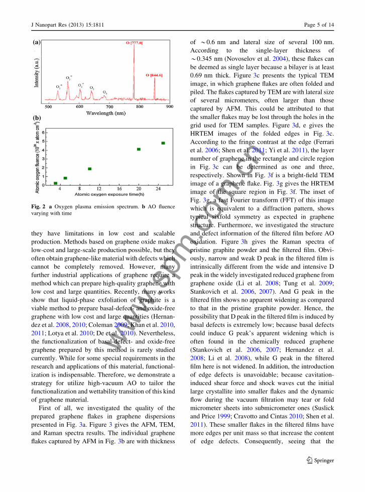
include O2, O2?, O, O?, e, etc., as shown in Fig. 2a. It
has been evidenced that the energy of AO was only
*0.04 eV while the energy of O2? could be as high as
15 eV (Zhao et al. 2001). AO exposure experiments
were carried out in the conditions of vacuum pressure
of 0.15 Pa, filament discharge voltage of 120 V, and
filament discharge current of 140 mA. Because the
erosion yield of Kapton under AO almost keeps
constant, the mass loss of Kapton in the AO exposure
experiment was used as a criterion to calculate the AO
flux (Reddy et al. 1993; Zhao et al. 2001; Banks et al.
2006). The calculation formula is Ft = DM/(qAEy) in
which F is the effective flow rate of AO onto the
sample surface and DM, q, A, t, Ey are mass loss,
density, surface area, exposure time, and erosion yield,
respectively. Ft is the accumulative AO flux exerting
on the sample surface. For Kapton, Ey is equal to
*3.0 9 10-24 cm3/atom (Zhao et al. 2001; Banks
et al. 2006), so according to the mass loss of Kapton,
the accumulative AO flux for the films can be
obtained, as indicated in Fig. 2b.
Results and discussion
Though micromechanical cleavage and chemical
vapor deposition can achieve high-quality graphene,
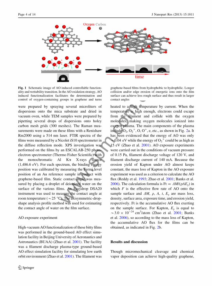
Fig. 1 Schematic image of AO induced controllable function-
ality and wettability transition. In the AO oxidation strategy, AO
induced functionalization facilitates the determination and
control of oxygen-containing groups in graphene and turns
graphene-based films from hydrophobic to hydrophilic. Longer
collision and/or edge erosion of energetic ions onto the film
surface can achieve less rough surface and thus result in larger
contact angles
Page 4 of 14 J Nanopart Res (2013) 15:1811
123
www.spm
.com
.cnthey have limitations in low cost and scalable
production. Methods based on graphene oxide makes
low-cost and large-scale production possible, but they
often obtain graphene-like material with defects which
cannot be completely removed. However, many
further industrial applications of graphene require a
method which can prepare high-quality graphene with
low cost and large quantities. Recently, many works
show that liquid-phase exfoliation of graphite is a
viable method to prepare basal-defect- and oxide-free
graphene with low cost and large quantities (Hernan-
dez et al. 2008, 2010; Coleman 2009; Khan et al. 2010,
2011; Lotya et al. 2010; De et al. 2010). Nevertheless,
the functionalization of basal-defect- and oxide-free
graphene prepared by this method is rarely studied
currently. While for some special requirements in the
research and applications of this material, functional-
ization is indispensable. Therefore, we demonstrate a
strategy for utilize high-vacuum AO to tailor the
functionalization and wettability transition of this kind
of graphene material.
First of all, we investigated the quality of the
prepared graphene flakes in graphene dispersions
presented in Fig. 3a. Figure 3 gives the AFM, TEM,
and Raman spectra results. The individual graphene
flakes captured by AFM in Fig. 3b are with thickness
of *0.6 nm and lateral size of several 100 nm.
According to the single-layer thickness of
*0.345 nm (Novoselov et al. 2004), these flakes can
be deemed as single layer because a bilayer is at least
0.69 nm thick. Figure 3c presents the typical TEM
image, in which graphene flakes are often folded and
piled. The flakes captured by TEM are with lateral size
of several micrometers, often larger than those
captured by AFM. This could be attributed to that
the smaller flakes may be lost through the holes in the
grid used for TEM samples. Figure 3d, e gives the
HRTEM images of the folded edges in Fig. 3c.
According to the fringe contrast at the edge (Ferrari
et al. 2006; Shen et al. 2011; Yi et al. 2011), the layer
number of graphene in the rectangle and circle region
in Fig. 3c can be determined as one and three,
respectively. Shown in Fig. 3f is a bright-field TEM
image of a graphene flake. Fig. 3g gives the HRTEM
image of the square region in Fig. 3f. The inset of
Fig. 3g, a fast Fourier transform (FFT) of this image
which is equivalent to a diffraction pattern, shows
typical sixfold symmetry as expected in graphene
structure. Furthermore, we investigated the structure
and defect information of the filtered film before AO
oxidation. Figure 3h gives the Raman spectra of
pristine graphite powder and the filtered film. Obvi-
ously, narrow and weak D peak in the filtered film is
intrinsically different from the wide and intensive D
peak in the widely investigated reduced graphene from
graphene oxide (Li et al. 2008; Tung et al. 2009;
Stankovich et al. 2006, 2007). And G peak in the
filtered film shows no apparent widening as compared
to that in the pristine graphite powder. Hence, the
possibility that D peak in the filtered film is induced by
basal defects is extremely low; because basal defects
could induce G peak’s apparent widening which is
often found in the chemically reduced graphene
(Stankovich et al. 2006, 2007; Hernandez et al.
2008; Li et al. 2008), while G peak in the filtered
film here is not widened. In addition, the introduction
of edge defects is unavoidable; because cavitation-
induced shear force and shock waves cut the initial
large crystallite into smaller flakes and the dynamic
flow during the vacuum filtration may tear or fold
micrometer sheets into submicrometer ones (Suslick
and Price 1999; Cravotto and Cintas 2010; Shen et al.
2011). These smaller flakes in the filtered films have
more edges per unit mass so that increase the content
of edge defects. Consequently, seeing that the
Fig. 2 a Oxygen plasma emission spectrum. b AO fluence
varying with time
J Nanopart Res (2013) 15:1811 Page 5 of 14
123

www.spm
.com
.cn
broadening of G band is unremarkable and the size of
laser point (1–2 lm) used in the Raman system will
inevitably cover the edges of graphene sheets in the
filtered film, the D band in the filtered film may be
largely attributed to the edge defects instead of the
basal defects. We can also look at the 2D band. The 2D
band in the filtered film is apparently distinct from 2D
band in pristine graphite, indicating the nature of few-
layer graphene (Ferrari et al. 2006; Hernandez et al.
2008; Malard et al. 2009). This indicates that though
aggregation of graphene flakes happens during the
filtration, the aggregation is not a process to drive
graphene flakes stacked in Bernal AB style which
exists in graphite. Therefore, the filtered film is neither
graphene nor graphite, but a graphene block consti-
tuted by numerous graphene flakes which are ran-
domly stacked.
Figures 4 and 5 give the FTIR and XPS results of
the films before and after AO exposure. The FTIR of
the pristine film in Fig. 4 shows no peaks associated
with oxygen-containing groups. The inset XPS spec-
trum in Fig. 5a also shows predominant C1s peak and
unobservable O1s peak. These results further verify
that the prepared graphene and thus the film are largely
free of basal defects and oxide, as reported by the
Coleman group (Hernandez et al. 2008; Coleman
2013). It can be seen from Fig. 4 that peaks associated
with oxygen-containing groups appear in the FTIR
after AO exposure. Because oxidation happens in the
high-vacuum condition where water or hydrogen does
not exist, the formation of hydroxyl and acid carboxyl
can be excluded. The absorption peaks at 778, 879,
1,010, and 1,280 cm-1 are attributed to the formation
of ether and/or epoxy groups (Hontoria-Lucas et al.
1995; van Dijk-Wolthuis et al. 1995; Mao and Gleason
2004; Titelman et al. 2005; Wang et al. 2009). The
determination of whether ether or epoxy forms in the
different exposure time needs further XPS analyses.
Meanwhile, it is clear that the C=O group (Bagri et al.
2010; Hu et al. 2012) at *1,750 cm-1 does not
presents observable peak until the AO exposure time is
over 10 h. This indicates the C=O group forms at the
deep oxidation stage. All the peaks in FTIR spectrum
(Fig. 4) evidence that AO can react with the film to
form different oxygen-containing groups. In addition,
by weighting the film mass, it was found that the mass
of each film nearly keeps constant during the whole
experiment. This is entirely different from the case of
polymers which are often used in spacecraft and can
react with AO to form volatile gas and induce severe
mass loss (Reddy et al. 1993; Zhao et al. 2001; Banks
et al. 2006; Bitetti et al. 2007). Thus, it can be
speculated that AO oxidation of these films only
generates oxygen-containing groups in the graphene
flakes, without gas formation.
In order to ascertain the formation process of
oxygen-containing groups during AO oxidation, we
performed XPS analysis, as shown in Fig. 5. Accord-
ing to the well-established relationship between C1s
components and possible oxygen-containing groups
forming during oxidation (Zielke et al. 1996; Ago et al.
1999; Ionescu et al. 2006; Larciprete et al. 2009),
fitting C1s spectra into several components contributes
to reveal the groups forming process. Based on the
experience of analyzing functional groups of carbon
material by XPS (Zielke et al. 1996; Ago et al. 1999;
Ionescu et al. 2006; Larciprete et al. 2009), we can fit
the C1s spectrum with five peaks using banding
energies of *284.80 (C1), *285.69 (C2), *286.37
(C3), *287.78 (C4), and *289.00 eV (C5), as show
in Fig. 5a. C1 and C2 are all related to C–C bond. C1
exists in the whole experiment and its binding energy
is the lowest, so we can attribute C1 to the undisturbed
sp2 carbon in graphene. C2 at *285.69 eV has been
reported in the research about the oxidation of graphite
material and is attributed to the sp3 carbon (Ago et al.
1999; Estrade-Szwarckopf 2004; Larciprete et al.
2009). Also, because of structural disorder induced by
oxidation, C1 (sp2 C) component widens with increas-
ing exposure time. These indicate that AO function-
alization can disturb sp2 hybridization and induce sp3
hybridization in graphene. C3, C4, and C5 are
attributed to single C–O–C bonds (epoxy, ether),
double C=O bond, and triple O–C=O bond. In the case
of O1s spectra, C=O groups often appear between the
binding energy of 531 and 532 eV and C–O bonds in
ether and hydroxyl appear between 533.8 and
534.6 eV (Zielke et al. 1996; Rjeb et al. 2000). Based
on these considerations and the results of thermal
decomposition of different oxygen-containing groups
under different temperature (Zielke et al. 1996; He
et al. 1998; McAllister et al. 2007; Barinov et al.
2009a), we have best-fitted the O1s spectra with three
components at *531.0 eV (O1), *532.7 eV (O2),
and *533.5 eV (O3). These components correspond
to double C=O bond, single epoxy bond, and single
ether (C–O–C) and etheric oxygen of the carboxyl
group (O–C=O), respectively.
Page 6 of 14 J Nanopart Res (2013) 15:1811
123

www.spm
.com
.cn
According to the above analyses of C1s and O1s
spectra, Fig. 5c gives the atomic percentage of differ-
ent oxygen-containing groups in C1s spectra as a
function of AO exposure time. Figure 5d gives the
O1 s component intensity (peak area) varying with
AO exposure time. Epoxy could disturb the pristine
sp2 carbon in graphene and induce sp3 hybridization in
the C atoms neighboring epoxy. So C2 components
related to sp3 C also appear. The formation of ether
groups may be attributed to three reasons: AO reacts
with point defects in the basal plane of graphene; AO
reacts with edge carbons; AO penetrates from edge
and intercalates between randomly stacked graphene
flakes to react with adjacent two carbon atoms from
different flakes. The above Raman, FTIR, and XPS
analyses of the pristine film have confirmed that the
prepared graphene is largely free of basal defects and
the film is constituted by numerous randomly stacked
graphene flakes. Large quantities of edges and disor-
der stacks exist in the film. So the possibility of the first
reason for ether formation is extremely low, and the
formation of ether may be mainly attributed to the
second and third reasons. Recently, some researchers
investigated the oxidation of single-layer graphene
prepared by chemical vapor deposition. They also
found that AO can penetrate under the graphene from
the graphene grain boundary and intercalate between
the metal substrate and graphene to form ether-like
groups (Vinogradov et al. 2011). This further evi-
dences the existence of the above-mentioned third
Fig. 3 a A photograph of the prepared graphene dispersions.
b A typical AFM image of several graphene flakes with a height
profile showing the graphene thickness. c Bright-field TEM
image of typical folded and piled graphene flakes. HRTEM
images of the flake edges indicating graphene flakes with
thickness of d single (the rectangle in c) and e three (the circle in
c) layers. f Bright-field TEM image of a graphene flake.
g HRTEM image of the square region in f. Inset FFT (equivalent
to an electron diffraction pattern) of the square region in
f showing typical sixfold symmetry. h Raman spectra of pristine
graphite and the filtered film made from graphene dispersions
J Nanopart Res (2013) 15:1811 Page 7 of 14
123

www.spm
.com
.cnreason. Based on the above discussion, the strategy of
AO functionalization shows advantages over the most
widely used Hummers method, because wet chemistry
strategy of functionalizing graphene based on Hum-
mers method often introduces miscellaneous groups
including hydroxyl and acid carboxyl and the type and
content of these groups are very difficult to tune
effectively. In this aspect, functionalization of graph-
ene based on AO provides possibility to tailor the
functional properties of graphene material made from
liquid-phase exfoliated graphene.
On the other hand, the study on interactions
between water and graphene material is also critically
important for applications of graphene in conformal
coatings. But currently there are very few works in this
aspect, and most investigation concentrates on tailor-
ing wettability by wet chemistry based methods. For
solid films used in electronic devices or energy
storage, their direct functionalization in dry environ-
ment is crucial and high-vacuum AO oxidation may
provide such a viable route. With these considerations,
we further investigated the wettability transition of
these prepared films. Because the wettability of a film
is closely related to the surface roughness, we firstly
used AFM to investigate the surface morphology of
these films and further to determine the average
surface roughness (Ra, root-mean-square roughness).
Figure 6 gives the typical 2D and 3D AFM images of
these films’ surfaces. It is obvious that graphene flakes
and their edges vertically protrude toward the surface
in the pristine film, making a very rough surface, as
shown in Fig. 6a. Nevertheless, as the AO exposure
time increases, the film surfaces tend to be more flat, as
shown in Fig. 6b–e. This phenomenon may be attrib-
uted to the collision of energetic ions onto the film
surface. It should be noted that our filament discharge
plasma-type ground-based AO effect simulation facil-
ity was designed for simulating low earth orbit
environment (Zhao et al. 2001). Because the AO
generated in our facility only has erosion effects, we
use the collision of energetic ions to simulate the
collision of AO in the real low earth orbit (Zhao et al.
2001). The energetic ions in our facility mainly
contain O2? and their energy is about 15 eV (Zhao
et al. 2001). Though some researchers have shown that
energetic ions with energy of several to several
hundred KeV could induce defects or react with
graphene (Ugeda et al. 2010; Akcoltekin et al. 2011;
Bubin et al. 2012), we still have no solid evidence that
ions of such a low energy (15 eV) would not react with
graphene in the experiment here. So some surface
erosion due to the action of the energetic ions (15 eV)
cannot be excluded. This maybe occurs when the
graphene surfaces are exposed directly to the O
plasma. Such a process would erode predominantly
the edges protruding out of the surface and this could
be the reason for the roughness smoothing revealed in
the AFM images as following. The other possibility is
that, these ions can exert physical collision on the film
surface and cause changes in the surface roughness. In
order to prove this speculation, we directly used
50 mL graphene dispersions to prepare a film by
vacuum filtration. This film is very loose, because the
dispersions volume is so small that the filtration time is
too short to obtain a compact film by the dynamic flow
during vacuum filtration. It was found that black
graphene flakes in the loose film disappeared and only
white substrate membrane remained just after AO
exposure of 2 h. However, as mentioned above, the
compact films prepared by diluted 1,000 mL disper-
sions kept constant in mass during the whole exper-
iment (25 h). So these analyses confirm that energetic
ions in AO atmosphere indeed collide with the film
surface. During the collision, graphene flakes and their
edges which vertically protrude toward the surface
would tend to be parallel with the surface, thus making
roughness surface more flat. We further determine the
Fig. 4 FTIR spectra of the pristine film and films with different
AO exposure time
Page 8 of 14 J Nanopart Res (2013) 15:1811
123

www.spm
.com
.cn
roughness of every film sample by averaging the
roughness values from 16 AFM scans in each sample,
as shown in Fig. 7 where the error bars are the standard
derivation relative to the average roughness. Appar-
ently, the film surface roughness decreases with the
AO exposure time, in accordance with the results in
Fig. 6.
Furthermore, we measured the contact angles of
these films, as shown in Fig. 7. The contact angle of
the pristine film is *107�, indicating hydrophobic
nature of graphene. However, after AO exposure,
these films turn to be hydrophilic. The wettability
transition is attributed to the AO induced functional
groups in graphene which increase the surface energy,
Fig. 5 Deconvolution of the a C1s and b O1s wide spectra of
films after AO exposure with different time. Inset of a shows the
XPS spectrum of the pristine film. The intensity of C1s spectrum
in 20 h is magnified by a factor of four to clearly present the
detail information. O–C=O means the etheric oxygen of the
carboxyl group. c Atomic percent of C1s components versus AO
exposure time. The dotted line in c refers to the right axis.
d Intensity of O1s components versus AO exposure time
J Nanopart Res (2013) 15:1811 Page 9 of 14
123

www.spm
.com
.cn
Fig. 6 Typical AFM
images of the pristine film
(a) and films with AO
exposure time of b 3 h,
c 5 h, d 10 h, and e 20 h.
Scale bar 4,000 nm
Page 10 of 14 J Nanopart Res (2013) 15:1811
123

www.spm
.com
.cnas evidenced in FTIR and XPS results. Most interest-
ingly, when the films are hydrophilic, the contact
angles increase with the exposure time, as shown in
Fig. 7. To explain these results, we consider the
Wenzel model (Wenzel 1936, 1949) which describes
the wetting behavior of water drops on the rough
surface. In the Wenzel model (Wenzel 1936, 1949),
the apparent contact angle on a rough surface, hw is
expressed as:
coshw ¼ rcosh ð1Þ
where h is the contact angle on the ideal flat surface
and r is the roughness ratio which is defined as the ratio
of the true area of the solid surface to its projection
area. Since r is always larger than 1 for rough surface,
according to the expression (1), hw is lower than h and
decreases with r when a surface is hydrophilic
(h\ 90�). Therefore, as the AO exposure time
increases, the decreasing surface roughness would
lead to a decreasing r and thus an increasing contact
angle hw, as shown in Fig. 7. So the Wenzel model can
perfectly explain these results.
For a more detailed analysis, examining the corre-
lation among surface content of the O-carrying groups,
morphology roughness, and wettability (contact
angle), we can find that as AO exposure time
increases, the O-containing groups content increases
(Fig. 5c, d), indicating an increase in surface energy.
This would result in a decrease in contact angle with
water. This may indicate that the wettability transition
is indeed attributed to the AO induced functional
groups which increase the surface energy. However,
after hydrophilicity has been achieved, we
experimentally found an increase in contact angle
(Fig. 7). From expression (1), if h holds constant and
r decreases, rcosh will decrease and thus hw will
increase. If r holds constant and h decreases, rcosh will
increase and thus hw will decrease. So r and h have
opposite effects on hw. The competition between r and
h will determine whether hw is decreased or not. The
results on experimentally measured contact angle
(Fig. 7) show that as AO exposure time increases,
r decreases while hw increases. This indicates that
though r decreases and h decreases, the product rcoshdecreases and thus leads to an increasing trend of hw
which varies with AO exposure time. Hence, it is
possible that once the graphene-based film has become
hydrophilic, the hydrophilic level (contact angle) will
be predominately determined by the Wenzel model in
the scope of surface physics (surface roughness) not
surface chemistry (surface functionalization).
Conclusions
In conclusion, we have demonstrated a high-vacuum
oxidation strategy by AO and investigated the AO
induced controllable functionalization and wettability
transition in graphene-based films which are made
from defect- and oxide-free graphene dispersions.
These films whose surface roughness can be tailored
are neither graphene nor graphite, but a randomly
stacked graphene blocks which are constituted by a
large number of graphene flakes in disordered arrays.
High-vacuum oxidation conditions facilitate the deter-
mination and control of oxygen-containing groups. It
is found that AO induced functionalization of these
films through the formation of epoxy groups, sp3
configuration, ether, and double and triple C–O
groups. AO induced functionalization turns these
films from hydrophobic to hydrophilic. The collision
and/or edge erosion effects of energetic ions in the AO
atmosphere can lower the film surface roughness, and
thus result in larger contact angles which can be
explained by the Wenzel model. The demonstrated
high-vacuum oxidation strategy shows many advan-
tages over Hummers method in controlling and
determining functional groups. This strategy provides
possibility to gain functionalization and wettability
transition in liquid-phase exfoliated basal-defect- and
oxide-free graphene in the dry environment. The study
is also very valuable for graphene-based electronics
Fig. 7 Surface roughness (root-mean-square roughness, Ra)
and contact angles of the pristine film and films with different
AO exposure time
J Nanopart Res (2013) 15:1811 Page 11 of 14
123

www.spm
.com
.cn
and sensors to be used in spacecraft in low earth orbit
where AO and energetic ions abound.
Acknowledgments This works was funded by Beijing Natural
Science Foundation (Grant No. 2132025), the Special Funds for
Co-construction Project of Beijing Municipal Commission of
Education, the Fundamental Research Funds for the Central
Universities, the Innovation Foundation of BUAA for Ph.D.
Graduates, and the Innovative Practice Foundation of BUAA for
Graduates (YCSJ01201309).
Open Access This article is distributed under the terms of the
Creative Commons Attribution License which permits any use,
distribution, and reproduction in any medium, provided the
original author(s) and the source are credited.
References
Ago H, Kugler T, Cacialli F, Salaneck WR, Shaffer MSP,
Windle AH, Friend RH (1999) Work functions and surface
functional groups of multiwall carbon nanotubes. J Phys
Chem B 103(38):8116–8121. doi:10.1021/jp991659y
Akcoltekin S, Bukowska H, Peters T, Osmani O, Monnet I,
Alzaher I, d’Etat BB, Lebius H, Schleberger M (2011)
Unzipping and folding of graphene by swift heavy ions.
Appl Phys Lett 98(10):103103. doi:10.1063/1.3559619
Allen MJ, Tung VC, Kaner RB (2010) Honeycomb carbon: a
review of graphene. Chem Rev 110(1):132–145. doi:10.1021/
cr900070d
Bagri A, Mattevi C, Acik M, Chabal YJ, Chhowalla M, Shenoy
VB (2010) Structural evolution during the reduction of
chemically derived graphene oxide. Nat Chem
2(7):581–587. doi:10.1038/nchem.686
Balog R, Jorgensen B, Nilsson L, Andersen M, Rienks E,
Bianchi M, Fanetti M, Laegsgaard E, Baraldi A, Lizzit S,
Sljivancanin Z, Besenbacher F, Hammer B, Pedersen TG,
Hofmann P, Hornekaer L (2010) Bandgap opening in
graphene induced by patterned hydrogen adsorption. Nat
Mater 9(4):315–319. doi:10.1038/nmat2710
Banks BA, Waters DL, Thorson SD, deGroh KK, Snyder A,
Miller S (2006) Comparison of atomic oxygen erosion
yields of materials at various energy and impact angles.
NASA/TM-2006-214363, E-15639
Barinov A, Gregoratti L, Dudin P, La Rosa S, Kiskinova M
(2009a) Imaging and spectroscopy of multiwalled carbon
nanotubes during oxidation: defects and oxygen bond-
ing. Adv Mater 21(19):1916–1920. doi:10.1002/
adma.200803003
Barinov A, Malcioglu OB, Fabris S, Sun T, Gregoratti L, Dal-
miglio M, Kiskinova M (2009b) Initial stages of oxidation
on graphitic surfaces: photoemission study and density
functional theory calculations. J Phys Chem C
113(21):9009–9013. doi:10.1021/jp902051d
Barone V, Hod O, Scuseria GE (2006) Electronic structure and
stability of semiconducting graphene nanoribbons. Nano
Lett 6(12):2748–2754. doi:10.1021/nl0617033
Bitetti G, Marchetti M, Mileti S, Valente F, Scaglione S (2007)
Degradation of the surfaces exposed to the space envi-
ronment. Acta Astronaut 60(3):166–174. doi:10.1016/
j.actaastro.2006.07.019
Boukhvalov D, Katsnelson M, Lichtenstein A (2008) Hydrogen
on graphene: electronic structure, total energy, structural
distortions and magnetism from first-principles calcula-
tions. Phys Rev B 77(3):035427. doi:10.1103/PhysRevB.
77.035427
Bubin S, Wang B, Pantelides S, Varga K (2012) Simulation of
high-energy ion collisions with graphene fragments. Phys
Rev B 85(23):235435. doi:10.1103/PhysRevB.85.235435
Choi BG, Park HS (2012) Superhydrophobic graphene/nafion
nanohybrid films with hierarchical roughness. J Phys Chem
C 116(5):3207–3211. doi:10.1021/jp207818b
Coleman JN (2009) Liquid-phase exfoliation of nanotubes and
graphene. Adv Funct Mater 19(23):3680–3695. doi:10.1002/
adfm.200901640
Coleman JN (2013) Liquid exfoliation of defect-free graphene.
Acc Chem Res 46(1):14–22. doi:10.1021/ar300009f
Cravotto G, Cintas P (2010) Sonication-assisted fabrication and
post-synthetic modifications of graphene-like materials.
Chem Eur J 16(18):5246–5259. doi:10.1002/chem.200
903259
Cui X, Zhang C, Hao R, Hou Y (2011) Liquid-phase exfoliation,
functionalization and applications of graphene. Nanoscale
3(5):2118–2126. doi:10.1039/c1nr10127g
De S, King PJ, Lotya M, O’Neill A, Doherty EM, Hernandez Y,
Duesberg GS, Coleman JN (2010) Flexible, transparent,
conducting films of randomly stacked graphene from sur-
factant-stabilized, oxide-free graphene dispersions. Small
6(3):458–464. doi:10.1002/smll.200901162
Deng W-Q, Xu X, Goddard W (2004) New alkali doped pillared
carbon materials designed to achieve practical reversible
hydrogen storage for transportation. Phys Rev Lett
92(16):166103. doi:10.1103/PhysRevLett.92.166103
Dhiman P, Yavari F, Mi X, Gullapalli H, Shi Y, Ajayan PM,
Koratkar N (2011) Harvesting energy from water flow over
graphene. Nano Lett 11(8):3123–3127. doi:10.1021/
nl2011559
Dreyer DR, Park S, Bielawski CW, Ruoff RS (2010) The
chemistry of graphene oxide. Chem Soc Rev 39(1):228–
240. doi:10.1039/b917103g
Elias DC, Nair RR, Mohiuddin TM, Morozov SV, Blake P, Halsall
MP, Ferrari AC, Boukhvalov DW, Katsnelson MI, Geim
AK, Novoselov KS (2009) Control of graphene’s properties
by reversible hydrogenation: evidence for graphane. Science
323(5914):610–613. doi:10.1126/science.1167130
Estrade-Szwarckopf H (2004) XPS photoemission in carbona-
ceous materials: a ‘‘defect’’ peak beside the graphitic
asymmetric peak. Carbon 42(8–9):1713–1721. doi:10.1016/
j.carbon.2004.03.005
Ferrari AC, Meyer JC, Scardaci V, Casiraghi C, Lazzeri M,
Mauri F, Piscanec S, Jiang D, Novoselov KS, Roth S, Geim
AK (2006) Raman spectrum of graphene and graphene
layers. Phys Rev Lett 97(18):187401. doi:10.1103/
PhysRevLett.97.187401
Fujita D (2011) Nanoscale synthesis and characterization of
graphene-based objects. Sci Technol Adv Mater 12(4):
044611. doi:10.1088/1468-6996/12/4/044611
Page 12 of 14 J Nanopart Res (2013) 15:1811
123

www.spm
.com
.cn
Geim AK (2009) Graphene: status and prospects. Science
324(5934):1530–1534. doi:10.1126/science.1158877
Geim AK, Novoselov KS (2007) The rise of graphene. Nat
Mater 6(3):183–191. doi:10.1038/nmat1849
Georgakilas V, Otyepka M, Bourlinos AB, Chandra V, Kim N,
Kemp KC, Hobza P, Zboril R, Kim KS (2012) Function-
alization of graphene: covalent and non-covalent approa-
ches, derivatives and applications. Chem Rev. doi:10.1021/
cr3000412
He H, Klinowski J, Forster M, Lerf A (1998) A new structural
model for graphite oxide. Chem Phys Lett 287(1–2):53–56.
doi:10.1016/s0009-2614(98)00144-4
Hernandez Y, Nicolosi V, Lotya M, Blighe FM, Sun Z, De S,
McGovern IT, Holland B, Byrne M, Gun’Ko YK, Boland
JJ, Niraj P, Duesberg G, Krishnamurthy S, Goodhue R,
Hutchison J, Scardaci V, Ferrari AC, Coleman JN (2008)
High-yield production of graphene by liquid-phase exfo-
liation of graphite. Nat Nanotechnol 3(9):563–568.
doi:10.1038/nnano.2008.215
Hernandez Y, Lotya M, Rickard D, Bergin SD, Coleman JN
(2010) Measurement of multicomponent solubility
parameters for graphene facilitates solvent discovery.
Langmuir 26(5):3208–3213. doi:10.1021/la903188a
Hontoria-Lucas C, Lopez-Peinado AJ, Lopez-Gonzalez JdD,
Rojas-Cervantes ML, Martın-Aranda RM (1995) Study of
oxygen-containing groups in a series of graphite oxides:
physical and chemical characterization. Carbon
33(11):1585–1592. doi:10.1016/0008-6223(95)00120-3
Hossain MZ, Johns JE, Bevan KH, Karmel HJ, Liang YT, Yo-
shimoto S, Mukai K, Koitaya T, Yoshinobu J, Kawai M,
Lear AM, Kesmodel LL, Tait SL, Hersam MC (2012)
Chemically homogeneous and thermally reversible oxida-
tion of epitaxial graphene. Nat Chem 4(4):305–309.
doi:10.1038/nchem.1269
Hu X, Mu L, Wen J, Zhou Q (2012) Covalently synthesized
graphene oxide-aptamer nanosheets for efficient visible-light
photocatalysis of nucleic acids and proteins of viruses. Car-
bon 50(8):2772–2781. doi:10.1016/j.carbon.2012.02.038
Hummers WS, Offeman RE (1958) Preparation of graphitic
oxide. J Am Chem Soc 80(6):1339–1440. doi:10.1021/
ja01539a017
Ionescu R, Espinosa EH, Sotter E, Llobet E, Vilanova X, Cor-
reig X, Felten A, Bittencourt C, Lier GV, Charlier JC, Pi-
reaux JJ (2006) Oxygen functionalisation of MWNT and
their use as gas sensitive thick-film layers. Sens Actuators
B 113(1):36–46. doi:10.1016/j.snb.2005.02.020
Katsnelson MI, Novoselov KS (2007) Graphene: new bridge
between condensed matter physics and quantum electrody-
namics. Solid State Commun 143(1–2):3–13. doi:10.1016/
j.ssc.2007.02.043
Khan U, O’Neill A, Lotya M, De S, Coleman JN (2010) High-
concentration solvent exfoliation of graphene. Small
6(7):864–871. doi:10.1002/smll.200902066
Khan U, Porwal H, O’Neill A, Nawaz K, May P, Coleman JN
(2011) Solvent-exfoliated graphene at extremely high
concentration. Langmuir 27(15):9077–9082. doi:10.1021/
la201797h
Larciprete R, Gardonio S, Petaccia L, Lizzit S (2009) Atomic
oxygen functionalization of double walled C nanotubes.
Carbon 47(11):2579–2589. doi:10.1016/j.carbon.2009.
05.008
Larciprete R, Lacovig P, Gardonio S, Baraldi A, Lizzit S (2012)
Atomic oxygen on graphite: chemical characterization and
thermal reduction. J Phys Chem C 116(18):9900–9908.
doi:10.1021/jp2098153
Lemme MC, Echtermeyer TJ, Baus M, Kurz H (2007) A
graphene field-effect device. IEEE Electron Device Lett
28(4):282–284. doi:10.1109/led.2007.891668
Li D, Muller MB, Gilje S, Kaner RB, Wallace GG (2008)
Processable aqueous dispersions of graphene nanosheets.
Nat Nanotechnol 3(2):101–105. doi:10.1038/nnano.
2007.451
Lin Y, Ehlert GJ, Bukowsky C, Sodano HA (2011) Superhy-
drophobic functionalized graphene aerogels. ACS Appl
Mater Inter 3(7):2200–2203. doi:10.1021/am200527j
Loh KP, Bao Q, Ang PK, Yang J (2010) The chemistry of
graphene. J Mater Chem 20(12):2277–2289. doi:10.1039/
b920539j
Lotya M, King PJ, Khan U, De S, Coleman JN (2010) High-
concentration, surfactant-stabilized graphene dispersions.
ACS Nano 4(6):3155–3162. doi:10.1021/nn1005304
Malard LM, Pimenta MA, Dresselhaus G, Dresselhaus MS
(2009) Raman spectroscopy in graphene. Phys Rep
473(5–6):51–87. doi:10.1016/j.physrep.2009.02.003
Mao Y, Gleason KK (2004) Hot filament chemical vapor
deposition of poly(glycidyl methacrylate) thin films using
tert-butyl peroxide as an initiator. Langmuir 20(6):
2484–2488. doi:10.1021/la0359427
McAllister MJ, Li J-L, Adamson DH, Schniepp HC, Abdala AA,
Liu J, Herrera-Alonso M, Milius DL, Car R, Prud’homme
RK, Aksay IA (2007) Single sheet functionalized graphene
by oxidation and thermal expansion of graphite. Chem
Mater 19(18):4396–4404. doi:10.1021/cm0630800
Mohanty N, Berry V (2008) Graphene-based single-bacterium
resolution biodevice and DNA transistor: interfacing
graphene derivatives with nanoscale and microscale bio-
components. Nano Lett 8(12):4469–4476. doi:10.1021/
nl802412n
Moriyama S, Morita Y, Watanabe E, Tsuya D, Uji S, Shimizu
M, Ishibashi K (2010) Fabrication of quantum-dot devices
in graphene. Sci Technol Adv Mater 11(5):054601.
doi:10.1088/1468-6996/11/5/054601
Nair RR, Wu HA, Jayaram PN, Grigorieva IV, Geim AK (2012)
Unimpeded permeation of water through helium-leak-tight
graphene-based membranes. Science 335(6067):442–444.
doi:10.1126/science.1211694
Novoselov KS, Geim AK, Morozov SV, Jiang D, Zhang Y,
Dubonos SV, Grigorieva IV, Firsov AA (2004) Electric
field effect in atomically thin carbon films. Science
306(5696):666–669. doi:10.1126/science.1102896
Novoselov KS, Geim AK, Morozov SV, Jiang D, Katsnelson
MI, Grigorieva IV, Dubonos SV, Firsov AA (2005) Two-
dimensional gas of massless Dirac fermions in graphene.
Nature 438(7065):197–200. doi:10.1038/nature04233
Park S, Ruoff RS (2009) Chemical methods for the production
of graphenes. Nat Nanotechnol 4(4):217–224. doi:10.1038/
nnano.2009.58
Rafiee J, Rafiee MA, Yu ZZ, Koratkar N (2010) Superhydrophobic
to superhydrophilic wetting control in graphene films. Adv
Mater 22(19):2151–2154. doi:10.1002/adma.200903696
Rao CNR, Subrahmanyam KS, Ramakrishna Matte HSS, Ab-
dulhakeem B, Govindaraj A, Das B, Kumar P, Ghosh A,
J Nanopart Res (2013) 15:1811 Page 13 of 14
123

www.spm
.com
.cn
Late DJ (2010) A study of the synthetic methods and
properties of graphenes. Sci Technol Adv Mater
11(5):054502. doi:10.1088/1468-6996/11/5/054502
Reddy MR, Srinivasamurthy N, Agrawal BL (1993) Atomic oxy-
gen protective coatings for Kapton film: a review. Surf Coat
Technol 58(1):1–17. doi:10.1016/0257-8972(93)90169-o
Rjeb A, Letarte S, Tajounte L, El Idrissi MC, Adnot A, Roy D,
Claire Y, Kaloustian J (2000) Polypropylene natural aging
studied by X-ray photoelectron spectroscopy. J Electron
Spectrosc Relat Phenom 107(3):221–230. doi:10.1016/
s0368-2048(00)00121-3
Schedin F, Geim AK, Morozov SV, Hill EW, Blake P, Kats-
nelson MI, Novoselov KS (2007) Detection of individual
gas molecules adsorbed on graphene. Nat Mater
6(9):652–655. doi:10.1038/nmat1967
Shen Z, Li J, Yi M, Zhang X, Ma S (2011) Preparation of
graphene by jet cavitation. Nanotechnology 22(36):
365306. doi:10.1088/0957-4484/22/36/365306
Sofo J, Chaudhari A, Barber G (2007) Graphane: a two-
dimensional hydrocarbon. Phys Rev B 75(15):153401.
doi:10.1103/PhysRevB.75.153401
Stankovich S, Piner RD, Chen X, Wu N, Nguyen ST, Ruoff RS
(2006) Stable aqueous dispersions of graphitic nanoplat-
elets via the reduction of exfoliated graphite oxide in the
presence of poly(sodium 4-styrenesulfonate). J Mater
Chem 16(2):155–158. doi:10.1039/b512799h
Stankovich S, Dikin DA, Piner RD, Kohlhaas KA, Kleinham-
mes A, Jia Y, Wu Y, Nguyen ST, Ruoff RS (2007) Syn-
thesis of graphene-based nanosheets via chemical
reduction of exfoliated graphite oxide. Carbon 45(7):
1558–1565. doi:10.1016/j.carbon.2007.02.034
Suslick KS, Price GJ (1999) Applications of ultrasound to
materials chemistry. Annu Rev Mater Sci 29(1):295–326.
doi:10.1146/annurev.matsci.29.1.295
Tanaka T, Iakoubovskii K (2010) Focus on advanced nanoma-
terials. Sci Technol Adv Mater 11(5):050201. doi:10.1088/
1468-6996/11/5/050201
Titelman GI, Gelman V, Bron S, Khalfin RL, Cohen Y, Bianco-
Peled H (2005) Characteristics and microstructure of
aqueous colloidal dispersions of graphite oxide. Carbon
43(3):641–649. doi:10.1016/j.carbon.2004.10.035
Tung VC, Allen MJ, Yang Y, Kaner RB (2009) High-throughput
solution processing of large-scale graphene. Nat Nano-
technol 4(1):25–29. doi:10.1038/nnano.2008.329
Ugeda MM, Brihuega I, Guinea F, Gomez-Rodrıguez JM (2010)
Missing atom as a source of carbon magnetism. Phys Rev
Lett 104(9):096804. doi:10.1103/PhysRevLett.104.09
6804
van Dijk-Wolthuis WNE, Franssen O, Talsma H, van Steen-
bergen MJ, Kettenes-van den Bosch JJ, Hennink WE
(1995) Synthesis, characterization, and polymerization of
glycidyl methacrylate derivatized dextran. Macromole-
cules 28(18):6317–6322. doi:10.1021/ma00122a044
Vinogradov NA, Schulte K, Ng ML, Mikkelsen A, Lundgren E,
Martensson N, Preobrajenski AB (2011) Impact of atomic
oxygen on the structure of graphene formed on Ir(111) and
Pt(111). J Phys Chem C 115(19):9568–9577. doi:10.1021/
jp111962k
Wakabayashi K, Sasaki K, Nakanishi T, Enoki T (2010) Elec-
tronic states of graphene nanoribbons and analytical solu-
tions. Sci Technol Adv Mater 11(5):054504. doi:10.1088/
1468-6996/11/5/054504
Wang X, Zhi L, Mullen K (2008) Transparent, conductive
graphene electrodes for dye-sensitized solar cells. Nano
Lett 8(1):323–327. doi:10.1021/nl072838r
Wang S, Zhang Y, Abidi N, Cabrales L (2009) Wettability and
surface free energy of graphene films. Langmuir
25(18):11078–11081. doi:10.1021/la901402f
Wenzel RN (1936) Resistance of solid surfaces to wetting by
water. Ind Eng Chem 28(8):988–994. doi:10.1021/
ie50320a024
Wenzel RN (1949) Surface roughness and contact angle. J Phys
Chem 53(9):1466–1467. doi:10.1021/j150474a015
Wu J, Pisula W, Mullen K (2007) Graphenes as potential
material for electronics. Chem Rev 107(3):718–747.
doi:10.1021/cr068010r
Yavari F, Kritzinger C, Gaire C, Song L, Gulapalli H, Borca-
Tasciuc T, Ajayan PM, Koratkar N (2010) Tunable band-
gap in graphene by the controlled adsorption of water
molecules. Small 6(22):2535–2538. doi:10.1002/
smll.201001384
Yi M, Li J, Shen Z, Zhang X, Ma S (2011) Morphology andstructure of mono- and few-layer graphene produced by jet
cavitation. Appl Phys Lett 99(12):123112. doi:10.1063/
1.3641863
Yi M, Shen Z, Ma S, Zhang X (2012a) A mixed-solvent strategy
for facile and green preparation of graphene by liquid-
phase exfoliation of graphite. J Nanopart Res 14(8):1003.
doi:10.1007/s11051-012-1003-5
Yi M, Shen Z, Zhang X, Ma S (2012b) Vessel diameter and
liquid height dependent sonication-assisted production of
few-layer graphene. J Mater Sci 47(23):8234–8244.
doi:10.1007/s10853-012-6720-8
Zhao X-H, Shen Z-G, Xing Y-S, Ma S-L (2001) A study of the
reaction characteristics and mechanism of Kapton in a
plasma-type ground-based atomic oxygen effects simula-
tion facility. J Phys D Appl Phys 34(15):2308–2314.
doi:10.1088/0022-3727/34/15/310
Zhou S, Siegel D, Fedorov A, Lanzara A (2008) Metal to
insulator transition in epitaxial graphene induced by
molecular doping. Phys Rev Lett 101(8):086402.
doi:10.1103/PhysRevLett.101.086402
Zhou J, Wang Q, Sun Q, Chen XS, Kawazoe Y, Jena P (2009)
Ferromagnetism in semihydrogenated graphene sheet.
Nano Lett 9(11):3867–3870. doi:10.1021/nl9020733
Zielke U, Huttinger KJ, Hoffman WP (1996) Surface-oxidized
carbon fibers: I. Surface structure and chemistry. Carbon
34(8):983–998. doi:10.1016/0008-6223(96)00032-2
Page 14 of 14 J Nanopart Res (2013) 15:1811
123
Top Related