


Languages
Pages
Legal


HAL Id: hal-02431903https://hal.univ-lorraine.fr/hal-02431903
Submitted on 8 Jan 2020
HAL is a multi-disciplinary open accessarchive for the deposit and dissemination of sci-entific research documents, whether they are pub-lished or not. The documents may come fromteaching and research institutions in France orabroad, or from public or private research centers.
L’archive ouverte pluridisciplinaire HAL, estdestinée au dépôt et à la diffusion de documentsscientifiques de niveau recherche, publiés ou non,émanant des établissements d’enseignement et derecherche français ou étrangers, des laboratoirespublics ou privés.
Distributed under a Creative Commons Attribution - NonCommercial - NoDerivatives| 4.0International License
Recovery of metals in a double-stage continuousbioreactor for acidic bioleaching of printed circuit
boards (PCBs)Agathe Hubau, Michel Minier, Alexandre Chagnes, Catherine Joulian,
Charline Silvente, Anne-Gwenaëlle Guezennec
To cite this version:Agathe Hubau, Michel Minier, Alexandre Chagnes, Catherine Joulian, Charline Silvente, et al..Recovery of metals in a double-stage continuous bioreactor for acidic bioleaching of printed cir-cuit boards (PCBs). Separation and Purification Technology, Elsevier, 2020, 238, pp.116481.�10.1016/j.seppur.2019.116481�. �hal-02431903�

1
Recovery of metals in a double-stage continuous bioreactor for acidic bioleaching of
printed circuit boards (PCBs)
Agathe HUBAU1,2 *
, Michel MINIER2, Alexandre CHAGNES
3, Catherine JOULIAN
1,
Charline SILVENTE1, Anne-Gwenaëlle GUEZENNEC
1
1BRGM, F-45060 Orléans, France
2Chimie ParisTech, PSL Research University, CNRS, Institut de Recherche de Chimie
Paris (IRCP), F-75005 Paris, France
3 Université de Lorraine, CNRS, GeoRessources, F- 54000 Nancy, France
* Corresponding author E-mail address: [email protected]
Abstract
Many studies are now focusing on bioleaching methods to recover metals from WEEE. The
efficiency of this process is highly dependent on microorganisms but also on the solid-liquid-
gas mass transfer, which is controlled by the reactor design. In this study, bioleaching of
comminuted spent printed circuit boards (PCBs) was performed in a stirred tank reactor
operated in batch mode and in a double-stage continuous bioreactor. The metal dissolution
kinetics were compared. The first stage of the continuous bioreactor was a bubble column in
which a BRGM-KCC acidophilic consortium comprising Leptospirillum ferriphilum and
Sulfobacillus benefaciens was used to oxidise Fe(II) into Fe(III). The resulting liquor was
used to leach out metals contained in PCBs in the second stage of the bioreactor with
mechanical stirring. The use of two distinct stages allowed the bacteria to adapt gradually to
the PCBs and reach high dissolution yields, i.e. 96% Cu, 73% Ni, 85% Zn and 93% Co when
1% (w/v) PCB scraps were added into the bioleaching reactor, with a hydraulic residence time
of 48 hours. By using the double-stage bioreactor, the concentration of PCB scraps could be
increased up to 1.8% (w/v) without reducing bioleaching performance. Biomass concentration
in the second stage and adaptation of the microorganisms to the toxicity of the metals were
sufficient for only the second stage to be used. Under these conditions, the dissolution kinetics
were stable, even when iron was provided only by the comminuted PCBs.
Keywords: Bioleaching / WEEE / acidophilic microorganisms / Printed Circuit Boards

2
1. Introduction
Hydrometallurgy to recover valuable metals involves three main stages: leaching, purification
and recovery. This technique has many advantages compared to pyrometallurgy, including
lower energy consumption, capital costs and gas emissions. (Tuncuk et al., 2012). However,
conventional hydrometallurgical processes consume large quantities of chemicals. The use of
microorganisms to recover metals, known as bio-hydrometallurgy, is an economic and
environmentally sound alternative. Bioleaching, which is one of the most frequently studied
unit operations in biohydrometallurgy, allows the amounts of reagents to be substantially
reduced because they are produced in situ by microorganisms under soft operating conditions,
i.e. low temperatures and a mild pH (Mrážiková et al., 2016). Bioleaching is an established
commercial technology that has been applied worldwide for over half a century to process
concentrates and mineral processing wastes. More recently, it has been recognised as a
promising technology for the treatment of e-wastes, and more particularly of spent Printed
Circuit Boards (PCBs). These wastes contain approximately 40% of metals, 30% of plastics
and 30% of glass and oxides (Sum, 1991). Metal concentrations in PCBs, including precious
and base metals, are generally higher than those found in primary resources. Therefore, there
is considerable interest in developing efficient, soft and sustainable methods to recover these
metals (Tuncuk et al., 2012).
The first study on PCB bioleaching was published by Brandl et al. (2001) and relied on the
use of acidophilic microorganisms. The two main reactions that occur are:
M0 + zFe
3+ → M
z+ + zFe
2+ [1]
2Fe2+
+ 0.5O2 + 2H+ → 2Fe
3+ + H2O (bacteria) [2]
Metals are oxidised by Fe(III), which is reduced into Fe(II) (Eq. (1)). Fe(III) is then
regenerated by the bacteria in the presence of dissolved oxygen according to Eq. (2). Fe(II)
oxidation in the absence of bacteria is slow but the presence of ferro-oxidising bacteria in the
bioleaching processes significantly accelerates Fe(III) regeneration. Brandl et al. (2001)
showed that one or more elements contained in PCBs may be sufficiently toxic to acidophilic
cultures to cause a dramatic decrease in bioleaching efficiency. Therefore, several works in
the literature have focused their studies on the feasibility of PCB bioleaching in batch mode
by using shake flasks and bioreactors (Bas et al., 2013; Guezennec et al., 2015; Ilyas et al.,
2010). A comparison of these studies brings out several points. Firstly, the time needed for
microorganisms to adapt to toxic elements in PCBs can vary depending on the PCB
composition and is dependent on the specificity of the microbial strains present. Secondly, it

3
is very difficult to draw clear conclusions from the studies reported in the literature because
the experiments were performed under very different operating conditions. For example, the
initial PCB content in the reactor varies from less than 1% (w/v) to more than 10% (w/v) and
the particle size distribution can be quite different from one study to another. Furthermore, the
very large discrepancies in bioleaching efficiency and bioleaching rates observed by
comparing the different studies may arise from:
Sampling issues: most of the experiments were performed with very small amounts of
PCBs, which can entail sample representativeness and reproducibility problems (Gy,
1992; Bryan et al., 2015).
Mass transfer issues due to the complex nature of bioleaching systems that involve
gas-liquid-solid interfaces. For instance, difficulties in controlling dioxygen
concentrations in the liquid phase may be responsible for discrepancies between
bioleaching rates, especially when the experiments are carried out in flasks rather than
in appropriate bioreactors (Hubau et al., 2018).
Consequently, PCB bioleaching should preferably be investigated in large stirred tank
reactors (>1L), in which significant amounts of samples can be processed and the transport of
reactants is more efficient.
Most of the studies also show that staggering the biological production of a lixiviant acidic
solution containing Fe(III) and the addition of PCBs increases bioleaching efficiency,
probably by lowering the toxicity of PCBs for microbial growth (Liang et al., 2013, Brandl et
al., 2001).
Figure 1 shows Cu dissolution kinetics determined in studies addressing PCB bioleaching
(Adhapure et al., 2013; Arshadi and Mousavi, 2014; Arshadi and Mousavi, 2015; Bai et al.,
2009; Bai et al., 2016; Bas et al., 2013; Brandl et al., 1999; Bryan et al., 2015; Chen et al.,
2015; Choi et al., 2004; Gu et al., 2014; Gu et al., 2017a; Gu et al., 2017b; Guezennec et al.,
2015; Ilyas et al., 2007; Ilyas et al., 2010; Ilyas et al., 2013; Ilyas et Lee, 2014; Işildar et al.,
2015; Liang et al., 2010; Liang et al., 2013; Liang et al., 2016; Mäkinen et al., 2015;
Mrážiková et al., 2013; Mrážiková et al., 2015; Mrážiková et al., 2016; Nie et al., 2014; Nie et
al., 2015; Priya and Hait, 2018; Rodrigues et al., 2015; Shah et al., 2014; Shah et al., 2015;
Silva et al., 2015; Sodha et al., 2017; Wang et al., 2009; Wang et al., 2016; Wang et al., 2018;
Willner 2013; Willscher et al., 2007; Xia et al., 2017; Xiang et al., 2010; Yamane et al., 2018;
Yang et al., 2009; Yang et al., 2014; Zhu et al., 2011). The figure shows that the highest Cu

4
dissolution kinetics are obtained when microorganisms and PCBs are added separately rather
than simultaneously. Several authors have suggested that the use of a two-stage process is a
promising option (Brandl et al., 2001). Such a process has already been used for processing
spent Ni-Cd batteries (Cerruti et al., 1998) but never to leach metals from PCBs, to our
knowledge.
Figure 1: Copper dissolution yields over time reported by several studies in batch mode
and depending on whether the study was carried out in a flask or in a reactor, and in
one stage (inoculum and PCBs added simultaneously) or two (inoculum and PCBs added
separately).
For this study, a double-stage bioreactor was designed in order to bioleach comminuted PCBs
in continuous mode. This configuration was chosen to reduce the impact of PCB toxicity on
microbial growth and activity and to improve bioleaching efficiency. The first stage dedicated
to iron bio-oxidation is described in our previous study (Hubau et al., 2018). The present
paper focuses on the second stage of the bioreactor where bioleaching of comminuted spent
PCBs is performed in order to optimise its operation in continuous mode and improve the
metal dissolution kinetics. Particular attention is given to the resistance of microorganisms to
PCBs by performing experiments in batch mode using shake flasks.
2. Material and methods
2.1 Growth medium and bacterial culture.
The BRGM-KCC bacterial acidophilic consortium was used for this study. The predominant
microorganisms were affiliated to the genera Leptospirillum, Acidithiobacillus and

5
Sulfobacillus, which oxidise either iron or sulphur, or both (Guezennec et al., 2017). The
inoculum used in the experiments originated from the BRGM cultures collection and was
subcultured several times on pyrite. The nutrient medium was denoted 0Cm and made up of
0.4 g.L-1
(NH4)2SO4 (extrapure, Merck), 0.81 g.L-1
H3PO4 85% (analytical grade, Merck),
0.48 g.L-1
KOH (pure pellets, Merck) and 0.52 g.L-1
MgSO4.7H2O (analytical grade, Merck).
This medium was chosen since it enabled to obtain high performances of biooxidation in the
bubble column, with stable biooxidation rate and yield, and microbial growth (Hubau et al.,
2018). Ferrous sulphate was added when needed as FeSO4.7H2O (99+%, Acros Organics).
Concentrated sulphuric acid (> 95%, analytical grade, Fisher Chemical) was used to adjust the
pH to 1.3.
2.2 Printed Circuit Board samples.
A sample of 526 kg of medium-grade spent PCBs in the Small Waste Electrical and
Electronic Equipment category (sWEEE) was collected from small appliances, such as IT
equipment, audio and video appliances, toys, personal care products, kitchen appliances, etc.,
all provided by the “Envie 2E Midi-Pyrénées” WEEE sorting centre (France). The PCBs were
shredded to 750 µm using a shear shredder (Bohmier Maschinen GmbH) and a laboratory
knife mill (Retsch SM-2000). The detailed procedure is described in Hubau et al., 2019. The
samples were divided successively to obtain smaller samples using a quartering methodology.
Figure 2 shows the particle size distribution of the final samples, determined by wet sieving of
290 g of comminuted PCBs.
6
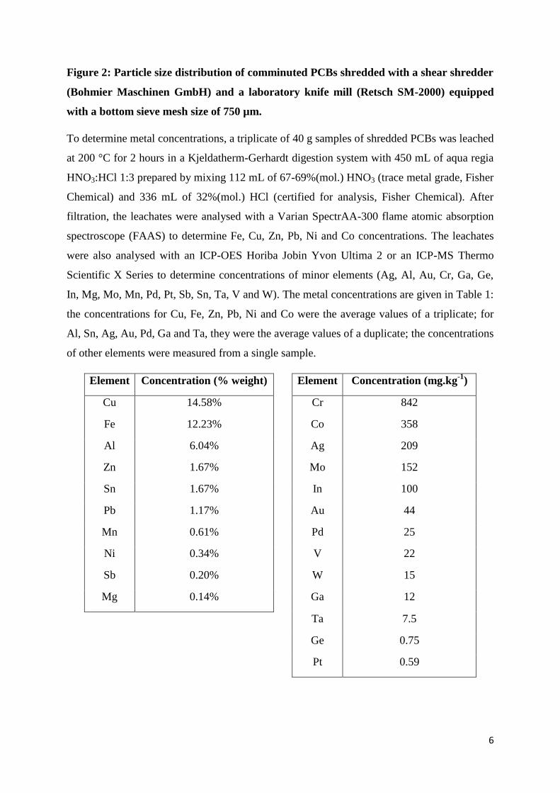
Figure 2: Particle size distribution of comminuted PCBs shredded with a shear shredder
(Bohmier Maschinen GmbH) and a laboratory knife mill (Retsch SM-2000) equipped
with a bottom sieve mesh size of 750 µm.
To determine metal concentrations, a triplicate of 40 g samples of shredded PCBs was leached
at 200 °C for 2 hours in a Kjeldatherm-Gerhardt digestion system with 450 mL of aqua regia
HNO3:HCl 1:3 prepared by mixing 112 mL of 67-69%(mol.) HNO3 (trace metal grade, Fisher
Chemical) and 336 mL of 32%(mol.) HCl (certified for analysis, Fisher Chemical). After
filtration, the leachates were analysed with a Varian SpectrAA-300 flame atomic absorption
spectroscope (FAAS) to determine Fe, Cu, Zn, Pb, Ni and Co concentrations. The leachates
were also analysed with an ICP-OES Horiba Jobin Yvon Ultima 2 or an ICP-MS Thermo
Scientific X Series to determine concentrations of minor elements (Ag, Al, Au, Cr, Ga, Ge,
In, Mg, Mo, Mn, Pd, Pt, Sb, Sn, Ta, V and W). The metal concentrations are given in Table 1:
the concentrations for Cu, Fe, Zn, Pb, Ni and Co were the average values of a triplicate; for
Al, Sn, Ag, Au, Pd, Ga and Ta, they were the average values of a duplicate; the concentrations
of other elements were measured from a single sample.
Element Concentration (% weight) Element Concentration (mg.kg-1
)
Cu 14.58% Cr 842
Fe 12.23% Co 358
Al 6.04% Ag 209
Zn 1.67% Mo 152
Sn 1.67% In 100
Pb 1.17% Au 44
Mn 0.61% Pd 25
Ni 0.34% V 22
Sb 0.20% W 15
Mg 0.14% Ga 12
Ta 7.5
Ge 0.75
Pt 0.59

7
Table 1: Concentrations (in % weight and mg.kg-1
) in PCB samples (weight = 40 g). The
samples were leached by aqua regia at 200 °C at a liquid/solid ratio of 11 mL.g-1
(N.A.=not analysed).
2.3 Evaluation of toxicity in shake flasks.
To evaluate the impact of cations from PCBs on the growth and activity of the bacteria, the
kinetics of Fe(II) bio-oxidation in the presence of different proportions of PCB leachate in the
culture medium were measured. The experiments were carried out in 250 mL Erlenmeyer
flasks. The leachate was prepared by abiotic leaching of PCBs (5%(w/v)) in the presence of 9
g.L-1
of synthetic Fe(III) added as Fe2(SO4)3.xH2O (reagent grade from Alfa Aesar). Leaching
was performed in a 2-L stirred tank reactor (STR) at 36 °C for 20 days. Analytical techniques
(described in Section 2.6) were used to determine the pH value, redox potential and metal
concentrations. After filtration through Whatman filters with 2.7 µm cotton fibres, the final
metal concentrations in the leachate were analysed (Table 2). Other metals such as Al were
assumed to be present in the leachate but their concentrations were not determined. This
leachate was then mixed in different proportions with water acidified to pH 1.3 with sulphuric
acid (0% v/v, 10% v/v, 30% v/v and 60% v/v). Nutrients were added into each flask (same
quantities for all tests, equivalent to 0Cm nutritive medium concentrations), as well as ferrous
sulphate so that the initial Fe(II) concentrations were above 6 g.L-1
. The flasks were then
inoculated with 10 mL (10% v/v) of BRGM-KCC consortium containing 1 g.L-1
Fe(II) and
107 cell.mL
-1. The flasks were maintained in an incubator at 40 °C and 107 rpm, with pH
values and redox potential measured on a daily basis.
Metal Leachate concentration (mg.L-1
)
Cu 9 416
Fe 16 840
Zn 934
Pb 16.8
Ni 18.7
Co 17.9
Table 2: Concentrations (in mg.L-1
) of Cu, Fe, Zn, Pb, Ni and Co in the leachate used for
evaluating toxicity in shake flasks.
2.4 PCB bioleaching in batch mode.

8
Experimental setup
The PCB bioleaching experiments in batch mode were performed in laboratory-scale glass
bioreactors with a working capacity of 2.25 L and jacketed for warm water circulation to
maintain a constant operating temperature (Figure 3). The temperature of the circulating water
was regulated by a cryothermostat and thermocouples placed in the bioreactors. The
bioreactors were equipped with four baffles mounted 90° apart and extending down to the
base of the vessel to optimise mixing of the pulp. The agitator was a dual impeller system
(axial/radial) consisting of a standard 6-blade Rushton turbine in combination with a 3-blade
45° axial flow impeller. The gas supply system was designed to accommodate air enriched
with 1% CO2, which was injected beneath the turbine at the bottom of the bioreactors via a
stainless steel pipe. The impellers and the gas injection pipe were positioned so as to keep to
the standard dimensions and thus optimise gas mass transfer and mixing in the bioreactors.
The top of the reactors was connected to a gas cooling system to prevent excessive
evaporation.
Figure 3: Schematic diagram of the experimental setup for bioleaching tests in batch
mode.

9
Methodology and operating conditions during the different experiments
Two types of tests were performed: abiotic and biotic. During the abiotic tests, acidified water
(pH 1.3 with sulphuric acid) and Fe(III) as ferric sulphate was introduced into the bioreactor
before adding different quantities of PCBs. When the biotic tests were performed, the
consortium (10% v/v) was inoculated in a nutrient medium with synthetic ferrous sulphate.
When the redox potential reached 800 mV vs SHE (corresponding to 90% of bio-oxidized
Fe(II)), different quantities of PCBs were added. The pH value was adjusted manually to 1.5
with sulphuric acid when it rose above 1.8. Ultrapure water was also added during daily
monitoring to compensate for water losses due to evaporation, despite the presence of a
condenser.
Different tests were performed to determine the influence of the presence of bacteria and the
impact of the initial concentration of Fe(III). The operating conditions that were used are
given in Table 3.
Solid
concentration
[Fe(III)]
(g.L-1
) Inoculation
Gas flow
(L.h-1
)
CO2
enrichment
Temperature
(°C)
Stirring
speed (rpm)
1% w/v PCBs
8 No 18 0% 40 600
Yes 60 1% 35 600
1
No 60 1% 35 600
Yes 60 1% 35 600
Yes 60 1% 35 600
0 No 60 1% 35 600
5% w/v PCBs 8 No 18 0% 40 600
Yes 60 1% 35 600
Table 3: Operating conditions during abiotic and biotic leaching experiments of
comminuted spent PCBs in 2.25 L-STR operated in batch mode.
Metal dissolution rates reported in this study were calculated as the maximum slope of metal
concentration vs. time obtained during batch experiments. For some experiments, two values
were reported since two distinct slopes, corresponding to two successive phenomena, were
observable.
2.5 PCBs bioleaching in continuous mode

10
Bioleaching experiments in continuous mode were carried out in the double-stage bioreactor,
illustrated in Figure 4.
Figure 4: Schematic diagram of the continuous double-stage bioreactor used for PCB
bioleaching.
The first stage was described in detail in our previous paper (Hubau et al., 2018). The 200
mL-bubble column was filled with 150 mL of 0Cm medium containing 1 g.L-1
Fe(II) from
ferrous sulphate at pH 1.1 and 15 g of activated charcoal (Organosorb11® from Desotec, high
specific surface area=950 m2.g
-1). Prior to use, the activated charcoal was washed with 5
mL.g-1
of water and 1 mL.g-1
of diluted sulphuric acid (pH 1.3), then dried at 40 °C for 20
hours and sieved to remove particles smaller than 1 mm. Bubble column stirring was
performed by means of a continuous air flow enriched with 1% of CO2 (air flow rate= 20-30
L.h-1
at 1 bar relative to atmospheric pressure and ambient temperature). The CO2 enrichment
was performed since it was previously proven that it favoured the growth and activity of
BRGM-KCC consortium (Guezennec et al., 2018). The temperature of the bubble column was
maintained at 36 °C by means of a thermostatic bath (Lauda), since this temperature was close
to the optimal temperature for the growth and activity of Leptospirillum ferriphilum, one of
the predominant strain of BRGM-KCC consortium that is able to oxidize iron (Coram and
Rawlings, 2002). The hydraulic residence time (HRT) was set to 3.2 hours. Continuous
feeding of the bioreactor and continuous removal of the effluent were performed at the
desired dilution rate by variable-speed pumps. The bubble column was first inoculated with

11
the BRGM-KCC consortium at 10% (v/v) in batch mode. Once the redox potential reached
900 mV vs SHE, the bubble column was run in continuous mode. HRT was gradually
decreased to 3.2 hours. All experiments were performed without sterilisation to be as
representative of industrial conditions as possible.
The second stage was a 2.25 L-STR fed continuously with the leaching solution produced in
the bubble column. This STR was the same as the one used in batch mode, described in
Section 2.4. The incoming flowrate was set at 47 mL.h-1
so that HRT in the STR was 48
hours. The STR was also fed with comminuted spent PCBs by means of a dosing screw (from
Dosapro Milton Roy) controlled with a programmable logic controller (Siemens LOGO!).
The flowrate of solids could vary between 0.5 and 0.85 g.h-1
so that the PCB concentration
varied from 1%(w/v) to 1.8%(w/v). The outflow (pulp) was removed with a peristaltic pump.
To adjust the pH to 1.5, sulphuric acid was added with a peristaltic pump at concentrations
ranging from 1.8 to 3.5 mol.L-1
and a flow rate of 2 to 5 mL.h-1
. STR stirring speed was set at
600 rpm, the temperature was maintained at 36 °C and air flowrate was set to 60 L.h-1
with
1% CO2 enrichment.
At the end of the experiments, the bubble column was by-passed to evaluate the dissolution
kinetics of metals from PCBs in a one-stage continuous bioreactor (2.25 L-STR). In this case,
the feeding solution did not contain Fe, so that PCBs were the only source of Fe. The feeding
solution was composed of 0Cm medium with diluted sulphuric acid to maintain the pH at 1.1.
2.6 Analytical techniques.
pH values and redox potentials were measured daily for all experiments. Metal concentrations
(Cu, Fe, Zn, Ni, Co, Pb) in liquid phase were determined by atomic absorption spectroscopy
(AAS) using a Varian SpectrAA-300. The Yue et al. (2016) correlation was used to determine
the Fe(III) to Fe(II) concentration ratio from the redox potential measurement and the Fe(II)
concentration from total Fe concentration. This correlation is not affected by the presence of
dissolved metals from PCBs.
When stable characteristics of STR outflow were obtained in continuous mode,
concentrations of Ag, Al, Ca, Cr, Ga, K, Mg, Mo, Na, P, S, Sb, Sn, Ta, V and W in the STR
liquid phase were measured with ICP-OES (following the ME-ICP02 protocol of the ALS
company in Loughrea, Ireland). NH4+ concentration was also measured in these samples by
colorimetric assays in accordance with the NF ISO 15923-1 standard (AFNOR, 2014).
Concentrations of solids were also determined to validate the experimental setup. XRD was

12
used on the bioleaching residues (Bruker D8 Advance diffractometer, 4-75 °2θ, 0.03 °2θ/s,
288 s each step, Cu tube) and the diffraction patterns were processed with Diffrac Suite
software.
2.7 Bacterial community monitoring.
2.4.1 Cell counting. The bacterial cells were counted in the liquid phase of the different
experiments with a Thoma cell counting chamber.
2.4.2 Scanning electron microscopy (SEM) with lyophilisation. SEM was used to observe
microorganisms being attached to PCB particles during the experiments in continuous mode.
For these observations, fresh aliquots of the outflow from the STR bioreactor were sampled.
Large particles of PCBs from these pulp samples were introduced onto a copper frame with
carbon paste and immersed in liquid nitrogen at -176 °C. Slush nitrogen at -210 °C was
reached by pressure depression. The samples were then set into a vacuum, transferred into the
SEM (Hitachi S-4500 field-emission) and slowly warmed up to room temperature. They were
then coated with Au-Pd alloy to obtain electrically conductive samples whatever the
composition of the PCB particle, and finally observed under a secondary electron beam at 2
kV. Energy dispersive X-ray spectroscopy (EDX) was used to characterise the sample
composition with SEM operated at 15 kV.
2.4.3 DNA extraction, 16S rRNA gene PCR (polymerase chain reaction) amplification and
bacterial community fingerprints. Samples of 0.5 mL solid medium or 2 mL liquid phase
were taken from the bubble column; 5 mL pulp samples were taken from the stirred tank
reactor. The solid samples, pulp and pellets (from centrifuging the liquid samples for 10
minutes at 14000 g) were washed by re-suspension in Tris buffer (100 mM, pH 8) until a pH
around 7 was reached. Genomic DNA was extracted with the FastDNA Spin Kit for Soil,
using the manufacturer’s protocol (MP Biomedicals) with a FastPrep®-24 at a speed of 5 m.s
-1
for 30 s (lysis was performed twice for the activated charcoal samples). PCR amplification of
the 16S rRNA genes (ca. 200 bp) from DNA extracts was performed with the w49 forward
primer and the 5’-end FAM-labelled w34 primer, in a C-1000 thermocycler (Biorad), as
described in Hedrich et al. (2016). The amplification products were analysed on agarose gel.
Community fingerprints were obtained by CE-SSCP (Capillary electrophoresis-single strand
conformational polymorphism). One microlitre of 5- to 500-fold diluted PCR product, 0.4 μL
of Genescan-600 LIZ internal standard (Applied Biosystems) and 18.6 µL of deionized
formamide HiDi (Applied Biosystems) were heat-denatured for 5 minutes at 95 °C and
immediately cooled on ice for 10 minutes. Fragment analyses were conducted with the ABI

13
Prism 310 (Applied Biosystems). GeneScan software (Applied Biosystems) was used to
realign the peak profiles based on internal standard migration, assign peak position, align
community peaks with peaks of known acidophilic strains, and calculate relative abundances
on the basis of peak areas.
3. Results
3.1 Microbial ecotoxicology
The microbial ecotoxicology tests performed in this study to evaluate the toxicity of dissolved
cations were carried out with PCB leachate rather than with solid PCBs or synthetic solutions
for two main reasons:
(i) When working at a small scale (a few hundred mL), the quantity of PCBs that can
be added to a flask is limited to a few grams to correspond to concentrations of
less than 10% solids (this is the maximum concentration used in the literature on
PCB bioleaching). However, samples of such small size are no longer
representative of the initial batch of PCBs and vary widely in their composition, so
that the results obtained are difficult to compare. The use of a leachate obtained
from a large quantity of PCBs allows work to be done at a small scale in a
repeatable and representative manner.
(ii) The toxicity evaluation performed with leachates takes the impacts of both major
metals and metal traces into account. This is a great advantage in comparison to
synthetic solutions that could be prepared to model PCB cations.
Figure 5 shows that biomass growth and Fe(II) bio-oxidation were delayed when the leachate
proportion increased. The duration of the lag phase increased with the quantity of leachate.
However, despite this delay, the maximum bio-oxidation rate and the final biomass
concentration were of the same order of magnitude as in the absence of leachate (around 200
mg.L-1
.h-1
of Fe(II) oxidized and 108 cell.mL
-1 respectively). Thus, regardless of the amount
of leachate added, the bacterial culture seemed to adapt progressively to increasing metal
concentrations. These results are consistent with those obtained by Choi et al. (2004). The
results of our study are promising as regards the ability of the BRGM-KCC consortium to
bioleach PCBs. They also confirmed the relevance of separating bacterial growth from the
bioleaching of PCBs, in order to limit the lag phase related to the toxicity of PCB cations.
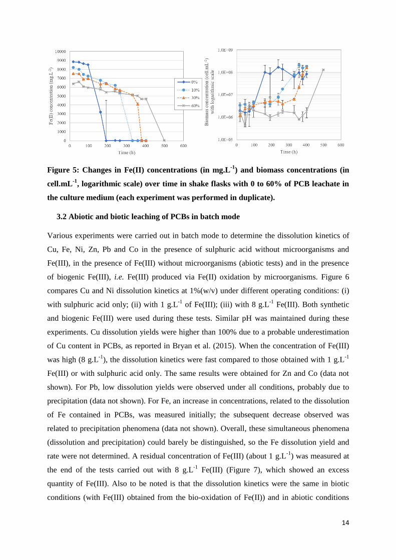
14
Figure 5: Changes in Fe(II) concentrations (in mg.L-1
) and biomass concentrations (in
cell.mL-1
, logarithmic scale) over time in shake flasks with 0 to 60% of PCB leachate in
the culture medium (each experiment was performed in duplicate).
3.2 Abiotic and biotic leaching of PCBs in batch mode
Various experiments were carried out in batch mode to determine the dissolution kinetics of
Cu, Fe, Ni, Zn, Pb and Co in the presence of sulphuric acid without microorganisms and
Fe(III), in the presence of Fe(III) without microorganisms (abiotic tests) and in the presence
of biogenic Fe(III), i.e. Fe(III) produced via Fe(II) oxidation by microorganisms. Figure 6
compares Cu and Ni dissolution kinetics at 1%(w/v) under different operating conditions: (i)
with sulphuric acid only; (ii) with 1 g.L-1
of Fe(III); (iii) with 8 g.L-1
Fe(III). Both synthetic
and biogenic Fe(III) were used during these tests. Similar pH was maintained during these
experiments. Cu dissolution yields were higher than 100% due to a probable underestimation
of Cu content in PCBs, as reported in Bryan et al. (2015). When the concentration of Fe(III)
was high (8 g.L-1
), the dissolution kinetics were fast compared to those obtained with 1 g.L-1
Fe(III) or with sulphuric acid only. The same results were obtained for Zn and Co (data not
shown). For Pb, low dissolution yields were observed under all conditions, probably due to
precipitation (data not shown). For Fe, an increase in concentrations, related to the dissolution
of Fe contained in PCBs, was measured initially; the subsequent decrease observed was
related to precipitation phenomena (data not shown). Overall, these simultaneous phenomena
(dissolution and precipitation) could barely be distinguished, so the Fe dissolution yield and
rate were not determined. A residual concentration of Fe(III) (about 1 g.L-1
) was measured at
the end of the tests carried out with 8 g.L-1
Fe(III) (Figure 7), which showed an excess
quantity of Fe(III). Also to be noted is that the dissolution kinetics were the same in biotic
conditions (with Fe(III) obtained from the bio-oxidation of Fe(II)) and in abiotic conditions

15
(in the presence of ferric sulphate). However, regeneration of Fe(III) by bio-oxidation of
Fe(II) was observed in the biotic experiments after a time-lag that was variable (Figure 7) but
longer than the time required for the chemical dissolution of metals. Fe(III) regeneration in
the abiotic experiments with 1 g.L-1
Fe(III) was also observed due to contamination.
Figure 6: Dissolution yields (in %) over time of (a) Cu and (b) Ni under abiotic and
biotic conditions (tests performed in STR operated in batch mode).

16
Figure 7: Fe(III) concentrations (in mg.L-1
) over time in STR operated in batch mode.
The kinetics obtained at 1 g.L-1
of Fe(III) were similar to those without Fe. However, in the
first hours of the experiments (zooms in the inlet of Figures 6A and 6B), Fe(III) accelerated
the dissolution kinetics for major metals but was quickly consumed. The metal dissolution
observed after the consumption of Fe(III) was likely to be an acidic hydrolysis. The average
proton consumption, measured between the addition of PCBs and the end of metal
dissolutions (around 300 h), was 0.4 mmol.L-1
.h-1
. Two different dissolution rates were thus
measured (Table 4): in the presence of an excess of Fe(III) and without Fe(III). Except for Pb,
the dissolution rates obtained in the presence of Fe(III) were higher than the dissolution rates
reached in the second leaching stage (acidic hydrolysis). In the case of Pb, the dissolution rate
was probably under-estimated in both leaching stages since Pb precipitated after dissolution.
For Cu, an increase followed by a decrease in Cu concentrations was observed at the start of
leaching, which could be explained by cementation phenomena.
Metal Fe(III) dissolution rate (mg.L-1
.h-1
) H2SO4 dissolution rate (mg.L-1
.h-1
)
Fe 1800 500-650
Cu 600 200-250
Zn 115 80
Ni 12 9.3
Pb 10 14

17
Co 2.7 1.2
Table 4: Dissolution rates (in mg.L-1
.h-1
) observed for Fe, Cu, Zn, Ni, Pb and Co with
excess Fe(III) or with sulphuric acid at pH 1.3 in the presence of 1%(w/v) PCBs in batch
mode.
Biotic and abiotic tests were also carried out in the presence of 5%(w/v) of solids and 8 g.L-1
of Fe(III). Table 5 shows the dissolution rates and yields reached during these experiments.
For most metals, very rapid initial dissolution was observed due to the Fe(III) reduction.
When all Fe(III) was consumed (low redox potential), dissolution slowed, which probably
corresponded to acidic hydrolysis.
Metal Dissolution yield (%) Maximum dissolution rate (mg.L-1
.h-1
)
Fe 99% 3200
Cu 104% 2500
Zn 93% 450
Ni 95% 70
Pb 3% 30
Co 92% 13
Table 5: Metal dissolution yields (in %) and rates (in mg.L-1
.h-1
) at 5%(w/v) PCBs with 8
g.L-1
of Fe(III) in batch mode.
3.3 Continuous bioleaching of PCBs in a double-stage bioreactor
Continuous bioleaching experiments were carried out with an initial concentration of 1 g.L-1
Fe(II) in the feed stream. This operating condition was selected (i) to ensure that Fe(III) was
not in excess and that the complete dissolution of PCB metals was made possible by bacterial
regeneration of Fe(III), and (ii) to minimise consumption of reagents and simplify the
downstream hydrometallurgical purification stages.
The HRT was set at 48 h in the second stage of the bioreactor (PCB leaching in stirred tank
reactor), which corresponds to an HRT of 3.2 h in the first stage (bubble column). In these
conditions, Fe(II) bio-oxidation performance was stable (100% bio-oxidation and 312 mg.L-
1.h
-1 of Fe(II) bio-oxidized) and biomass concentration was constant over time (2.10
7 cell.mL
-
1) in the first stage (Hubau et al., 2018); the leaching solution that fed the second stage
contained 1 g.L-1
of Fe(III). This residence time was also selected to allow bacteria to grow in
the second stage, since the microbial generation time was shorter than the HRT: Coram and

18
Rawlings (2002) and Johnson et al. (2008) reported a generation time of 12 - 15 h and 3.1 h
for Leptospirillum ferriphilum and Sulfobacillus benefaciens respectively.
Changes in dissolution yields over time in the presence of 1% PCBs
Although the bioreactor was run at constant operating conditions for 2500 hours, significant
changes in redox potential and metal dissolution yields (Figure 8) and pH (Table 6) were
observed after 500 and 1600 hours of operation.
Figure 8: (A) Changes in the redox potential (in mV) in the bioleaching reactor run in
continuous mode over time and (B) Metal dissolution yields (in %) in the double-stage
bioreactor in the presence of 1%(w/v) of PCBs (continuous mode) depending on the
period (error bars depict uncertainties resulting from elemental analyses).
Period pH H
+ consumption in the
2nd
stage (mmol.h-1
)
Feeding solution - 1.1 -
Outlet of 1st stage - 1.2 -
Outlet of 2nd
stage
Period 1 (0 – 400 h) 1.5 1.7
Period 2 (600 – 1000 h) 1.8 2.3
Period 3 (1600 – 2400 h) > 2.0 so acid added 4.8

19
Table 6: pH and proton consumption (in mmol.h-1
) in the feed solution and at the outlet
of the 1st and 2
nd stage.
Three leaching periods were defined:
Period 1 (0 – 400 h): The whole amount of incoming Fe(III) from the first stage was
reduced as Fe(II) in the 2nd
stage (Eh = 530 mV vs SHE). The pH, which was set at 1.1
in the feed solution and was 1.2 at the outlet of the 1st stage, reached 1.5 in the second
stage. Cu was not dissolved (<1%), while high yields were obtained for Fe and Co
(respectively 76% and 73% dissolution). Limited dissolution yields were obtained for
Zn and Ni (respectively 33% and 55%).
Period 2 (600 – 1000 h): Fe was still in the Fe(II) oxidation state but redox potential
was slightly higher (Eh = 560 mV vs SHE). In the meantime, acid consumption
increased from 1.7 to 2.3 mmol.h-1
of protons. The pH reached 1.8 and metal
concentrations in the leachate increased significantly. Dissolution yields were
respectively 81% Cu, 72% Fe, 91% Zn, 83% Ni and 86% Co.
Period 3 (1600 – 2400 h): Fe(III) was fully regenerated, the redox potential was
880 mV vs SHE. Acid consumption was even greater than during period 2, with a pH
value above 2.0. An addition of sulphuric acid was required to limit Fe precipitation
(the pH value was maintained at 1.5). Acid consumption reached 4.8 mmol.h-1
.
Dissolution yields were 96% Cu, 59% Fe, 85% Zn, 73% Ni, 93% Co.
The dissolution rates during these different periods were compared (Table 7). Whatever the
period, similar results were obtained for Al dissolution kinetics, with 46% to 54% of Al
bioleached. For certain metals in smaller quantities, such as Ag and Mo, a large increase in
the bioleaching yield was obtained during the third period; their concentrations were too low
to calculate their dissolution rates during the first two periods. In addition, a decrease in the
concentration of Mn in the leachate was observed over time and the dissolution yield
decreased from 55% in the first period to 10% in the third period, corresponding to a large
decrease in the dissolution rate. This result was not yet understood. For Cr, Sb, Sn and Pb,
concentrations in the leachate and dissolution rates remained low regardless of the period
(dissolution yield <10%). Finally, the limits of detection of Ta, V, W and Ga were higher than
the maximum concentration that would be reached if they were completely dissolved, which
did not allow us to conclude on the ability to dissolve these metals in these operating
conditions.

20
Cu Fe Zn Ni Co Pb Ag Al Cr Mn Mo Sb Sn
Period 1 0.26 19.2 1.14 0.41 0.052 0.18 - 6.63 0.015 0.72 - 0.035 0.52
Period 2 23.8 17.9 3.07 0.57 0.063 0.15 - 5.65 0.008 0.37 - 0.013 0.04
Period 3 28.3 12.2 2.71 0.45 0.067 0.28 0.012 6.04 0.010 0.14 0.01 0.027 0.40
+/- 0.02 0.02 0.02 0.02 0.02 0.21 0.008 0.02 0.020 0.02 0.02 0.083 0.21
Table 7: Dissolution rates (in mg.L-1
.h-1
) and errors due to the uncertainties on the
analysis of leachate concentrations during PCBs bioleaching in continuous mode in the
presence of 1%(w/v) of solids.
Consumption of sulphates and precipitation phenomena
In these complex environments, precipitation phenomena are regularly reported (Adhapure et
al., 2013, Ilyas et al., 2010). The X-ray diffractogram of the bioleaching residues (see
Supplementary information) revealed the presence of a large quantity of amorphous material,
as evidenced by the broad peak located between 2θ = 12° and 2θ = 30°. A significant amount
of anglesite (PbSO4) was also observed. This explains the low Pb dissolution yields (between
6% and 12%) and is consistent with the literature (Guezennec et al., 2015). Other elements,
such as Al and Sn, were also observed in these residues: these were partially dissolved
(between 46% and 54% of Al was dissolved and between 1% and 15% of Sn). For Sn,
precipitation of SnO during PCB bioleaching has been reported in different studies (Brandl et
al., 2001; Bryan et al., 2015). For Al, we can assume that its speciation was very
heterogeneous in the sample, with aluminium present as a metal or oxide. Bioleaching was
probably not suited to dissolving Al as an oxide, which would account for its detection in the
residues.
The X-ray analyses were supplemented by SEM imagery of the residues of bioleached spent
PCBs (Figure 9). Crystallization was observed on some particles. EDX analysis suggested
that this was potassium jarosite (KFe3(OH)6(SO4)2) that had crystallized on a particle of
alumina (Al2O3). Traces of Cu were also detected, which could have come from lyophilisation
of the leachate containing a high Cu concentration.

21
Figure 9: SEM image and EDX analysis of a comminuted spent PCB particle covered
with a layer of crystallized precipitate (the particle came from STR reactor during 3rd
period).
Jarosite precipitation is common during bioleaching. Fe(III) and sulphates are consumed
during this precipitation, according to the following reaction (with A+ a monovalent cation):
A+ + 3Fe
3+ + 2SO4
2- + 6H2O → AFe3(SO4)2(OH)6 + 6H
+ [3]
The precipitation rate was calculated from the concentration of sulphur (only present in
solution as sulphates) in the output streams and in the feed stream, with the hypothesis that
Fe(III) precipitates mainly in the form of jarosite, with other precipitates, if any, being
negligible. This hypothesis was supported by the X-Ray analysis of the leaching residues,
which did not detect any other Fe precipitates. During the first two periods, there was no
precipitation or very little, due to a combination of low pH and low Fe(III) concentration in
the liquid phase. During the third period, precipitation was substantial with 0.16 mmol.h-1
of
sulphates consumed. The amount of Fe(III) precipitating as jarosite was estimated from the
consumption of sulphates, given that two moles of sulphates are involved in the formation of
jarosite for 3 moles of Fe(III). During the 3rd
period, 6.0 mg.L-1
.h-1
of Fe(III) was consumed
by the precipitation phenomena.
Generation of Fe(III) by bio-oxidation of Fe(II)
To estimate the biological Fe(II) bio-oxidation rate, a mass balance for Fe(III) during steady
state was calculated for the second stage of the continuous bioreactor:
Fe3+
from bio-oxidation = Fe3+
consumed for leaching + Fe3+
precipitated + Fe3+
in the outlet flow - Fe3+
in the feed [4]
Fe(III) concentrations in the feed and the outlet flow (Fe3+
in the feed and Fe3+
in the outlet flow) were
estimated with the Yue et al. (2016) correlation and AAS results (see Section 2.6). The rate of
Fe(III) precipitation (Fe3+
precipitated) was estimated from the consumption of sulphates as
explained above. The Fe3+
consumed for leaching was estimated from the dissolution rates of the

22
different metals and the stoichiometric requirements associated with each of these metals (2
moles of Fe(III) for the dissolution of one mole of Cu, 3 moles of Fe(III) per one mole of Al,
etc.). This calculation was based on the hypothesis that metal were dissolved only by
oxidation reactions and acidic hydrolysis was negligible. This hypothesis was supported by
the results obtained in the batch bioleaching experiments showing that acidic dissolution is
much slower than oxidative dissolution. Concerning Fe dissolution, the Fe contained in PCBs
was considered to dissolve as Fe(II) cations, which were potentially re-oxidized; we did not
consider Fe dissolution as Fe(III), since the redox potential of the Fe/Fe(III) couple is higher
than for Fe/Fe(II). Fe dissolution rate was calculated with the following mass balance on the
STR reactor during steady state :
Fedissolved = Feprecipitated + Fein the leachate - Fein the feed [5]
With Fedissolved, Fein the feed and Fein the leachate the Fe dissolution rate and flows of Fe that came
from the bubble column and that went out from STR reactor, respectively. The Fe dissolution
rates are shown on Table 8. The estimated Fe dissolution yield reached 93% during the third
period. However, the calculated rate of Fe dissolution for the same period of time did not
greatly increase. This was due to a small deviation in the solid concentration in the STR,
which decreased slowly due to a probable partial obstruction of the dosing screw (less PCBs
were added); this deviation was taken into account in our yield calculations.
Consequently, the Fe(III) demand for the dissolution reactions (Fe3+
consumed for leaching) varied
from 3.63 mmol.h-1
(period 1) to 4.96 mmol.h-1
(period 3).
Table 8 shows the estimated Fe(II) bio-oxidation rate for each period. These rates increased
significantly over the different periods. It should be noted that the balance was calculated on a
macroscopic scale. The values will probably vary on a microscopic scale with, for example,
areas of high microbial concentration and therefore higher bio-oxidation rates than in our
estimations.
Estimated Fe dissolution yield
(%) and rate (mg.L-1
.h-1
)
Fe(II) bio-oxidation rate
(mg.L-1
.h-1
)
Period 1 76% - 18.7 69
Period 2 62% - 17.2 105
Period 3 93% - 18.5 142
Table 8: Estimation of Fe dissolution kinetics (in % and mg.L-1
.h-1
) and Fe(II) bio-
oxidation rates (in mg.L-1
.h-1
) over time in the bioreactor used for continuous
bioleaching spent PCBs, in the presence of 1%(w/v) PCBs and HRT=48 h.

23
Changes in the bacterial community over time at 1% solids
To understand the structure of the acidophilic culture and its impact on the different
phenomena, various tools were used to characterise the bacterial populations. First, the
biomass concentration in the liquid phase was determined with a Thoma cell counting
chamber. Figure 10 shows how cell concentrations in the 1st stage and 2
nd stage changed over
time. During period 1, bacteria were transferred from the bubble column to the second stage
of the bioreactor but no growth was observed in the latter. Their concentration in the PCB
leaching bioreactor was lower than in the liquid phase at the outlet of the 1st stage. During
period 2, the bacteria seemed more active when observed with Thoma cell counting (fast
motions on the plate) and slightly more numerous. Finally, during period 3, the number of
cells increased sharply in the liquid phase.
Figure 10: Biomass concentration over time (in cell.mL-1
) in the liquid phases of the 1st
and 2nd
stages of the bioreactor.
DNA extraction in the liquid and solid phases (activated charcoal from the bubble column and
comminuted PCB residues in the 2nd
stage) enabled us to determine the microbial distribution
between the two phases (Figure 11). In the first stage (bubble column), DNA was mainly
extracted from activated carbon (99.83%). In the 2nd
stage (STR with PCBs), 17% of the
DNA was in the liquid phase, while 83% came from the PCBs. Bacteria were mostly attached.

24
Figure 11: DNA distribution (in % weight) between the liquid and solid phases in the
two stages of the continuous bioreactor used for bioleaching spent PCBs.
Microbial diversity profiles in the bioreactors were determined during the 3rd
period by 16S
rRNA gene CE-SSCP fingerprints. In the first stage (bubble column), the iron-oxidizer
Leptospirillum ferriphilum was detected at average proportions of 70.4% in the solid phase
and 69.3% in the liquid phase. The sulphur and iron oxidizer Sulfobacillus benefaciens and an
unknown strain were also detected on the solid phase at a relative abundance of 16.5% and
13.1% respectively, and 15.0% and 15.6% respectively on the liquid phase. In the second
stage (STR), the growth of Leptospirillum ferriphilum was favoured, since only this strain
was detected on the fingerprints in the pulp, on the solid phase and in the liquid phase.
Despite the fact that Acidithiobacillus caldus and Sulfobacillus thermosulfidooxidans were
known to be members of BRGM-KCC consortium inoculated into the bioreactor, they
remained undetected. For Acidithiobacillus caldus, which is a sulphur-oxidizing bacterium,
this can be explained by the absence of reduced sulphur compounds in the bioreactor.
Likewise, the temperature maintained at 35 °C in the bioreactor was probably too low for
Sulfobacillus thermosulfidooxidans since the optimum growth temperature is 51 °C for this
bacterium (Watling et al., 2008).
25
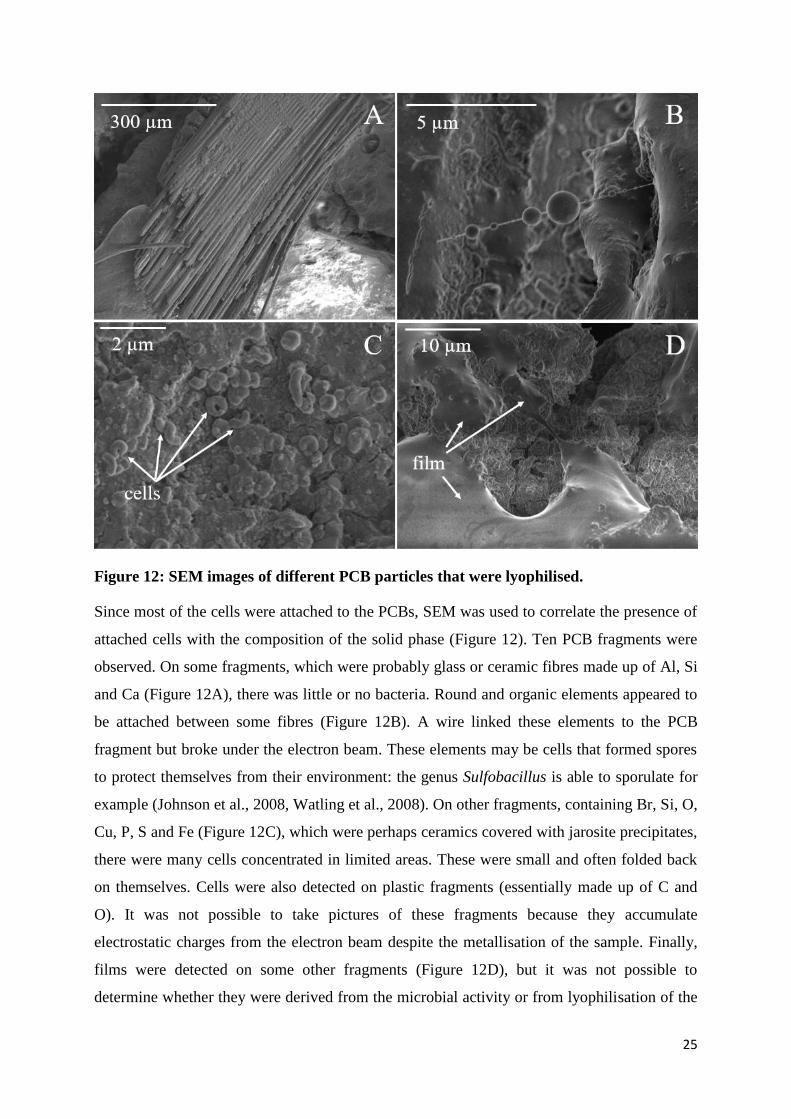
Figure 12: SEM images of different PCB particles that were lyophilised.
Since most of the cells were attached to the PCBs, SEM was used to correlate the presence of
attached cells with the composition of the solid phase (Figure 12). Ten PCB fragments were
observed. On some fragments, which were probably glass or ceramic fibres made up of Al, Si
and Ca (Figure 12A), there was little or no bacteria. Round and organic elements appeared to
be attached between some fibres (Figure 12B). A wire linked these elements to the PCB
fragment but broke under the electron beam. These elements may be cells that formed spores
to protect themselves from their environment: the genus Sulfobacillus is able to sporulate for
example (Johnson et al., 2008, Watling et al., 2008). On other fragments, containing Br, Si, O,
Cu, P, S and Fe (Figure 12C), which were perhaps ceramics covered with jarosite precipitates,
there were many cells concentrated in limited areas. These were small and often folded back
on themselves. Cells were also detected on plastic fragments (essentially made up of C and
O). It was not possible to take pictures of these fragments because they accumulate
electrostatic charges from the electron beam despite the metallisation of the sample. Finally,
films were detected on some other fragments (Figure 12D), but it was not possible to
determine whether they were derived from the microbial activity or from lyophilisation of the

26
ionic solution. These observations did not enable us to confirm that bacteria became
preferentially attached to certain fragments, as asserted by Silva et al. (2015).
Bioleaching kinetics at higher concentrations of solids (1.8% w/v)
When the second stage of the bioreactor reached a permanent regime (stabilisation of all the
chemical, physical and biological parameters), the concentration of solids was increased from
1% to 1.8%(w/v). The metal dissolution rates reached with the new concentration are shown
in Figure 13: after the change in concentration, they increased gradually and stabilised after
1000 hours. The dissolution rates increased with the concentration of solids for almost all
metals (Cu, Fe, Ni, Zn, Sn, Pb, Mn, Co, Al, Cr and Mo) except Ag and Sb. The dissolution
rate of Sb remained constant. The dissolution rate of Ag was halved while its concentration in
the leachate remained constant. This may be due to precipitation phenomena.
Figure 13: Bioleaching rates (in mg.L-1
.h-1
) according to the concentration of solids in
the double-stage bioreactor run in continuous mode.

27
Figure 14: Bioleaching yields (in %) according to the concentration of solids in the
double-stage bioreactor run in continuous mode. Error bars depict uncertainties in the
leachate analysis.
Figure 14 shows the maximum metal dissolution yields reached after changing the
concentration of solids. Some metals such as Ta, W, Ga and V are not reported. In the case of
Ta, W and Ga, the analytical uncertainties were still higher than the maximum concentration
that would be reached if they were completely dissolved. In the case of V, the limit of
detection was 0.1 mg.L-1
. This value corresponds to a dissolution of 25% of V from PCBs. As
V remained undetected, its dissolution yield was thus lower than 25%. The dissolution yields
of the other metals were of the same order of magnitude as those reached with the 1%(w/v)
concentration of solids, except for Cu whose dissolution yield was above 100% (as explained
in batch experiments in Section 3.2, Cu content in PCBs was probably underestimated).
The redox potential was higher than 880 mV vs ENH for both concentrations of solids (data
not shown), which shows that all Fe(II) produced by the dissolution of metals in the PCBs
was re-oxidized by the microorganisms present in the reactor, the bio-oxidation rate being
higher than the rate of Fe(III) consumption.
Using equations [4] and [5], the Fe mass balance was established and the rates of jarosite
precipitation and Fe(II) bio-oxidation were estimated (Table 9). For both concentrations of
solids, the Fe dissolution yields were very high. They even reached more than 100% at
1.8%(w/v), probably because of an overestimation of jarosite precipitation in our mass
balance based on equation [5]: sulphates were apparently involved not only in the
precipitation of jarosite but also of anglesite (PbSO4), as shown previously. These high Fe
dissolution yields are promising as regards the ability to run the bioreactor without any Fe in
the feeding solution, using PCBs as the only source of Fe.

28
Moreover, the Fe(II) bio-oxidation rate at 1.8%(w/v) was approximately twice the rate
obtained at 1%(w/v).
PCB
concentration
Fe dissolution rate
(mg.L-1
.h-1
)
Fe dissolution
yield (%)
Fe(II) bio-oxidation
rate (mg.L-1
.h-1
)
1%(w/v) 18.2 93% 142
1.8%(w/v) 61.9 >100% 328
Table 9: Estimation of the Fe(II) bio-oxidation rate (in mg.L-1
.h-1
) and dissolution
kinetics of Fe (taking into account jarosite precipitation) according to PCB
concentrations.
Figure 15 shows the consumption of protons and the difference in biomass concentrations
between the 1st and 2
nd stages for different concentrations of solids. The values increased with
increasing PCB concentrations. The consumption of protons was due to the high alkalinity of
the PCBs and the high bio-oxidation rates. Concerning biomass concentrations, the PCBs did
not inhibit bacterial growth. The significant increase in biomass concentrations probably
resulted from larger quantities of substrate and from better adaptation of microbial cells to
their environment. Leptospirillum ferriphilum was the only strain detected in the second stage
of the bioreactor (STR) with CE-SSCP fingerprints, as at the 1%(w/v) PCB concentration.

29
Figure 15: Proton consumption (in mmol.h-1
) and difference in bacterial concentrations
between the liquid phases of the 1st and 2
nd stages (in cell.mL
-1, logarithmic scale) for
different concentrations of solids.
3.4 Bioleaching of PCBs in continuous mode in a single-stage bioreactor
When the bioreactor reached a stationary state after changing the concentration of solids, the
first stage (bubble column) was by-passed and the STR was fed only with the comminuted
PCBs and the 0Cm nutritive medium at pH 1.2, without Fe. The aim was to test the ability of
the bacteria in the bioreactor to use PCBs as the only source of Fe, since the Fe content in the
PCBs had reached 12.2% (in weight). The concentration of solids was maintained at 1.8%
(w/v). The dissolution yields reached in the single-stage bioreactor were compared to those
obtained during the double-stage experiment and are given in Figure 16. Proton consumption
and biomass concentration in the liquid phase are shown in Figure 17. Redox potential is not
shown but remained the same after the first stage was by-passed. These results clearly indicate
that there was no major difference between the two modes of operation. A slight improvement
was even observed in the dissolution yields of Cu, Fe, Zn, Ni and Co. Slightly higher
consumption of protons was observed when the 1st stage was no longer being operated. It also
seemed that the culture was less robust to possible changes in operating conditions, such as air
shutdowns: when a sudden change in operating conditions occurred, the time needed to obtain
similar dissolution yields after favourable conditions were restored was longer than when the
first stage was not by-passed (data not shown).

30
Figure 16: Cu, Fe, Zn, Ni, Pb and Co dissolution yields (in %) in the presence of
1.8%(w/v) PCBs in the double-stage bioreactor with 1 g.L-1
Fe(II) in the feed and in the
single-stage bioreactor (by-pass of the bubble column) without Fe in the feed.
Figure 17: Proton consumption (in mmol.h-1
) and difference in biomass concentrations
between the 1st and 2
nd stage (in cell.mL
-1, logarithmic scale) in the presence of
1.8%(w/v) PCBs in the double-stage bioreactor with 1 g.L-1
Fe(II) in the feed and in the
single-stage bioreactor (by-pass of the bubble column) without Fe in the feed.
4. Discussion
Toxicity test and bioleaching in batch mode
Although the bioleaching kinetics in batch mode were quite conclusive, the contribution of the
biological compartment to the leaching reactions was not fully demonstrated: the biological
activity in the dissolution of metals was almost undetectable during these experiments in
batch mode. High initial concentrations of Fe(III) (either biogenic or synthetic) were therefore
necessary to obtain rapid kinetics: 7 g.L-1
of Fe(III) were necessary in the presence of
1%(w/v) PCBs and 35 g.L-1
of Fe(III) would be necessary in the presence of 5%(w/v) of
solids. These high Fe concentrations would be difficult to manage in downstream
hydrometallurgical operations. Fe(II) bio-oxidation was nevertheless detected after a long lag
phase (approximately 300 h), which was also the case during the microbial ecotoxicology
tests. The culture would need to be adapted to the e-waste, but this can hardly be done on an
industrial scale because of the highly variable composition of e-waste. The transition to the
operation in continuous mode eliminated the time-lag as a steady-state mode was established.
This allowed metal dissolution and Fe(II) bio-oxidation to be observed simultaneously.

31
PCB bioleaching in continuous mode
At the beginning of the study on PCB bioleaching in continuous mode with 1%(w/v) PCBs,
three distinct periods were observed, which could be attributed to phases in cell adaptation:
Period 1 (0 – 400 h): Bacteria entering the second stage (STR) were not able to grow
or to oxidize Fe(II) noticeably. Metal dissolution was mainly related to chemical
leaching by incoming Fe(III) (1 g.L-1
) and sulphuric acid. Thus, consumption of 1.7
mmol.h-1
of protons was observed in the second stage. Our estimation of the Fe(II)
bio-oxidation rate (Table 8) gave 69 mg.L-1
.h-1
. It could be assumed that there is a bias
on this value due the fact that during this period, leaching was partly caused by
hydrolysis reactions with sulphuric acid. This phenomenon, which was not considered
in our mass balance, artificially increased Fe(III) demand due to the dissolution of
metals and distorted both the mass balance and the evaluation of the bio-oxidation
rate.
Period 2 (600 – 1000 h): The bacteria were slightly more active (cells were more
mobile on the counting chamber). Metal dissolution rates and yields increased during
this period compared to the previous ones, probably because of the bio-oxidation of
Fe(II). However, only Fe(II) was detected: it is likely that the kinetics of Fe(III)
generation (bio-oxidation of Fe(II)) were slower than the consumption kinetics. The
consumption of protons also increased, which was consistent with the bio-oxidation
reaction [2]. In addition, very low consumption of sulphates was observed, which
corresponded to a very low rate of jarosite precipitation.
Period 3 (1600 – 2400 h) : The number of cells increased exponentially in the liquid
phase. The microbial culture bio-oxidized Fe(II) much faster than the consumption of
Fe(III) by the metal dissolution reactions, so that only Fe(III) was detected. Moreover,
the addition of sulphuric acid to maintain a low pH (<1.8), SO42-
and the higher Fe(III)
concentrations than during the previous periods favoured the precipitation phenomena
and jarosite formation.
Thus, the adaptation time of bacteria was relatively long, but the culture eventually developed
in the second stage within a reasonable time. The time to reach period 2 (600 h), when
bacterial activity began, was longer than that observed in the shake flasks for the toxicity tests
and in batch mode (approximately 300 h). It should be noted, however, that management
problems with the experimental reactor in continuous mode (such as air shutdowns) may have

32
slowed down the adaptation process. Moreover, toxicity tests were performed with PCBs
leachates only, while solid particles could extend the adaptation time.
When increasing the concentration of solids to 1.8%(w/v), a gradual increase in dissolution
rates was observed. This was probably linked to the gradual adaptation of the bacteria to the
new concentration of solids. After 1000 hours, the bacteria had adapted well, so that
dissolution yields reached values equivalent to those at 1%(w/v) for most metals and
dissolution rates increased overall.
As a result of the tests in the presence of 1.8%(w/v) PCBs in continuous mode in the double-
stage bioreactor, it seemed that biomass concentration in the 2nd
stage (STR) was sufficient to
eliminate the 1st stage and thus simplify the process. The same dissolution kinetics were
observed in the double-stage bioreactor supplied with 1 g.L-1
Fe(II) and in the single-stage
bioreactor supplied with the nutrient solution. The yields obtained were even slightly higher,
probably due to better adaptation of bacteria to their environment. Thus, once the bioleaching
reaction was established in the bioreactor, the first stage was no longer necessary. Bacteria
were able to grow in the second stage. The intake of Fe contained in the PCBs was sufficient,
so that no Fe(II) needed to be added to the feeding solution. Consequently, the Fe
concentration in the leachates was lower. This is of significant benefit to simplify downstream
hydrometallurgical processes, since large quantities of Fe(III) in leachates raise many issues.
The structure of the bacterial population showed that the operating conditions were unsuitable
for the growth of Acidithiobacillus caldus and Sulfobacillus thermosulfidooxidans, as
explained in Hubau et al. (2018), although they are known members of the BRGM-KCC
consortium inoculated into the bioreactor, while Sulfobacillus benefaciens was detected only
in the first stage (bubble column) and may sporulate. The iron-oxidizer Leptospirillum
ferriphilum appeared to be the species most suited to PCB bioleaching conditions in the STR.
A high rate of cell attachment to PCB fragments was also apparent. This may be related to
their usual disposition: for example, Leptospirillum ferriphilum is most often present in larger
proportions on solid fractions than in plankton form (Guezennec et al., 2017). It was also
consistent with the study by Silva et al. (2015), which indicated a high rate of cell attachment
during bioleaching of PCBs.
Finally, it appeared that the rates of metal dissolution obtained in continuous mode at
1%(w/v) (Table 7) were much lower than the maximum rates obtained in batch mode (Table
4) in the presence of excess Fe(III) (factor 21 for Cu and Ni, 45 for Fe, 57 for Zn, 36 for Fe,
Pb and 40 for Co). This was also confirmed at 1.8% (w/v). It can therefore be assumed that

33
operating conditions in continuous mode could be modified to increase the dissolution rates
further: either the hydraulic residence time could be decreased, or the PCB concentration
could be increased. This is also consistent with the fact that the estimated Fe(II) bio-oxidation
rates in continuous mode were very low (142 mg.L-1
.h-1
at 1%(w/v) and 328 mg.L-1
.h-1
at
1.8%(w/v)) compared to the bio-oxidation rate obtained in the bubble column (1400 mg.L-1
.h-
1, Hubau et al., 2018). It seems that limits of dissolution kinetics were not reached in our
experiments, which is a promising result for further work.
PCBs contain metals in concentrations much higher than those of conventional ores. This
innovative work has demonstrated that the bioleaching of this resource in continuous mode
was efficient to recover metals.
5. Conclusion
Acidophilic bioleaching of PCBs in a stirred tank reactor in batch mode gave promising
results in the presence of 1% or 5%(w/v) PCBs. The study was conducted in two stages:
bacterial growth in the presence of Fe(II) oxidized into Fe(III), followed by the addition of
PCBs. Metals were quickly dissolved in the presence of an excess of Fe(III). However, the
study could not show the impact of microbial Fe(III) regeneration on metal dissolution
kinetics because their lag phase was longer than the time typically required for their chemical
dissolution. Moreover, in batch mode, the bacteria were able to oxidize Fe(II) in the presence
of 1%(w/v) PCBs but not 5%(w/v).
Setting up a continuous double-stage bioreactor enabled us to adapt the culture to PCBs so
that Fe(III) was regenerated: at start-up, the culture could not become active in the PCB
reactor, but after a few weeks, biomass concentrations greatly exceeded concentrations in the
incoming flow and all the Fe was in the form of Fe(III). Consequently, although Fe(III) was
not in excess in the feeding solution, the main metals were dissolved in 48 hours with high
yields: 96% Cu, 85% Zn, 73% Ni, 93% Co and an estimated 93% of Fe (taking into account
its precipitation as jarosite). The Pb dissolution yield only reached 12% because it precipitated
as anglesite (PbSO4). Bacteria were mainly attached to solid particles, perhaps to better
withstand the stress of their environment containing high concentrations of metal cations.
Their attachment did not seem to be specific to the composition of the particle (plastics,
ceramics, etc.). Leptospirillum ferriphilum seemed to be better adapted and more resistant
than other species in such conditions.

34
Increasing the concentration of solids to 1.8%(w/v) resulted in higher dissolution rates. The
culture adapted rapidly to the higher concentration of PCBs and the biomass concentration
was even greater. After several months of operation, this made it possible to withdraw the 1st
stage without affecting the bioleaching kinetics. Moreover, the experiments showed that it
was no longer necessary to provide Fe(II) in the feeding solution, as the bacteria were able to
use Fe from PCBs only, even though the culture seemed to be more sensitive to sudden
variations in operating conditions. These points were beneficial for the economy of the
process. This study opens up new perspectives about continuous bioleaching of PCBs. It
would be necessary to study the impacts on dissolution kinetics of solids at higher
concentrations or at shorter residence times.
6. Acknowledgements
This study was supported by the “Mines Urbaines” Chair of the ParisTech foundation
supported by Eco-systèmes.
7. References
Adhapure, N.N., Waghmare, S.S., Hamde, V.S., Deshmukh, A.M., 2013. Metal Solubilization
from Powdered Printed Circuit Boards by Microbial Consortium from Bauxite and Pyrite
Ores, Applied Biochemistry and Microbiology, 49, 3, 256–262.
AFNOR, 2014. Water quality - Determination of selected parameters by discrete analysis
systems - Part 1: Ammonium, nitrate, nitrite, chloride, orthophosphate, sulfate and silicate
with photometric detection, international standard ISO 15923-1:2014, 25 p.
Arshadi, M., Mousavi, S.M., 2014. Simultaneous recovery of Ni and Cu from computer
printed circuit boards using bioleaching: Statistical evaluation and optimization, Bioresource
Technology, 174, 233–242.
Arshadi, M., Mousavi, S.M., 2015. Multi-objective optimization of heavy metals bioleaching
from discarded mobile phone PCBs: Simultaneous Cu and Ni recovery using
Acidithiobacillus ferrooxidans, Separation and Purification Technology, 147, 210–219.
Bai, J., Wang, J., Xu, J., Zhou, M., Guan, J., Zhang, C., 2009. Microbiological recovering of
metals from printed circuit boards by Acidithiobacillus ferrooxidans, Proceedings for the
IEEE International Symposium on Sustainable Systems and Technology.

35
Bai, J., Gu, W., Dai, J., Zhang, C., Yuan, W., Deng, M., Luo, X., Wang, J., 2016. The
catalytic role of nitrogen-doped carbon nanotubes in bioleaching copper from waste printed
circuit boards, Pol. J. Environ. Stud., 25 (3), 951-957.
Bas, A.D., Deveci, H., Yazici, E.Y., 2013. Bioleaching of copper from low grade scrap TV
circuit boards using mesophilic bacteria, Hydrometallurgy, 138, 65–70.
Brandl, H., Bosshard, R., Wegmann, M., 2001. Computer-munching microbes: metal leaching
from electronic scrap by bacteria and fungi. Hydrometallurgy 59, 319–326.
Bryan, C.G., Watkin, E.L., McCredden, T.J., Wong, Z.R., Harrison, S.T.L., Kaksonen, A.H.,
2015. The use of pyrite as a source of lixiviant in the bioleaching of electronic waste,
Hydrometallurgy, 152, 33-43.
Cerruti, C., Curutchet, G., Donati, E., 1998. Bio-dissolution of spent nickel–cadmium
batteries using Thiobacillus ferrooxidans, Journal of Biotechnology, 62, 209-219.
Chen, S., Yang, Y., Liu, C., Dong, F., Liu, B., 2015. Column bioleaching copper and its
kinetics of waste printed circuit boards (WPCBs) by Acidithiobacillus ferrooxidans,
Chemosphere, 141, 162-168.
Choi, M.S., Cho, K.S., Kim, D.S., Kim, D.J., 2004. Microbial recovery of copper from
printed circuit boards of waste computer by Acidithiobacillus ferrooxidans, Journal of
environmental science and health, 39:11-12, 2973-2982.
Coram, N.J., Rawlings, D.E., 2002. Molecular relationship between two groups of the genus
Leptospirillum and the finding that Leptospirillum ferriphilum sp. nov. dominates South
African commercial biooxidation tanks that operate at 40°C, Applied and environmental
microbiology, 838-845.
Gu, W., Bai, J., Dai, J., Zhang, C., Yuan, W., Wang, J., Wang, P., Zhao, X., 2014.
Characterization of extreme acidophile bacteria (Acidithiobacillus ferrooxidans) bioleaching
copper from flexible PCB by ICP-AES, Journal of Spectroscopy, 269351.
Gu, W., Bai, J., Dong, B., Zhuang, X., Zhao, J., Zhang, C., Wang, J., Shih, K., 2017a.
Catalytic effect of graphene in bioleaching copper from waste printed circuit boards by
Acidithiobacillus ferrooxidans, Hydrometallurgy, 171, 172-178.
Gu, W., Bai, J., Dong, B., Zhuang, X., Zhao, J., Zhang, C., Wang, J., Shih, K., 2017b.
Enhanced bioleaching efficiency of copper from waste printed circuit board driven by

36
nitrogen-doped carbon nanotubes modified electrode, Chemical Engineering Journal, 324,
122-129.
Guezennec, A.G., Bru, K., Jacob, J., d’Hugues, P., 2015. Co-processing of sulfidic mining
wastes and metal-rich post-consumer wastes by biohydrometallurgy, Minerals Engineering,
75, 45–53.
Guezennec, A.G., Joulian, C., Delort, C., Bodénan, F., Hedrich, S., d’Hugues, P., 2018. CO2
mass transfer in bioleaching reactors: CO2 enrichment applied to a complex copper
concentrate, Hydrometallurgy, 180, 277-286.
Guezennec, A.G., Joulian, C., Jacob, J., Archane, A., Ibarra, D., de Buyer, R., Bodénan, F.,
d’Hugues, P., 2017. Influence of dissolved oxygen on the bioleaching efficiency under
oxygen enriched atmosphere, Minerals Engineering, 106, 64–70.
Gy, P.M. 1992. Sampling of heterogeneous and dynamic material systems, theories of
heterogeneity, sampling and homogeneizing, Elsevier, Amsterdam, 653p.
Hedrich, S., Guezennec, A.-G., Charron, M., Schippers, A., Joulian, C., 2016. Quantitative
Monitoring of Microbial Species during Bioleaching of a Copper Concentrate, Frontiers in
Microbiology, 07, 2044.
Hubau, A., Chagnes, A., Minier, M., Touzé, S., Chapron, S., Guezennec, A.G., 2019.
Recycling-oriented methodology to sample and characterize the metal composition of waste
Printed Circuit Boards, Waste Management, 91, 62-71.
Hubau, A., Minier, M., Chagnes, A., Joulian, C., Perez, C., Guezennec, A.G., 2018.
Continuous production of a biogenic ferric iron lixiviant for the bioleaching of printed circuit
boards (PCBs), Hydrometallurgy, 180, 180-191.
Ilyas, S., Anwar, M.A., Niazi, S.B., Ghauri, M.A., 2007. Bioleaching of metals from
electronic scrap by moderately thermophilic acidophilic bacteria, Hydrometallurgy, 88, 180–
188.
Ilyas, S., Lee, J.C., 2014. Bioleaching of metals from electronic scrap in a stirred tank reactor,
Hydrometallurgy, 149, 50–62.
Ilyas, S., Lee, J.C., Chi, R.A., 2013. Bioleaching of metals from electronic scrap and its
potential for commercial exploitation, Hydrometallurgy, 131-132, 138–143.

37
Ilyas, S., Ruan, C., Bhatti, H. N., Ghauri, M. A., Anwar, M. A., 2010. Column bioleaching of
metals from electronic scrap, Hydrometallurgy 101, 135–140.
Işildar, A., van de Vossenberg, J., Rene, E.R., van Hullebusch, E.D., Lens, P.N.I., 2015. Two-
step bioleaching of copper and gold from discarded printed circuit boards (PCB), Waste
Management, 57, 149-157.
Johnson, D. B., Joulian, C., d’Hugues, P., Hallberg, K. B., 2008. Sulfobacillus benefaciens sp.
nov., an acidophilic facultative anaerobic Firmicute isolated from mineral bioleaching
operations, Extremophiles, 12, 789–798.
Liang, G., Mo, Y., Zhou, Q., 2010. Novel strategies of bioleaching metals from printed circuit
boards (PCBs) in mixed cultivation of two acidophiles, Enzyme and Microbial Technology,
47, 322–326.
Liang, G., Tang, J., Liu, W., Zhou, Q., 2013. Optimizing mixed culture of two acidophiles to
improve copper recovery from printed circuit boards (PCBs), Journal of Hazardous Materials,
250-251, 238–245.
Liang, G., Ti, P., Liu, W., Wang, B., 2016. Enhanced bioleaching efficiency of copper from
waste printed circuit boards (PCBs) by dissolved oxygen-shifted strategy in Acidithiobacillus
ferrooxidans, J Mater Cycles Waste Manag, 18, 742-751.
Mäkinen, J., Bachér, J., Kaartinen, T., Wahlström, M., Salminen, J., 2015. The effect of
flotation and parameters for bioleaching of printed circuit boards, Minerals Engineering, 75,
26–31.
Mrážiková, A., Marcinčáková, R., Kaduková, J., Velgosová, O., 2013. Influence of bacterial
culture to copper bioleaching from printed circuit boards, J. Polish Mineral Engineering
Scoiety, 59-62.
Mrážiková, A., Marcinčáková, R., Kaduková, J., Velgosová, O., Balintova, M., 2015.
Influence of used bacterial culture on zinc and aluminium bioleaching from printed circuit
boards, Nova Biotechnologica et Chimica, 14-1, 45-51.
Mrážiková, A., Kaduková, J., Marcinčáková, R., Velgosová, O., Willner, J., Fornalczyk, A.,
Saternus, M., 2016. The effect of specific conditions on Cu, Ni, Zn and Al recovery from
PCBs waste using acidophilic bacterial strains, Arch. Metall. Mater., 61, 1, 261–264.

38
Nie, H., Yang, C., Zhu, N., Wu, P., Zhang, T., Zhang, Y., Xing, Y., 2014. Isolation of
Acidithiobacillus ferrooxidans strain Z1 and its mechanism of bioleaching copper from waste
printed circuit boards, J. Chem. Technol. Biotechnol., 90, 714-721.
Nie, H., Zhu, N., Cao, Y., Xu, Z., Wu, P., 2015. Immobilization of Acidithiobacillus
ferrooxidans on Cotton Gauze for the Bioleaching ofWaste Printed Circuit Boards, Appl
Biochem Biotechnol, 177, 675–688.
Priya, A., Hait, S., 2018. Extraction of metals from high grade waste printed circuit board by
conventional and hybrid bioleaching using Acidithiobacillus ferrooxidans, Hydrometallurgy,
177, 132-139.
Rodrigues, M.L.M., Leão, V.A., Gomes, O., Lambert, F., Bastin, D., Gaydardzhiev, S., 2015.
Copper extraction from coarsely ground printed circuit boards using moderate thermophilic
bacteria in a rotating-drum reactor, Waste Management, 41, 148-158.
Shah, M.B., Tipre, D.R., Dave, S.R., 2014. Chemical and biological processes for multi-metal
extraction from waste printed circuit boards of computers and mobile phones, Waste
Management & Research, 32(11), 1134-1141.
Shah, M.B., Tipre, D.R., Purohit, M.S., Dave, S.R., 2015. Development of two-step process
for enhanced biorecovery of Cu-Zn-Ni from computer printed circuit boards, Journal of
Bioscience and Bioengineering, 120(2), 167-173.
Silva, R.A., Park, J., Lee, E., Park, J., Choi, S.Q., Kim, H., 2015. Influence of bacterial
adhesion on copper extraction from printed circuit boards, Separation and Purification
Technology, 143, 169–176.
Sodha, A.B., Qureshi, S.A., Khatri, B.R., Tipre, D.R., Dave, S.R., 2017. Enhancement in Iron
Oxidation and Multi-metal Extraction from Waste Television Printed Circuit Boards by Iron
Oxidizing Leptospirillum ferriphilum Isolated from Coal Sample, Waste Biomass Valor, 1-10.
Sum, E.Y.L., 1991. The recovery of metals from electronic scrap, Review of Extractive
Metallurgy, JOM, 53-61.
Tuncuk, A., Stazi, V., Akcil, A., Yazici, E. Y., Deveci, H., 2012. Aqueous metal recovery
techniques from e-scrap: Hydrometallurgy in recycling, Miner. Eng., 25, 28–37.
Wang, J., Bai, J., Liang, B., 2009. Bioleaching of metals from printed wire boards by
Acidithiobacillus ferrooxidans and Acidithiobacillus thiooxidans and their mixture, Journal of
Hazardous Materials, 172, 1100-1105.

39
Wang, L., Li, Q., Li, Y., Sun, X., Li, J., Shen, J., Han, W., Wang, L. 2018. A novel approach
for recovery of metals from waste printed circuit boards and simultaneous removal of iron
from steel pickling waste liquor by two-step hydrometallurgical method, Waste Management,
71, 411-419.
Wang, S., Zheng, Y., Yan, W., Chen, L., Mahadevan, G.D., Zhao, F., 2016. Enhanced
bioleaching efficiency of metals from E-wastes driven by biochar, Journal of Hazardous
Materials, 320, 393-400.
Watling, H. R., Perrot, F. A., Shiers, D. W., 2008. Comparison of selected characteristics of
Sulfobacillus species and review of their occurrence in acidic and bioleaching environments.
Hydrometallurgy, 93, 57–65.
Willner, J., 2013. Influence of physical and chemical factors on biological leaching process of
copper from printed circuit boards, Metabk, 52(2), 189-192.
Willscher, S., Katzschner, M., Jentzsch, K., Matys, S., Pöllmann, H., 2007. Microbial
leaching of metals from printed circuit boards, Advanced Materials Research, 20-21, 99-102.
Xia, M.C., Wang, Y.P., Peng, T.J., Shen, L., Yu, R.L., Liu, Y.D., Chen, M., Li, J.K., Wu,
X.L., Zeng, W.M., 2017. Recycling of metals from pretreated waste printed circuit boards
effectively in stirred tank reactor by a moderately thermophilic culture, Journal of Bioscience
and Bioengineering, 123(6), 714-721.
Xiang, Y., Wu, P., Zhu, N., Zhang, T., Liu, W., Wu, J., Li, P., 2010. Bioleaching of copper
from waste printed circuit boards by bacterial consortium enriched from acid mine drainage,
Journal of Hazardous Materials, 184, 812–818.
Yamane, L.H., Moraes, V.T., Tenório, J.A.S., Espinosa, D.C.R., 2018. Influence of Bacterial
Adaptation on Copper Bioleaching from Printed Circuit Boards, Adv Biotech & Micro, 9(3),
555761.
Yang, T., Xu, Z., Wen, J., Yang, L., 2009. Factors influencing bioleaching copper from waste
printed circuit boards by Acidithiobacillus ferrooxidans, Hydrometallurgy, 97, 29-32.
Yang, Y., Chen, S., Li, S., Chen, M., Chen, H., Liu, B., 2014. Bioleaching waste printed
circuit boards by Acidithiobacillus ferrooxidans and its kinetics aspect, Journal of
Biotechnology, 173, 24–30.

40
Yue, G., Guezennec, A.G., Asselin, E., 2016. Extended validation of an expression to predict
ORP and iron chemistry: Application to complex solutions generated during the acidic
leaching or bioleaching of printed circuit boards, Hydrometallurgy, 164, 334-342.
Zhu, N., Xiang, Y., Zhang, T., Wu, P., Dang, Z., Li, P., Wu, J., 2011. Bioleaching of metal
concentrates of waste printed circuit boards by mixed culture of acidophilic bacteria, Journal
of Hazardous Materials, 192, 2, 614-619.
Top Related