


Languages
Pages
Legal


RABBIT EARS is a second-whorl repressor of AGAMOUS thatmaintains spatial boundaries in Arabidopsis flowers
Beth A. Krizek1,*, Michael W. Lewis1,† and Jennifer C. Fletcher2
1Department of Biological Sciences, University of South Carolina, 700 Sumter Street, Columbia, SC 29208, USA, and2Department of Plant and Microbial Biology, University of California, Berkeley and USDA Plant Gene Expression Center, 800
Buchanan Street, Albany, CA 94710, USA
Received 5 September 2005; accepted 13 October 2005.*For correspondence (fax 803-777-4002; e-mail [email protected]).†Present address: Department of Biology, University of North Carolina, Chapel Hill, NC 27599, USA.
Summary
The RABBIT EARS (RBE) gene has been identified as a regulator of petal development in Arabidopsis thaliana.
We find that second-whorl petals in rbemutants can be replaced with staminoid organs, stamens or filaments
and that some rbe flowers have increased numbers of sepals and exhibit fusion of sepals. We show that these
rbe defects are due to AGAMOUS (AG) misexpression in the second whorl. Consistent with its role in
maintaining the spatial boundary of AG expression, rbe enhanced the second-whorl defects present in ap2-1,
lug-1 and clf-2 mutants. In the development of second-whorl organs, RBE acts in the same pathway and
downstream of UNUSUAL FLORAL ORGANS (UFO). Enhanced first-whorl organ fusion in ap2-2 rbe-3, ant-4
rbe-3 and cuc2-1 rbe-3 doublemutants supports an additional role forRBE in organ separation.RBE thus acts to
maintain two different types of spatial boundaries in young flowers: boundaries between organ primordia
within a whorl and boundaries of homeotic gene expression between whorls.
Keywords: AGAMOUS, boundaries, cell division, floral patterning, UNUSUAL FLORAL ORGANS (UFO).
Introduction
Arabidopsis thaliana flowers consist of four whorls of or-
gans arranged in a characteristic pattern. Four first-whorl
sepals arise in a cruciform pattern, with four second-whorl
petals arising between the sepals. The third whorl consists
of six stamens: four tall medial stamens and two short lateral
stamens. Two congenitally fused carpels make up the inner
fourth-whorl gynecium. Organ primordia acquire their dis-
tinct identities through the region-specific activities of the
ABC class genes (reviewed in Jack, 2004). Sepal identity is
specified by the class A genes APETALA1 (AP1) and APET-
ALA2 (AP2). Petal identity is specified by the combined
activity of the class A genes and the class B genesAPETALA3
(AP3) and PISTILLATA (PI). Stamen identity is specified by
the combined activity of the class B genes and the class C
gene AGAMOUS (AG). Carpel identity is conferred by AG
activity. The SEPALLATA genes act together with the A, B
and C class genes to specify sepal, petal, stamen and carpel
identities in the flower (Ditta et al., 2004; Pelaz et al., 2000).
In addition to specifying the identity of second-whorl
organs, AP1 and AP2 are also required for the initiation of
these organs. In ap1 and ap2 mutants, second-whorl organ
primordia are not initiated. Because organ initiation is at
least partly restored in ap2 ag and ap1 ag double mutants,
AG is responsible for the absence of these organs and has
been proposed to act as a suppressor of growth in the
second whorl (Bowman et al., 1991b, 1993). AG is misex-
pressed in the outer two floral whorls of ap2, resulting in the
conversion of first-whorl sepals into carpels in addition to
the absence of second-whorl organ primordia (Drews et al.,
1991). Besides AP2, other genes act to repress AG expres-
sion in the first and second whorls including LEUNIG (LUG),
SEUSS (SEU), CURLY LEAF (CLF), STERILE APETALA (SAP),
BELLRINGER (BLR) and AINTEGUMENTA (ANT; Bao et al.,
2004; Byzova et al., 1999; Franks et al., 2002; Goodrich
et al., 1997; Krizek et al., 2000; Liu and Meyerowitz, 1995;
Liu et al., 2000).
Other genes that promote the initiation and development
of second-whorl organ primordia include UNUSUAL FLOR-
AL ORGANS (UFO), PETAL LOSS (PTL), RABBIT EARS (RBE)
and ROXY1. One class of ufo alleles exhibit an almost
ª 2006 The Authors 369Journal compilation ª 2006 Blackwell Publishing Ltd
The Plant Journal (2006) 45, 369–383 doi: 10.1111/j.1365-313X.2005.02633.x

complete loss of second-whorl organs (Durfee et al., 2003).
Since UFO encodes an F-box protein that is a component of
an SCF complex, it has been proposed that UFO may target
for degradation a repressor of whorl-two cell proliferation
(Durfee et al., 2003; Samach et al., 1999). In ptl mutants,
second-whorl petals can be absent, tubular, staminoid or
filamentous in appearance and petal orientation is some-
times altered (Griffith et al., 1999). PTL encodes a trihelix
transcription factor that is expressed in four domains
corresponding to regions between initiating sepal primordia
(Brewer et al., 2004).
rbe mutants have been described previously as having
alterations in petal morphology, reduced numbers of petals
and conversion of petals to filaments (Takeda et al., 2003).
RBE encodes a SUPERMAN (SUP)-like zinc-finger protein
that is expressed in second-whorl cells from floral stages 3–6
(Takeda et al., 2003). Recently described roxy1 mutants
show reductions in petal number and alterations in petal
morphology similar to ptl and rbe mutants (Xing et al.,
2005). ROXY1 encodes a glutaredoxin that may post-trans-
lationally regulate proteins involved in petal development.
UFO has been proposed to function downstream of AP1 in
whorl-two proliferation (Durfee et al., 2003) while RBE has
been proposed to function downstream of both AP1 and PTL
(Takeda et al., 2003).
Several genes involved in maintaining boundaries within
developing flowers have been identified. SUP has been
proposed to maintain a boundary between the third and
fourth whorls of the flower (Sakai et al., 1995). Mutations in
SUP result in misexpression of the class B gene AP3 in the
center of the flower and the formation of additional stamens
at the expense of fourth-whorl carpels (Bowman et al.,
1992). SUP may prevent expansion of AP3 expression into
the fourth whorl by regulating the balance of cell division in
the third and fourth whorls (Sakai et al., 1995).
The development of discrete organ primordia in the first
and third whorls of Arabidopsis flowers requires the activity
of the CUP-SHAPED COTYLEDON (CUC) genes. Fusion
between sepals and between stamens can occur in cuc1
and cuc2 single mutants but is more common and more
dramatic in cuc1 cuc2 double mutants (Aida et al., 1997).
Consistent with their roles in the establishment of bound-
aries, CUC1 and CUC2 are expressed between floral whorls
and between individual organ primordia within a whorl
(Ishida et al., 2000; Takada et al., 2001). Recently, PTL has
been shown to repress growth between sepal primordia,
thus contributing to the formation of boundaries between
these organs (Brewer et al., 2004). Sepals are sometimes
fused in ptlmutants and more often fused in ptl cuc1 and ptl
cuc2 double mutants (Brewer et al., 2004).
Our work described here reveals that RBE patterns young
flowers by maintaining two different types of boundaries.
RBE promotes second-whorl development by maintaining a
boundary between the second and third whorls of the flower
that restricts AG expression to whorls three and four.
Although the initial boundary of AG expression may be
established independently of RBE, the continued existence
of this boundary requires RBE. Our data also suggest that
RBE acts to maintain a second type of boundary, one that
separates individual sepal primordia within the first whorl.
Molecular and genetic evidence suggests that UFO and RBE
function in the same pathway to promote the development
of second-whorl organs and that UFO is required for normal
levels of RBE expression. In addition, we describe the
expression pattern of RBE during the development of
the ovule and show that RBE also acts to regulate growth
of the outer integument.
Results
Identification of a new rbe allele
We identified a mutant displaying loss of petal identity and
reduced fertility that is allelic to rabbit ears (rbe; Takeda
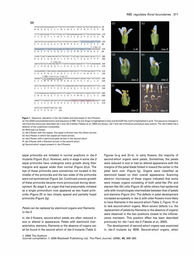
et al., 2003). This allele, rbe-3, contains a single nucleotide
change (C to T) within RBE that converts Q106 to a stop
codon (Figure 1a) and fails to complement the rbe-2 T-DNA
insertion allele. rbe-1 and rbe-2 had been described previ-
ously as having defects in development of the second whorl
of the flower. Second-whorl petals were often missing,
reduced in size, altered in shape (described as spoon-
shaped) or replaced by filaments (Takeda et al., 2003).
rbe-3 mutants have disrupted development of first-whorl
organs
We noticed several phenotypes in rbe-3 that had not been
described previously for the other two alleles. rbe-3 flowers
sometimes producemore than four sepals (Figure 1b–d) and
exhibit fusion between adjacent sepals (Figures 1d and 2a).
More than four sepals were observed in approximately 7% of
rbe-3 flowers, while some degree of sepal fusion was ob-
served in 35% of flowers (corresponding to 10% of all sepals,
Table 1). In rbe-3 flowers with more than four sepals, the
‘extra’ sepal was thinner than the remaining four sepals
(Figure 1c). In some cases, one of the adjacent sepals was
also somewhat reduced in width. Sepal fusion can occur in
the absence of more than four sepals. Sepal fusion was
observed most often at the base of these organs (Figure 2a)
and more rarely along their entire length (Figure 1d). We
measured the width of mature sepals to determine whether
sepal fusionmight be due to wider organs. rbe-3 sepals were
12% wider than Landsberg erecta (Ler) sepals at maturity
(Table 1).
Examination of rbe-3 flowers by scanning electron micr-
oscopy (SEM) suggests that sepal fusion in rbe-3 mutants is
due to altered patterns of growth within sepal primordia
after initiation. During stage 3 of flower development, four
370 Beth A. Krizek et al.
ª 2006 The AuthorsJournal compilation ª 2006 Blackwell Publishing Ltd, The Plant Journal, (2006), 45, 369–383
sepal primordia are initiated in normal positions in rbe-3
mutants (Figure 2b,c). However, early in stage 4 some rbe-3
sepal primordia have undergone extra growth along their
margins and appear wider than normal (Figure 2d,e). The
tips of these primordia were sometimes not located in the
middle of the primordia and the two sides of the primordia
were not symmetrical (Figure 2e). Continued uneven growth
of these primordia became more pronounced during devel-
opment. By stage 5, an organ that had presumably initiated
as a single primordium now appeared as two fused prim-
ordia (Figure 2f) or two closely spaced and partially fused
primordia (Figure 2g).
Petals can be replaced by staminoid organs and filaments
in rbe-3
In rbe-3 flowers, second-whorl petals are often reduced in
size or altered in appearance. Petals with staminoid char-
acteristics, stamens, filaments or the absence of organs can
all be found in the second whorl of rbe-3 mutants (Table 2;
Figures 1e–g and 2h–k). In early flowers, the majority of
second-whorl organs were petals. Sometimes, the petals
were reduced in size or had an altered appearance with the
margins of the petal blade folded in toward the center or the
petal bent over (Figure 1g). Organs were classified as
staminoid based on their overall appearance. Scanning
electron microscopy of these organs indicated that some
were mosaic organs consisting of both petal-like (Pe) and
stamen-like (St) cells (Figure 2l) while others had epidermal
cells with morphologies intermediate between that of petals
and stamens (Figure 2m). The defects in petal development
increased acropetally in rbe-3, with older flowers more likely
to have filaments in the second whorl (Table 2, Figure 1f) or
to lack second-whorl organs. More severe defects (i.e. the
replacement of petals by filaments or the absence of organs)
were observed in the two positions closest to the inflores-
cence meristem. This position effect has been described
previously for rbe-1 and rbe-2 (Takeda et al., 2003).
The development of second-whorl organs was examined
in rbe-3 mutants by SEM. Second-whorl organs, when
Figure 1. Sequence alteration in the rbe-3 allele and phenotype of rbe-3 flowers.
(a) The cDNA and predicted amino acid sequence of RBE. The zinc finger is highlighted in blue and the EAR-likemotif is highlighted in pink. The sequence changes in
rbe-3 and the previously described rbe-1 and rbe-2 alleles (Takeda et al., 2003) are shown. rbe-1 and rbe-3 introduce premature stop codons. The rbe-2 allele has a
deletion of the underlined nucleotides.
(b) Wild-type Ler flower.
(c) rbe-3 flower with five sepals. One sepal is thinner than the others (arrow).
(d) rbe-3 flower in which two sepals are fused (arrow).
(e) rbe-3 flower with a staminoid petal (arrow) in the second whorl.
(f) rbe-3 flower with a filament (arrow) in the second whorl.
(g) Second-whorl organs present in rbe-3 flowers.
RBE regulates floral boundaries 371
ª 2006 The AuthorsJournal compilation ª 2006 Blackwell Publishing Ltd, The Plant Journal, (2006), 45, 369–383

Figure 2. Scanning electron micrographs of rbe-3.
(a) rbe-3 flower in which two sepals are fused at their base (arrow).
(b) Stage 3 Ler flower.
(c) Stage 3 rbe-3 flower.
(d) Ler inflorescence.
(e) rbe-3 inflorescence. Arrows point to regions of abnormal growth in stage 4 sepal primordia.
(f) Close-up of a rbe-3 stage 5 flower in which one sepal primordium is split at its tip (arrow).
(g) Close-up of a rbe-3 stage 5 flower with five sepal primordia. The arrow points to a small sepal primordium located close to a medial sepal.
(h) Ler flower in which the sepals have been removed. Second-whorl petals are visible.
(i) rbe-3 flower containing a second-whorl staminoid organ (arrow).
(j) rbe-3 flower with a second-whorl staminoid petal (arrow).
(k) rbe-3 mosaic second-whorl organ.
(l) Close-up of organ in (k) showing stamen-like (St) and petal-like (Pe) cells.
(m) Epidermal cells present on a rbe-3 second-whorl organ.
(n) Stage 9 Ler flower.
(o) Stage 9 rbe-3 flower. The second-whorl organ primordia (arrows) are smaller than those of the wild type.
(p) Stage 9 rbe-3 flower containing a filament (arrow) in the second whorl.
(q) Ler stage 2-V ovules.
(r) rbe-3 stage 2-V ovules.
(s) Mature Ler ovules.
(t) Mature rbe-3 ovules. The outer and inner integuments (oi and ii) do not fully enclose the nucellus.
(u) Ler ovule showing the narrow width of the outer integument (oi) upon completion of growth around the nucellus.
(v) The outer integument (oi) is not tightly appressed to the inner integument (ii) in some rbe-3 ovules (arrow).
Scale bars correspond to 500 lm in (a), (h) and (i), 200 lm in (j) and (k), 100 lm in (n)–(p), (s) and (t), 50 lm in (d)–(g), (q), (r), (u) and (v), 20 lm in (b), (c) and (l) and
10 lm in (m).
Table 1 Sepal width and extent of sepalfusion in Ler, rbe-3, cuc2-1 and cuc2-1 rbe-3 flowers
GenotypeSepal width(mm)
Fraction of total sepal length exhibiting fusion
Total no. offlowers counted 0 <1/4 1/4–1/2 1/2–3/4 3/4–1
Ler 0.66 � 0.069rbe-3 0.74 � 0.095 100 89.8 9.75 0.5 0.0 0.0cuc2-1 0.68 � 0.065 100 62.0 19.2 13.2 4.2 1.5cuc2-1 rbe-3 0.69 � 0.076 128 10.9 26.6 38.1 17.0 7.4
372 Beth A. Krizek et al.
ª 2006 The AuthorsJournal compilation ª 2006 Blackwell Publishing Ltd, The Plant Journal, (2006), 45, 369–383
initiated in rbe-3 mutants, had a normal appearance. How-
ever, by stage 9 of flower development differences in the
overall size and shape of wild-type and rbe-3 second-whorl
organs were apparent (Figure 2n–p). Second-whorl rbe-3
organs were not as wide as wild-type petals (Figure 2o) and
in some cases remained radially symmetric rather than
expanding laterally and becomingflat (Figure 2p). To investi-
gate whether the defect in second-whorl organ development
might be the result of smaller floralmeristems, wemeasured
the diameter of stage 3 floral meristems in Ler and rbe-3
flowers. No difference in floralmeristemwidthwas observed
(35.2 � 2.81 lM in Ler versus 35.3 � 4.85 lM in rbe-3).
Because several rbe-3 phenotypes (extra sepals, fusion of
first-whorl sepals and replacement of petals by staminoid
organs) had not been reported for rbe-1 or rbe-2 (Takeda
et al., 2003), we examined rbe-2 to investigate whether our
observations were allele-specific. We found first- and sec-
ond-whorl defects in the rbe-2 allele similar to those
described here for rbe-3, indicating that our observations
are not particular to the rbe-3 allele.
rbe-3 mutants show defects in integument growth
To better define the reduced fertility of rbe-3 mutants, we
examined ovule development by SEM. Ovule development
was normal up to stage 2-V (Schneitz et al., 1995) with both
integuments initiating from the chalazal region and growing
toward the nucellus (Figure 2q,r). However, later growth of
the outer integument in rbe-3 mutants is abnormal with
premature cessation of growth. The extent of growth of the
outer integument in rbe-3 is somewhat variable, but in most
cases the outer integument does not grow to surround the
nucellus (Figure 2s,t). In addition, the outer integument is
not tightly appressed to the inner integument (Figure 2u,v).
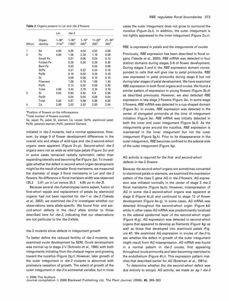
RBE is expressed in petals and the integuments of ovules
Previously, RBE expression has been described in floral or-
gans (Takeda et al., 2003). RBE mRNA was detected in four
distinct domains during stages 3–6 of flower development.
During stages 3 and 4, the RBE expression domain corres-
ponded to cells that will give rise to petal primordia. RBE
was expressed in petal primordia during stage 6 but not
during later stages of petal development.We have examined
RBE expression in both floral organs and ovules. We found a
similar pattern of expression in young flowers (Figure 3b,d)
as described previously. However, we also detected RBE
expression in late stage 2 flowers (Figure 3a). In some stage
3 flowers, RBE mRNA was detected in a cup-shaped domain
(Figure 3c). In ovules, RBE expression was detected in the
center of elongated primordia at the time of integument
initiation (Figure 3e). RBE mRNA was initially detected in
both the inner and outer integument (Figure 3e,f). As the
integuments grow around the nucellus, RBE expression is
maintained in the inner integument but not the outer
integument (Figure 3g,h). Prior to its disappearance in the
outer integument, RBE becomes confined to the adaxial side
of the outer integument (Figure 3g).
AG activity is required for the first- and second-whorl
defects in rbe-3 flowers
Because rbe second-whorl organs are sometimes converted
to staminoid petals or stamens, we examined the expression
pattern of the class C gene AG in rbe-3 flowers. AG expres-
sion was initiated normally in the center of stage 3 rbe-3
floral meristems (Figure 4a,b). However, misexpression of
AG in some rbe-3 second-whorl organs was apparent at
stage 6 (Figure 4c,d) and continued to stage 12 of flower
development (Figure 4e–g). In some cases, AG mRNA was
detected throughout the second-whorl organ (Figure 4d)
while in other cases AG mRNA was predominantly localized
to the adaxial epidermal layer of the second-whorl organ
(Figure 4f,g). AG expression was detected in second-whorl
organs that appeared to develop as filaments (Figure 4g) as
well as those that developed into staminoid petals (Fig-
ure 4f). We examined AG expression in ovules of rbe-3 to
see whether the defect in growth of the outer integument
might result from AG misexpression. AG mRNA was found
in a normal pattern in rbe-3 ovules, first appearing
throughout ovule primordia and later becoming restricted to
the endothelium (Figure 4h,i). This expression pattern mat-
ches that described earlier for AG (Bowman et al., 1991a).
To determine whether the rbe second-whorl defect was
due entirely to ectopic AG activity, we made an ag-1 rbe-3
Table 2 Organs present in Ler and rbe-3 flowers
WhorlOrganidentity
Ler rbe-3
1–30a
(114)b1–30a
(150)b1–10a
(50)b11–20a
(50)b21–30a
(50)b
1 Se 4.00 4.05 4.04 4.02 4.082 Pe 4.00 1.36 2.34 1.16 0.58
Small Pe 0.07 0.06 0.04 0.10Folded Pe 0.29 0.28 0.28 0.30Bent Pe 0.03 0.04 0.06St/Pe 0.21 0.22 0.24 0.16Pe/St 0.18 0.02 0.34 0.18St 0.09 0.06 0.10 0.10Filament 1.08 0.76 1.08 1.40Pe/fil 0.13 0.02 0.06 0.30Total 4.00 3.44 3.76 3.34 3.18
3 St 5.82 5.93 5.92 5.9 5.96Pe/St 0.04 0.04 0.06 0.04Total 5.82 5.97 5.96 5.96 6.00
4 Ca 2.00 2.02 2.02 2.00 2.04
aPosition of flowers on the inflorescence.bTotal number of flowers counted.Se, sepal; Pe, petal; St, stamen; Ca, carpel; St/Pe, staminoid petal;Pe/St, petaloid stamen; Pe/fil, petaloid filament.
RBE regulates floral boundaries 373
ª 2006 The AuthorsJournal compilation ª 2006 Blackwell Publishing Ltd, The Plant Journal, (2006), 45, 369–383
Figure 3. RBE expression in Ler flowers and ovules.
(a) RBE expression in a late stage two flower.
(b, c) RBE expression in stage 3 flowers.
(d) RBE expression in a stage 4 and stage 6 flower.
(e) RBE expression in the chalazal region of young ovule primordia (op). Arrows indicate RBE expression in the inner (black) and outer (white) integuments that have
just initiated from the chalazal region in one of these ovule primordia. The base of the ovule primordia are indicated by op.
(f) RBE expression in the inner (black arrow) and outer (white arrow) integuments of ovule primordia.
(g) RBE is expressed in the inner integument (black arrows) and on the adaxial side of the outer integument (white arrows) as the integuments grow around the
nucellus (stage 2-V).
(h) During stage 2-V, RBE expression becomes restricted to the inner integument. Black arrows point to the inner integument of two ovule primordia oriented with
the future micropylar end facing forward.
Scale bars correspond to 50 lm in (a)–(h).
Figure 4. AG expression in rbe-3.
(a) AG is expressed in the center of a Ler stage 3 floral meristem.
(b) AG is expressed in the center of a rbe-3 stage 3 floral meristem.
(c) AG mRNA is detected in the stamen and carpel primordia of a stage 6 flower, but not in the second-whorl petal primordia (arrow).
(d) AG mRNA is present in a rbe-3 second-whorl organ primordia (arrow).
(e) Transverse section of a stage 12 Ler flower showing AG expression in the stamens but not the petal.
(f) Transverse section of a stage 12 rbe-3 flower in which AG is expressed in the adaxial epidermis of the second-whorl organ (arrow).
(g) Transverse section showing AG mRNA in the epidermal layers of two second-whorl organ primordia (arrows).
(h) In mature Ler ovules, AGmRNA is restricted to the endothelium (black arrow). The white arrow points to the endothelium of a second ovule that was sectioned at
an oblique angle.
(i) AG mRNA is restricted to the endothelium in rbe-3 mature ovules (black arrow).
Scale bars correspond to 50 lm in (a)–(i).
374 Beth A. Krizek et al.
ª 2006 The AuthorsJournal compilation ª 2006 Blackwell Publishing Ltd, The Plant Journal, (2006), 45, 369–383
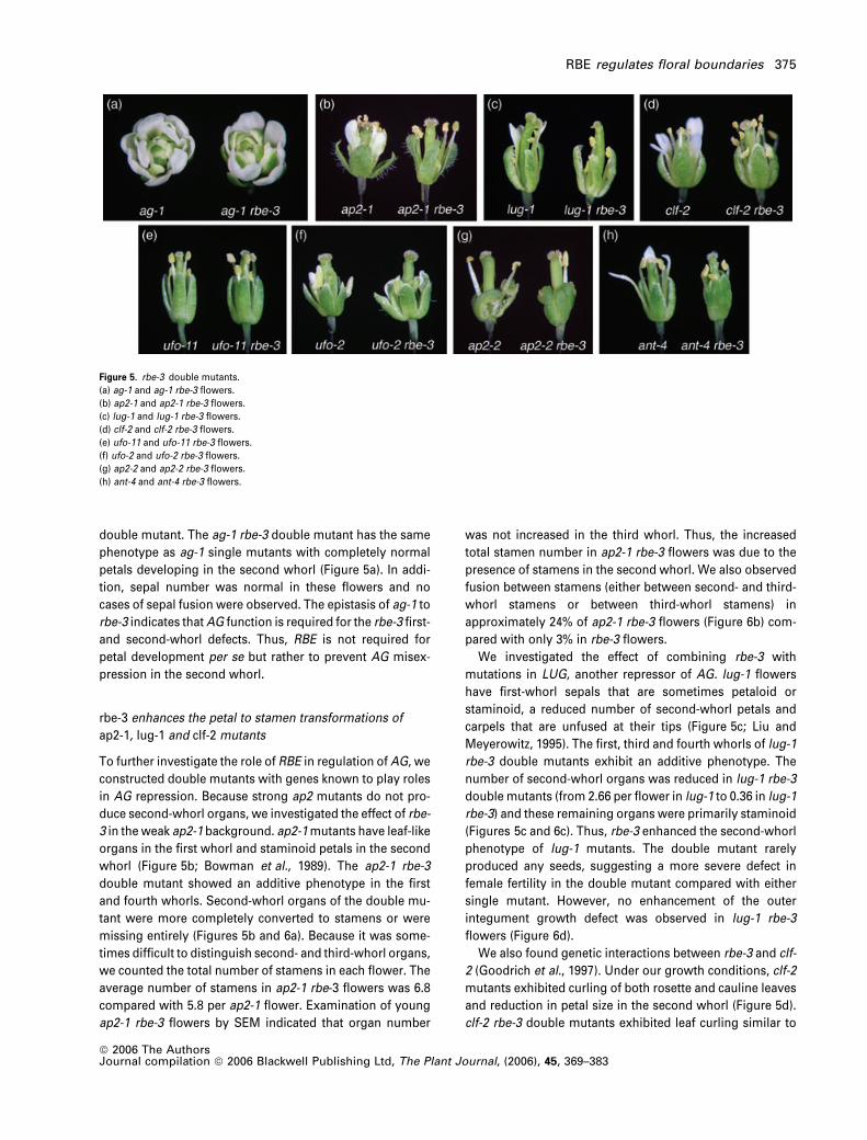
double mutant. The ag-1 rbe-3 double mutant has the same
phenotype as ag-1 single mutants with completely normal
petals developing in the second whorl (Figure 5a). In addi-
tion, sepal number was normal in these flowers and no
cases of sepal fusion were observed. The epistasis of ag-1 to
rbe-3 indicates thatAG function is required for the rbe-3 first-
and second-whorl defects. Thus, RBE is not required for
petal development per se but rather to prevent AG misex-
pression in the second whorl.
rbe-3 enhances the petal to stamen transformations of
ap2-1, lug-1 and clf-2 mutants
To further investigate the role of RBE in regulation of AG, we
constructed double mutants with genes known to play roles
in AG repression. Because strong ap2 mutants do not pro-
duce second-whorl organs, we investigated the effect of rbe-
3 in the weak ap2-1 background. ap2-1mutants have leaf-like
organs in the first whorl and staminoid petals in the second
whorl (Figure 5b; Bowman et al., 1989). The ap2-1 rbe-3
double mutant showed an additive phenotype in the first
and fourth whorls. Second-whorl organs of the double mu-
tant were more completely converted to stamens or were
missing entirely (Figures 5b and 6a). Because it was some-
times difficult to distinguish second- and third-whorl organs,
we counted the total number of stamens in each flower. The
average number of stamens in ap2-1 rbe-3 flowers was 6.8
compared with 5.8 per ap2-1 flower. Examination of young
ap2-1 rbe-3 flowers by SEM indicated that organ number
was not increased in the third whorl. Thus, the increased
total stamen number in ap2-1 rbe-3 flowers was due to the
presence of stamens in the second whorl. We also observed
fusion between stamens (either between second- and third-
whorl stamens or between third-whorl stamens) in
approximately 24% of ap2-1 rbe-3 flowers (Figure 6b) com-
pared with only 3% in rbe-3 flowers.
We investigated the effect of combining rbe-3 with
mutations in LUG, another repressor of AG. lug-1 flowers
have first-whorl sepals that are sometimes petaloid or
staminoid, a reduced number of second-whorl petals and
carpels that are unfused at their tips (Figure 5c; Liu and
Meyerowitz, 1995). The first, third and fourth whorls of lug-1
rbe-3 double mutants exhibit an additive phenotype. The
number of second-whorl organs was reduced in lug-1 rbe-3
double mutants (from 2.66 per flower in lug-1 to 0.36 in lug-1
rbe-3) and these remaining organs were primarily staminoid
(Figures 5c and 6c). Thus, rbe-3 enhanced the second-whorl
phenotype of lug-1 mutants. The double mutant rarely
produced any seeds, suggesting a more severe defect in
female fertility in the double mutant compared with either
single mutant. However, no enhancement of the outer
integument growth defect was observed in lug-1 rbe-3
flowers (Figure 6d).
We also found genetic interactions between rbe-3 and clf-
2 (Goodrich et al., 1997). Under our growth conditions, clf-2
mutants exhibited curling of both rosette and cauline leaves
and reduction in petal size in the second whorl (Figure 5d).
clf-2 rbe-3 double mutants exhibited leaf curling similar to
Figure 5. rbe-3 double mutants.
(a) ag-1 and ag-1 rbe-3 flowers.
(b) ap2-1 and ap2-1 rbe-3 flowers.
(c) lug-1 and lug-1 rbe-3 flowers.
(d) clf-2 and clf-2 rbe-3 flowers.
(e) ufo-11 and ufo-11 rbe-3 flowers.
(f) ufo-2 and ufo-2 rbe-3 flowers.
(g) ap2-2 and ap2-2 rbe-3 flowers.
(h) ant-4 and ant-4 rbe-3 flowers.
RBE regulates floral boundaries 375
ª 2006 The AuthorsJournal compilation ª 2006 Blackwell Publishing Ltd, The Plant Journal, (2006), 45, 369–383
that in clf-2. Both clf-2 and clf-2 rbe-3 produced an average of
4.0 second-whorl organs per flower. In clf-2 mutants these
were almost exclusively small petals (3.97 per flower) while
the average number of small petals in clf-2 rbe-3 was 1.37.
Second-whorl petals were most often replaced by spoon-
shaped petals (0.29 per flower), staminoid petals (0.91 per
flower), petaloid stamens (0.78 per flower) or stamens (0.48
per flower; Figure 5d). Thus, rbe-3 enhanced the transfor-
mation of clf-2 second-whorl organs to stamens and
stamen-like organs.
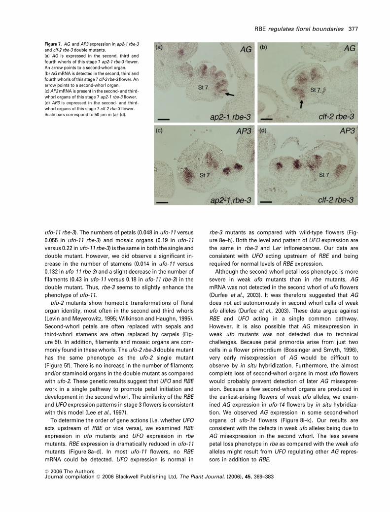
We examined the expression ofAG in ap2-1 rbe-3 and clf-2
rbe-3 plants to determine whether the enhancement of the
second-whorl phenotypes in these double mutants resulted
from AG misexpression. High levels of AG mRNA were
detected not only in the third and fourth whorls of these
flowers but also ectopically in the second whorl, confirming
our hypothesis (Figure 7a,b). In contrast, the class B gene
AP3 was correctly expressed in second- and third-whorl
organs in ap2-1 rbe-3 and clf-2 rbe-3 double mutant flowers
(Figure 7c,d). This normal pattern of AP3 expression paral-
lels that observed previously in rbe-1 singlemutants (Takeda
et al., 2003).
ufo is epistatic to rbe in development of the second whorl
The UFO gene has been shown to promote petal initiation
and development by inhibiting AG activity in the second
whorl (Durfee et al., 2003). To determine whether RBEmight
function in a common pathway with UFO, we crossed rbe-3
to the weak ufo-11 and strong ufo-2 alleles. ufo-11 is a weak
allele specifically affecting petal development (Durfee et al.,
2003). Petals are usually missing in ufo-11 flowers or are
sometimes replaced by filaments or petal/stamen mosaic
organs. ufo-11 flowers have fewer second-whorl organs
compared with rbe-3 mutants and also sometimes produce
mosaic petaloid stamens in the third whorl. ufo-11 rbe-3
double-mutant flowers have a phenotype that is similar to
ufo-11 flowers (Figure 5e). The total number of second-
whorl organs is approximately the same (0.69 second-whorl
organs in ufo-11 flowers compared with 0.58 organs in
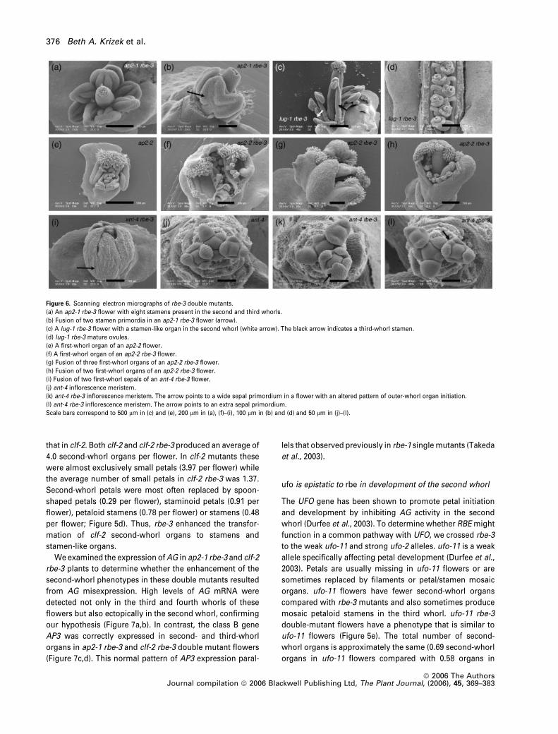
Figure 6. Scanning electron micrographs of rbe-3 double mutants.
(a) An ap2-1 rbe-3 flower with eight stamens present in the second and third whorls.
(b) Fusion of two stamen primordia in an ap2-1 rbe-3 flower (arrow).
(c) A lug-1 rbe-3 flower with a stamen-like organ in the second whorl (white arrow). The black arrow indicates a third-whorl stamen.
(d) lug-1 rbe-3 mature ovules.
(e) A first-whorl organ of an ap2-2 flower.
(f) A first-whorl organ of an ap2-2 rbe-3 flower.
(g) Fusion of three first-whorl organs of an ap2-2 rbe-3 flower.
(h) Fusion of two first-whorl organs of an ap2-2 rbe-3 flower.
(i) Fusion of two first-whorl sepals of an ant-4 rbe-3 flower.
(j) ant-4 inflorescence meristem.
(k) ant-4 rbe-3 inflorescence meristem. The arrow points to a wide sepal primordium in a flower with an altered pattern of outer-whorl organ initiation.
(l) ant-4 rbe-3 inflorescence meristem. The arrow points to an extra sepal primordium.
Scale bars correspond to 500 lm in (c) and (e), 200 lm in (a), (f)–(i), 100 lm in (b) and (d) and 50 lm in (j)–(l).
376 Beth A. Krizek et al.
ª 2006 The AuthorsJournal compilation ª 2006 Blackwell Publishing Ltd, The Plant Journal, (2006), 45, 369–383
ufo-11 rbe-3). The numbers of petals (0.048 in ufo-11 versus
0.055 in ufo-11 rbe-3) and mosaic organs (0.19 in ufo-11
versus 0.22 in ufo-11 rbe-3) is the same in both the single and
double mutant. However, we did observe a significant in-
crease in the number of stamens (0.014 in ufo-11 versus
0.132 in ufo-11 rbe-3) and a slight decrease in the number of
filaments (0.43 in ufo-11 versus 0.18 in ufo-11 rbe-3) in the
double mutant. Thus, rbe-3 seems to slightly enhance the
phenotype of ufo-11.
ufo-2 mutants show homeotic transformations of floral
organ identity, most often in the second and third whorls
(Levin and Meyerowitz, 1995; Wilkinson and Haughn, 1995).
Second-whorl petals are often replaced with sepals and
third-whorl stamens are often replaced by carpels (Fig-
ure 5f). In addition, filaments and mosaic organs are com-
monly found in these whorls. The ufo-2 rbe-3 double mutant
has the same phenotype as the ufo-2 single mutant
(Figure 5f). There is no increase in the number of filaments
and/or staminoid organs in the double mutant as compared
with ufo-2. These genetic results suggest that UFO and RBE
work in a single pathway to promote petal initiation and
development in the second whorl. The similarity of the RBE
and UFO expression patterns in stage 3 flowers is consistent
with this model (Lee et al., 1997).
To determine the order of gene actions (i.e. whether UFO
acts upstream of RBE or vice versa), we examined RBE
expression in ufo mutants and UFO expression in rbe
mutants. RBE expression is dramatically reduced in ufo-11
mutants (Figure 8a–d). In most ufo-11 flowers, no RBE
mRNA could be detected. UFO expression is normal in
rbe-3 mutants as compared with wild-type flowers (Fig-
ure 8e–h). Both the level and pattern of UFO expression are
the same in rbe-3 and Ler inflorescences. Our data are
consistent with UFO acting upstream of RBE and being
required for normal levels of RBE expression.
Although the second-whorl petal loss phenotype is more
severe in weak ufo mutants than in rbe mutants, AG
mRNA was not detected in the second whorl of ufo flowers
(Durfee et al., 2003). It was therefore suggested that AG
does not act autonomously in second whorl cells of weak
ufo alleles (Durfee et al., 2003). These data argue against
RBE and UFO acting in a single common pathway.
However, it is also possible that AG misexpression in
weak ufo mutants was not detected due to technical
challenges. Because petal primordia arise from just two
cells in a flower primordium (Bossinger and Smyth, 1996),
very early misexpression of AG would be difficult to
observe by in situ hybridization. Furthermore, the almost
complete loss of second-whorl organs in most ufo flowers
would probably prevent detection of later AG misexpres-
sion. Because a few second-whorl organs are produced in
the earliest-arising flowers of weak ufo alleles, we exam-
ined AG expression in ufo-14 flowers by in situ hybridiza-
tion. We observed AG expression in some second-whorl
organs of ufo-14 flowers (Figure 8i–k). Our results are
consistent with the defects in weak ufo alleles being due to
AG misexpression in the second whorl. The less severe
petal loss phenotype in rbe as compared with the weak ufo
alleles might result from UFO regulating other AG repres-
sors in addition to RBE.
Figure 7. AG and AP3 expression in ap2-1 rbe-3
and clf-2 rbe-3 double mutants.
(a) AG is expressed in the second, third and
fourth whorls of this stage 7 ap2-1 rbe-3 flower.
An arrow points to a second-whorl organ.
(b) AGmRNA is detected in the second, third and
fourth whorls of this stage 7 clf-2 rbe-3 flower. An
arrow points to a second-whorl organ.
(c) AP3mRNA is present in the second- and third-
whorl organs of this stage 7 ap2-1 rbe-3 flower.
(d) AP3 is expressed in the second- and third-
whorl organs of this stage 7 clf-2 rbe-3 flower.
Scale bars correspond to 50 lm in (a)–(d).
RBE regulates floral boundaries 377
ª 2006 The AuthorsJournal compilation ª 2006 Blackwell Publishing Ltd, The Plant Journal, (2006), 45, 369–383
Figure 8. RBE, UFO, and AG expression in wild
type, ufo, and rbe mutants.
(a–d) RBE expression in Columbia (Col) and ufo-
11.
(a) RBE expression in a stage 3 Col flower.
(b) No RBE mRNA was detected in this stage 3
ufo-11 flower.
(c) RBE expression in a stage 5 Col flower.
(d) No RBE mRNA was detected in this stage 5
ufo-11 flower.
(e–h) UFO expression in Ler and rbe-3 inflores-
cences.
(e) In Ler plants, UFO mRNA is present in the
inflorescence meristem (IM) and in a cup-shaped
domain in stage 3 flowers.
(f) In rbe-3 plants, UFO is expressed in the
inflorescence meristem (IM) and in a cup-shaped
domain in stage 3 flowers.
(g)UFO is expressed between the sepals (Se) and
petals in this stage 7 Ler flower. The arrow points
to a second-whorl petal primordium.
(h) UFO is expressed between the sepals and
second-whorl organs in this stage 7 rbe-3 flower.
The arrow points to a second-whorl organ prim-
ordium.
(i–k) AG expression in ufo-14.
(i) AGmRNA is detected in a second-whorl organ
of this ufo-14 flower (indicated by an arrow).
(j) Close-up of the second-whorl organ shown in
(i).
(k) AG mRNA is detected in a second-whorl
staminoid petal of this ufo-14 flower (arrow).
Scale bars correspond to 50 lm in (a)–(k).
378 Beth A. Krizek et al.
ª 2006 The AuthorsJournal compilation ª 2006 Blackwell Publishing Ltd, The Plant Journal, (2006), 45, 369–383
ap2-2 rbe-3 and ant-4 rbe-3 double mutants show
enhanced first-whorl organ fusion
Strong ap2-2 alleles have carpels or staminoid carpels in
medial first-whorl positions and leaf-like organs in the lateral
positions (Figure 5g; Bowman et al., 1991b). The first-whorl
staminoid carpels are mosaic organs consisting of a central
region of carpel tissue fused on one or both sides with sta-
men tissue (Figure 6e). Second-whorl organs are missing in
ap2-2 flowers and there are a reduced number of third-whorl
stamens. In ap2-2 rbe-3 double mutants, first-whorl carpels
and staminoid carpels were often wider than in ap2-2 (Fig-
ure 6f). These organs had the appearance of several fused
organs but were only connected to the base of the flower in
the central part of the organ and were in a position normally
occupied by a single organ. In addition, first-whorl organs
were often fused along much of their length (Figures 5g and
6g). All of the first-whorl organs were sometimes fused such
that the fourth whorl carpel poked through a ring of tissue
(Figure 6h). Cells present along the margin of fusion
resembled the epidermal cells normally present on the ab-
axial replum of wild-type carpels. Thus, the function of RBE
in the separation of floral organs does not depend on the
identify of the organs in the first whorl. The number of first-
whorl organs in the double mutant was slightly increased
compared with ap2-2 single mutants (3.96 in ap2-2 rbe-3
compared with 3.56 in ap2-2). No second-whorl organs were
present in the doublemutant and the third and fourth whorls
had an additive phenotype.
ant-4 flowers have the normal pattern of organ identity
but show reductions in both organ number and size
(Figure 5h; Elliott et al., 1996; Klucher et al., 1996). ant-4
rbe-3 double mutants had an increased number of sepals
compared with either singlemutant (4.42 sepals in ant-4 rbe-
3 compared with 3.98 in ant-4). In early arising flowers as
many as eight sepals were present. In addition, fusion
between sepals was observed in ant-4 rbe-3 mutants (Fig-
ures 5h and 6i). Sepals in the double mutant were often
fused along their entire length rather than just at their base
as usually seen in rbe-3. The initiation pattern of sepal
primordia was often disrupted in ant-4 rbe-3 double
mutants. One or more of the primordia were sometimes
initiated in positions shifted from the typical medial and
lateral positions (Figure 6j,k). This resulted in the four
primordia not being equally spaced from each other. In
some cases, the positions of four sepal primordia were fairly
normal but an additional sepal primordium arose in a region
between a medial sepal and a lateral sepal (Figure 6l).
ant-4 rbe-3 flowers also show a reduction in the overall
number of second-whorl organs (3.8 in rbe-3 compared with
2.5 in ant-4 rbe-3). In addition, fewer filaments (1.2 in rbe-3
compared with 0.7 in ant-4 rbe-3) and staminoid organs (0.5
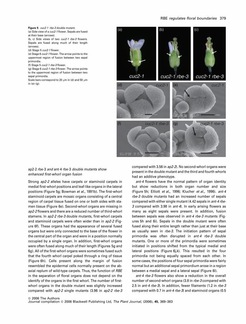
Figure 9. cuc2-1 rbe-3 double mutant.
(a) Side view of a cuc2-1 flower. Sepals are fused
at their base (arrows).
(b, c) Side views of two cuc2-1 rbe-3 flowers.
Sepals are fused along much of their length
(arrows).
(d) Stage 5 cuc2-1 flower.
(e) Stage 6 cuc2-1 flower. The arrow points to the
uppermost region of fusion between two sepal
primordia.
(f) Stage 5 cuc2-1 rbe-3 flower.
(g) Stage 6 cuc2-1 rbe-3 flower. The arrow points
to the uppermost region of fusion between two
sepal primordia.
Scale bars correspond to 20 lm in (d) and 50 lmin (e)–(g).
RBE regulates floral boundaries 379
ª 2006 The AuthorsJournal compilation ª 2006 Blackwell Publishing Ltd, The Plant Journal, (2006), 45, 369–383
in rbe-3 compared with 0.04 in ant-4 rbe-3) were present in
the double mutant compared with rbe-3 single mutants.
Reduced stamen number in the third whorl of ant-4 mutants
was partially rescued by rbe-3. ant-4 mutants have 4.3
stamens per flower whereas ant-4 rbe-3 flowers produce 5.6
stamens per flower.
cuc2-1 rbe-3 double mutants show increased frequency and
extent of sepal fusion
To further investigate the roleofRBE inorganseparation, rbe-
3 was crossed to cuc2-1. In the cuc2-1 single mutant, sepals
are sometimes fused at their base (Aida et al., 1997;
Figure 9a). In rbe-3 cuc2-1 flowers, sepal fusion occurred
moreoftenand toagreater extent than ineither singlemutant
(Figure 9b,c; Table 1). The width of cuc2-1 and cuc2-1 rbe-3
sepals is the same at maturity (Table 1), suggesting that the
increasedamountof fusion in thisdoublemutant isnotdue to
increased organwidth. Examination of sepal development in
the cuc2-1 rbe-3 double mutant by SEM showed that four
distinct sepal primordia are initiated in a normal pattern
during stage 3 of flower development aswas seenpreviously
for cuc2-1 and rbe-3 single mutants. Later growth of sepal
primordia in the cuc2-1 rbe-3double is uneven, similar to that
seen in rbe-3 single mutants. An increased extent of sepal
fusion in the cuc2-1 rbe-3 double mutant as compared with
cuc2-1was first observed during stage 6 (Figure 9d–g).
Discussion
RBE is a new repressor of AG expression
Our studies have identified RBE as a new repressor of AG
expression in Arabidopsis flowers. The defects observed in
the second whorl of rbe mutants (reduced organ number,
replacement of petals by filaments and staminoid organs)
result from ectopic expression of AG in second-whorl cells.
In the absence of AG activity, development of both first- and
second-whorl organs is normal in rbemutants. RBE encodes
a potential transcriptional repressor. It contains a single
Cys2His2 zinc-finger DNA-binding domain and an ethylene-
responsive element binding factor (ERF)–associated
amphiphilic repression (EAR)-like domain (Takeda et al.,
2003). The related protein SUPERMAN (SUP), which con-
tains a similarly arranged single zinc-finger domain and
EAR-like motif, has been shown to possess both DNA-
binding and transcriptional repression activities (Dathan
et al., 2002; Hiratsu et al., 2002). SUP and RBE are similar in
that mutations in either gene disrupt the ability to maintain
the boundaries of floral homeotic gene expression.
All but one of the previously identified AG repressors
prevent AG transcription in both the first and second whorls
of the flower.We have never observed carpelloid features on
first-whorl sepals or misexpression of AG in first-whorl
organs of rbe mutants, suggesting that RBE acts only within
the second whorl to repressAG expression. This is similar to
the proposed function of ANT in AG repression (Krizek et al.,
2000). The spatially restricted expression pattern of RBE also
contrasts with that of other known AG repressors that are
transcribed in all four whorls of the flower (Conner and Liu,
2000; Goodrich et al., 1997; Jofuku et al., 1994). The recent
demonstration thatAP2 is regulated at the translational level
by miRNA172 suggests one possible mechanism for restric-
tion of AG repressor activity to the outer whorls of the flower
(Aukerman and Sakai, 2003; Chen, 2004). Another possibility
is that globally expressed AG repressors require whorl-
specific factors like RBE for their activity.
Plant Cys2His2 zinc-finger proteins containing the con-
served QALGGH sequence have been shown to bind 5¢-AGT-
3¢ sequences. In SUP, the single zinc-finger domain and
adjacent basic residues on either side of the zinc finger are
sufficient to bind such sequences (Dathan et al., 2002). Basic
residues are also present on either side of the RBE zinc
finger; those at the carboxy-terminal end of the zinc finger
are present at conserved positions with those in SUP. Cis-
acting regulatory sequences required for proper spatial
expression of AG are located in the AG 3 kb second intron
(Busch et al., 1999; Deyholos and Sieburth, 2000; Sieburth
and Meyerowitz, 1997). We examined this intron for the
presence of AGT elements and found a total of 102 such
sequences on either strand. The expected number of AGT
sequences on either strand of a 3 kb piece of DNA is 94.
Although the total number of AGT sequences is not signi-
ficantly greater than the expected number, six of these
sequences are present within regions conserved among 29
Brassicaceae species (Hong et al., 2003). Further work will be
necessary to determine whether RBE binds directly to AG
regulatory sequences.
Pathways regulating petal development
Mutations in UFO, RBE or PTL cause somewhat similar de-
fects in the outer two whorls of the flower. Sepal fusion can
occur in the first whorl of ufo-11 (B. Krizek, unpublished
data), similar to that observed in rbe-3 and ptl-1 (Griffith
et al., 1999). In the second whorl, mutations in any of these
three genes results in loss of petals and/or their replacement
with filaments or staminoid organs. Our results suggest that
RBE and UFO are components of a common pathway that
regulates petal initiation and development. RBE transcripts
are dramatically reduced in ufomutants, indicating thatUFO
is required for RBE expression. Furthermore, AG misex-
pression in the second whorl occurs in both rbe and weak
ufo alleles, and the second-whorl defects in these mutants
can be eliminated by the loss of AG activity.
UFO has been shown to function as part of a SCFUFO
complex in flowers. Such complexes act as E3 ubiquitin
ligating enzymes in the ubiquitin–proteasome protein
380 Beth A. Krizek et al.
ª 2006 The AuthorsJournal compilation ª 2006 Blackwell Publishing Ltd, The Plant Journal, (2006), 45, 369–383

degradation pathway. Interestingly, mutations in other
components of the SCFUFO complex result in carpelloid
sepals, sepal fusion and dramatic reductions in petal num-
ber (Ni et al., 2004). These phenotypes are present in axr6-2
and ask1/ask1 ask2/ASK2 plants and require AG activity.
Thus SCF complexes have been proposed to repress AG
function in the outer whorls and to play a role in organ
separation by regulating cell division in each whorl (Ni et al.,
2004). Our results are consistent with the SCFUFO complex
mediating proteolysis of a repressor of RBE expression in
the second whorl.
Whether PTL is a component of theUFO/RBE pathway or a
separate pathway remains unclear. RBE has been proposed
to function downstream of PTL in the development of
second-whorl organs, as RBE is not expressed in ptlmutants
(Takeda et al., 2003). However, AG activity is not responsible
for the ptl second-whorl defects as they are still present in ptl
ag double mutants (Griffith et al., 1999). This argues against
the idea of RBE acting directly downstream of PTL. An
alternative possibility is that PTL may act as a growth
suppressor in regions between adjacent sepals, and its effect
on second-whorl development (and RBE expression)may be
an indirect consequence of excessive growth within the
inter-sepal domain (Brewer et al., 2004). Further experi-
ments will be required to determine whether PTL and RBE
act together in organ formation in the first and/or second
whorls. PTL and RBE are expressed in adjacent groups of
cells in the first and second whorls. The similarity of the ptl
and rbe phenotypes suggest that signaling between these
two populations of cells is critical for regulating organ
growth during early stages of flower development.
RBE may be a component of an inter-whorl signaling
pathway that regulates growth in the outer two floral whorls
RBE is only expressed in second-whorl cells (Takeda et al.,
2003), yet mutations in this gene alter growth in both the first
and second whorls. Decreased growth in the second whorl
of rbe mutants appears to be accompanied by increased
growth in an adjacent region of the first whorl. We propose
that signaling pathways act between adjacent groups of cells
in the first and second whorls to regulate cellular prolifer-
ation. In our model (Figure 10), UFO and RBE act in one
pathway by promoting proliferation of second-whorl cells
through repression of AG, which acts as a suppressor of
growth when misexpressed in the second whorl (Bowman
et al., 1991b, 1993). Proliferating second-whorl cells at the
sites of petal initiation generate a signal (S) that moves to
adjacent first-whorl cells, activating a first-whorl inhibitor (I)
of growth in these regions. Such a pathway results in petal
initiation and growth as well as the establishment of
boundaries between adjacent sepal primordia. In thismodel,
the amount of second-whorl cell proliferation and thus the
amount of S generated is controlled by the activities of UFO
and RBE. ROXY1, a glutaredoxin protein (Xing et al., 2005),
may also play a role here as a potential post-translational
regulator of RBE. In rbe flowers, expression of AG in the
second whorl inhibits cell proliferation in the second whorl.
This results in reduction or elimination of the growth inhi-
bition signal to the first whorl, leading to increased growth in
the inter-sepal zone and consequently sepal–sepal fusion
and the occasional formation of extra sepals.
Experimental procedures
Plant growth conditions, mutant isolation and strain
construction
Arabidopsis plants were grown in a 4:1:1 mix of Fafard 4P:per-lite:vermiculite at 22�C in continuous light. The rbe-3 allele was
Figure 10. Model for RBE function in development of the first and second
whorls.
Solid arrows and bars describe interactions supported by genetic and/or
molecular evidence. Dotted arrows indicate proposed interactions. The green
ellipses represent sepal primordia and the yellow circle represents a petal
primordium. RBE promotes the development of second-whorl organs by
acting as a repressor of AG in second-whorl cells. Negative regulation of AG
by RBE results in increased proliferation of second-whorl cells since AG is an
inhibitor of the development of second-whorl organs. RBE acts downstream
of UFO in this pathway. RBE may be regulated post-translationally by the
glutaredoxin protein ROXY1 (Xing et al., 2005). RBE inhibits growth of the first
whorl in regions adjacent to RBE-expressing second-whorl cells. Division of
second-whorl cells could generate a signal that moves to the first whorl and
activates an inhibitor (I) of first-whorl growth in regions adjacent to initiating
petal primordia. In rbe mutants, reduced second-whorl proliferation due to
ectopic AG activity would lead to reduced amounts of the first-whorl growth
inhibition signal.
RBE regulates floral boundaries 381
ª 2006 The AuthorsJournal compilation ª 2006 Blackwell Publishing Ltd, The Plant Journal, (2006), 45, 369–383
isolated from a mutagenized population of 35S::PI seeds in theNossen (No-O) ecotype. The rbe-3 allele was backcrossed four timesto Ler prior to phenotypic analyses. All doublemutants were createdby manual cross-pollination using homozygous mutant strains,except for ag-1. We believe the weak ufo allele used in our geneticstudies is ufo-11. Polymerase chain reaction (PCR) genotypingand sequencing of PCR products from this allele indicated thatthe ufo-12, ufo-13 and ufo-14molecular lesions are not present. Thegenotype of ufo-11 rbe-3 double mutants was confirmed usingcleaved amplified polymorphic sequences (CAPS) markers for therbe-3 mutation and crossing of the plants to ufo-11.
Scanning electron microscopy
Samples for SEM were fixed, dried, dissected and coated as des-cribed previously (Krizek, 1999). Images were collected on a XL30.To measure the widths of stage 3 floral meristems, images werecaptured at a magnification of 800·, printed and measured with aruler. The width of the dome of the floral meristem was measuredfrom the middle of one medial sepal to the middle of the oppositemedial sepal. Ten stage 3 floral meristems were measured for Lerand 13 for rbe-3.
Sepal width measurements
All sepals were removed from mature flowers corresponding topositions 1–12 on the inflorescences of four individual plants ofeach genotype. The sepals were laid on their adaxial side and theirwidth measured with an ocular micrometer.
In situ hybridization
Inflorescences were fixed, embedded, sectioned, hybridized andwashed as described previously (Krizek, 1999). Digoxigenin-labe-led RNA probes were synthesized by in vitro transcription usingT7 RNA polymerase. The AG antisense probe was made bydigestion of pCIT565 (Yanofsky et al., 1990). For the RBE antisenseprobe, a fragment of RBE corresponding to nucleotides 273–580was cloned into pCR2.1 and linearized with BamHI. The UFOantisense probe was made by digestion of pDW221.1 with ClaI(Lee et al., 1997).
Acknowledgements
We thank the Salk Institute Genomic Analysis laboratory for provi-ding the Arabidopsis T-DNA insertion mutant, the ArabidopsisBiological Resource Center for seeds of the T-DNA insertion mutant(SALK_037010) and cuc2-1, DetlefWeigel for theUFO in situ plasmidpDW221.1 and Patricia Zambryski for the weak ufo alleles. This workwas supported by a University of South Carolina Research andProductive Scholarship award to BAK and by a United StatesDepartment of Agriculture CRIS grant to JCF.
References
Aida, M., Ishida, T., Fukaki, H., Fujisawa, H. and Tasaka, M. (1997)Genes involved in organ separation in Arabidopsis: an analysis ofthe cup-shaped cotyledon mutant. Plant Cell, 9, 841–857.
Aukerman, M.J. and Sakai, H. (2003) Regulation of flowering timeand floral organ identity by a microRNA and its APETALA2-liketarget genes. Plant Cell, 15, 2730–2741.
Bao, X., Franks, R.G., Levin, J.Z. and Liu, Z. (2004) Repression ofAGAMOUS by BELLRINGER in floral and inflorescence meri-stems. Plant Cell, 16, 1478–1489.
Bossinger, G. and Smyth, D.R. (1996) Initiation patterns of flowerand floral organ development in Arabidopsis thaliana. Develop-ment, 122, 1093–1102.
Bowman, J.L., Smyth, D.R. and Meyerowitz, E.M. (1989) Genesdirecting flower development in Arabidopsis. Plant Cell, 1, 37–52.
Bowman, J.L., Drews, G.N. and Meyerowitz, E.M. (1991a) Expres-sion of the Arabidopsis floral homeotic gene AGAMOUS isrestricted to specific cell types late in flower development. PlantCell, 3, 749–758.
Bowman, J.L., Smyth, D.R. and Meyerowitz, E.M. (1991b) Geneticinteractions among floral homeotic genes of Arabidopsis.Development, 112, 1–20.
Bowman, J.L., Sakai, H., Jack, T., Weigel, D., Mayer, U. and
Meyerowitz, E.M. (1992) SUPERMAN, a regulator of floralhomeotic genes in Arabidopsis. Development, 114, 599–615.
Bowman, J.L., Alvarez, J., Weigel, D., Meyerowitz, E.M. and Smyth,
D.R. (1993) Control of flower development inArabidopsis thalianaby APETALA1 and interacting genes. Development, 119, 721–743.
Brewer, P.B., Howles, P.A., Dorian, K., Griffith, M., Ishida, T., Kaplan-
Levy, R.N., Kilinc, A. and Smyth, D.R. (2004) PETAL LOSS, a tri-helix transcription factor gene, regulates perianth architecture inthe Arabidopsis flower. Development, 131, 4035–4045.
Busch, M.A., Bomblies, K. andWeigel, D. (1999) Activation of a floralhomeotic gene in Arabidopsis. Science, 285, 585–587.
Byzova, M.V., Franken, J., Aarts, M.G.M., de Almeida-Engler, J.,
Engler, G., Mariana, C., Van Lookeren Campagne, M.M. and
Angenent, G.C. (1999) Arabidopsis STERILE APETALA, a multi-functional gene regulating inflorescence, flower, and ovuledevelopment. Genes Dev. 13, 1002–1014.
Chen, X. (2004) A microRNA as a translational repressor of APET-ALA2 in Arabidopsis flower development. Science, 303, 2022–2025.
Conner, J. and Liu, Z. (2000) LEUNIG, a putative transcriptionalcorepressor that regulates AGAMOUS expression during flowerdevelopment. Proc. Natl Acad. Sci. USA 97, 12902–12907.
Dathan, N., Zaccaro, L., Esposito, S., Isernia, C., Omichinski, J.G.,
Riccio, A., Pedone, C., Di Blasio, B., Fattorusso, R. and Pedone,
P.V. (2002) The Arabidopsis SUPERMAN protein is able to spe-cifically bind DNA through its single Cys2-His2 zinc finger motif.Nucleic Acids Res. 30, 4945–4951.
Deyholos, M.K. and Sieburth, L.E. (2000) Separable whorl-specificexpression and negative regulation by enhancer elements withinthe AGAMOUS second intron. Plant Cell, 12, 1799–1810.
Ditta, G., Pinyopich, A., Robles, P., Pelaz, S. and Yanofsky, M.F.
(2004) The SEP4 gene of Arabidopsis thaliana functions in floralorgan and meristem identity. Curr. Biol. 14, 1935–1940.
Drews, G.N., Bowman, J.L. and Meyerowitz, E.M. (1991) Negativeregulation of the Arabidopsis homeotic gene AGAMOUS by theAPETALA2 product. Cell, 65, 991–1002.
Durfee, T., Roe, J.L., Sessions, R.A., Inouye, C., Serikawa, K.,
Feldmann, K.A., Weigel, D. and Zambryski, P.C. (2003) The F-boxcontaining protein UFO and AGAMOUS participate in antagon-istic pathways governing early petal development in Arabidopsis.Proc. Natl Acad. Sci. USA 100, 8571–8576.
Elliott, R.C., Betzner, A.S., Huttner, E., Oakes, M.P., Tucker, W.Q.J.,
Gerentes, D., Perez, P. and Smyth, D.R. (1996) AINTEGUMENTA,an APETALA2-like gene of Arabidopsis with pleiotropic roles inovule development and floral organ growth. Plant Cell, 8, 155–168.
Franks, R.G., Wang, C., Levin, J.Z. and Liu, Z. (2002) SEUSS, amember of a novel family of plant regulatory proteins, represses
382 Beth A. Krizek et al.
ª 2006 The AuthorsJournal compilation ª 2006 Blackwell Publishing Ltd, The Plant Journal, (2006), 45, 369–383

floral homeotic gene expressionwith LEUNIG.Development, 129,253–263.
Goodrich, J., Puangsomlee, P., Martin, M., Long, D., Meyerowitz,
E.M. and Coupland, G. (1997) A Polycomb-group gene regulateshomeotic gene expression in Arabidopsis. Nature, 386, 44–51.
Griffith, M.E., da Silva Conceicao, A. and Smyth, D.R. (1999) PETALLOSS gene regulates initiation and orientation of second whorlorgans in the Arabidopsis flower. Development, 126, 5635–5644.
Hiratsu, K., Ohta, M., Matsui, K. and Ohme-Takagi, M. (2002)The SUPERMAN protein is an active repressor whose carboxy-terminal repression domain is required for the development ofnormal flowers. FEBS Lett. 514, 351–354.
Hong, R.L., Hamaguchi, L., Busch, M.A. and Weigel, D. (2003) Reg-ulatory elements of the floral homeotic AGAMOUS identified byphylogenetic footprinting and shadowing. Plant Cell, 15, 1296–1309.
Ishida, T., Aida, M., Takada, S. and Tasaka, M. (2000) Involvement ofCUP-SHAPED COTYLEDON genes in gynoecium and ovuledevelopment in Arabidopsis thaliana. Plant Cell Physiol. 41, 60–67.
Jack, T. (2004) Molecular and genetic mechanisms of floral control.Plant Cell, 16, S1–S17.
Jofuku, K.D., den Boer, B.G.W., Van Montagu, M. and Okamuro,
J.K. (1994) Control of Arabidopsis flower and seed developmentby the homeotic gene APETALA2. Plant Cell, 6, 1211–1225.
Klucher, K.M., Chow, H., Reiser, L. and Fischer, R.L. (1996) TheAINTEGUMENTA gene of Arabidopsis required for ovule andfemale gametophyte development is related to the floral home-otic gene APETALA2. Plant Cell, 8, 137–153.
Krizek, B.A. (1999) Ectopic expression of AINTEGUMENTA in Ara-bidopsis plants results in increased growth of floral organs. Dev.Genet. 25, 224–236.
Krizek, B.A., Prost, V. and Macias, A. (2000) AINTEGUMENTA pro-motes petal identity and acts as a negative regulator of AGA-MOUS. Plant Cell, 12, 1357–1366.
Lee, I., Wolfe, D.S., Nilsson, O. and Weigel, D. (1997) A LEAFY co-regulator encoded by UNUSUAL FLORAL ORGANS. Curr. Biol. 7,95–104.
Levin, J.Z. and Meyerowitz, E.M. (1995) UFO: an Arabidopsis geneinvolved in both floral meristem and floral organ development.Plant Cell, 7, 529–548.
Liu, Z. and Meyerowitz, E.M. (1995) LEUNIG regulates AGAMOUSexpression in Arabidopsis flowers. Development, 121, 975–991.
Liu, Z., Franks, R.G. and Klink, V.P. (2000) Regulation of gynoeciummarginal tissue formation by LEUNIG and AINTEGUMENTA.Plant Cell, 12, 1879–1891.
Ni, W., Xie, D., Hobbie, L., Feng, B., Zhao, D., Akkara, J. and Ma, H.
(2004) Regulation of flower development in Arabidopsis by SCFcomplexes. Plant Physiol. 134, 1574–1585.
Pelaz, S., Ditta, G.S., Baumann, E., Wisman, E. and Yanofsky, M.F.
(2000) B and C floral organ identity functions require SEPALLATAMADS-box genes. Nature, 405, 200–203.
Sakai, H., Medrano, L.J. and Meyerowitz, E.M. (1995) Role ofSUPERMAN in maintaining Arabidopsis floral whorl boundaries.Nature, 378, 199–203.
Samach, A., Klenz, J.E., Kohalmi, S.E., Risseeuw, E., Haughn, G.W.
and Crosby,W.L. (1999) TheUNUSUAL FLORALORGANS gene ofArabidopsis thaliana is an F-box protein required for normalpatterning and growth in the floral meristem. Plant J. 20, 433–445.
Schneitz, K., Hulskamp, M. and Pruitt, R.E. (1995) Wild-type ovuledevelopment inArabidopsis thaliana: a light microscopic study ofcleared whole-mount tissue. Plant J. 7, 731–749.
Sieburth, L.E. and Meyerowitz, E.M. (1997) Molecular dissection ofthe AGAMOUS control region shows that cis elements for spatialregulation are located intragenically. Plant Cell, 9, 355–365.
Takada, S., Hibara, K.-i., Ishida, T. and Tasaka, M. (2001) The CUP-SHAPED COTYLEDON1 gene of Arabidopsis regulates shootapical meristem formation. Development, 128, 1127–1135.
Takeda, S., Matsumoto, N. and Okada, K. (2003) RABBIT EARS,encoding a SUPERMAN-like zinc finger protein, regulates petaldevelopment in Arabidopsis thaliana. Development, 131, 425–434.
Wilkinson, M.D. and Haughn, G.W. (1995) UNUSUAL FLORAL OR-GANS controls meristem identity and organ primordia fate inArabidopsis. Plant Cell, 7, 1485–1499.
Xing, S., Rosso, M.G. and Zachgo, S. (2005) ROXY1, a member ofthe plant glutaredoxin family, is required for petal development inArabidopsis thaliana. Development, 132, 1555–1565.
Yanofsky, M.F., Ma, H., Bowman, J.L., Drews, G.N., Feldman, K.A.
and Meyerowitz, E.M. (1990) The protein encoded by the Ara-bidopsis homeotic gene AGAMOUS resembles transcriptionfactors. Nature, 346, 35–39.
ª 2006 The AuthorsJournal compilation ª 2006 Blackwell Publishing Ltd, The Plant Journal, (2006), 45, 369–383
RBE regulates floral boundaries 383
Top Related