


Languages
Pages
Legal

Close this window to return to IVIS http://www.ivis.org
Proceeding of the Belgian Equine Practitioners Society
15 November 2014 Leuven, Belgium
GOLDEN SPONSOR
SILVER SPONSOR
Next Meeting:
Reprinted in the IVIS website with the permission of BEPS
Belgian Equine Practitioners Society Study Days 14 Nov. 2015 - Leuven, Belgium

DIEPE INFECTIES VAN DE VOET
DIEP FOOT INFECTIONS
LES INFECTIONS PROFONDES DU PIED
DR. HANS WILDERJANS
DIERENKLINIEK DE BOSDREEF, MOERBEKE-WAAS, BELGIUM
Deep penetrating foot injuries:How to approach in the field
Dr. Hans Wilderjans, Diplomate ECVSDierenkliniek De Bosdreef
BelgiumBEPS 2014www.bosdreef.be
Deep penetrating foot injuries
Most common:
Nail punctures
Deep foot abscess
Coronary band injuries
Deep penetrating foot injuries
Foot punctures
Question to be answered (punctures in general):Assess potential severityCan I treat this safely at home ? Now your limits.Does it need referral ?
Deep penetrating foot injuries
Nail punctures
Question: is there a synovial cavity or tendon involved ?
If yes = emergency and referral
Deep penetrating foot injuries
Nail punctures
Aim: Prevent disasters
Several months old nail punctures of the navicular bursa
Oeps, did I miss somethingor was he just unlucky ?
Deep penetrating foot injuries
Nail punctures
Anatomy – Danger zone
Published in IVIS with the permission of BEPS Close this window to return to IVIS
Proceedings of the Belgian Equine Practitioners Society (BEPS) - 2014 - Leuven, Belgium
Deep penetrating foot injuriesNail punctures
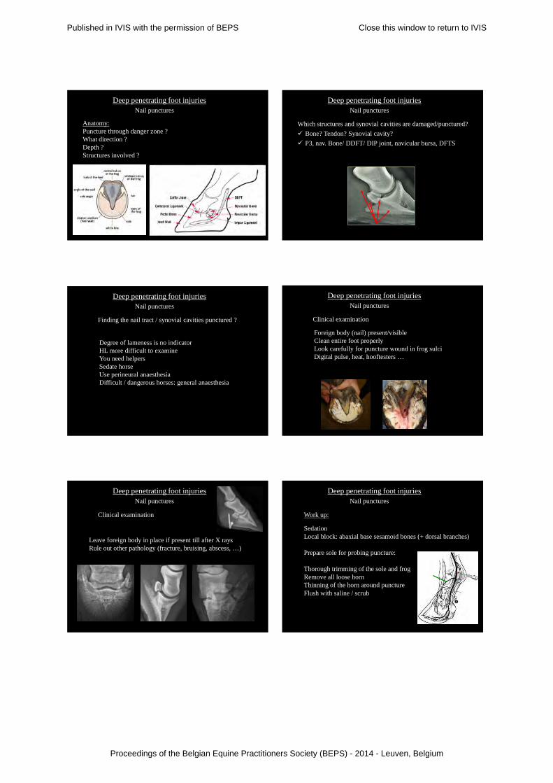
Anatomy:Puncture through danger zone ?What direction ?Depth ?Structures involved ?
Deep penetrating foot injuriesNail punctures
Which structures and synovial cavities are damaged/punctured? Bone? Tendon? Synovial cavity? P3, nav. Bone/ DDFT/ DIP joint, navicular bursa, DFTS
Deep penetrating foot injuriesNail punctures
Finding the nail tract / synovial cavities punctured ?
Degree of lameness is no indicatorHL more difficult to examineYou need helpersSedate horseUse perineural anaesthesiaDifficult / dangerous horses: general anaesthesia
Deep penetrating foot injuriesNail punctures
Clinical examination
Foreign body (nail) present/visibleClean entire foot properlyLook carefully for puncture wound in frog sulciDigital pulse, heat, hooftesters …
Deep penetrating foot injuriesNail punctures
Clinical examination
Leave foreign body in place if present till after X raysRule out other pathology (fracture, bruising, abscess, …)
Deep penetrating foot injuriesNail punctures
Work up:
SedationLocal block: abaxial base sesamoid bones (+ dorsal branches)
Prepare sole for probing puncture:
Thorough trimming of the sole and frogRemove all loose hornThinning of the horn around punctureFlush with saline / scrub
Published in IVIS with the permission of BEPS Close this window to return to IVIS
Proceedings of the Belgian Equine Practitioners Society (BEPS) - 2014 - Leuven, Belgium
Deep penetrating foot injuriesNail punctures
Work up:
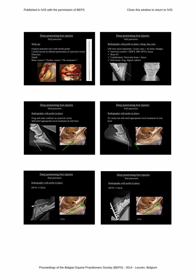
Explore puncture tract with sterile probeCareful (avoid accidental penetration of synovial cavity)DirectionDepthBone contact ? Tendon contact ? No resistance ?
Deep penetrating foot injuriesNail punctures
Radiography with probe in place: cheap, fast, easy
LM view most important / Acute cases + no bony changes Synovial cavities ? DDFT, DIP, DFTS, bursa Bone P3 Combination: Navicular bone + bursa Soft tissue: frog, digital cushion
Deep penetrating foot injuriesNail punctures
Radiography with probe in place:
Frog and solar cushion/ no synovial cavityStill need appropriate local treatment of sole horn
Deep penetrating foot injuriesNail punctures
Radiography with probe in place:
P3, lucky but still need appropriate local treatment of sole horn
Deep penetrating foot injuriesNail punctures
Radiography with probe in place:
DFTS ? Check
lucky
Deep penetrating foot injuriesNail punctures
Radiography with probe in place:
DFTS ? Check
lucky
Published in IVIS with the permission of BEPS Close this window to return to IVIS
Proceedings of the Belgian Equine Practitioners Society (BEPS) - 2014 - Leuven, Belgium
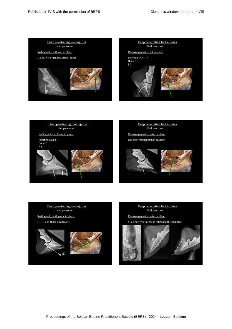
Deep penetrating foot injuriesNail punctures
Radiography with nail in place:
Digital flexor tendon sheath: check
Deep penetrating foot injuriesNail punctures
Radiography with nail in place:
Insertion DDFT ?Bursa ?P 3
Deep penetrating foot injuriesNail punctures
Radiography with nail in place:
Insertion DDFT ?Bursa ?P 3
Deep penetrating foot injuriesNail punctures
Radiography with probe in place:
DIP joint through impar ligament
Deep penetrating foot injuriesNail punctures
Radiography with probe in place:
DDFT and Bursa navicularis
Deep penetrating foot injuriesNail punctures
Radiography with probe in place:
Make sure your probe is following the right trac
Published in IVIS with the permission of BEPS Close this window to return to IVIS
Proceedings of the Belgian Equine Practitioners Society (BEPS) - 2014 - Leuven, Belgium

Deep penetrating foot injuriesNail punctures
Radiography with probe in place:
Make sure your probe is following the right trac
Deep penetrating foot injuriesNail punctures
Other imaging:
Ultrasonography CT scan MRI
Limited use in acute casesMore value in chronic cases / complications
Deep penetrating foot injuriesNail punctures
Nice indication for CT – sequester localisation:Long term complication after sole punctureChronic pain and fistulation due to sequester formationTakes weeks before sequester becomes visible
Deep penetrating foot injuriesNail punctures
No synovial penetration:
Possible treatment at yard
Deep penetrating foot injuriesNail punctures
No synovial penetration:
Standing treatment in stable (character horse, hind limbs, follow up)Wide thinning of the surrounding horn + debridementUse sharp hoof knives, curette, scalpel blade
Use nerve block + tourniquetNo blood !Use leg supportClean surface in case foot goes down
Foto hoofknives
Deep penetrating foot injuriesNail punctures
No synovial penetration: Standing treatment at yard/farm
tourniquet No blood
Leg supportClean surface
Published in IVIS with the permission of BEPS Close this window to return to IVIS
Proceedings of the Belgian Equine Practitioners Society (BEPS) - 2014 - Leuven, Belgium

Deep penetrating foot injuries
Thinning and debridement – general rules in foot surgery:
Minimal but enough !Work clean = surgery = avoid contaminationAll loose horn should be removed until nice attachment with coriumResect all infected/contaminated/hypergranulating coriumMinimal debridement/curettage tendon (DDFT) Remove all contaminated bone till healthy bleeding bone
ebone
coriumhorn
No visualisationHypergranulation/ pressure
Dirth catcher
Opening frog sulcusThinning of the sole and frog hornExposure coriumSolid attachment horn on corium
Deep penetrating foot injuriesNail punctures
Thinning and debridement – exposing the puncture hole:
Solid attachment horn on corium, horn should slowly cover a healthy granulation bed
Deep penetrating foot injuries
Thinning and debridement – what is the aim ?
Hoof cancer: incomplete trimming
Deep penetrating foot injuries
Thinning and debridement – what is enough ?
Hoof cancer: complete trimming
Hoof cancer: 2 days later, bandage
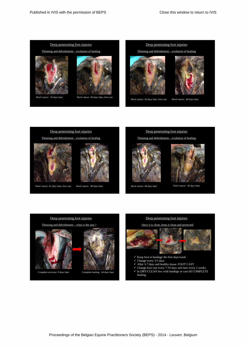
Deep penetrating foot injuries
Thinning and debridement – evolution of healing
Hoof cancer: 10 days later, foot cast
Hoof cancer: remove all unhealthy horn and corium until a solid attachment horn on corium is found (dermo-epidermal junction)
Deep penetrating foot injuries
Thinning and debridement – what is the aim ?
Published in IVIS with the permission of BEPS Close this window to return to IVIS
Proceedings of the Belgian Equine Practitioners Society (BEPS) - 2014 - Leuven, Belgium
Hoof cancer: 10 days later
Deep penetrating foot injuries
Thinning and debridement – evolution of healing
Hoof cancer: 20 days later, foot cast
Hoof cancer: 40 days later
Deep penetrating foot injuries
Thinning and debridement – evolution of healing
Hoof cancer: 20 days later, foot cast
Hoof cancer: 48 days later
Deep penetrating foot injuries
Thinning and debridement – evolution of healing
Hoof cancer: 45 days later, foot cast
Hoof cancer: 56 days later
Deep penetrating foot injuries
Thinning and debridement – evolution of healing
Hoof cancer: 48 days later
Complete healing: 64 days later
Deep penetrating foot injuries
Thinning and debridement – what is the aim ?
Complete excision: 0 days later
Keep foot in bandage the first days/week Change every 3-5 days After 3-7 days and healthy tissue: FOOT CAST Change foot cast every 7-10 days and later every 2 weeks. In DRY/CLEAN box with bandage or cast till COMPLETE
healing
Deep penetrating foot injuries
Once it is clean, keep it clean and protected
Published in IVIS with the permission of BEPS Close this window to return to IVIS
Proceedings of the Belgian Equine Practitioners Society (BEPS) - 2014 - Leuven, Belgium

Antibiotics locally ? Silver dressing ? Alginaat ?Rolls of gauze over defect / frog = counter pressureBandage (soffban/cotton wool/crepe/tape)
Deep penetrating foot injuries
Bandage
Foot cast + picture
Deep penetrating foot injuries
Bandage
Antibiotics systemically: may be the first 5-7 daysTake swabs of wound if delayed wound healing (Ab gram)
Deep penetrating foot injuries
Antibiotics ?
Infected woundNo healing
Swab/ Ab gramAg Alginaat
Not very healthy fresh granulation tissueSmall cracks, not 100% attachment horn to coriumNidus of infection despite foot cast
Deep penetrating foot injuries
Antibiotics
Debride / openNo Ab syst.Locally:- Ag Alginaat- Fucidine
Deep penetrating foot injuriesFoot punctures
Synovial penetration:
Refer to hospital
Deep penetrating foot injuriesFoot punctures
Synovial penetration:Detailed examination and treatment under general anesthesiaDifficult to treat at the yard / referral
Standing:Examination as above + X-rays & blunt probe
General anaesthesia:Synoviocentesis of:
DIP jointNavicular bursaDigital flexor tendon sheath
Synovial fluid sample: wbc/TP/bacterio/ blood culture medium
Published in IVIS with the permission of BEPS Close this window to return to IVIS
Proceedings of the Belgian Equine Practitioners Society (BEPS) - 2014 - Leuven, Belgium

Flush DIP joint/DFTS/bursa: exit through wound ? False negative.If in doubt: Contrast study (omnipaque)
Contrast study of DIP joint and navicular bursa with iohexol (omnipaque). Check for leakage.
Deep penetrating foot injuriesFoot punctures
Synovial penetration:
Deep penetrating foot injuries
Synovial penetration: DFTS
Deep penetrating foot injuries
Synovial penetration: DFTS contrast study
Courtesy Dr F. Pille
Deep penetrating foot injuriesFoot punctures
Synovial penetration: treatment
Flush Joint / bursa / tendon sheatho Needle through and through lavageo Arthroscopy / bursoscopy / tenoscopyo André “street nail surgery” = history, too aggressive
Deep penetrating foot injuriesFoot punctures
Synovial penetration: treatment
Navicular bursa punctured:
Non transthecal approach or direct bursoscopy:Avoid the neighbouring synovial cavities
Synovitis (hyperaemic, proliferative and fibrinopurulent). Foreign material. Puncture site through DDFT. Damage to Nav. bone: fibrocartilage / subchondral bone.
Deep penetrating foot injuriesFoot punctures
Synovial penetration bursa: endoscopic findings
Published in IVIS with the permission of BEPS Close this window to return to IVIS
Proceedings of the Belgian Equine Practitioners Society (BEPS) - 2014 - Leuven, Belgium

Puncture of the navicular bursa: endoscopic treatment
Nav bone
DDFT
Deep penetrating foot injuriesFoot punctures
Synovial penetration: treatment
Bursoscopy: probe through DDFT defect
Puncture of the navicular bursa: endoscopic treatment
High pressure lavage. Remove fibrin, dirt and other foreign bodies. Debride defect on flexor surface and in DDFT. Remove torn tendon fibrils. Debride the puncture tract and use this tract as instrument
portal. If this is not possible create a medial instrument portal
(similar to arthroscope portal). Evaluate and treat DIP joint and DFTS if needed
Deep penetrating foot injuriesFoot punctures
Synovial penetration: medical treatment after flushing Local medical treatment:
Regional IV perfusion (several times?) Intra-synovial antibiotics (several times?)
Deep penetrating foot injuriesFoot punctures
Synovial penetration: medical treatment after flushing
Systemic medical treatment:
Antibiotics IV/IM + NSAID IV + ATS
Deep penetrating foot injuriesFoot punctures
Synovial penetration: treatment
Important:
Bandage / foot cast till complete healing of the defect.
Published in IVIS with the permission of BEPS Close this window to return to IVIS
Proceedings of the Belgian Equine Practitioners Society (BEPS) - 2014 - Leuven, Belgium

Deep penetrating foot injuriesFoot punctures
Hoof cancer: treatment and outcome
DebridementPrednisolone per os 3 weeksEarly treatment gives better resultsDuration treatment: 1-3 months
Outcome: ± 40 % cured± 70 % controllable
Deep penetrating foot injuriesFoot punctures: Outcome
Superficial punctures: good Digital cushion: good P3: good Synovial cavities:
Navicular bursa puncture, needle lavage + RIP, n = 23 (Pille et al, 2005) : 73 % full recovery
Navicular bursa puncture, endoscopic lavage, n = 16 (Wright et al , 1999): 62.5 % full recovery
Bosdreef: 2004-2012: n = 31, bursa + synovial cavities74 % full recoveryAll failures > 3 days standing before referral
Deep penetrating foot injuries
Deep punctures wounds
Prevent disasters
Deep penetrating foot injuries
Cases
www.bosdreef.be
Published in IVIS with the permission of BEPS Close this window to return to IVIS
Proceedings of the Belgian Equine Practitioners Society (BEPS) - 2014 - Leuven, Belgium
Top Related