


Languages
Pages
Legal


PRELIMINARY HSC PDHPE
CQ1 – How do the musculoskeletal and cardiorespiratory systems of the body influence and respond to movement?

How do the musculoskeletal and cardiorespiratory systems of the body influence and respond to movement?
Students learn about: skeletal system − major bones involved in movement
structure and function of synovial joints joint actions, eg extension and flexion
Students learn to: • identify the location and type of major bones
involved in movement, eg long bones articulate at hinge joints for flexion and extension
muscular system − major muscles involved in movement − muscle relationship (agonist, antagonist) − types of muscle contraction (concentric, eccentric,
isometric)
• identify the location of the major muscles involved in movement and related joint actions
• perform and analyse movements, eg overarm throw, by examining:
− bones involved and the joint action − muscles involved and the type of contraction
respiratory system − structure and function − lung function (inspiration, expiration) − exchange of gases (internal, external)
• analyse the various aspects of lung function through participation in a range of physical activities
circulatory system − components of blood − structure and function of the heart, arteries, veins,
capillaries − pulmonary and systemic circulation − blood pressure.
• analyse the movement of blood through the body and the influence of the circulatory and respiratory systems on movement efficiency and performance.

SKELETAL SYSTEM – watch video 1
Function of the skeletal system - watch video 2
The skeletal system consists of bone tissue, bone marrow, cartilage and the periosteum (the membrane around bones). The functions of the skeleton and bone tissue include: Support — they provide a framework for attachment of soft connective
tissue, such as muscles Protection — they protect internal organs; for example, the ribs protect
the heart and lungs Movement — when muscles contract they pull on bones and produce
movement Mineral storage — in particular they store calcium and phosphorus,
which are released when needed Blood cell production — most blood cell formation occurs within the red
bone marrow Storage of energy — yellow bone marrow is a stored source of lipids in
the bones.

Types of bones
Axial skeleton
The axial skeleton provides the central structure (or long axis) of the skeletal system. Many of the bones in the axial skeleton do not move, or move only minimally. They provide the main structure of the overall skeleton, and the core stability of the axial skeleton allows the bones of the joined appendicular skeleton (especially the long bones) to move efficiently, with both parts of the skeleton relying on the muscles that are attached to perform their appropriate function of either supporting stability or driving the movement. The axial skeleton includes the cranium, vertebral column and rib cage.
Axial skeleton
Cranium (skull) It is formed by two sets of bones— the cranial bones and the
facial bones—numbering 22 in total. Vertebral column (spine)
Cervical vertebrae—the seven vertebrae of the neck Thoracic vertebrae—the next 12 vertebrae Lumbar vertebrae—the five vertebrae supporting the lower
back Sacrum—the five fused vertebrae that connect to the pelvis Coccyx—the four fused vertebrae, also known as the tailbone
Rib cage The sternum and 12 pairs of ribs make up the rib cage.

Axial skeleton

The appendicular skeleton
Shoulder girdle consists of two bones: the clavicle (also known as the
collarbone) and scapula (also known as the shoulder blade).
Upper limbs Thirty bones comprise the upper limbs, also known as the
arm, forearm and hand. Pelvic girdle (hip) Lower limbs
Three segments make up the lower limb: the thigh, leg and foot.
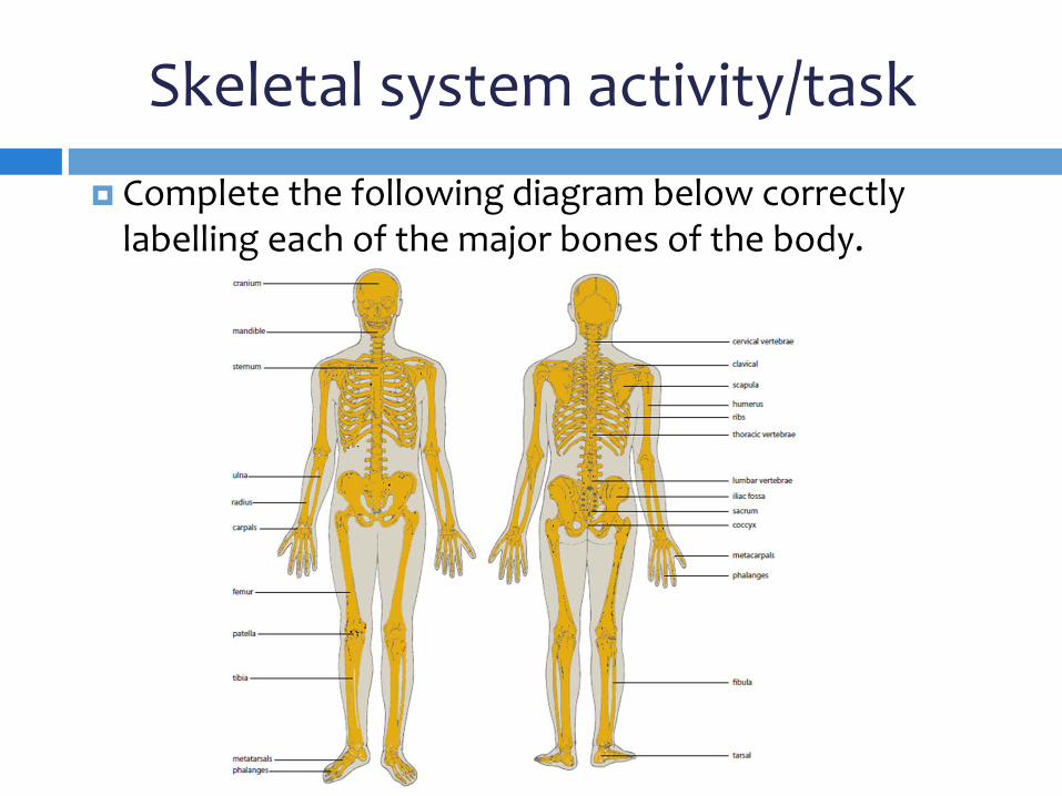
Skeletal system activity/task Complete the following diagram below correctly
labelling each of the major bones of the body.

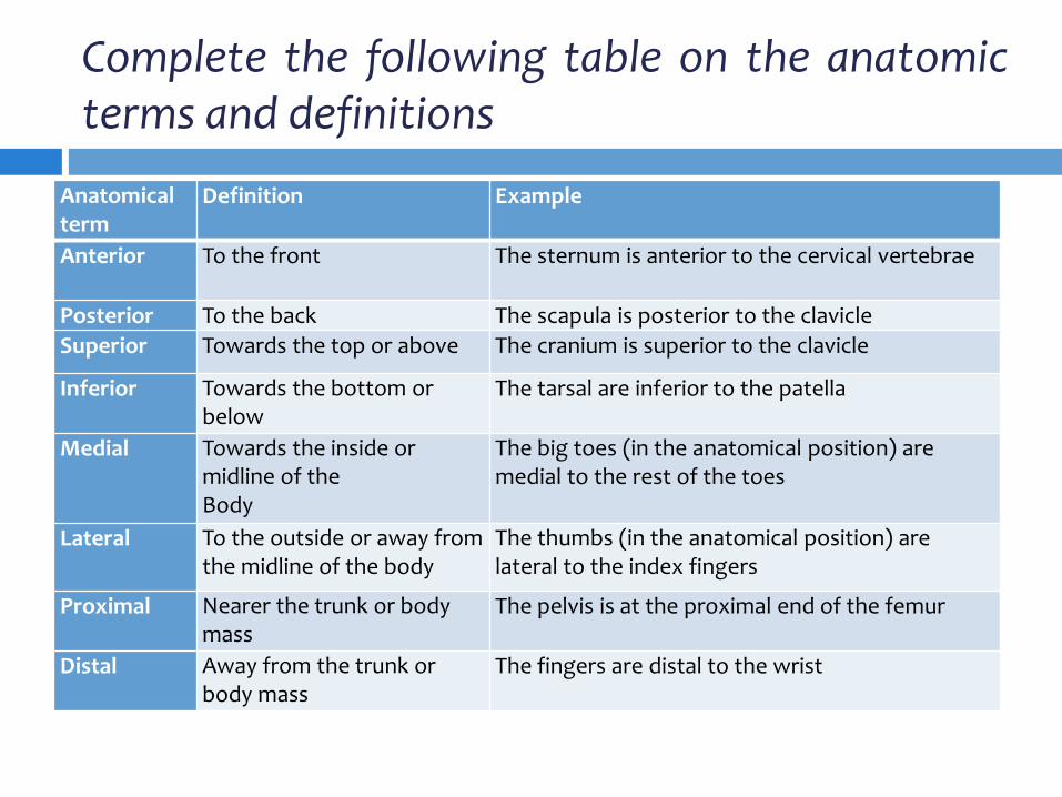
Complete the following table on the anatomic terms and definitions
Anatomical term
Definition Example
Anterior To the front The sternum is anterior to the cervical vertebrae
Posterior To the back The scapula is posterior to the clavicle Superior Towards the top or above The cranium is superior to the clavicle
Inferior Towards the bottom or below
The tarsal are inferior to the patella
Medial Towards the inside or midline of the Body
The big toes (in the anatomical position) are medial to the rest of the toes
Lateral To the outside or away from the midline of the body
The thumbs (in the anatomical position) are lateral to the index fingers
Proximal Nearer the trunk or body mass
The pelvis is at the proximal end of the femur
Distal Away from the trunk or body mass
The fingers are distal to the wrist

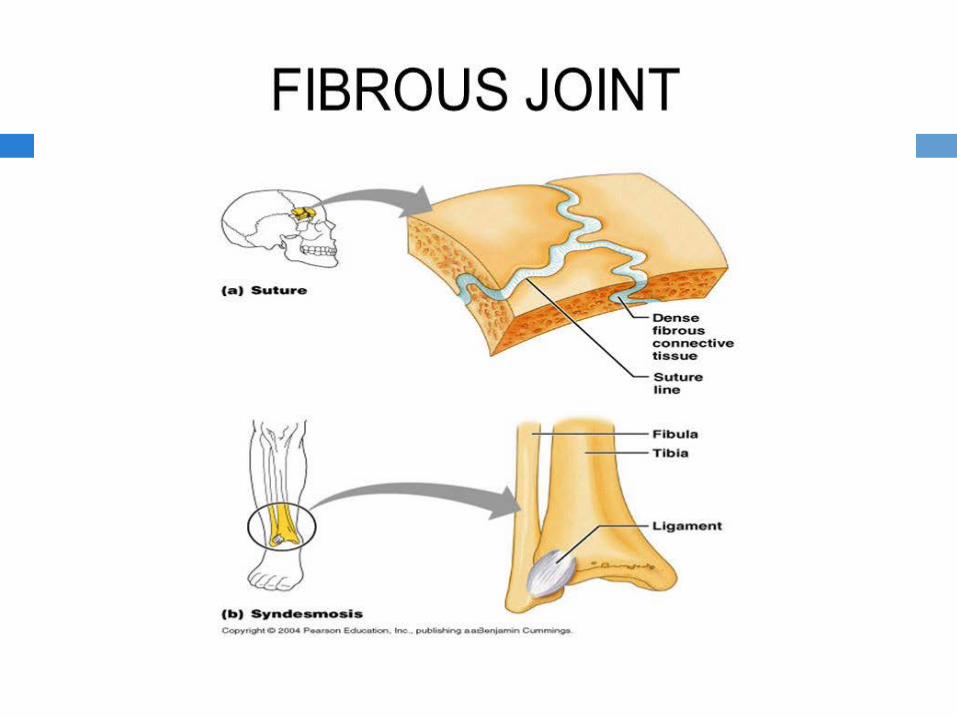
Fibrous Joints
These joints are characterised by fibres joining the ends or parts of bones together. Fibrous joints use various fibres to connect bones, including short fibres (sutures), long fibres (syndesmosis) and ligaments (gomphosis). No joint cavity is present in these types of joints. These fibres make movement difficult.

Cartilaginous Joints
These joints are characterised by cartilage joining the ends or parts of bones together. They contain no joint cavity and, therefore, allow only slight movement.

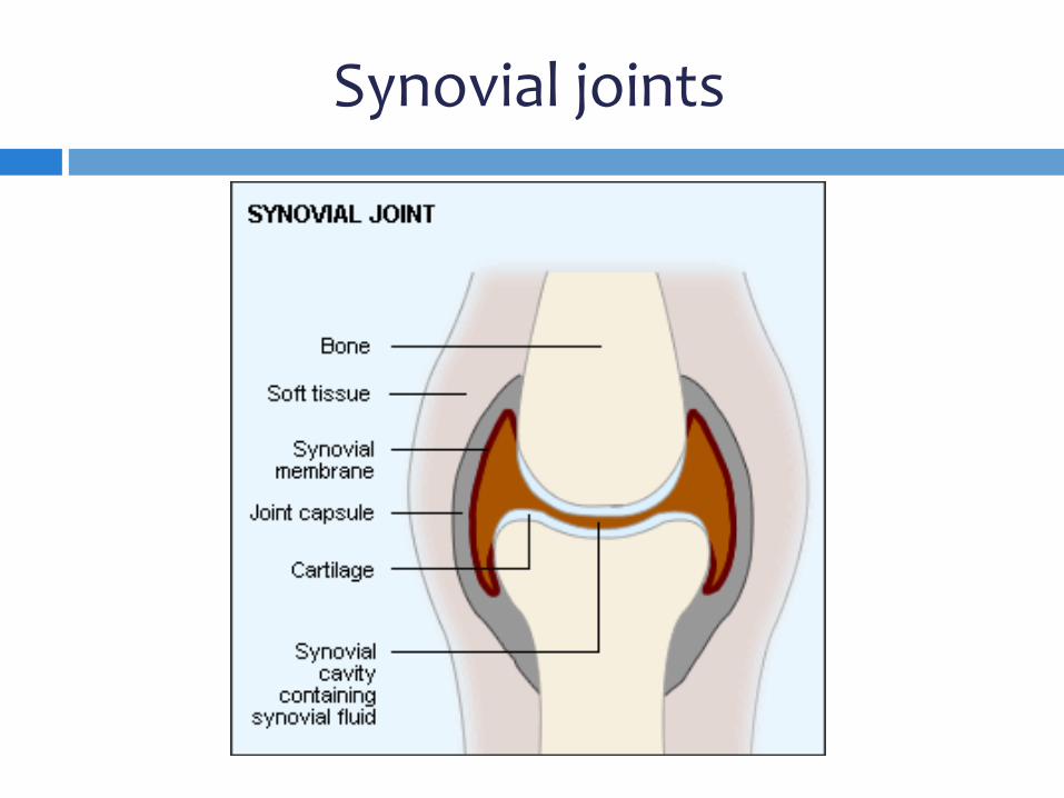
Synovial Joints
These joints are enclosed in a capsule and covered with cartilage and a synovial membrane to allow free movement.
• Synovial cavity — a space where two bones meet • Synovial fluid — a rich viscous fluid that acts as lubrication to the
bones in the synovial cavity. It also plays a role in the clean-up of tissue debris from daily wear and tear on the joint
• Articular cartilage — covers the end of the bones, provides cushioning and reduces friction between the bones in the joint
• Joint capsule — encloses the cavity and keeps the fluid contained • Fibrous capsule — encloses the bones, tissues and fluid • Synovial membrane — secretes the fluid into the joint • Meniscus — cartilage that grows inwards from the joint capsules to
absorb shock, reduce pressure and enhance stability.
Synovial joints

Types of Synovial joints (six types) - watch videos 3, 4 and 5
gliding joint — side-to-side or back-and-forth movement is permitted across these simple, usually flat surfaces; for example, between the carpals and the tarsals
hinge joint — the convex surface of one bone fits into the concave surface of another, and movement occurs in one plane. Examples are the elbow joint and knee joint
pivot joint — the primary movement is rotation, where the rounded or pointed surface of one bone articulates with the depression or opening of another; for example, between the atlas and axis, or at the proximal end of the radius and ulna
ellipsoid (or condyloid) joint — an oval-shaped condyle of one bone fits into the elliptical depression of another, and movement can occur in two planes. Examples are the joints between the metacarpals and the phalanges
saddle joint — the articular surface of one bone is saddle shaped and the other bone sits on it like a rider. This is a modified ellipsoid joint; for example, between the carpal and the metacarpal of the thumb
ball and socket joint — a rounded ball-like surface of one bone fits into a cup-like depression of another. These are very movable joints. Examples are the shoulder joint and hip joint.
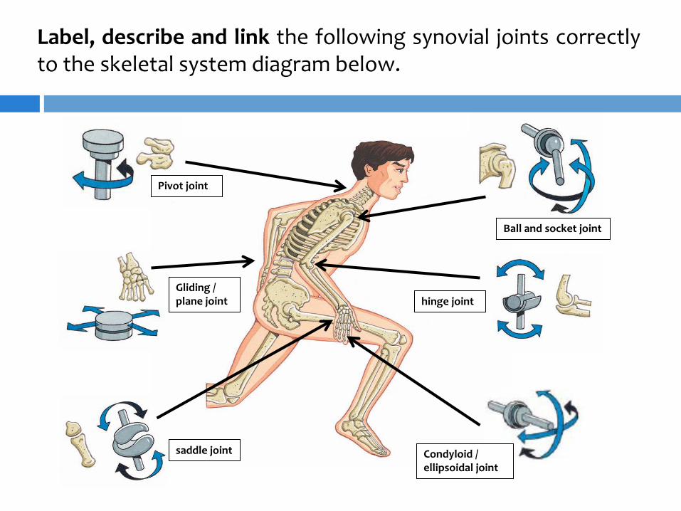
Label, describe and link the following synovial joints correctly to the skeletal system diagram below.
Pivot joint
Gliding / plane joint
saddle joint
Ball and socket joint
hinge joint
Condyloid / ellipsoidal joint
Joint actions
The majority of joints in the body are synovial joints. They are very mobile, and permit a great deal of movement because of the contraction of the muscles that pull bones together. All our muscles are attached to bones or other connective tissue in at least two places. The origin of the muscle is attached to the less movable bone, and the insertion is attached to the movable bone. The types of movements that can occur at joints depend on the type of joint. Not all joints have the same possible movements.

Activity/task – Complete the following table and define each joint action with examples.
Joint action example Description Flexion Flexion is the bending movement that causes a decrease in the angle between the bones at the joint. Bending the leg at the knee and performing the upward phase of a biceps curl are examples of flexion. Extension Extension is the straightening movement that causes an increase in the angle between the bones at the joint. Straightening the leg at the knee and performing the downward phase of a biceps curl are examples of extension. Hyperextension excessive extension of a body part.

Joint actions Joint action example Description
Pronation Pronation is the movement that occurs when the radius rotates around the ulna (the thinner, longer bone of the forearm), so that the hand is facing with the palm downwards.
Supination Supination is the movement that occurs when the radius rotates around the ulna, so that the hand is facing with the palm upwards. Putting the hand out to receive some loose change is an example of supination.

Joint action example Description Inversion Inversion is the rotation of the foot so that the sole turns inwards.
Eversion Eversion is the rotation of the foot so that the sole turns outwards.
Joint actions

Joint action example Description Abduction Abduction is moving the body part in the lateral plane away from the mid-line of the body. An example of abduction is performing a star jump where a person starts with the legs together and arms by the side and then extending the arms and legs so that they are apart in their finishing position. Adduction Adduction is moving the body part in the lateral plane back towards the midline of the body. An example of abduction is completing a star jump from the arms and legs in the apart position to the finishing position of arms by the side and legs together.
Joint actions

Joint action example Description Dorsi Flexion Dorsi flexion is simply the flexion of the ankle. Pulling the toes back towards the shin when stretching the hamstring muscles is an example of dorsiflexion at the ankle.
Plantar Flexion Plantar flexion is actually the extension of the foot at the ankle joint and is the opposite of dorsiflexion. A dancer standing up on their toes during a dance routine is an example of plantar flexion.
Joint actions

Joint action example Description Circumduction is the combination of other movements that results in a circular or cone-like pattern. Performing arm circles or a softball pitcher’s wind up and release are examples of circumduction.
Rotation Rotation is the movement of a bone turning on a central axis. A full rotation is usually not possible because there are ligaments and muscles that restrict the movement. An example of rotation is the neck turning from one side to the other.
Joint actions

Joint action example Description Elevation raising of a body part
Depression lowering of a body part
Joint actions

Joint action example Description Retraction withdrawing of a body part
Retraction withdrawing of a body part
Joint actions

Revision/Homework task – Answer / complete the following statements/questions:
Identify the six different types of synovial joint. Describe the function of synovial fluid. Compare the structures and functions of the elbow and
knee joints. Explain how the joint movements of flexion and extension
differ from those of abduction and adduction. Explain how rotation differs from circumduction. Demonstrate with a partner and then list the kinds of
movements that can occur in the neck, jaw, hip, wrist, knee, shoulder and ankle.
Explain how issues such as arthritis that can affect joint mobility.

In pairs, use the following skills to identify the joint actions that occur throughout the movement. Sitting on a chair Bowling a cricket ball Jumping over a hurdle Kicking a soccer ball Hitting a tennis ball with a racquet Writing on a page Performing a forward roll
Revision/Homework task Answer/complete the following statements/questions:
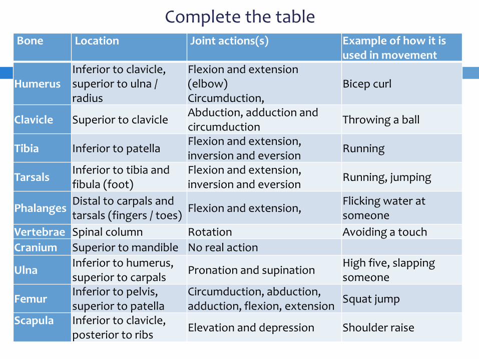
Complete the table Bone
Location Joint actions(s) Example of how it is used in movement
Humerus Inferior to clavicle, superior to ulna / radius
Flexion and extension (elbow) Circumduction,
Bicep curl
Clavicle Superior to clavicle Abduction, adduction and circumduction Throwing a ball
Tibia Inferior to patella Flexion and extension, inversion and eversion Running
Tarsals Inferior to tibia and fibula (foot)
Flexion and extension, inversion and eversion Running, jumping
Phalanges Distal to carpals and tarsals (fingers / toes) Flexion and extension, Flicking water at
someone Vertebrae Spinal column Rotation Avoiding a touch Cranium Superior to mandible No real action
Ulna Inferior to humerus, superior to carpals Pronation and supination High five, slapping
someone
Femur Inferior to pelvis, superior to patella
Circumduction, abduction, adduction, flexion, extension Squat jump
Scapula
Inferior to clavicle, posterior to ribs Elevation and depression Shoulder raise
Summary - watch videos 6 and 7
The shape and length of bones and how they join other bones influences the movements those bones can make.
Bones can be classified as long, flat, short or irregular bones.
The interface between bones (the joints) play a significant role in human movement.
There are three categories of joints: immoveable, cartilaginous and synovial joints.
Synovial joints have common features that influence their efficiency of movement; these include hyaline cartilage, synovial membranes, joint capsules and synovial fluid.
There are six types of synovial joints in the body: ball and socket, hinge, condyloid, saddle, gliding and pivot.
The bones of the axial skeleton (cranium, vertebral column, rib, sacrum and coccyx) provide bases of attachment for the bones of the appendicular skeleton (bones of the pelvis, legs, shoulder girdle and arms).
The major bones of the body articulate at synovial (movable) joints and allow movement to occur.