


Languages
Pages
Legal


Prof Andrew M Scott, MD, FRACP, FAANMS
Department of Molecular Imaging and Therapy Austin Health, Melbourne, Australia
World Federation of Nuclear Medicine and Biology
PET in CNS Conditions
IPET 2015 Vienna, Austria
October 5th – 9th, 2015

• no relevant disclosures
• research funding from Abbvie, Daiichi-Sankyo, Avipep
• grant support from NHMRC, Cancer Australia, Victorian Cancer Agency, Cure Brain Cancer, Prostate Cancer Foundation, Movember, National Breast Cancer Foundation
• consultant to Life Science Pharmaceuticals
Disclosures

• PET imaging in brain tumours
• PET imaging in epilepsy
• PET imaging in neurodegenerative disorders
• Case Reviews
Overview
Learning Objectives:
• to understand the clinical use of PET in the evaluation of patients with benign and malignant CNS conditions
• to evaluate appropriate PET tracers and imaging techniques required for imaging neurological patients with brain tumours, epilepsy and dementia
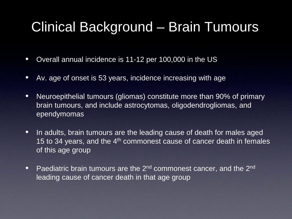
• Overall annual incidence is 11-12 per 100,000 in the US
• Av. age of onset is 53 years, incidence increasing with age
• Neuroepithelial tumours (gliomas) constitute more than 90% of primary brain tumours, and include astrocytomas, oligodendrogliomas, and ependymomas
• In adults, brain tumours are the leading cause of death for males aged 15 to 34 years, and the 4th commonest cause of cancer death in females of this age group
• Paediatric brain tumours are the 2nd commonest cancer, and the 2nd leading cause of cancer death in that age group
Clinical Background – Brain Tumours

WHO Classification of Gliomas
Grade Tumour Type Characteristic
I Low-grade astrocytoma Pilocytic astrocytoma
II Low-grade astrocytoma
Diffuse infiltrating lesions without enhancement
III Anaplastic Variable enhancement with oedema
IV Glioblastoma multiforme
Heterogeneous enhancement with oedema
Heiss et al J Nuc Med 2011; 52:1-16

Wester HJ Clin Cancer Res 2007 ;13:3470-81
FDG PET in Brain Tumours
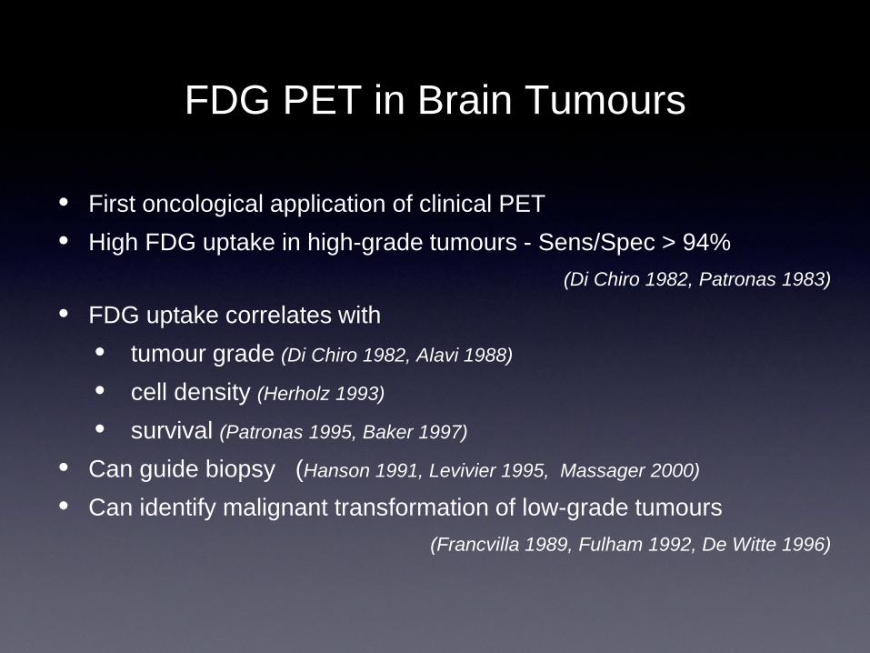
• First oncological application of clinical PET • High FDG uptake in high-grade tumours - Sens/Spec > 94%
(Di Chiro 1982, Patronas 1983)
• FDG uptake correlates with • tumour grade (Di Chiro 1982, Alavi 1988)
• cell density (Herholz 1993)
• survival (Patronas 1995, Baker 1997)
• Can guide biopsy (Hanson 1991, Levivier 1995, Massager 2000)
• Can identify malignant transformation of low-grade tumours (Francvilla 1989, Fulham 1992, De Witte 1996)

FLAIR T1 Gd+ FDG
T1 Gd+ FDG FDG-MRI
WHO II
WHO IV

FDG PET in Grading of Brain Tumours
• FDG PET in 58 patients, 32 (20 gliomas) high and 26 (18 gliomas) low grade tumours (Delbeke 1995)
• FDG uptake in low grade tumours ≤ white matter
• FDG uptake in high grade tumours > white matter
• Differentiation of low and high grade tumours
• Tumour / White matter > 1.5 (Sens 94% / Spec 77%)
• Tumour / Gray matter > 0.6
• 6 low grade gliomas tumours had T/WM > 1.5
• No high grade gliomas had T/WM < 1.5

FDG PET in radiation necrosis • Radiation injury may be difficult to differentiate from tumour on MRI
• Early studies report sensitivity 81-86% & specificity 40-94% for FDG PET in distinguishing recurrence from radiation necrosis (Langleben 2000)
• In 117 post radiotherapy patients, FDG PET can achieve a sensitivity of 96% & specificity of 77% (Wang 2006)
• FDG PET-MR registration is essential
• Any FDG activity greater than expected background is considered suggestive of tumour
• Optimal timing of PET - at least 6 wk following radiotherapy recommended
Chen J Nucl Med 2007;48:1468-81
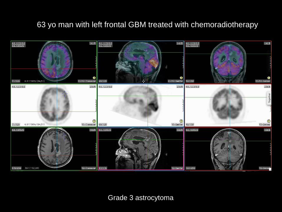
Grade 3 astrocytoma
63 yo man with left frontal GBM treated with chemoradiotherapy

Differential Diagnosis of Focal FDG Uptake in Brain
High Grade Glioma Metastatic Tumour Pilocytic Astrocytoma Primary CNS Lymphoma Pituitary Adenoma Ictal State Abscess Multiple Sclerosis Severe Radiation Necrosis Radiation Induced Meningioma Pleomorphic Xanthoastrocytoma Cerebral Whipple's disease Congenital Hemiplegia Lhermitte-Duclos disease Acute Stroke Sydenham's Chorea

Limitations of FDG PET in Brain Tumours
• High normal brain activity decreases the detection of low-grade and some recurrent high grade tumours
• FDG uptake in high grade tumours varies greatly and can be slightly above white matter (Wong 2002)
• MR-PET coregistration greatly improves performance of PET (Wong 2004)
• Delayed imaging improves distinction of tumour from normal gray matter (Spence 2004)
11C-Methionine (MET) in Brain Tumours
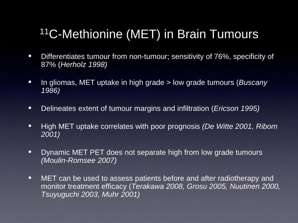
• Differentiates tumour from non-tumour; sensitivity of 76%, specificity of 87% (Herholz 1998)
• In gliomas, MET uptake in high grade > low grade tumours (Buscany 1986)
• Delineates extent of tumour margins and infiltration (Ericson 1995)
• High MET uptake correlates with poor prognosis (De Witte 2001, Ribom 2001)
• Dynamic MET PET does not separate high from low grade tumours (Moulin-Romsee 2007)
• MET can be used to assess patients before and after radiotherapy and monitor treatment efficacy (Terakawa 2008, Grosu 2005, Nuutinen 2000, Tsuyuguchi 2003, Muhr 2001)
18F-Fluoroethyltyrosine (FET) in Brain Tumours
• FET was more sensitive than FDG (86% cf 35%) in detection of brain tumours, and better at delineating tumour extent (Pauleit 2009)
• FET combined with MRI can guide biopsy with high diagnostic yield (Pauleit 2005)
• Dynamic FET PET can differentiate low from high grade tumours with 92% sensitivity & specificity (Pöpperl 2007)
• Post-operative FET tumour volume is predictive of overall survival and disease free survival in patients undergoing chemoradiotherapy (Piroth 2011)
• FET early after chemoradiotherapy may stratify responders from nonresponders (Piroth 2011)

Piroth et al Int J Radiat Oncol Biol Phys 2011;80:176-84
18F-FET PET early after chemoradiotherapy

18F-Fluoro-L-Dopa (F-DOPA) in Brain Tumours
• 81 patients with brain tumours evaluated
• 30 patients, newly diagnosed or previously treated brain tumours had both F-DOPA & FDG PET
• 51 patients F-DOPA PET only
• F-DOPA more sensitive (98%) than FDG (61%) for detection of brain tumours
• No significant difference in F-DOPA uptake between low and high grade tumours (p=0.04), or between contrast-enhancing or non-enhancing tumours (p=0.97)
• F-DOPA able to distinguish radiation necrosis from tumour recurrence
Chen et al J Nucl Med 2006;47:904-911

(A) Glioblastoma (B) Grade II oligodendroglioma
Newly diagnosed tumours Recurrent tumours
(A) Recurrent glioblastoma (B) Recurrent grade II oligodendroglioma
Chen et al J Nucl Med 2006;47:904-911
F-DOPA in brain tumours
FDG FDG FDOPA FDOPA

18F-Fluorothymidine in Brain Tumours
• PET nucleosides are indicators of cellular proliferation
• FLT is a marker of tumour cell proliferation, uptake correlates with thymidine-1-kinase activity
• FLT & MET have similar sensitivity (>80%) in detection of brain tumours; low grade tumours false-negative on MET are also false negative on FLT (Hatakeyama 2008)
• FLT should not be considered for recurrent low grade tumours (Tripathi 2009)
• FLT response predictive of survival in patients treated with Avastin (Chen 2007)
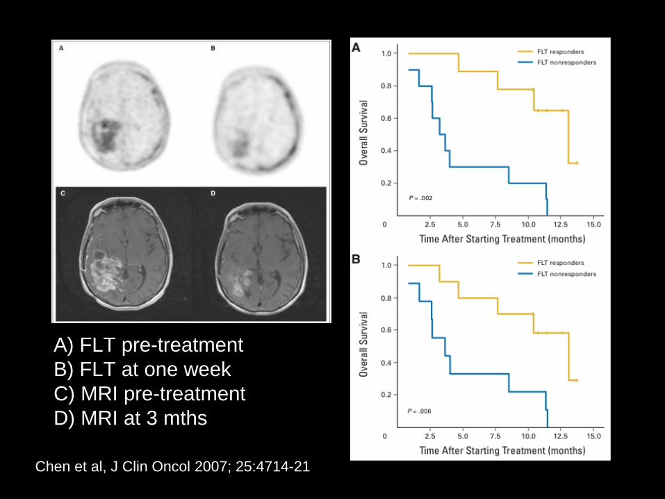
Chen et al, J Clin Oncol 2007; 25:4714-21
A) FLT pre-treatment B) FLT at one week C) MRI pre-treatment D) MRI at 3 mths

Hypoxia Imaging in Brain Tumours
• Hypoxia is associated with tumour progression and resistance to radiotherapy
• 18F-Fluoromisonidazole (FMISO) is a nitroimidazole derivative, metabolites of FMISO are trapped in hypoxic cells
• FMISO uptake correlates with tumour grade, with FDG PET can identify areas of aerobic and anaerobic glycolysis
• Significant association between hypoxia (FMISO uptake) & Ki-67, VEGF-R1 and HIF-1α
• may be predictive of sites of relapse post radiotherapy
• principally used for biologic characterisation of brain tumours
Cher et al J Nucl Med 2006;43:410-418

MRI 18F-FDG PET 18F-FMISO PET
18F-FMISO in high grade glioma
Lee ST & Scott AM, Sem Nucl Med 2007;37:451-461
• Metastatic brain tumours are the most common brain tumour
• A single lesion in nearly 50%, two lesions in 21%, three in 13%, and four or more in the remaining cases.
• Cerebral metastases are FDG-avid in 68-79% of cases
• In up to 20% of cases, cerebral metastases are the initial presenting feature of malignant disease
• FDG PET identifies the primary site in 80-90% of cases
• Most common cerebral metastases arise from lung, breast, melanoma, colon and kidney cancers
Metastatic Brain Tumours
(Gupta 1999, Jeong 2002)

FDG PET - Metastatic Brain Tumour

PET in Brain Tumours Case Studies

In this case of a newly diagnosed left parietal lesion, A) FDG PET and B) MRI, which of the following is correct: A) the lesion is likely to be a high grade lesion
B) the lesion is typical of a cerebral lymphoma
C) a low grade tumour (glioma) may be present
D) the appearance is typical of multiple sclerosis
- correct
Case 1

A 12 yr old boy with symptoms of persisting headache was evaluated with A) FDG PET and B) MRI. Which of the following is correct:
A) the lesion is likely to be a meningioma
B) the lesion is most likely a cerebral lymphoma
C) a low grade tumour (glioma) is present
D) the appearance is typical of pilocytic astrocytoma - correct
Case 2

T2 MRI FDG
23/3/06
24/1/08
54 yr old man with low grade left frontal lesion Case 3
In this patient, is the likely diagnosis:
A) post-radiation inflammatory response
B) local tumour recurrence
C) focal dysplasia post therapy
D) partial metabolic response to treatment
- correct

In this 68Ga-68 DOTATATE PET study, which of the following is correct:
A) the cerebral uptake is an artifact
B) the lesion is suggestive of a meningioma
C) a cerebral aneurysm is present
D) the appearance is typical of multiple sclerosis
- correct
Case 4

Epilepsy
• Common chronic neurological disorder characterised by recurrent, unprovoked seizures
• Affects 3% of the population during their lifetime
• After the first seizure, about 80% of patients experience another seizure within the first 3 years
• About 60-70% of patients experience focal or partial seizures
• Epilepsy is controlled with medication in 70% of cases
• When seizures are intractable, resection of the epileptogenic cortex may be considered

Presurgical workup of patients with intractable epilepsy
• Seizure history
• Physical & neurological examination
• Neuropsychological assessment
• Scalp EEG
• MRI
• Video-EEG
• Interictal and ictal EEG
• Interictal and ictal SPECT
• Interictal FDG PET

The Epileptogenic Zone
• The epileptogenic zone is the cortex generating the seizures which needs to be removed to render the patient seizure free
• Is a theoretical construct consisting of
• the seizure onset zone (ictal SPECT)
• the epileptogenic lesion (MRI)
• the symptomatogenic zone (Video-EEG)
• the functional deficit zone (Interictal PET)
• Epilepsy surgery has the best results if the different cortical zones are concordant
• Surgery renders 60-90% of patients with unilateral temporal lobe epilepsy (TLE), and up to 70% of patients with a focal cortical malformation seizure free
Brain 2001;124:1683-1700

Epileptogenic lesion (MRI)
Symptomatic zone (Video EEG)
Functional deficit zone (Interictal PET)
Seizure onset zone (Ictal SPECT)

Interictal FDG PET
• The epileptogenic zone is usually contained within the cortex with the most profound hypometabolism on interictal FDG PET
• In a series of 89 patients, the sensitivity of FDG-PET in patients with refractory epilepsy and normal MRI was 44% (Lee et al, Ann Neurol 2005;58:525-532)
• In temporal lobe epilepsy (TLE), hypometabolism ipsilateral to the seizure focus is present in 60-90% (Casse et al, Mol Imaging Biol 2002;4:338-351)

Temporal lobe epilepsy
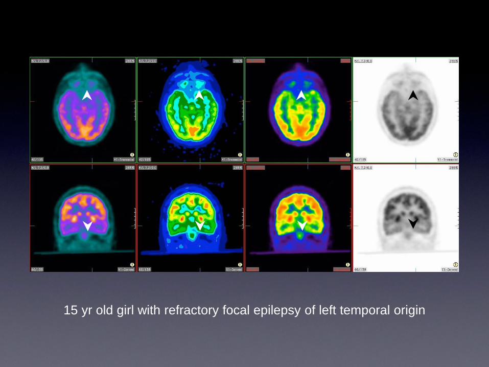
15 yr old girl with refractory focal epilepsy of left temporal origin

20 year old man with right parietal lobe seizures
20 year old man with right parietal lobe seizures
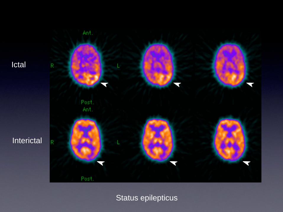
Ictal
Interictal
Status epilepticus
FDG PET

FDG-PET & surgical outcome
• FDG-PET localisation of the ictal focus to a lobe correlates with seizure free surgical outcome (Yun et al, Epilepsia 2006;47:574-579)
• Concordance of 2 or more presurgical evaluations also correlates with seizure freedom (Lee et al, Ann Neurol 2005; 58: 525-532)
• Extent of hypometabolism predictive of surgical outcome
• In TLE, greater maximal asymmetry is associated with decreased chance of seizure freedom (Lin et al, J Nucl Med 2007; 48: 776-782)
• PET hypometabolism is more extensive than the pathological abnormality

Localising value of individual modalities in seizure free patients
Ann Neurol 2005;58:525-532
Interictal EEG Ictal EEG PET Ictal SPECT
Frontal 7/15 12/15 4/14 3/7
Temporal 9/17 13/17 14/16 6/8
Parietal 0/3 1/3 1/3 0/3
Occipital 4/7 7/7 4/7 1/6
Subtotal 20/42 33/42 23/40 10/24
p value 0.365 0.132 0.01 0.062
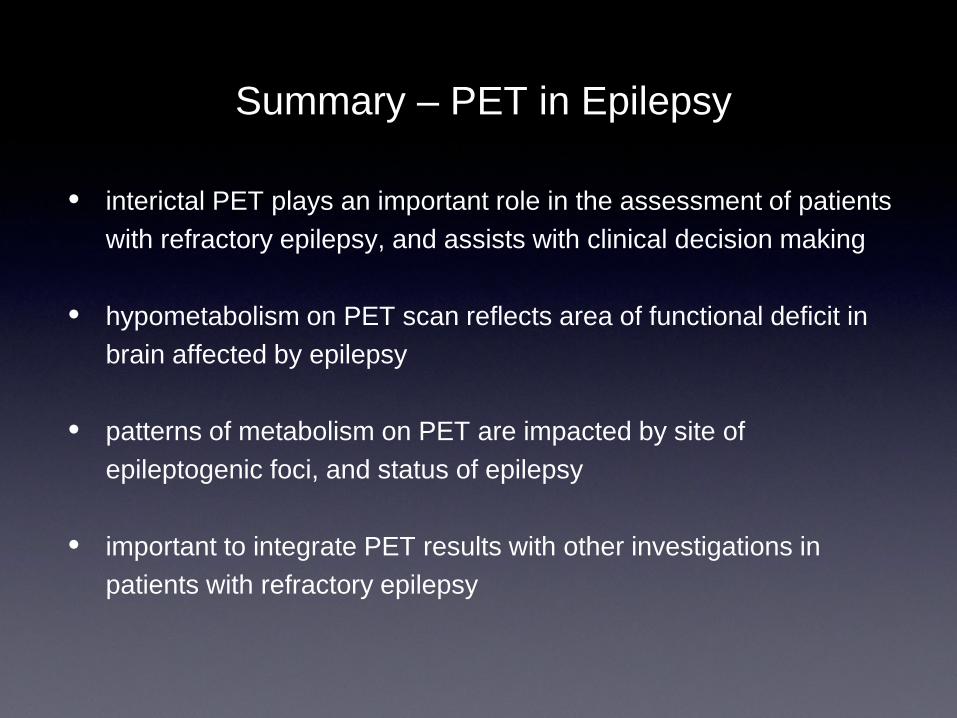
• interictal PET plays an important role in the assessment of patients with refractory epilepsy, and assists with clinical decision making
• hypometabolism on PET scan reflects area of functional deficit in brain affected by epilepsy
• patterns of metabolism on PET are impacted by site of epileptogenic foci, and status of epilepsy
• important to integrate PET results with other investigations in patients with refractory epilepsy
Summary – PET in Epilepsy
PET in Epilepsy Case Studies

18 year old young woman with left parietal lobe seizures
Case 5

In this patient, the most likely diagnosis is:
A) high grade glioma in left parietal lobe
B) possible seizure focus in left parietal lobe
C) possible seizure focus in right parietal lobe
D) cerebral lymphoma
- correct
Case 5

52 year old woman with cerebral abscess surgically treated at age of 16 presents with myoclonus
Case 6
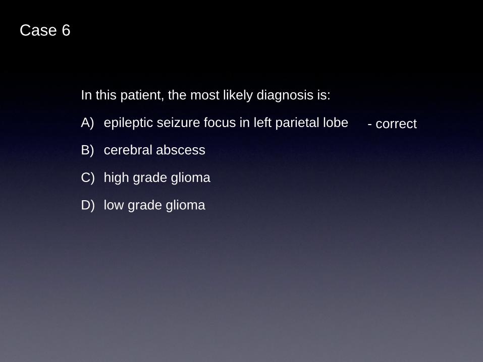
In this patient, the most likely diagnosis is:
A) epileptic seizure focus in left parietal lobe
B) cerebral abscess
C) high grade glioma
D) low grade glioma
- correct
Case 6

28 year old woman with right temporal lobe seizures
Case 7

The FDG PET scan shows:
A) a high grade glioma in right temporal lobe
B) mid brain focus of epilepsy
C) hypothalamic tumour
D) right parahippocampal hypermetabolism
- correct
Case 7

Dementia and Alzheimer’s disease
• Dementia is defined as cognitive impairment of sufficient severity that it prevents independent function in the patient’s usual occupation or daily activities
• Alzheimers disease (AD) is an irreversible, progressive neurodegenerative disorder clinically characterized by memory loss and other cognitive and functional decline
• AD invariably leads to death, within 7 to 10 years of diagnosis
• At this time, there is no cure for AD, and no proven way to slow the rate of neurodegeneration
• Clinical diagnosis is 80-85% sensitive with a specificity of 70%
• Pathology of Alzheimer’s disease includes intracellular neurofibrillary tangles, ß-amyloid deposition in the form of extracellular senile plaques, synaptic reductions, neuronal loss and volume loss

IWG-2 research diagnostic criteria for Alzheimer’s disease
• Dementia NOT required
• Objective memory impairment
plus
• Pathophysiological biomarker for AD
i.e. CSF (low Ab42 with high tau) or positive Aβ PET
• FDG and MRI (for neuronal damage) are markers for disease severity and progression
Dubois B, et al. Lancet Neurology 2014

FDG PET & Alzheimer’s Disease
• Characteristic findings include hypometabolism in temporoparietal, posterior cingulate and prefrontal cortex, and preservation of the sensorimotor strip
Reiman EM, et al. N Engl J Med. 1996: 334(12): 752-758.

Sensitivity Specificity
1990’s pooled analysis (Silverman 1999) 92% 71%
Hoffman JM JNM 2000 93% 63%
Silverman D JAMA 2001 94% 73%
Jagust W Neurology 2007 84% 74%
Foster NL Brain 2007 (Neurostat & AD vs FTD only) 86% 98%
FDG PET vs. Pathological Diagnosis for AD

Quantitative tools (Neurostat 3D-SSP) – Brain Imaging Council
• Z-score images of the reduction in metabolism of the patient compared to normal subjects
• Surface rendering of brain depending on how many SD away from normal mean
Quantitation of FDG PET Brain Metabolism

Visual Neurostat
Reader Sens. Spec. Sens. Spec.
1 expert 84% 97% 83% 97%
2 novice 70% 63% 88% 91%
3 novice 67% 84% 83% 97%
AD (n = 68) vs Normal (n=32)
Quantification can turn a beginner into an expert

Alzheimer’s Disease (Neurostat-SSP)
Less marked change is found in the very elderly (atrophy)
Clear example of typical AD findings
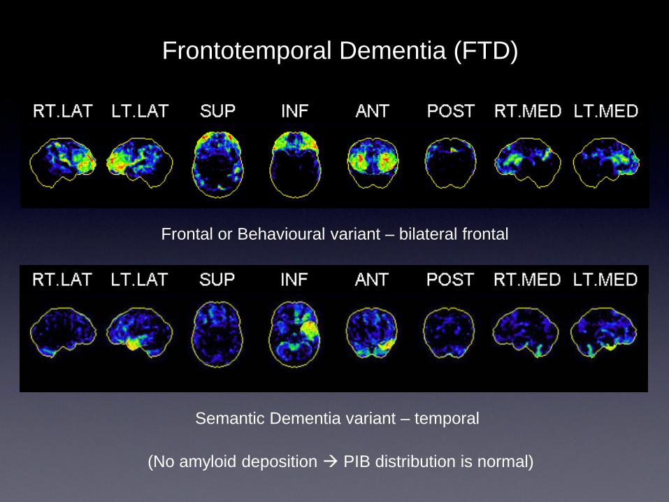
Frontotemporal Dementia (FTD)
Frontal or Behavioural variant – bilateral frontal
Semantic Dementia variant – temporal
(No amyloid deposition PIB distribution is normal)

Dementia with Lewy Bodies
Cingulate Island Sign (100% specific in 14 DLB & 10 AD pts)
Temporal, parietal & occipital hypometabolism (sensitive)
• diagnosis is based on progressive cognitive decline plus visual hallucinations, cognitive decline, gait or Parkinsonian symptoms
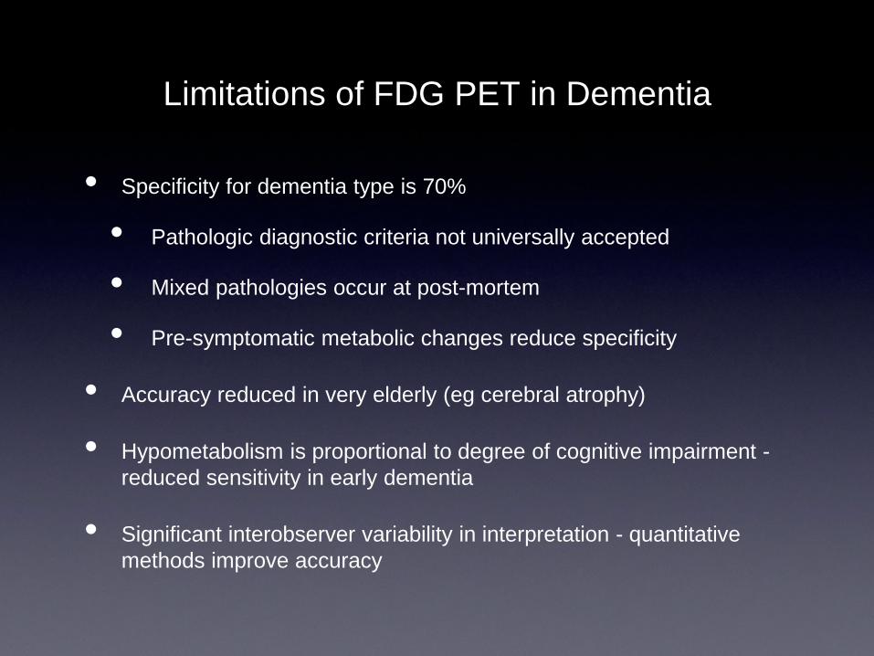
• Specificity for dementia type is 70%
• Pathologic diagnostic criteria not universally accepted
• Mixed pathologies occur at post-mortem
• Pre-symptomatic metabolic changes reduce specificity
• Accuracy reduced in very elderly (eg cerebral atrophy)
• Hypometabolism is proportional to degree of cognitive impairment - reduced sensitivity in early dementia
• Significant interobserver variability in interpretation - quantitative methods improve accuracy
Limitations of FDG PET in Dementia
Rowe et al Neurology 2007;68:1718–1725
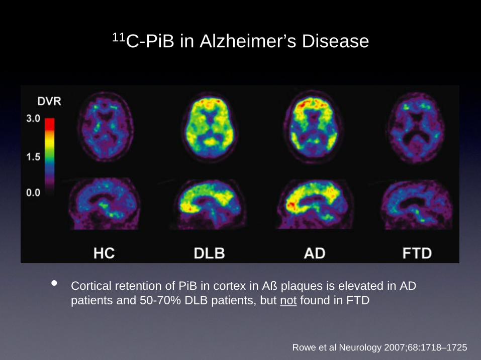
11C-PiB in Alzheimer’s Disease
• Cortical retention of PiB in cortex in Aß plaques is elevated in AD patients and 50-70% DLB patients, but not found in FTD
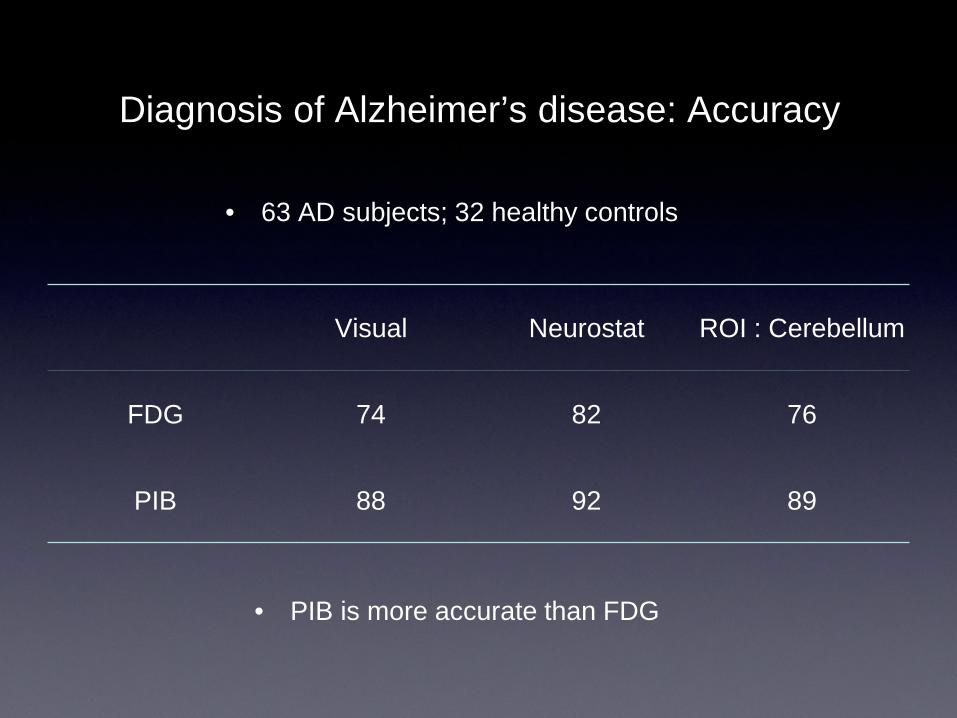
Visual Neurostat ROI : Cerebellum
FDG 74 82 76
PIB 88 92 89
• 63 AD subjects; 32 healthy controls
• PIB is more accurate than FDG
Diagnosis of Alzheimer’s disease: Accuracy
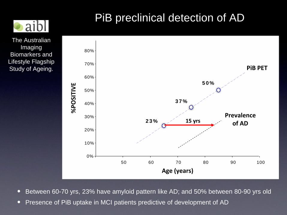
PiB preclinical detection of AD
The Australian Imaging
Biomarkers and Lifestyle Flagship Study of Ageing.
• Between 60-70 yrs, 23% have amyloid pattern like AD; and 50% between 80-90 yrs old
• Presence of PiB uptake in MCI patients predictive of development of AD
Age (years)
%PO
SITI
VE
PiB PET
Prevalence of AD
0%
10%
20%
30%
40%
50%
60%
70%
80%
50 70 80 90 100 60
15 yrs 23%
50%
37%

18F-labelled Amyloid Imaging Ligands
• three 18F-labelled amyloid imaging ligands are approved for clinical use in US
• Florbetapir (Amyvid™, Lilly)
• Flutemetamol (GE Healthcare)
• Florbetaben (Bayer)
• white matter uptake is greater for all 3 compared to 11C-PiB
• in AD patients, uptake similar to or less than uptake in white matter, in contrast to PiB, which is 30% greater in cortex than white matter

Rowe et al J Nucl Med 2011; 52:1733–1740
• 11C-PiB, 18F-florbetaben, 18F-florbetapir, and 18F-flutemetamol images of healthy subjects (HC) and Alzheimer’s disease (AD) patients
• Left: negative for brain amyloid (distinctive pattern of retention in white matter)
• Right: positive scans show uptake in cortical gray matter which obscures normal white matter pattern and binding extends to outer edge of brain
Comparison of 18F-labelled amyloid tracers HC AD

Development of Evidence for 18F-labelled Amyloid Imaging Ligands

TAU PET Imaging
SUVR
2.0
1.0
• 70 y/o MCI • MMSE = 28 • PiB +ve
2.0
1.4
SUVR
AT8 Histochemistry
Braak 2006

0
0 VMAT
VMAT
0
PET VMAT Imaging
• Vesicular Monoamine Transporters (VMAT) are found in the terminals of dopaminergic, serotonergic and noradrenergic neurones
• Like the Dopamine transporter (DAT), VMAT are reduced with loss of nigrostriatal neurones in disorders such as Parkinson’s disease and diffuse lewy body disease
F-18 dihydrotetrabenazine (AV-133) PET
HC PD AD DLB

Future of Diagnosis & Treatment of AD
Subjective memory complaint
Abnormal memory on objective testing
Tailored Imaging
Biomakers
Early treatment to reduce ß-Amyloid
MRI
FDG
ß-Amyloid
DAT / VMAT

PET in Dementia Case Studies

• 59 year old businessman with mild memory concern volunteered for a study of aging
• Found to have low scores on psychometric tests (-1.5 s.d. below mean in most areas), MMSE 29/30
• No other symptoms
• Normal neurological exam
• No family history of dementia
• FDG PET performed
Case 8 Case 8


What does the FDG PET scan suggest?
A) no evidence of a neurodegenerative disease
B) Alzheimer’s disease
C) Dementia with Lewy Bodies
D) Frontotemporal Dementia
E) Vascular disease
- correct
Case 8

PiB PET and MRI Case 9

Which statement is correct?
A) The PiB scan shows extensive cortical amyloid deposition
B) The MRI shows hippocampal atrophy
C) Amyloid is not present in Dementia with Lewy Bodies
D) MRI shows marked occipital lobe atrophy in DLB
E) Dopamine transporter imaging reliably distinguishes DLB from AD
- correct
Case 9

• 53 year old female with 18 months of worsening personality change, repetitive behaviours, marked reduction in speech
• Neuropsychology identified language and executive dysfunction worse than memory impairment
Case 10


What is the diagnosis?
A) Normal study
B) Frontotemporal dementia
C) Frontal variant of Alzheimer’s disease
D) Schizophrenia
E) Progressive supranuclear palsy
- correct
Case 10

PiB

Acknowledgements
• Prof. Christopher Rowe
• Dr. Sam Berlangieri
• A/Prof. Sze Ting Lee
• Dr. Aurora Poon
• Staff and Colleagues of Department of Molecular Imaging and Therapy, Department of Medical Oncology, Olivia Newton-John Cancer Research Institute, and Department of Neurology, Austin Health, Melbourne, Australia
• Patients and their families
Top Related