


Languages
Pages
Legal


RENAL CASE REPORT
1
Renal Case Report
Penina Langer
Department of Family, Nutrition, and Exercise Sciences, Queens College (CUNY)
FNES 366: Medical Nutrition Therapy
Professor Allison Charney, MSEd, RD, CDE, CDN
October 21, 2019
RENAL CASE REPORT
2
Pathophysiology
1. Describe the 4 basic pathophysiological functions of the kidneys. (4)
The four basic pathophysiological functions of the kidneys are to remove waste products
from the body, to regulate red blood cell production and blood formation, to control the
body’s chemical and fluid balance, and to release the hormones that regulate blood
pressure (National Kidney Institute, 2017).
The kidneys main function is to maintain the body’s chemical and fluid balance. Healthy
kidneys filter ~1600 L/day of blood and produce 180 L of ultrafiltrate, which is
ultimately changed into the 1.5 L of urine excreted a day. Each kidney has about 1
million nephrons, which consists of a glomerulus connected to a series of tubules. Each
nephron contributes to the final urine. If part of a nephron is destroyed, the nephron is no
longer functioning. The glomerulus produces the ultrafiltrate, which is a mostly passive
process. The tubules reabsorb most of the components that make up the ultrafiltrate,
which is a mostly active process. The tubule produces the final urine, which is funneled
into collective tubules and the renal pelvis, which goes to a ureter that carries urine into
the bladder. The kidneys are able to regulate water homeostasis. Water excretion is
regulated by vasopressin, an antidiuretic hormone. The amount of water in the body
controls how much vasopressin is secreted (Wilkens et al., 2017).
Another pathophysiological function of the kidneys is to secrete waste. Renal function is
the ability of the kidney to eliminate nitrogenous waste. Nitrogenous waste is the end
product of protein metabolism that is excreted in the urine. This contains urea, uric acid,
creatinine and ammonia. On average, urine production is 1.5 L/day. A minimum of 500
ml of urine/day needs to be eliminated to get rid daily waste (Wilkens et al., 2017).

RENAL CASE REPORT
3
The kidneys also have the pathophysiological function of regulating red blood cell
production by secreting the hormone erythropoietin, which stimulates the synthesis of red
blood cell formation and release in bone marrow (VanPutte et al., 2017).
The fourth pathophysiological function of the kidneys is to regulate blood pressure. The
kidneys perform the renin-angiotensin mechanism (Lewis, 2018), which controls blood
pressure. When blood volume is decreased, glomerulus cells release renin, which acts on
angiotensin in the plasma to form angiotensin I, which is converted to angiotensin II.
Angiotensin II is a vasoconstrictor and a stimulus of aldosterone secretion. Because of
this, sodium and fluid are reabsorbed, causing blood pressure to return to normal
(Wilkens et al., 2017).
2. List the primary diseases/conditions that most commonly lead to chronic kidney
disease (CKD)? Explain the role of diabetes in the development of CKD. (4)
The primary diseases/conditions that most commonly lead to chronic kidney disease
(CKD) are diabetes, hypertension and glomerulonephritis (Wilkens et al., 2017). Other
conditions that can lead to CKD are lupus, inherited diseases such as polycystic kidney
disease, malformations that happen to a baby in utero, and repeated urinary infections
(National Kidney Institute, 2019). Kidney damage caused by diabetes happens slowly.
High blood glucose can damage the blood vessels in the kidneys. These blood vessels
don’t work as well when damaged because they cannot properly clean your blood,
causing the body to retain more salt and water than usual, which can result in weight gain
and swelling. There can also be protein buildup in your urine and a buildup of waste in
your blood. Diabetes can also cause nerve damage, which can make urinating more

RENAL CASE REPORT
4
difficult. When the bladder is full it can cause pressure to back up and damage the
kidneys and can also cause an infection from rapid bacteria growth in urine with a high
sugar level (National Kidney Institute, 2017). Hypertension is commonly developed in
those with diabetes, which can damage the kidneys as well. Preventing diabetes related
CKD is possible by having healthy lifestyle habits and taking medication to help you
reach your blood pressure and blood glucose goals (National Institute of Diabetes and
Digestive and Kidney Diseases, 2017).
3. State each stage of CKD, basic descriptions and GFR (chart format). From your
reading of Mrs. Joaquin’s history and physical, what signs and symptoms did she
have that correlate with her chronic kidney disease? (4)
Stage of CKD Basic Description GFR
Stage 1 There is kidney damage;
kidney function is normal. 90-130 ml/min
Stage 2 Kidney function is mildly
decreased. 60-89 ml/min
Stage 3 Kidney function is
moderately decreased. 30-59 ml/min
Stage 4 Kidney function is severely
decreased. 15-29 ml/min
Stage 5
There is kidney failure,
which requires dialysis,
transplantation or medical
management.
< 15 ml/min
(Wilkens et al., 2017).
From my reading of Mrs. Joaquin’s chart, the signs and symptoms she had that correlate
with her CKD are: fatigue, the inability to urinate, a 4 kg weight gain, edema in
extremities, face and eyes, she’s Native American, has type 2 diabetes mellitus and
doesn’t take her medication, has a declining GFR (4 ml/min), is obese, has increasing

RENAL CASE REPORT
5
urea and creatinine concentrations, elevated serum phosphate, muscle weakness, and
hypertension (Nahikian Nelms, 2017).
4. In addition to the possibility of a kidney transplant, medical treatment options for
Stage 5 CKD (renal failure / ESRD) include HD and PD. Define hemodialysis and
peritoneal dialysis (CAPD and CCPD). Which of Mrs. Joaquin’s symptoms would
you expect to begin to improve when she starts dialysis? (4)
Hemodialysis (HD) is when blood is removed from the body and pumped by a machine
outside the body into a dialyzer/an artificial kidney. Metabolic waste products are filtered
out of the blood by the dialyzer, and the dialyzer then returns the purified blood to the
person’s body. The amount of fluid returned can be adjusted. Doctors usually create an
arteriovenous fistula in patients on hemodialysis to make long-term access to the blood
stream easier. The radial artery is usually joined with the cephalic vein in the forearm,
resulting in the cephalic vein enlarging and blood flow increasing, which allows for
repeated puncture with a needle. If a fistula can’t be made, a surgeon can create a graft in
the patient’s arm. A technician will place a needle into a person’s fistula or graft to let the
blood to be removed for cleaning (Hechanova, 2017).
Peritoneal dialysis (PD) is when the peritoneum, a membrane lining the abdomen and
covering the abdominal organs, acts as a filter. The peritoneum has a large surface area
and a lot of blood vessels. Matter from the blood can pass through the peritoneum into
the abdominal cavity easily. In PD, dialysate is injected into a peritoneal space in the
abdomen through a catheter inserted into the abdominal wall. The dialysate is left in the
abdomen for a while to allow waste products from the blood to slowly go into it; it is then
RENAL CASE REPORT
6
drained out and replaced with fresh dialysate. Dialysate is done through a catheter, which
can be temporary or permanent through surgery (Hechanova, 2017). There are two types
of peritoneal dialysis- continuous ambulatory peritoneal dialysis (CAPD) and continuous
cycler-assisted peritoneal dialysis (CCPD). CAPD does not use a machine to administer
dialysis; the exchanges are done manually. Each exchange takes 30-40 minutes to
administer, and you leave the dialysate in your stomach for 4-6 hours. The solution is
usually changed four times a day and you sleep with it in your stomach at night (NIDDK,
2018). CCPD is an automated peritoneal dialysis, meaning a machine does the exchanges
while you’re asleep. A machine, called a cycler, fills and empties your stomach 3-5 times
throughout the night, and you have fresh dialysate in your stomach in the morning. You
can leave the dialysate in your stomach all day or do a manual exchange once during the
day. Both CAPD and CCPD can be done anywhere you wish; you do not need to go to a
dialysis center (NIDDK, 2018).
Once Mrs. Joaquin begins hemodialysis, a few of her symptoms will start to improve.
She should have an increased energy level and her anorexia should go away. Her edema
should reduce significantly since HD reduces salt and fluid buildup. She should also
return to her dry weight once beginning HD, which will help control her blood pressure
(NIDDK, 2018).

RENAL CASE REPORT
7
Assessment
5. Calculate Mrs. Joaquin’s BMI based on current body weight on admission.
Calculate her BMI based on dry weight (UBW). How does edema affect
interpretation?
The formula for BMI is: 𝐵𝑀𝐼 =𝑤𝑒𝑖𝑔ℎ𝑡 (𝑙𝑏𝑠)
(ℎ𝑒𝑖𝑔ℎ𝑡 (𝑖𝑛))2 𝑥 703 (Demarest-Litchford, 2017)
BMI based on current body weight on admission: 𝐵𝑀𝐼 = 170
(60)2 𝑥 703 = 33.2
BMI based on dry weight (UBW): 𝐵𝑀𝐼 = 161
(60)2 𝑥 703 = 31.4
Edema affects interpretation of BMI because the accumulation of fluid causes a
significant increase in weight, which causes an increase in BMI, which is not an accurate
representation of the patient’s actual BMI. In both cases, Mrs. Joaquin is obese so the
numbers don’t matter as much, but in a patient with a different weight edema can be the
difference between a normal BMI/overweight or between overweight/obese, etc. which
can make a difference in treatment of the patient. When someone has edema, it is difficult
to use weight alone to assess overall nutrition status (Demarest-Litchford, 2017).
Define edema-free weight. State the equation that can be used to calculate the
edema-free adjusted body weight as per NFK KDOQI guidelines. (4)
Edema-free weight is the actual body weight of a patient without the fluid accumulation.
This body weight should be used for assessing or prescribing protein and energy intake.
For patients on HD, this should be obtained after dialysis. For patients on PD, this should
be obtained after the dialysate is drained (Bailie et al., 2007).

RENAL CASE REPORT
8
The equation used to calculate the edema-free adjusted body weight as per NKF KDOQI
guidelines is: 𝑎𝐵𝑊𝑒𝑓 = 𝐵𝑊𝑒𝑓 + [(𝑆𝐵𝑊 − 𝐵𝑊𝑒𝑓) 𝑥 0.25] (National Kidney Foundation,
n.d.).
6. What are the energy requirements for CKD (adults not on dialysis and adults on
dialysis)? Calculate what Mrs. Joaquin’s energy needs will be once she begins
hemodialysis. (4)
The energy requirement for adults with CKD not dialysis is 30-35 kcal/kg IBW (Wilkens
et al., 2017). The energy requirement for adults on hemodialysis is 35 kcal/kg IBW
(Wilkens et al., 2017). The energy requirement for adults on peritoneal dialysis is 30-35
kcal/kg IBW (Wilkens et al., 2017). Once Mrs. Joaquin begins hemodialysis, her energy
needs will be: 35 𝑘𝑐𝑎𝑙/𝑘𝑔 𝑥 𝐼𝐵𝑊 (Wilkens et al., 2017) = 35 𝑘𝑐𝑎𝑙 𝑥 45.5 = 1592.5 kcal.
I would round this to 1600 kcal because it is a simpler number for Mrs. Joaquin to aim
for.
For stages 1 and 2 CKD predialysis, stages 3 and 4 CKD predialysis, patients state
the protein requirements and rationale in chart format. Calculate Mrs. Joaquin’s
protein needs on dialysis. (4)
Stages CKD Protein Requirement Rationale
1 and 2 CKD predialysis 0.8 g/kg with 60% HBV
(Wilkens et al., 2017)
Studies have shown that
dietary protein increases
glomerular pressure and
leads to quicker loss of
renal function, so protein
restriction may slow this
down. The NIDDKD
recommends that those
with a GFR greater than 55
ml/min have 0.8 g/kg/day
(Wilkens et al., 2017).

RENAL CASE REPORT
9
3 and 4 CKD predialysis 0.6 g/kg with 60% HBV
(Wilkens et al., 2017)
As mentioned above,
studies have shown that
decreasing protein intake
may decrease the rate of
progression of CKD. The
NIDDKD recommends
that those with a GFR of
25-55 ml/min have 0.6
g/kg/day (Wilkens et al.,
2017).
Hemodialysis 1.2 g/kg IBW
(Wilkens et al., 2017)
Protein recommendations
for patients on dialysis are
higher than for those not
on dialysis. Hemodialysis
patients require increased
protein because of losses
through the artificial
kidney membrane and
blood loss (Karupaiah,
2018).
Peritoneal dialysis
1.2 – 1.5 g/kg BW, at least
50% HBV
(Wilkens et al., 2017)
Protein intake needs to be
increased because dialysis
is a drain on the body.
Patients with PD can have
protein losses of 20-
30g/day (Wilkens et al.,
2017).
Once she begins dialysis, Mrs. Joaquin will have a protein requirement of 1.2 g/kg IBW
(Wilkens et al., 2017). Her needs will be: 1.2𝑔
𝑘𝑔 𝑥 𝐼𝐵𝑊 = 1.2
𝑔
𝑘𝑔 𝑥 45.5 = 54.6 𝑔. I
would round her needs to 55 g of protein/day to make it a round number, which is simpler
to keep track of.
7. Mrs. Joaquin has a PO4 restriction. Why? What foods have the highest levels of
phosphorus? (4)
Mrs. Joaquin has a PO4 restriction. Normally, excess phosphate (~99%) is excreted in the
urine. When GFR decreases, phosphorus is retained in the plasma. Patients will gain

RENAL CASE REPORT
10
about half of the phosphate they consume since phosphate is not easily removed by
dialysis because of its large molecular weight (Wilkens et al., 2017). An overload of
phosphate and hyperphosphatemia are risk factors for cardiovascular mortality, vascular
calcification, left ventricular hypertrophym and progression of CKD (Gonzalez-Parra et
al., 2013). High protein-foods have the highest levels of phosphorus. These foods
include milk products, dried beans, nuts and meats. Phosphorus in animal foods is more
readily absorbed than phosphorus in plant foods (National Kidney Institute, 2019).
Highly processed foods contain a lot of phosphate additives that are about 100%
absorbed, compared to naturally occurring phosphorus, which is about 60% absorbed.
This makes it more challenging to follow a low phosphorus diet (Wilkens et al., 2017).
Common processed foods with high levels of phosphorus include canned and bottled
drinks, fast foods, enhanced meats and chocolate (National Kidney Institute, 2019).
8. Mrs. Joaquin is on a fluid restriction. What foods are considered to be fluids? What
fluid restriction is generally recommended for someone on hemodialysis? Is there a
standard guideline for maximum fluid gain between dialysis visits? If a patient must
follow a fluid restriction, what can be done to help reduce his or her thirst? (4)
Foods that are considered to be fluids are anything that is liquid at room temperature (ex:
coffee, soup, ice cream) (Wilkens et al., 2017). The generally recommended fluid
restriction for someone on hemodialysis is 750 ml/day, plus the amount equal to urine
output (Wilkens et al., 2017). The standard guideline for maximum fluid gain between
dialysis visits is 4-5 pounds or 2-3 kg. The goal is to have a fluid gain of less than 4%
body weight (Wilkens et al., 2017). If a patient must follow a fluid restriction, the best
RENAL CASE REPORT
11
way to reduce their thirst is to have them follow a low-sodium diet, because eating salt
makes us thirsty. Other ways to deal with thirst without drinking fluids include sucking
on ice chips, eating sour candies, having cold fruit, or even using artificial saliva
(Wilkens et al., 2017).
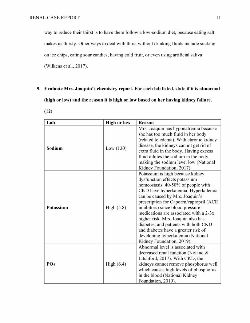
9. Evaluate Mrs. Joaquin’s chemistry report. For each lab listed, state if it is abnormal
(high or low) and the reason it is high or low based on her having kidney failure.
(12)
Lab High or low Reason
Sodium Low (130)
Mrs. Joaquin has hyponatremia because
she has too much fluid in her body
(related to edema). With chronic kidney
disease, the kidneys cannot get rid of
extra fluid in the body. Having excess
fluid dilutes the sodium in the body,
making the sodium level low (National
Kidney Foundation, 2017).
Potassium High (5.8)
Potassium is high because kidney
dysfunction effects potassium
homeostasis. 40-50% of people with
CKD have hyperkalemia. Hyperkalemia
can be caused by Mrs. Joaquin’s
prescription for Capoten/captopril (ACE
inhibitors) since blood pressure
medications are associated with a 2-3x
higher risk. Mrs. Joaquin also has
diabetes, and patients with both CKD
and diabetes have a greater risk of
developing hyperkalemia (National
Kidney Foundation, 2019).
PO4 High (6.4)
Abnormal level is associated with
decreased renal function (Noland &
Litchford, 2017). With CKD, the
kidneys cannot remove phosphorus well
which causes high levels of phosphorus
in the blood (National Kidney
Foundation, 2019).

RENAL CASE REPORT
12
Total CO2 High (32)
Kidneys help maintain normal total CO2
levels. Abnormal results are due to
kidney disease. Changes in total CO2
levels are due to Mrs. Joaquin’s fluid
retention (MedlinePlus, 2019)
Albumin Low (3.3)
Low albumin is an indicator of kidney
disease (MedlinePlus, 2019). It can be
caused by not getting enough protein or
calories in the diet (National Kidney
Foundation, 2018). It can also be due to
metabolic acidosis (DaVita, n.d.)
Protein Low (5.9)
There is low protein in Mrs. Joaquin’s
blood because protein is being excreted
in her urine – she has high urine protein
levels (National Kidney Foundation,
2017).
BUN High (69)
Renal failure (Sanna-Gouin, 2018);
BUN is increased due to poor filtration
of urea (Wilkens et al., 2017). When
kidney function slows down, BUN
levels rise (National Kidney Foundation,
2018) Once Mrs. Joaquin begins
dialysis, the excess urea nitrogen will be
removed from the blood (Wilkens et al.,
2017).
Creatinine High (12)
Creatinine, a waste product in the blood,
is high because it is usually removed
from the blood by the kidneys. When
kidney function decreases, creatinine
levels increase (National Kidney
Foundation, 2018).
Calcium Low (8.2)
Normally, the kidneys turn vitamin D
into calcitriol, which increases calcium
absorption into the blood. With CKD,
patients are lacking this, and they are
normally on a low phosphorus diet,
which means they are not eating enough
calcium either (DaVita, n.d.). Both of
these factors contribute to low blood
calcium.
RBC, Hgb, Hct Low (3.1,
10.5, 33)
Low levels of RBC, Hgb and Hct are
symptoms of anemia (National Kidney
Foundation, 2018). We know that Mrs.
Joaquin has normochromic, normocytic
anemia, which is causing these levels to
be low.

RENAL CASE REPORT
13
Urine protein, glucose High (+2, +1)
Urine protein is high because they
kidneys are not filtering the blood
properly. The protein is being excreted
instead of kept in the blood (National
Kidney Foundation, 2018) – we see that
she has low blood protein levels. She
has high urine glucose because of her
uncontrolled diabetes (MedlinePlus,
2019).
GFR Low (4)
There is a low GFR because she is in
renal failure (National Kidney
Foundation, 2018).
All lab values were taken from Nahikian-Nelms (2018).
10. The following medications were prescribed for Mrs. Joaquin. Explain why each was
prescribed (the indications/mechanism) and describe any nutritional concerns and
dietary recommendations related to the medication. (10)
Medication
Indications
Nutritional Concerns
(Food/Nutrient – Drug
Interaction)
Capoten/ captopril
Prescribed to treat high
blood pressure. Also treats
kidney disease. Captopril is
an ACE inhibitor, which
means it causes blood to
flow more smoothly and
allows the heart to pump
blood more efficiently
(MedlinePlus, 2017).
Avoid salt substitutes containing
potassium. If prescribed a low-
sodium diet, follow carefully.
This medication may cause a
salty/metallic taste and a
decreased ability to taste
(MedlinePlus, 2017).
Erythropoietin
This medication is
prescribed to treat anemia
in patients with chronic
kidney failure. This
medication is an
erythropoiesis-stimulating
agent; it causes the bone
marrow to increase red
blood cell production
(MedlinePlus, 2019).
It’s important to try and control
your blood pressure and increase
iron levels so this medication
can work well. Erythropoietin
can cause nausea, vomiting,
weight loss and mouth sores
(MedlinePlus, 2019).

RENAL CASE REPORT
14
Sodium bicarbonate
This medication is
normally prescribed to
patients with metabolic
acidosis to make the blood
and urine less acidic
(MedlinePlus, 2017). A
small group of studies has
shown that taking sodium
bicarbonate with CKD can
help prevent CKD from
getting worse (National
Kidney Foundation, 2019).
Need to be careful when on a
low-sodium diet, as sodium
bicarbonate increases the amount
of sodium in your body. Sodium
bicarbonate can cause increased
thirst, stomach cramps and gas
(MedlinePlus, 2017).
Renal caps
This is a multivitamin
given to people on dialysis,
since it can affect your
vitamin needs. You might
have a poor appetite,
limited food options on a
kidney diet, medications
may change vitamin
absorption, and vitamins
can be lost during dialysis
treatment (DaVita, n.d.)
There are no nutritional concerns
with Renal Caps.
Renvela
Prescribed to control high
levels of phosphorus in
people with CKD who are
on dialysis. Renvela is a
phosphate binder, which
means that it binds
phosphorus from foods you
eat and prevents it from
being absorbed in the
blood stream (MedlinePlus,
2017)
Follow a low-phosphorus diet.
This medication may cause
diarrhea, nausea, vomiting,
stomach pain, gas, heartburn or
constipation (MedlinePlus,
2017).
Hectorol
This medication is
prescribed to patients
receiving dialysis. Hectorol
is a vitamin D analog,
meaning it helps the body
utilize calcium found in
foods/supplements by
regulating the body’s
production of parathyroid
hormone (MedlinePlus,
2016).
For this medication to work, you
need to be eating the right
amount of calcium. If eating
calcium is too difficult, a
supplement can be prescribed. A
low-phosphate diet may also be
needed. Hectorol may cause
heartburn, fluid retention, and
weight gain (MedlinePlus,
2016).

RENAL CASE REPORT
15
Glucophage
This medication is used to
treat type 2 diabetes.
Glucophage is a
biguanides; it helps control
the amount of glucose in
your blood, decreases the
amount of glucose you
absorb from food and the
amount of glucose made by
your liver. It also increases
your body’s response to
insulin (MedlinePlus,
2019).
A major concern is that
Glucophage will cause lactic
acidosis, which is dangerous in
those with kidney disease.
Make sure to follow diet and
exercise recommendations made
by a health care professional.
This medication can cause
diarrhea, bloating, stomach pain,
gas, indigestion, constipation,
metallic taste in mouth, heart
burn and muscle pain
(MedlinePlus, 2019).
Diagnosis
11. Choose two high-priority nutrition problems and complete a PES statement for
each. (4)
a. Excessive sodium intake NI-5.10.2 related to intake of foods high in sodium as
evidenced by diet recall, hypertension, and 3+ pitting edema in the extremities, face
and eyes
b. Decreased protein needs NI-5.3 related to renal dysfunction as evidenced by
increased phosphorus, BUN, creatinine and potassium, decreased GFR and 3+ pitting
edema
Intervention
12. Why is it recommended for patients to have at least 50% of their protein from
sources that have high biological value? (2)
It is recommended that patients have at least 50% of their protein from sources that have
high biological value because it allows for optimal protein use - the body is able to easily
digest it and use the amino acids (Wilkens et al., 2017).

RENAL CASE REPORT
16
13. Explain the reasons for the following components of Mrs. Joaquin’s medical
nutrition therapy: (12)
Nutrition Therapy Rationale
35 kcal/kg
Mrs. Joaquin is on a 35 kcal/kg diet because she is
starting hemodialysis, and patients on hemodialysis
need 35 kcal/kg IBW to meet their energy needs
(Wilkens et al., 2017).
1.2 g protein/kg
Mrs. Joaquin will need 1.2 g protein/kg once she starts
hemodialysis. Hemodialysis patients require increased
protein because of losses through the artificial kidney
membrane and blood loss (Karupaiah, 2018).
2 g K+
Patients on dialysis have a restricted potassium diet to
2 g K+/day (NCM, n.d.), compared to the average
American, who consumes 3-4g/day (Wilkens et al.,
2017). When the kidneys aren’t working properly, they
don’t remove enough potassium from the body which
can lead to a dangerous buildup of potassium in the
blood (National Kidney Foundation, 2017).
1 g phosphorus
Mrs. Joaquin’s phosphorus is limited to 1 g/day (AND
Evidence Analysis Library, n.d.) because renal patients
do not excrete enough phosphorus, which leads to a
harmful buildup in the blood (Wilkens et al., 2017).
2 g Na
Dialysis patients need a restricted sodium diet as excess
sodium intake can lead to thirst, fluid gain, and
hypertension. Patients on HD have both a sodium and
fluid restriction to allow for a weight gain of 4-5
pounds between dialysis (Wilkens et al., 2017).
1000 mL fluid + urine output
Fluid is limited to prevent large interdialytic fluid gains
(Wilkens et al., 2017). Normally, the kidneys balance
fluid in the body but cannot remove enough when on
dialysis. Limiting fluid (and salt) helps your body
maintain the right amount of fluid, which can prevent
swelling, discomfort, high blood pressure, shortness of
breath and heart problems (National Kidney
Foundation, 2018).

RENAL CASE REPORT
17
14. What resources (i.e. renal diet education guides) and counseling techniques (i.e.
motivational interviewing, others) would you use to help Mrs. Joaquin with her
diet? (4)
I would use nutrition counseling based on problem solving strategy (eNCPT C-2.4) to
work with Mrs. Joaquin and identify the barriers preventing her from following the diet
she was taught two years ago. I would also find out why she stopped taking her
prescribed hypoglycemic agent. I would then work with her to come up with a solution
for her to be able to follow the newly prescribed renal diet. We would create a meal plan
together based on foods that she likes, would be willing to eat, and would be willing to
prepare. This is essential since Mrs. Joaquin purchases and prepares her own food. I
would also provide her with renal diet education guides, (eNCPT E-1.1) which would
include handouts on sodium, potassium, phosphorus, fluid, and protein levels in different
foods. If I felt she would be receptive towards it, I would also educate Mrs. Joaquin on
the exchange lists so that she can switch up her diet daily. I would also encourage Mrs.
Joaquin to be more active throughout the day; she works as a secretary, so she is mostly
sedentary. It’s important for people with CKD to get in some physical activity as studies
have shown that physical activity may decrease the catabolic effects of protein restriction
and improve quality of life (AND Evidence Analysis Library, n.d.). Lastly, I would
transfer her nutrition care to the RD at the dialysis center (eNCPT RC-2.3). Having this
constant follow-up would hopefully be motivation for her to stick to her diet and improve
her health.
RENAL CASE REPORT
18
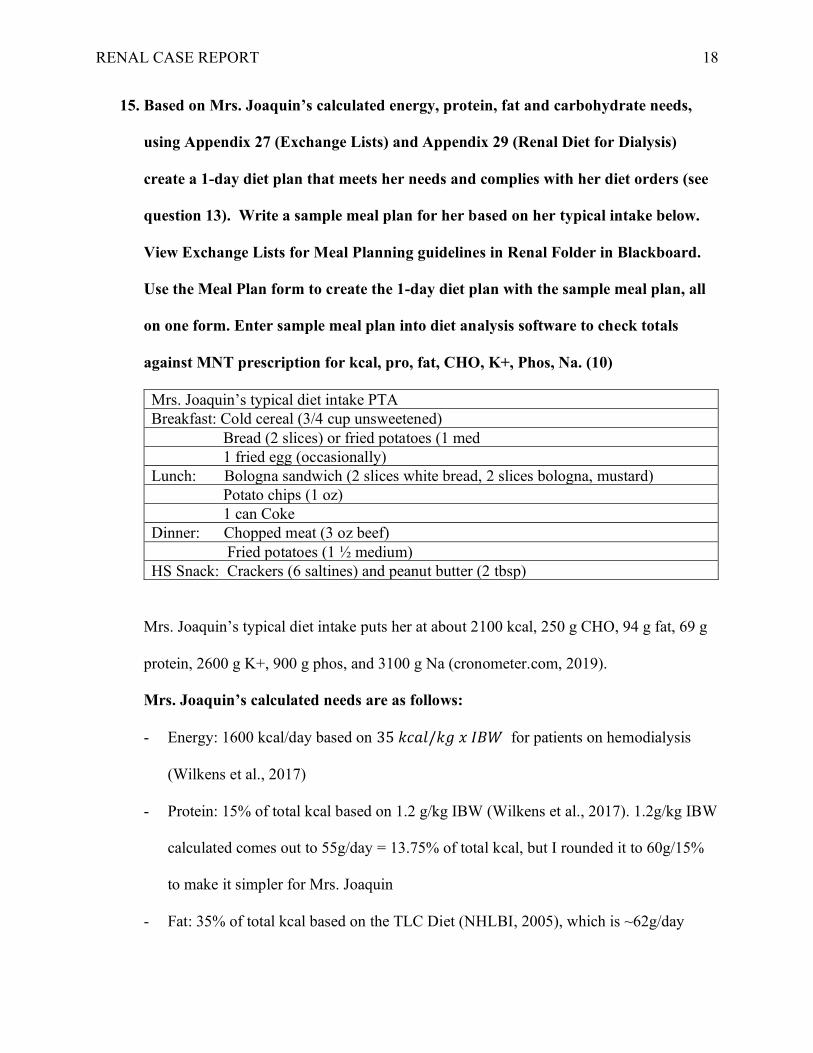
15. Based on Mrs. Joaquin’s calculated energy, protein, fat and carbohydrate needs,
using Appendix 27 (Exchange Lists) and Appendix 29 (Renal Diet for Dialysis)
create a 1-day diet plan that meets her needs and complies with her diet orders (see
question 13). Write a sample meal plan for her based on her typical intake below.
View Exchange Lists for Meal Planning guidelines in Renal Folder in Blackboard.
Use the Meal Plan form to create the 1-day diet plan with the sample meal plan, all
on one form. Enter sample meal plan into diet analysis software to check totals
against MNT prescription for kcal, pro, fat, CHO, K+, Phos, Na. (10)
Mrs. Joaquin’s typical diet intake PTA
Breakfast: Cold cereal (3/4 cup unsweetened)
Bread (2 slices) or fried potatoes (1 med
1 fried egg (occasionally)
Lunch: Bologna sandwich (2 slices white bread, 2 slices bologna, mustard)
Potato chips (1 oz)
1 can Coke
Dinner: Chopped meat (3 oz beef)
Fried potatoes (1 ½ medium)
HS Snack: Crackers (6 saltines) and peanut butter (2 tbsp)
Mrs. Joaquin’s typical diet intake puts her at about 2100 kcal, 250 g CHO, 94 g fat, 69 g
protein, 2600 g K+, 900 g phos, and 3100 g Na (cronometer.com, 2019).
Mrs. Joaquin’s calculated needs are as follows:
- Energy: 1600 kcal/day based on 35 𝑘𝑐𝑎𝑙/𝑘𝑔 𝑥 𝐼𝐵𝑊 for patients on hemodialysis
(Wilkens et al., 2017)
- Protein: 15% of total kcal based on 1.2 g/kg IBW (Wilkens et al., 2017). 1.2g/kg IBW
calculated comes out to 55g/day = 13.75% of total kcal, but I rounded it to 60g/15%
to make it simpler for Mrs. Joaquin
- Fat: 35% of total kcal based on the TLC Diet (NHLBI, 2005), which is ~62g/day

RENAL CASE REPORT
19
- Carbohydrates: 50% of total kcal (DaVita, n.d.), which is ~200g/day
Mrs. Joaquin’s macronutrient breakdown in her usual diet recall is pretty similar to the
calculated one- 47% carbohydrates, 13% protein, and 40% fat. She is having too much
sodium and potassium. The major discrepancy is that she is eating too many calories, so
even though her macronutrient breakdown is similar she is having too much protein,
carbs, and fat. The foods she includes in her diet need to be altered to include foods lower
in potassium and sodium.
SAMPLE MEAL PLAN:
EXCHANGE LISTS FOR MEAL PLANNING
Carbohydrate
200 g
800 kcal
50%
Protein
60 g
240 kcal
15%
Fat
62 g
560
kcal
35%
Total
kcal
=
1600
Sample Meal Plan
Group Exchanges Food Carbohydrate Protein Fat Kcal
Morning Meal
CHO Group 1 ¾ cup cold
cereal
15 3 1 80
Starch
Fruit 1 ¾ cup
blueberries
15 0 0 60
Vegetable
Milk .5 ½ cup fat-
free milk
6 4 0 50
Meat group 2 Hard-boiled
egg + egg
white
0 11 7 100
Fat group
Morning Snack
CHO Group
Starch

RENAL CASE REPORT
20
Fruit 2 1 cup
unsweetened
applesauce
30 0 0 120
Vegetable
Milk
Meat group
Fat group
Noon Meal
CHO Group 1 100 cal pack
of cookies
15 3 4 100
Starch 2 2 slices
bread
30 6 2 160
Fruit 1 1 ¼ cup
whole
strawberries
15 0 0 60
Vegetable
Milk
Meat group 1 1 oz bologna 0 7 8 100
Fat group 1 1 tbsp
reduced fat
mayo
0 0 5 45
Afternoon Snack
CHO Group
Starch
Fruit
Vegetable 1 1 cup raw
green beans
5 2 0 25
Milk
Meat group
Fat group 2 12 almonds 0 0 10 90
Evening Meal
CHO Group
Starch 2 ½ baked
potato
without skin
30 6 2 120
Fruit
Vegetable 1 1 cup mixed
veg with
corn and
peas
15 3 1 80
Milk
Meat group 2 2 oz
chopped
meat, lean
0 14 4 90
RENAL CASE REPORT
21
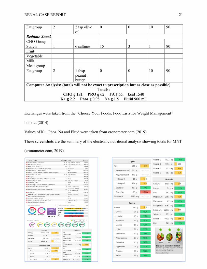
Fat group 2 2 tsp olive
oil
0 0 10 90
Bedtime Snack
CHO Group
Starch 1 6 saltines 15 3 1 80
Fruit
Vegetable
Milk
Meat group
Fat group 2 1 tbsp
peanut
butter
0 0 10 90
Computer Analysis: (totals will not be exact to prescription but as close as possible)
Totals:
CHO g 191 PRO g 62 FAT 65 kcal 1540
K+ g 2.2 Phos g 0.98 Na g 1.5 Fluid 900 mL
Exchanges were taken from the “Choose Your Foods: Food Lists for Weight Management”
booklet (2014).
Values of K+, Phos, Na and Fluid were taken from cronometer.com (2019).
These screenshots are the summary of the electronic nutritional analysis showing totals for MNT
(cronometer.com, 2019).

RENAL CASE REPORT
22
The computer analysis, cronometer.com (2019) was very similar to the exchange list. It showed
her eating 20 less calories than calculated based on the exchange list, and 3 g for carbohydrates,
protein and fat. Overall, I would say that it was very accurate.
Monitoring & Evaluation and Documentation
16. Write an initial ADIME note for your consultation with Mrs. Joaquin. Use the
ADIME form provided with the renal case questions. (10)
A - Assessment
S - Subjective
Chief Complaint: anorexia, N/V; 4-kg weight gain in the past 2 weeks; edema in extremities, face and eyes; inability to urinate
UBW: 161 lbs
Weight change: gain / loss
Appetite: anorexia
Chewing / swallowing problem / sore mouth none
Nausea / vomiting / diarrhea / constipation
Food intolerance / allergies: none
Diet prior to admit: diet recall was obtained
Nutritional supplement: none
Vitamins / herbs: none
Food preparation: self
Factors affecting food intake: anorexia, N/V
Social / cultural / religious / financial
Native American, married with 1 child, works 9-5, Catholic
Other:1-2 12oz beers daily, sedentary job
O - Objective
Current Diet Order:
35 kcal/kg, 2 g K, 1 g Phos, 2 g Na, 1000 mL fluid + urine output per day
(prior was low simple sugar, 0.8 g protein/kg, 2-3g Na)
Medical Diagnosis:
CKD (renal failure)
T2DM
Pertinent Medical History:
Poorly compliant with T2DM treatment
Infant weighed 10 lbs at birth
Stopped taking prescribed hypoglycemic agent
Never filled prescription for anti-hypertensive medication
Progressive decompensation of kidney function
Nutrition Focused Physical Assessment – General (Appendix 21, p 973 Krause text)
General survey: obese
Skin: dry and yellowish brown
Eyes: normal
Nose: noncontributory
Neck: supple
Head: normocephalic, equal carotid pulses
Chest/lungs: patient breathes with poor effort
Heart: regular rate/rhythm; I/VI systolic ejection murmur
Abdomen: rounded, obese
Musculoskeletal: weakness
Neurologic: oriented, mild asterixis

RENAL CASE REPORT
23
Age:
24
Gender: Male
Female •
Ht:
5’0” Wt: Admit •
170 lbs Current
DBW:
100 lbs (100 + 5(0)) 10%
BMI:
33.2 (𝟏𝟕𝟎
(𝟔𝟎)𝟐 𝒙 𝟕𝟎𝟑)
% UBW: 106% (170/161 x 100)
% wt : 6% (161-170/161 x 100) % DBW: 170% (170/100 x 100) Other: obese class I
(Calculations based on weight
at admission)
Nutritionally Relevant Laboratory Data:
Sodium – 130
Potassium – 5.8
PO4 – 6.4
Total CO2 – 32
Albumin – 3.3
Protein – 5.9
BUN – 69
Creatinine – 12
Calcium – 8.2
RBC – 3.1
Hgb – 10.5
Hct – 33
Urine protein – +2
Urine glucose – +1
GFR – 4
Drug Nutrient Interaction:
Capoten/captopril (ACE inhibitor) – avoid salt substitutes with K, follow low sodium diet; may cause salty/metallic taste in mouth and
decreased ability to taste
Erythropoietin (ESA) – may cause N/V, weight loss
Sodium bicarbonate (antacid) – follow low sodium diet; may cause increased thirst, cramps, gas
Renal caps (multivitamin)
Renvela (phosphate binder) – follow low phos diet; may cause N/V/D/C, stomach pain, gas, heartburn
Hectorol (vitamin D analog) – control calcium intake, low phos diet; may cause heartburn, fluid retention, weight gain
Glucophage (biguanides) – may cause C/D, stomach pain, gas, indigestion, metallic taste, heartburn
Estimated Energy Need:
1600 kcal / day
Based on: 35 kcal/kg IBW
Estimated Protein Need:
55 g/day
Based on: 1.2 g/kg IBW
Estimated Fluid Need:
1000 ml + urine output / day
Based on: HD fluid restriction
A - Assessment (A)
Nutrition Focused Physical Assessment – to support dx of malnutrition (see Appendix 21, p 974-975 Krause text)
1) % of estimated energy intake: < 75% < 50% other – know intake is poor due to anorexia but don’t know for how long or
how much of usual dietary she is eating each day, diet recall is ~2100 kcal/day
2) Weight loss interpretation: 0 % Time undetermined due to edema
Physical findings: for each below, choose Mild, Moderate or Severe (based on text description) or None
3) Body fat: none
4) Muscle mass: none
5) Fluid accumulation: severe (3+ edema)
6) Reduced grip strength: none
Malnutrition Assessment: Minimum of 2 of the 6 characteristics above is indicative of
______Severe OR _______Non-severe Malnutrition of _____Social/Environmental OR _____Chronic Illness OR __1___Acute Illness or Injury
There is no malnutrition noted because she only has one criteria (we do not know that she has reduced grip strength even though she has
muscle weakness, and we do not know exactly how poor her intake is over what period of time, and we don’t know if she has lost weight
because of the edema. There is also nothing noted about body fat or muscle mass)
Nutrition Diagnosis (D)

RENAL CASE REPORT
24
State no more than 2 priority Nutrition Diagnosis statements in PES Format. Use Nutrition Diagnosis Terminology sheet
ND Term (Problem) related to (Etiology) as evidenced by (Signs and Symptoms):
1. Excessive sodium intake NI-5.10.2 related to intake of foods high in sodium as evidenced by diet recall, hypertension, and 3+
pitting edema in the extremities, face and eyes
2. Decreased protein needs NI-5.3 related to renal dysfunction as evidenced by increased phosphorus, BUN, creatinine and
potassium, decreased GFR and 3+ pitting edema
Nutrition Intervention (I)
P - Plan
List Nutrition Interventions. Use Nutrition Intervention Terminology sheet. (The intervention(s) must address the problems (diagnoses).
ND-1.2.3.3 Decreased protein diet
ND-1.2.11.7.2 Decreased sodium diet
E-1.1 Content related nutrition education (renal diet education, drug-nutrient interactions for medications listed above)
C-2.4 Nutrition counseling based on problem solving strategy (address why previously prescribed T2DM diet was not followed)
RC-2.3 Discharge and transfer of care from nutrition professional to another nutrition professional (consistent follow-ups with dialysis
center RD)
Goal(s) (SMART):
1. Patient will substitute high sodium foods with unsalted alternatives by follow-up with dialysis center RD (within 1 week)
2. Patient will decrease protein intake by reducing amount of meat eaten at dinner by 1 oz over the next 2 weeks
Plan for Monitoring and Evaluation (M E)
List indicators for monitoring and evaluation. Use Nutrition Assessment and Monitoring & Evaluation sheets. (Upon follow-up, the plan for
monitoring would indicate if interventions are addressing the problems).
FH-1.5.3.2 Measured protein intake
FH-1.6.2.2 Measured mineral intake, sodium (13190)
FH-2.1.1 Diet order (renal)
FH-5.1.1 Self-reported adherence score
BD-1.2.1 Electrolyte and renal profile
BD-1.12 Urine profile
Signature: Penina Langer Date: 10/21/2019

RENAL CASE REPORT
25
References
Academy of Nutrition and Dietetics, & American Diabetes Association. (2014). Choose Your
Foods: Food Lists for Weight Management.
Albumin Blood Test: MedlinePlus Lab Test Information. (2019, October 2). Retrieved from
https://medlineplus.gov/lab-tests/albumin-blood-test/.
Bailie, G. R., Johnson, C. A., Mason, N. A., & St. Peter, W. L. (Eds.). (2007). KDOQI Clinical
Practice Guidelines and Clinical Practice Recommendations for Diabetes and Chronic
Kidney Disease. American Journal of Kidney Diseases, 49(2). doi:
10.1053/j.ajkd.2006.12.005
Calcium and Chronic Kidney Disease. (n.d.). Retrieved from https://www.davita.com/diet-
nutrition/articles/basics/calcium-and-chronic-kidney-disease.
Captopril: MedlinePlus Drug Information. (2017, July 15). Retrieved from
https://medlineplus.gov/druginfo/meds/a682823.html.
Carbohydrates and the kidney diet. (n.d.). Retrieved from https://www.davita.com/diet-
nutrition/articles/basics/carbohydrates-and-the-kidney-diet.
Chronic Kidney Disease. (n.d.). Retrieved from
https://www.andeal.org/topic.cfm?menu=5303&pcat=3927&cat=3929.
CO2 Blood Test: MedlinePlus Medical Encyclopedia. (2019, April 29). Retrieved from
https://medlineplus.gov/ency/article/003469.htm.
Demarest Litchford, M. (2017). Clinical: Biochemical, Physical, and Functional Assessment.
In Krause's Food & The Nutrition Care Process (14th ed., pp. 98–121). Elsevier.
Diabetes - A Major Risk Factor for Kidney Disease. (2017, February 3). Retrieved October 13,
2019, from https://www.kidney.org/atoz/content/diabetes.

RENAL CASE REPORT
26
Diabetic Kidney Disease. (2017, February 1). Retrieved October 13, 2019, from
https://www.niddk.nih.gov/health-information/diabetes/overview/preventing-
problems/diabetic-kidney-disease#how.
Doxercalciferol Injection : MedlinePlus Drug Information. (2016, November 15). Retrieved from
https://medlineplus.gov/druginfo/meds/a616044.html.
Epoetin Alfa, Injection: MedlinePlus Drug Information. (2019, September 15). Retrieved from
https://medlineplus.gov/druginfo/meds/a692034.html.
Facts About Chronic Kidney Disease. (2019, July 19). Retrieved October 12, 2019, from
https://www.kidney.org/atoz/content/about-chronic-kidney-disease#causes.
Facts About High Potassium in Patients with Kidney Disease. (2019, July 19). Retrieved October
18, 2019, from https://www.kidney.org/atoz/content/hyperkalemia/facts.
Fluid Overload in a Dialysis Patient. (2018, March 30). Retrieved from
https://www.kidney.org/atoz/content/fluid-overload-dialysis-patient.
Glucose in Urine Test: MedlinePlus Lab Test Information. (2019, April 15). Retrieved from
https://medlineplus.gov/lab-tests/glucose-in-urine-test/.
González-Parra, E., Gracia-Iguacel, C., Egido, J., & Ortiz, A. (2013). Phosphorus and Nutrition
in Chronic Kidney Disease. Clinical Nutrition, 141–153. doi: 10.1201/b16308-11
Hechanova, L. A. (2017, April). Dialysis - Kidney and Urinary Tract Disorders. Retrieved
October 18, 2019, from https://www.merckmanuals.com/home/kidney-and-urinary-tract-
disorders/dialysis/dialysis#v764296.
Hemodialysis. (2018, January). Retrieved from https://www.niddk.nih.gov/health-
information/kidney-disease/kidney-failure/hemodialysis#working.
RENAL CASE REPORT
27
High Potassium (Hyperkalemia). (2017, December 20). Retrieved from
https://www.kidney.org/atoz/content/hyperkalemia.
How High Blood Pressure Can Lead to Kidney Damage or Failure. (2016, October 31).
Retrieved from https://www.heart.org/en/health-topics/high-blood-pressure/health-
threats-from-high-blood-pressure/how-high-blood-pressure-can-lead-to-kidney-damage-
or-failure.
How Your Kidneys Work. (2017, May 10). Retrieved October 11, 2019, from
https://www.kidney.org/kidneydisease/howkidneyswrk.
Hyponatremia. (2017, May 17). Retrieved October 18, 2019, from
https://www.kidney.org/atoz/content/hyponatremia.
Intake (NI). (n.d.). Retrieved from https://www.ncpro.org/pubs/encpt-en/page-037.
Karupaiah, T. (2018). Kidney Disease. In The Essential Pocket Guide for Clinical Nutrition (2nd
ed., pp. 385–412). Wolters Kluwer.
Lewis, J. L. (2018, September). Overview of Disorders of Fluid Volume - Endocrine and
Metabolic Disorders. Retrieved from
https://www.merckmanuals.com/professional/endocrine-and-metabolic-disorders/fluid-
metabolism/overview-of-disorders-of-fluid-volume?query=renin angiotensin.
Mahan, L. K., MS, RDN, CD, & Raymond, J. L., MS, RDN, CD, CSG. (2017). Krause's Food &
The Nutrition Care Process (14th ed). Elsevier.
Metabolic Acidosis. (2019, August 21). Retrieved from
https://www.kidney.org/atoz/content/metabolic-acidosis.
Metformin: MedlinePlus Drug Information. (2019, July 15). Retrieved from
https://medlineplus.gov/druginfo/meds/a696005.html.

RENAL CASE REPORT
28
Nahikian Nelms, M. (2017). Chronic Kidney Disease (CKD) Treated with Dialysis. In Medical
Nutrition Therapy: A Case Study Approach (5th ed., pp. 217–229). Cengage Learning.
NCP Step 2: Nutrition Diagnosis. (n.d.). Retrieved from https://www.ncpro.org/pubs/encpt-
en/page-036.
NCP Step 3: Nutrition Intervention. (n.d.). Retrieved from https://www.ncpro.org/pubs/encpt-
en/page-036.
NCP Step 4: Nutrition Monitoring and Evaluation. (n.d.). Retrieved from
https://www.ncpro.org/pubs/encpt-en/page-015a.
NKF KDOQI GUIDELINES. (n.d.). Retrieved from
https://kidneyfoundation.cachefly.net/professionals/KDOQI/guidelines_nutrition/nut_a12
.html.
Noland, D., & Litchford, M. (2017). APPENDIX 22: Laboratory Values for Nutritional
Assessment and Monitoring. In Krause's Food & The Nutrition Care Process (14th ed.,
pp. 981–1001). Elsevier.
Peritoneal Dialysis. (2018, January). Retrieved from https://www.niddk.nih.gov/health-
information/kidney-disease/kidney-failure/peritoneal-dialysis.
Phosphorus and Your CKD Diet. (2019, August 30). Retrieved October 17, 2019, from
https://www.kidney.org/atoz/content/phosphorus.
Renal Diet - Nutrition Care Manual. (n.d.). Retrieved from
https://www.nutritioncaremanual.org/topic.cfm?ncm_category_id=33&lv1=273483&lv2
=273574&lv3=273576&ncm_toc_id=273576&ncm_heading=Diet Manual.
Renal Vitamins for People on Dialysis. (n.d.). Retrieved from https://www.davita.com/diet-
nutrition/articles/basics/renal-vitamins-for-people-on-dialysis.

RENAL CASE REPORT
29
Sanna-Gouin, K. (2018). Appendix A: Laboratory Assessment. In The Essential Pocket Guide
for Clinical Nutrition (2nd ed., pp. 435–454). Wolters Kluwer.
Sevelamer: MedlinePlus Drug Information. (2017, July 15). Retrieved from
https://medlineplus.gov/druginfo/meds/a601248.html.
Sodium Bicarbonate: MedlinePlus Drug Information. (2017, April 15). Retrieved from
https://medlineplus.gov/druginfo/meds/a682001.html.
Track Nutrition & Count Calories. (n.d.). Retrieved from https://cronometer.com/.
U.S. DEPARTMENT OF HEALTH AND HUMAN SERVICE, & National Institutes of Health
National Heart, Lung, and Blood Institute. (2005). Your Guide to Lowering your
Cholesterol with TLC. PsycEXTRA Dataset. doi: 10.1037/e480152008-001
Understanding Your Lab Values. (2018, September 17). Retrieved from
https://www.kidney.org/atoz/content/understanding-your-lab-values.
VanPutte, C. L., Regan, J. L., Russo, A. F., Seeley, R. R., Stephens, T., & Tate, P..
(2017). Seeley’s Anatomy & Physiology (11th ed.). McGraw-Hill Education.
What Is Albumin? (n.d.). Retrieved from https://www.davita.com/diet-
nutrition/articles/basics/what-is-albumin.
Wilkens, K. G., Juneja, V., & Shanaman, E. (2017). Medical Nutrition Therapy for Renal
Disorders. In Krause's Food & The Nutrition Care Process (14th ed., pp. 700–728).
Elsevier.
Top Related