


Languages
Pages
Legal


Order ASCARIDIDA
(Ascarids)• Adult worms in small intestine are large!
– mouth surrounded by 3 fleshy lips
• Host-specific, adult stage– Toxocara canis in dogs
– Toxocara cati in cats
– Ascaris suum in pigs
– Parascaris in horses
– Baylisascaris in raccoons

Impaction from massive worm
burden in foal

Order ASCARIDIDA
(Ascarids)
• Eggs are thick-walled (highly resistant),
distinctive, contain a single cell. Can
persist in soil for years!
Ascaris suum egg
thick rough shell
Toxocara egg
thick rough shell

Pot-belly
typical of
large worm-
burden in
young
Toxocara canis in duodenum

Toxocara canis
• very common parasitic problem in dogs
• thick, white, large 50-180 mm adult
worms
• anterior end: cervical alae are
expanded i.e. ‘arrowhead’ worms
cervical alae at
anterior end
posterior end of female

Life-cycle of T. canis• Adult worms live in the small intestine
• Female worms produce a large number of
eggs..…
• 1 cell develops into an infective larva within
the egg shell in ~4 weeks
‘EGG’ is ingested
egg from fresh feces
L2 hatching in lab experiment

Routes of infection:
ADULT Dog
1. Direct: Ingestion of infective egg containing larva. AscaridL2 are infective
2. Indirect: Ingestion of paratenic host which contains larva

Incoming
Larva
“somatic migration”
leads to larval ARREST in
tissues
“tracheal migration”
leads to adult development

Routes of infection: FETUS or
Newborn
Reactivation of arrested larvae
TRANSUTERINE infection in
dogs

Prepatent period:
3 - 5 weeks
..........
~5 weeks if infection starts with ‘egg’ stage
~3 weeks if in utero infection or if ingestion
of paratenic host

Pathogenesis & Clinical Signs:
Gastroenteritis - inflammation
hypersensitivity
Abdominal pain, pot-bellied, poor coat
Fetid, mucoid diarrhea
Respiratory signs are rare

• Diagnosis:
– Adult worms in vomit or in feces
– Clinical signs
– Fecal
negative unless > 3-5 weeks

Treatment and control
• Adults and larvae in intestines - many drugs
effective
• Arrested larvae - drugs less effective
• Deworm dam (timing of monthly prophylaxis)
• Deworm newborn puppies...start at 2-3
weeks till monthly heartworm preventative
started
• Environment
– wash hands thoroughly after handling
– use bleach to clean, dispose of feces

Ascarids:
Toxocara canis

Zoonosis: Visceral larva migrans
– ingestion of 'infective egg' -- migration of
larvae in tissues of aberrant host
– ocular larvae migrans: children with
granulomatous reaction to larvae in eye
– 14% of people have antibodies to
Toxocara


Toxocara cati• small intestine of cats
• similar to T. canis but …
– prominent cervical alae
– Transmission to kittens:
• transmammary transmission is important but queen
must have been infected during pregnancy
• no transuterine transmission
– PPP ~ 8weeks from ingested egg
Cervical alae
Toxocara egg
thick rough shell

Toxocara cati
• Treatment of kittens from 6-8 weeks of
age
– Pyrantel, fenbendazole, ivermectin
• Visceral larva migrans in humans,
sand boxes and gardens

Toxascaris leonina
• <1% prevalence - dogs, cats
• eggs oval, smooth shell
• infection: ingestion of eggs or infected paratenic host only
• PPP 8-10 weeks
• mild clinical signs
• no visceral larva migrans

Baylisascaris procyonis
• Raccoons
• May infect dogs exposed to raccoon
latrines
• Very aggressive visceral larva migrans,
neurological signs
Baylisascaris egg
Granular shell surface

Parascaris equorum
• small intestine of young horses < 2 yrs
• adult worms are large, thick-bodied

Parascaris equorum
• only ONE route of infection i.e.
ingestion of infective egg

Egg containing infective larva (takes ~10-14
days)
Larvae migrate to liver, lungs, coughed up
and swallowed, returning to the small
intestine 2-4 weeks after ingestion
Prepatent period ~ 80 days

Pathogenesis:
1. Respiratory problems
- congestion due to parasite
antigens/allergy
- migration of larvae
2. Intestinal problems
- enteritis, obstruction, perforation

Clinical signs:
diarrhea - odorous
potbellied appearance
rough hair coat *Suboptimal Growth*
respiratory signs

Treatment & Control:
• clean environment - adult worms are very
fecund, eggs are very resistant and sticky!
• mare: clean teats & udder
• deworm foal at 2 months, q 2 months till ~1
year of age

If you suspect a heavy infection, do NOT
use a potent drug at full dosage e.g.
benzimidazole or ivermectin
WHY?

LARGE worms causing impaction, anaphylaxis
So, use a lower dose or mild drug + mineral oil
Assigned reading for class discussion and exam
questions: “What is your diagnosis?” in
supplemental course materials at
http://parasitology.cvm.ncsu.edu

Ascaris suum – PIGS
• Eggs: thick shelled, rough, brownish, oval
• 1 female 200,000 eggs/day
Impaction of pig jejunem with Ascaris suum egg from fresh pig feces

Life-cycle of A. suum
Only 1 route of infection:
INGESTION of infective egg
Larvae migrate, coughed up and swallowed
back into the small intestine in 7-8 days p.i.
Prepatent period ~ 60 days
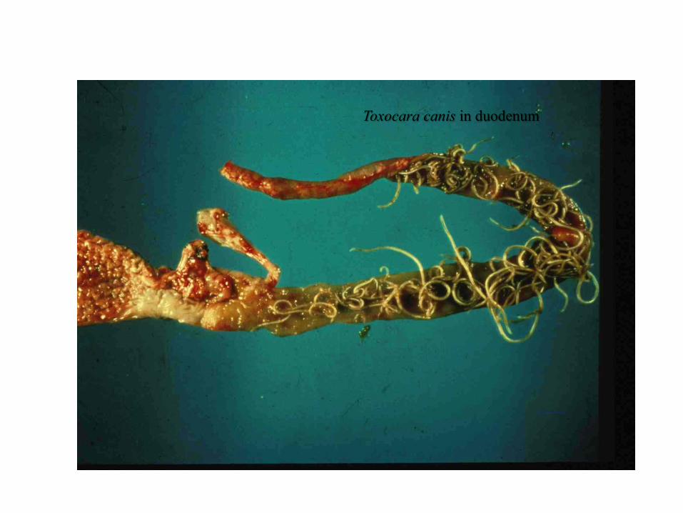
PATHOGENESIS
Especially with repeated infections
Lungs - hemorrhage, edema, eosinophils/cells
Liver - focal fibrosis ‘milk spots’,
$ loss, even though edible
Intestine - hypertrophy of muscle layer
( poor nutrient absorption)
Focal fibrosis

CLINICAL SIGNS
– coughing =‘thumps’, rapid, shallow expiration
– stunted growth
– diarrhea
Treatment & Control
– clean environment - adult worms are very
fecund, eggs are very resistant and sticky!
– deworm sows 2 weeks before farrowing &
wash thoroughly to get rid of those sticky
eggs
– most drugs work
• PYRANTEL kills newly hatched larvae use
as feed additive

Ascaridia sp. small intestine
ascarid of birds• Relatively large worm, 5 – 10cm, males
have distintive pre-anal sucker, adults in
the small intestine
• Only route of infection is by ingestion of
egg, prepatent time = 4-8 weeks
• Pathology is in young birds (< 3 months),
hemorrhagic enteritis, anemia and
diarrhea, blockage with heavy burdens

Ascaridia galli
Smooth thick shell egg
Ascaridia in small intestine of a bird
Posterior of male Ascaridia
Pre-cloacal sucker

Heterakis gallinarum “CECAL WORM”
• cecum of chicken, turkeys, etc.
• bird is infected either by:
ingestion of egg containing infective
larva
OR infected transport host - earthworm
• Nematode is relatively NON-pathogenic
but.....

Heterakis gallinarum
eggs & larvae are
carriers of a protozoa, Histomonas meleagridis
severe ‘blackhead’ disease in turkeys only;
(not in chickens)
inflammation/necrosis of cecum & liver
high mortality

To control ‘blackhead’ disease, must
control Heterakis nematode infections:
* deworm
* clean up the environment
* don’t house turkeys with chicken, or use
areas that previously housed chickens
Top Related