


Languages
Pages
Legal

Oncologic Emergencies in the ICU
Rhonda D’Agostino, ACNP-BC, FCCM
NP Coordinator, Critical Care (ICU/PACU/RRT)
Memorial Sloan Kettering Cancer Center

Commonality of Oncologic ICU Emergencies
Common
• Neutropenic Septic Shock
• Cytokine Release Syndrome
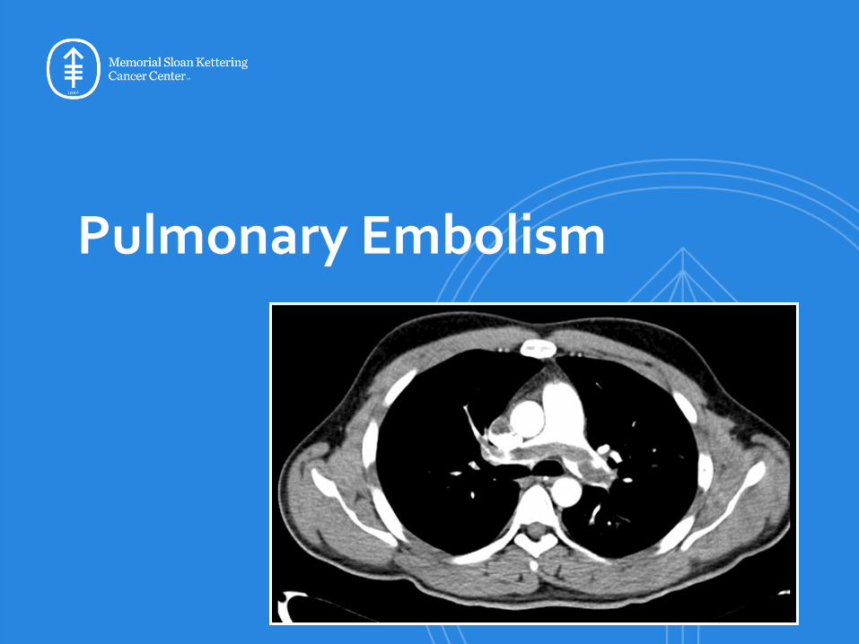
• Pulmonary Embolism
• Bleeding (hemoptysis, DIC, tumor invasion)
Uncommon (predicted)
• Pericardial Tamponade
• Acute Tumor Lysis
• SVC Syndrome
• Spinal Cord Compression
from my 10 yr experience at MSKCC

Neutropenic Septic Shock
Question # 1
What patient below meets criteria for sepsis?
• Neutropenic patient with a Sofa score of 4
• Neutropenic patient with 2 or more SIRS criteria
• Neutropenic pt with an elevated lactate level of 5.0
• Neutropenic pt with BP 70/40

The Third International Consensus Definitions for Sepsis and Septic Shock (Sepsis-3)
• Sepsis is defined as life-threatening organ dysfunction caused by a dysregulated host response to infection
JAMA 2016;315(8):801-810

…it was the host, not the germ, that drove the pathogenesis of sepsis.
Angus DC., & van der Poll, T. Critical Care Medicine, August 29, 2013

Host Response in Septic Shock
Management
Recognize & Act Quickly!
• IV antimicrobials within the first hour of recognition
• Combination therapy with extended spectrum β-lactam and either an aminoglycoside, fluoroquinolone or Azithromycin for neutropenic pts plus Vancomycin 1 g Q 12 hrs or Linezolid
• liposomal amphotericin B, voriconazole or micafungin with hx of HSCT and/or not responding to therapy (fluconazole-abdominal surgery)
• Antivirals (CMV, RSV) and Anti-PCP (steroids)
• Crystalloids/Colloids
• Norepinephrine or Epinephrine +/- Vasopressin
• GCSF
• Daily procalcitonin levels
Dellinger RP, et al. Crit Care Med 2013;41:580-637
Cytokine Release Syndrome & Neurotoxicity

Cancer Evolution Simplified
Cancer cell
Normal Cell
Normal Cell Normal Cell
Normal Cell
Cancer cell
Cancer cell
Killer T Cell
Cancer cell
Cancer cell
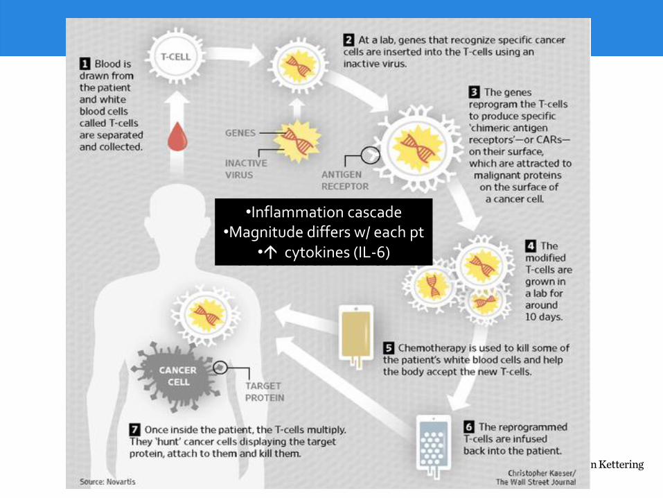
•Inflammation cascade •Magnitude differs w/ each pt
• cytokines (IL-6)

CRS Management
Davila et al. Sci Trans Med 2014

CRS
Severe CRS Better outcomes
tumor burden CAR- T cell proliferation dose of CAR-T infused

MSK Led trials • Leukemia
• Lymphoma
• Ovarian
• Head and Neck
• Mesothelioma
• Gliomas (future?)
• Juno Rocket Trail- Phase II relapsed or refractory B cell acute lymphoblastic leukemia put on hold d/t severe CRS and neurotoxicity resulting in death
• (removed Fludarabine as part of pretreatment and are only doing cyclophosphamide)
• Suicide gene

Cytokine Release Syndrome (CRS) Management Algorithm (for Grade ≥2)
• Hypotension SBP< 90 refractory to IVF challenge requiring vasopressors OR• Respiratory distress/hypoxia requiring increasing supplemental oxygen or ventilatory support OR • Acute coronary syndrome with positive troponin, clinically significant arrhythmia, and/or ECG changes
Tocilizumab 8mg/kg IV once
Worsening CRS within 12 hours• Increasing vasopressors dose OR• Increasing oxygen requirement OR• Cardiac arrhythmia, evolving EKG
Dexamethasone 10-20mg IV q12h
Clinical improvement < 24 hours• Decreasing vasopressor dose OR• Decreasing oxygen requirement
Observe
Worsening CRS
Taper over 3-5 days or longer if clinically indicated
No clinical improvement ≥ 24 hours
Worsening Symptoms Clinical Improvement
• Start higher dose of corticosteroid• Consider activating suicide gene if applicable
Cytokine Release Syndrome Management Algorithm
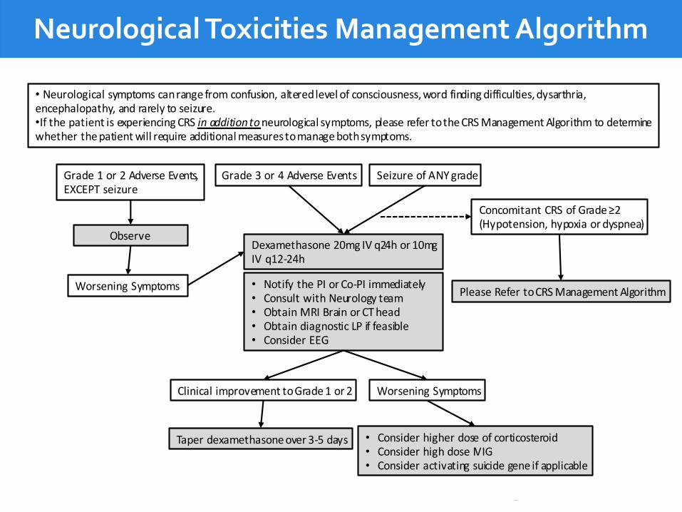
Neurological Toxicities Management Algorithm
• Neurological symptoms can range from confusion, altered level of consciousness, word finding difficulties, dysarthria, encephalopathy, and rarely to seizure. •If the patient is experiencing CRS in addition to neurological symptoms, please refer to the CRS Management Algorithm to determine whether the patient will require additional measures to manage both symptoms.
Grade 1 or 2 Adverse Events, EXCEPT seizure
• Notify the PI or Co-PI immediately• Consult with Neurology team • Obtain MRI Brain or CT head• Obtain diagnostic LP if feasible • Consider EEG
Seizure of ANY grade
Worsening Symptoms
Taper dexamethasone over 3-5 days
Grade 3 or 4 Adverse Events
Observe
Clinical improvement to Grade 1 or 2
Dexamethasone 20mg IV q24h or 10mg IV q12-24h
Worsening Symptoms
• Consider higher dose of corticosteroid• Consider high dose IVIG• Consider activating suicide gene if applicable
Concomitant CRS of Grade ≥2(Hypotension, hypoxia or dyspnea)
Please Refer to CRS Management Algorithm
Neurological Toxicities Management Algorithm

Pathophysiology of Major PE
Konstantinides et al. European Heart Journal , 2014
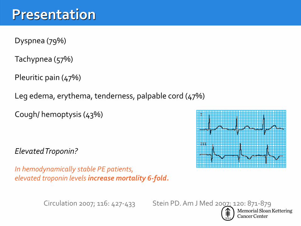
Presentation
Dyspnea (79%)
Tachypnea (57%)
Pleuritic pain (47%)
Leg edema, erythema, tenderness, palpable cord (47%)
Cough/ hemoptysis (43%)
Elevated Troponin?
In hemodynamically stable PE patients, elevated troponin levels increase mortality 6-fold.
Stein PD. Am J Med 2007; 120: 871-879 Circulation 2007; 116: 427-433

Treatment Algorithm for Suspected PE
Tapson VF. NEJM 2008;358:1037
Stabilize the patient
Anticoagulation contraindicated?
Yes
Diagnostic Eval
(Echo)
IVC filter
(consider AC)
No
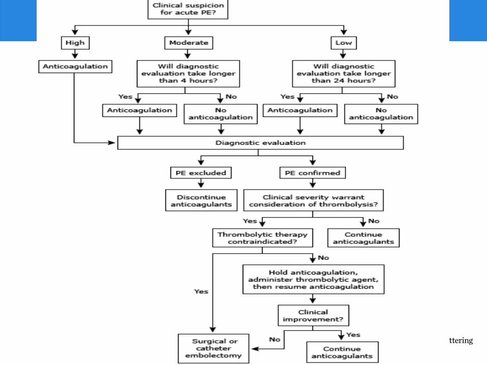

Embolectomy
Surgical • Operative mortality: 10-75%; 50-95% in pts who have had cardiopulmonary
arrest
• Complications: ARDS, acute renal failure, mediastinitis, severe neurologic sequelae
• Critical to have an experienced and aggressive CT surgery team that can be rapidly mobilized
Catheter • Transvenous insertion of embolectomy catheter suction and pulverization
by a high-speed rotor or fluid jet, and physical fragmentation with the catheter tip
• Limited studies, but survival rate ~70%-90%
Timsit JF, et al. Chest 1991;100(3):655-8 & Fava M, et al. J Vasc Interv Radiol 1997;8:261-66

Disseminated Intravascular Coagulation

DIC
DX: •Thrombocytopenia • PT and APTT • Fibrinogen • Fibrin degradation products or (FDP) or fibrin split products
Levi M, Ten Cate H. Disseminated intravascular coagulation. N Engl J Med. 1999 Aug 19. 341(8):586-92.
• Persistent activation of the coagulation cascade
• Widespread (micro) vascular thrombosis
• Ongoing activation exhausts factors and platelets resulting in profuse bleeding

DIC: Cancer
• 10-15% of patients with metastasized tumors have evidence of DIC
– Mucin-secreting adenocarcinomas; prostate, lung, breast, pancreas, stomach, bladder
• 15% of patients with acute leukemia have evidence of DIC (promyelocytic leukemia)
• Underreporting of DIC in cancer patients with sepsis
• DIC scoring systems not reliable in cancer patients
Levi M, Ten Cate H. Disseminated intravascular coagulation. N Engl J Med. 1999 Aug 19. 341(8):586-92.

DIC: Management
• Treat the cause
• PRBC (hgb >8d/dl), platelet transfusion (maintain platelet count > 20,000/ul (50,000 preferred)
• Fibrinogen > 100
• PT, APTT q 4-6 hrs

Question # 2
What is the difference between FFP and Cryoprecipitate?
• Cryoprecipitate is 10 times more concentrated than FFP
• Cryoprecipitate contains additional factors including fibrinogen and Von Wilbrand
• There is no difference except for manufacturer
• FFP does not include Factors II and X; Cryoprecipitate does

FFP vs Cryoprecipitate
• Primary indication for Cryo is Fibrinogen < 100
• Transfused in pools of 6-10 units which will increase fibrinogen 30-80 mg/dL
FFP Cryo
Factors II, V, VII, VIII (70%), X, XII, XIII
II, V, VII, VIII, X, XII, XIII
Fibrinogen No Yes
vWB No Yes

Pericardial Tamponade
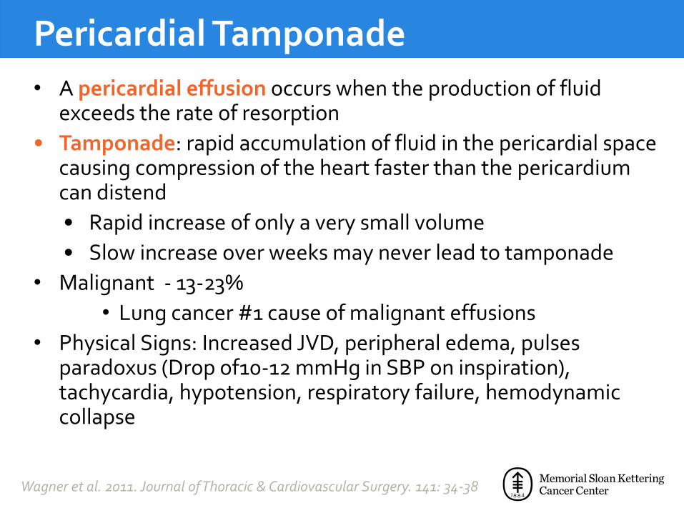
Pericardial Tamponade
• A pericardial effusion occurs when the production of fluid exceeds the rate of resorption
• Tamponade: rapid accumulation of fluid in the pericardial space causing compression of the heart faster than the pericardium can distend
• Rapid increase of only a very small volume
• Slow increase over weeks may never lead to tamponade
• Malignant - 13-23%
• Lung cancer #1 cause of malignant effusions
• Physical Signs: Increased JVD, peripheral edema, pulses paradoxus (Drop of10-12 mmHg in SBP on inspiration), tachycardia, hypotension, respiratory failure, hemodynamic collapse
Wagner et al. 2011. Journal of Thoracic & Cardiovascular Surgery. 141: 34-38

Pericardial Tamponade
• Echo Findings – Right atrial systolic collapse (RASC)
• Right atrial inversion
• About 100% sensitive, but low specificity (33-94% in some series)
– Right ventricular diastolic collapse (RVDC) • 75-93% sensitive and 85-100% specific
Singh S. et al. 1986 Gilliam LD et al 1983
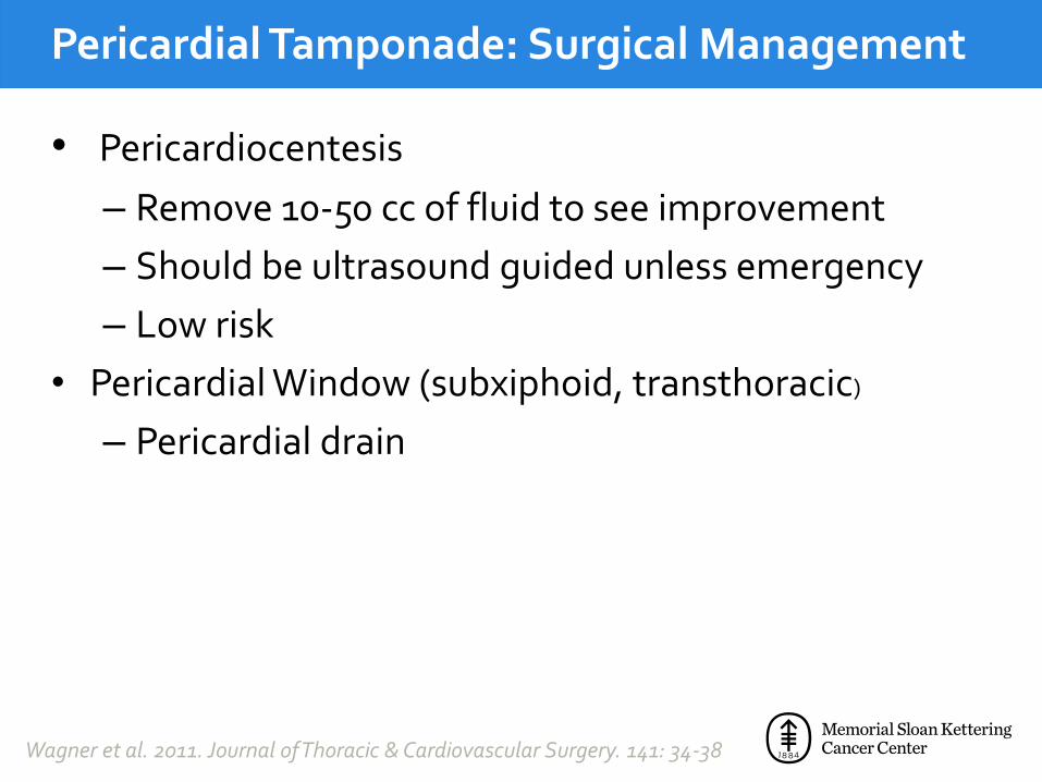
Pericardial Tamponade: Surgical Management
• Pericardiocentesis
– Remove 10-50 cc of fluid to see improvement
– Should be ultrasound guided unless emergency
– Low risk
• Pericardial Window (subxiphoid, transthoracic)
– Pericardial drain
Wagner et al. 2011. Journal of Thoracic & Cardiovascular Surgery. 141: 34-38
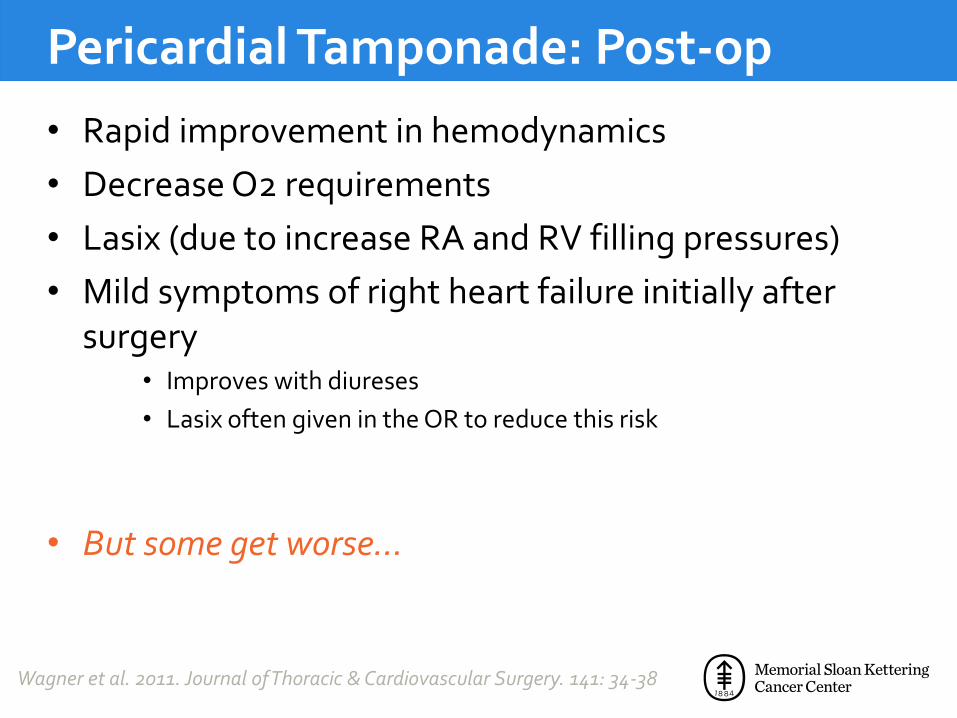
Pericardial Tamponade: Post-op
• Rapid improvement in hemodynamics
• Decrease O2 requirements
• Lasix (due to increase RA and RV filling pressures)
• Mild symptoms of right heart failure initially after surgery
• Improves with diureses
• Lasix often given in the OR to reduce this risk
• But some get worse…
Wagner et al. 2011. Journal of Thoracic & Cardiovascular Surgery. 141: 34-38

Pericardial Tamponade
• Paradoxical hemodynamic Instability – Instead of improving, patients worsen after tamponade is released
– Unknown etiology
– Poor prognosis
• Review of MSKCC data on pericardial effusions over 5 years – Evaluated survival, predictive factors and incidence of paradoxical
hemodynamic instability (PHI)
Wagner et al. JTCVS 2011
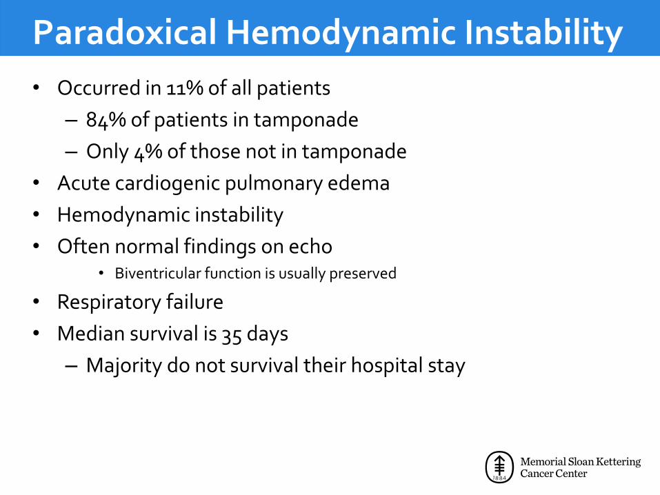
Paradoxical Hemodynamic Instability
• Occurred in 11% of all patients
– 84% of patients in tamponade
– Only 4% of those not in tamponade
• Acute cardiogenic pulmonary edema
• Hemodynamic instability
• Often normal findings on echo • Biventricular function is usually preserved
• Respiratory failure
• Median survival is 35 days
– Majority do not survival their hospital stay
Wagner et al. JTCVS 2011

Paradoxical Hemodynamic Instability
Wagner et al. JTCVS 2011
Malignant cells Larger effusion Pre-op tamponade (did not reach significance)

Paradoxical Hemodynamic Instability
• Prevention of PHI – Slow drainage of fluid
• Similar to re-expansion pulmonary edema phenomenon
– Early prophylactic drainage of effusions • Only 4% of non-tamponade patients developed PHI
– Massive hemodynamic support in the first 24-48 hrs • If no early improvement, unlikely to every recover
• Must explain this all to the family
Wagner et al. JTCVS 2011

Acute Tumor Lysis
Acute Tumor Lysis
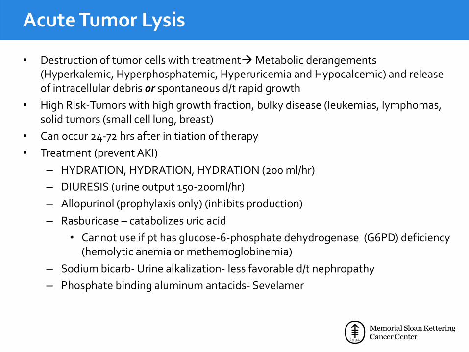
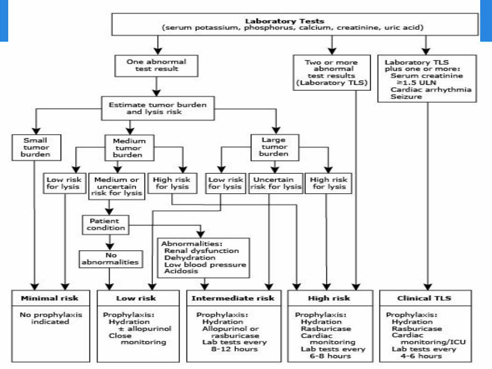
• Destruction of tumor cells with treatment Metabolic derangements (Hyperkalemic, Hyperphosphatemic, Hyperuricemia and Hypocalcemic) and release of intracellular debris or spontaneous d/t rapid growth
• High Risk-Tumors with high growth fraction, bulky disease (leukemias, lymphomas, solid tumors (small cell lung, breast)
• Can occur 24-72 hrs after initiation of therapy
• Treatment (prevent AKI)
– HYDRATION, HYDRATION, HYDRATION (200 ml/hr)
– DIURESIS (urine output 150-200ml/hr)
– Allopurinol (prophylaxis only) (inhibits production)
– Rasburicase – catabolizes uric acid
• Cannot use if pt has glucose-6-phosphate dehydrogenase (G6PD) deficiency (hemolytic anemia or methemoglobinemia)
– Sodium bicarb- Urine alkalization- less favorable d/t nephropathy
– Phosphate binding aluminum antacids- Sevelamer
SVC Syndrome

Pathophysiology
• Occlusion of the SVC resulting in an increase in venous pressure which leads to venous stasis and engorgement
• Causes- Lung Cancer (Small Cell- 65-80%)
– Catheters, thrombosis, aneurysm, vasculitis, sarcoidosis
• Symptoms: periorbital edema, facial fullness, dyspnea, cough, arm swelling, chest pain, dysphagia
• Late signs: Dysphagia and hoarseness due to the entrapment of the laryngeal nerve and laryngeal edema, Increasing intercranial pressure, cerebral edema, heart failure
• Treatment: chemotherapy, radiation, stenting, thrombosis
• Adjunct management: diuretics, steroids, avoid CVC insertion
43

Kumaer & Hosn, 2014, NEJM
Spinal Cord Compression

High Risk for SCC
• High Risk: Primary Intramedullary Tumors (Ependymoma, Astrocytoma, Glioma, Hemangioblastoma), Metastatic Intramedullary Tumors (Lymphoma, Neuroblastoma) Metastatic Bone/Epidural Tumors (Breast, Lung, Prostate, Renal, Myeloma, Thyroid, Sarcoma) , vertebral fractures or intraspinal abscesses
• Symptoms: Pain parethesiasmotor lossloss of proprioception
• Treatment: High dose corticosteriods,
laminectomy, radiation, chemo

Case Study
47
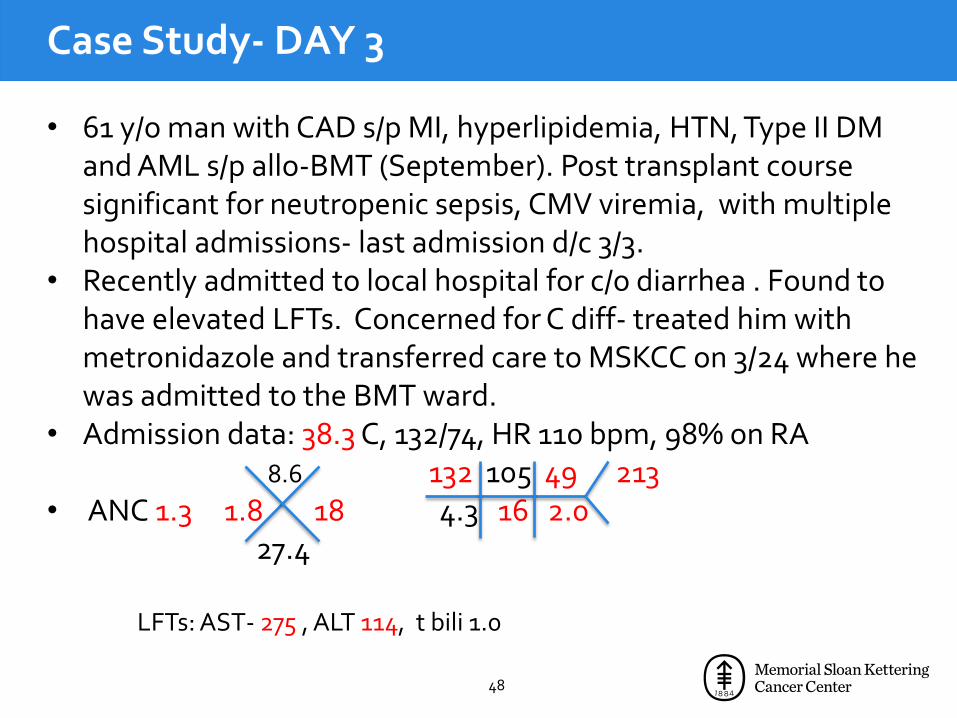
Case Study- DAY 3
• 61 y/o man with CAD s/p MI, hyperlipidemia, HTN, Type II DM and AML s/p allo-BMT (September). Post transplant course significant for neutropenic sepsis, CMV viremia, with multiple hospital admissions- last admission d/c 3/3.
• Recently admitted to local hospital for c/o diarrhea . Found to have elevated LFTs. Concerned for C diff- treated him with metronidazole and transferred care to MSKCC on 3/24 where he was admitted to the BMT ward.
• Admission data: 38.3 C, 132/74, HR 110 bpm, 98% on RA 8.6 132 105 49 213 • ANC 1.3 1.8 18 4.3 16 2.0
27.4 LFTs: AST- 275 , ALT 114, t bili 1.0
48

Imaging

Imaging

Case Study- DAY 2
• Doing OK • Ongoing diarrhea • Afebrile next day • BP 98/60, HR 70, RR 18, 97% RA • Imipenem, Linezolid (VRE surveillance rectal swab was +) • Micafungin, posaconazole and acyclovir for fungal and viral
prophylaxis • D/C posaconazole d/t increased LFTs • MRI to evaluate liver lesions • 35.0 10.2 136 112 33 192 3.12 • ANC 1.4 1.8 18 3.6 12 1.9
31.9 58.7 LFTs: AST- 753 , ALT 261, t bili 1.8 , LDH 1041
51
Case Study- Day 3
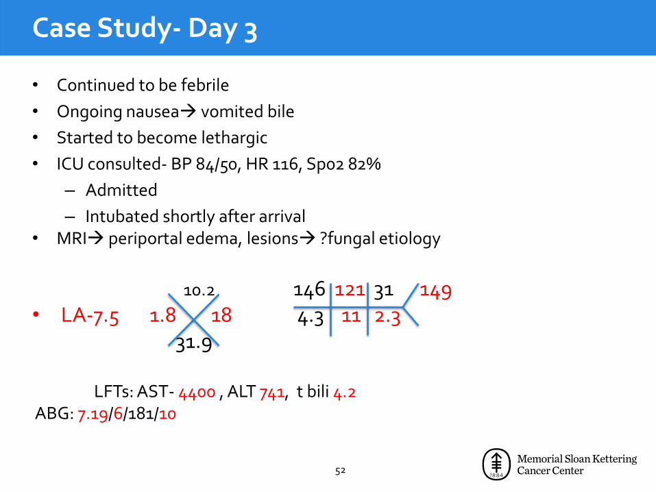
• Continued to be febrile
• Ongoing nausea vomited bile
• Started to become lethargic
• ICU consulted- BP 84/50, HR 116, Sp02 82%
– Admitted
– Intubated shortly after arrival • MRI periportal edema, lesions ?fungal etiology
10.2 146 121 31 149 • LA-7.5 1.8 18 4.3 11 2.3
31.9 LFTs: AST- 4400 , ALT 741, t bili 4.2 ABG: 7.19/6/181/10
52

Case Study: Day 3- ICU
• Admitted to ICU for…
– 1. Hepatic failure
– 2. Respiratory Failure
– 3.Renal failure
– 4. Coagulopathy
• Why?
• Severe Neutropenic Sepsis • Fluids but continued to drop his BP
• Levophed->quickly titrated up
• Attempted radial a-line but too hypotensive- femoral a line
• Hematuria
53
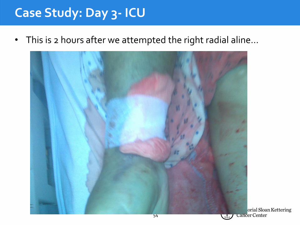
Case Study: Day 3- ICU
• This is 2 hours after we attempted the right radial aline…
54

Case Study: Day 3- ICU
• Upgraded his antibiotics
• Overnight need massive transfusion
• BP started to drop despite being on Levophed
• Bleeding now from everywhere!
55

56

57

Case Study: Day 3- ICU
• 10.1(2 hrs) 8.0(30 mins) 6.9 (5 hrs)3.7
• Looking back…
• Severe neutropenic septic shock with MSOF
• Fungal hepatitis (despite being on prophylaxis)
• DIC

Thank you!
Top Related