


Languages
Pages
Legal

Observer Performance Evaluation of a Digital Camera for Acquiring
Radiographic Images for Teleradiology
Elizabeth A. Krupinski, PhD
University of Arizona

Goal Conduct a feasibility study to
determine if a commercially available digital camera can be used to acquire photographs of radiologic images to transmit them via store-forward technology over a telemedicine network.

Arizona Telemedicine Program (ATP)Sites
Tuba City
Kingman
Cottonwood
Ganado
PaysonSpringerville
Whiteriver
DOC - Phoenix
DOC - Yuma
NogalesPatagonia
Douglas
AHSCKinoTucson VA
AHSC Hub
Telemedicine
Telepathology
TeleradiologyDOC - Florence
DOC - Buckeye
DOC - Douglas
Study Rationale Teleradiology is an important part of an
increasing number of Radiology & Telemedicine programs
Saves time and feedback to referring clinician is faster than with other methods (e.g., courier) - 85% of our teleradiology cases have a “wet read” report generated within 6 hours
Rationale Continued
It can, however, be expensive! For digital modalities a merge box is
needed For plain film a digitizer or video capture
system is required Some sites just cannot afford these options A low-cost alternative had to be explored
The Test Site The Patagonia Family Health Center in rural
Arizona needed consults, especially in orthopedics & bone radiology
The clinic has no x-ray facilities so sends patients to Nogales’ Mariposa Clinic, the report & films are then sent over either with the patients or later
Problem - Mariposa only has a general radiologist so sub-specialty consults are often required
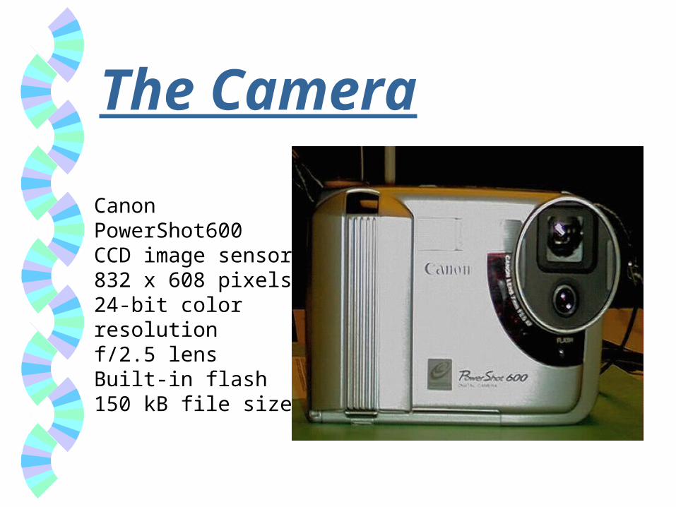
The Camera
Canon PowerShot600CCD image sensor832 x 608 pixels24-bit color resolutionf/2.5 lensBuilt-in flash150 kB file size
The Cases 40 bone trauma cases (e.g., fracture,
subluxation, soft tissue damage) from the Patagonia Family Health Clinic
Cases that required a consult in the past & were representative of the types of cases they would be sending for teleconsults in the future - typically subtle lesions
The Photographs Film images were placed on standard
viewbox with extraneous light blocked out The physician identified region(s) of
interest based on clinical history and nature of problem
At least one global and one close up shot were acquired for each image using the Canon PowerShot camera

The Displays
Images sent over ARTN to the MedVision workstation
Color monitor, 1024 x 768 pixels Window/level, zoom/pan available for use
during viewing Film images displayed on standard viewbox

Example of a Close-Up Shot

Example of an Original Film Image
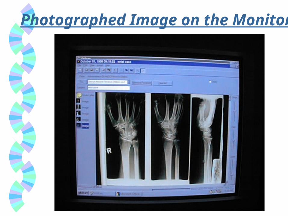
Photographed Image on the Monitor

Protocol 2 orthopedic surgeons & 2 bone radiologists
reviewed the 40 cases on film & using the digital photographs displayed on the monitor
Film reading 6 months after monitor reading Reported:
• Diagnosis
• Decision confidence using 6-level scale
• Image quality using 4-level scale

Results - Image Quality
0
1020
3040
50P
ER
CE
NT
Film Photo
ExcellentGoodFairPoor
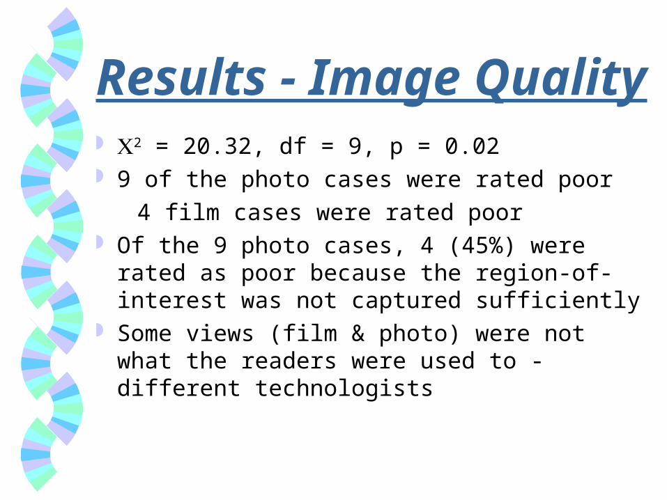
Results - Image Quality 2 = 20.32, df = 9, p = 0.02 9 of the photo cases were rated poor
4 film cases were rated poor Of the 9 photo cases, 4 (45%) were rated as poor
because the region-of-interest was not captured sufficiently
Some views (film & photo) were not what the readers were used to - different technologists

Results - Diagnoses* Reader Agreement (Kappa)*
1 0.89
2 0.92
3 0.94
4 0.88
* Agreement with truth. Truth was determined from the film images by 2 radiologists not participating in the study.
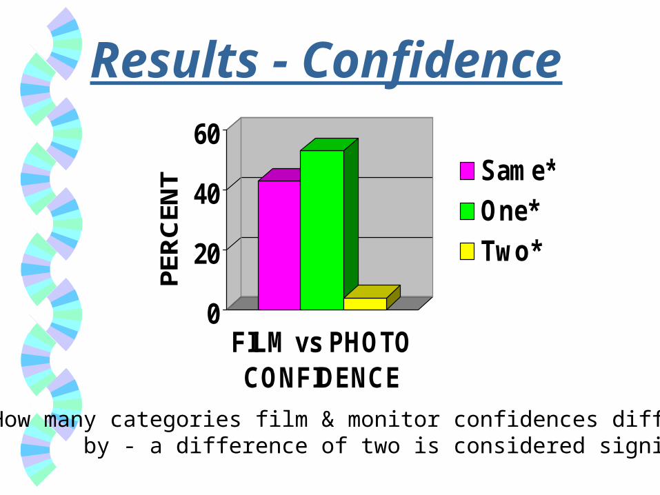
Results - Confidence
0
20
40
60PE
RC
ENT
FILM vs PHOTOCONFIDENCE
Same*
One*
Two*
* How many categories film & monitor confidences differ by - a difference of two is considered significant
Conclusions A digital still camera can be used effectively to
capture images of bone trauma radiographs for store-forward telemedicine consults
Well-framed close-up shots of the region-of-interest are essential
Good quality original films are essential
Other Applications? Other non-screening applications where a
specific suspicious region-of-interest can be identified for close-up photographs
Unless the lesion is fairly gross, the global shots are not adequate for consultation - close-up shots are required in the majority of cases
Top Related