


Languages
Pages
Legal


Muscles of the thigh
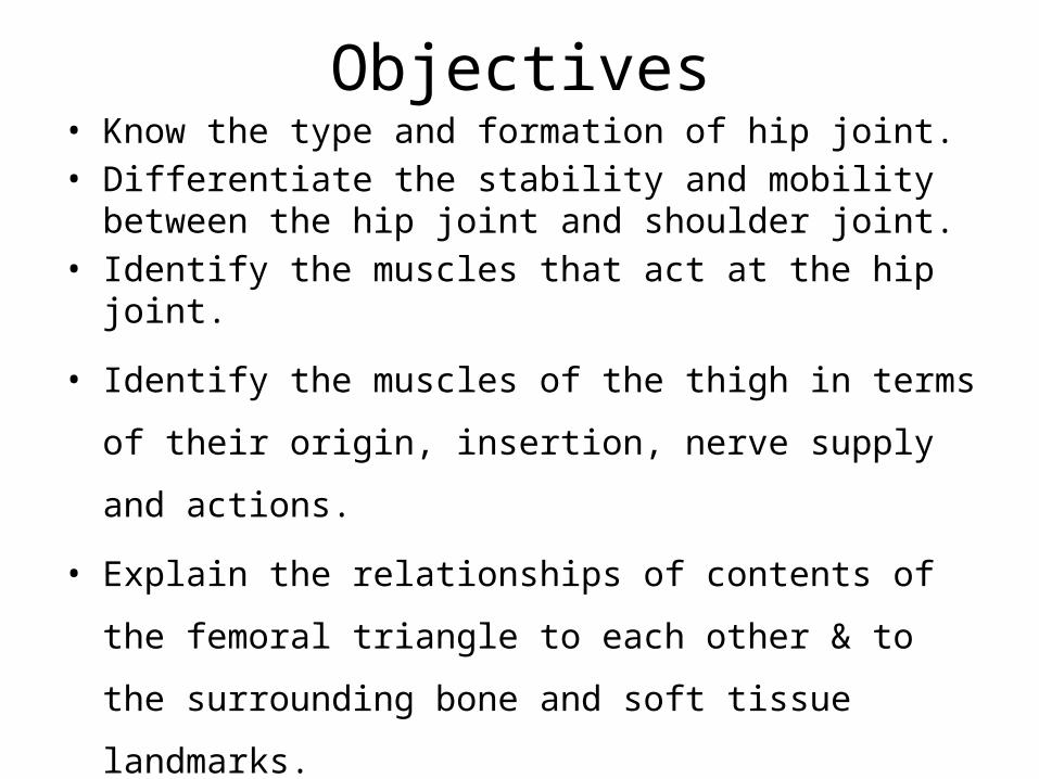
Objectives• Know the type and formation of hip joint.• Differentiate the stability and mobility between the hip joint
and shoulder joint. • Identify the muscles that act at the hip joint.
• Identify the muscles of the thigh in terms of their origin,
insertion, nerve supply and actions.
• Explain the relationships of contents of the femoral triangle to
each other & to the surrounding bone and soft tissue
landmarks.
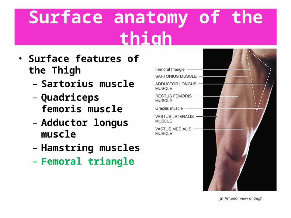
Surface anatomy of the thigh
• Surface features of the Thigh– Sartorius muscle– Quadriceps femoris
muscle– Adductor longus muscle– Hamstring muscles– Femoral triangle

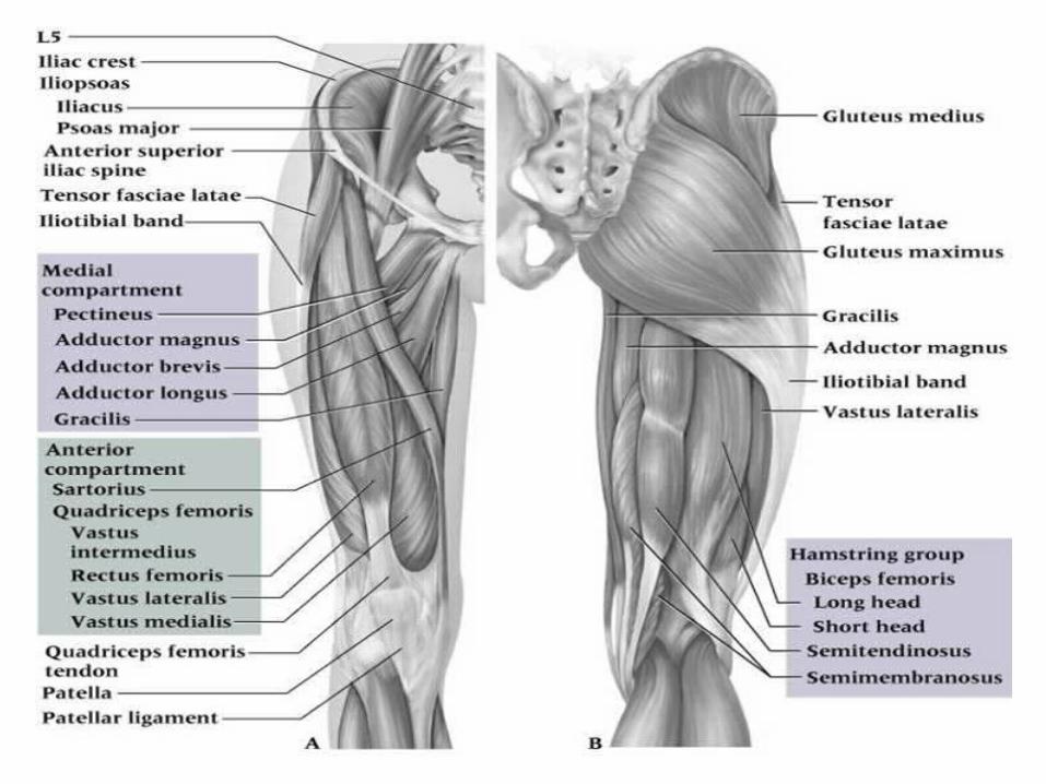
Compartments of the thigh
Thigh is divided to 3 groups of muscles called compartments.
• Anterior compartment• Posterior compartment• Medial compartment
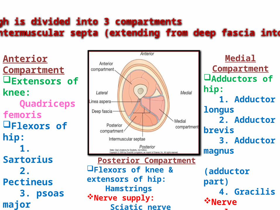
The thigh is divided into 3 compartments by 3 intermuscular septa (extending from deep fascia into femur)
Anterior CompartmentExtensors of knee: Quadriceps femorisFlexors of hip: 1. Sartorius 2. Pectineus 3. psoas major 4. IliacusNerve supply: Femoral nerve
Medial Compartment
Adductors of hip: 1. Adductor longus 2. Adductor brevis 3. Adductor magnus (adductor part) 4. GracilisNerve supply: Obturator nervePosterior Compartment
Flexors of knee & extensors of hip: HamstringsNerve supply: Sciatic nerve
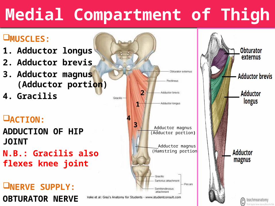
MUSCLES:1. Adductor longus2. Adductor brevis3. Adductor magnus
(Adductor portion)4. Gracilis
ACTION:ADDUCTION OF HIP JOINTN.B.: Gracilis also flexes knee joint
NERVE SUPPLY:OBTURATOR NERVE
Adductor magnus(Adductor portion)
Medial Compartment of Thigh
1
2
34
Adductor magnus(Hamstring portions)
1
2
34
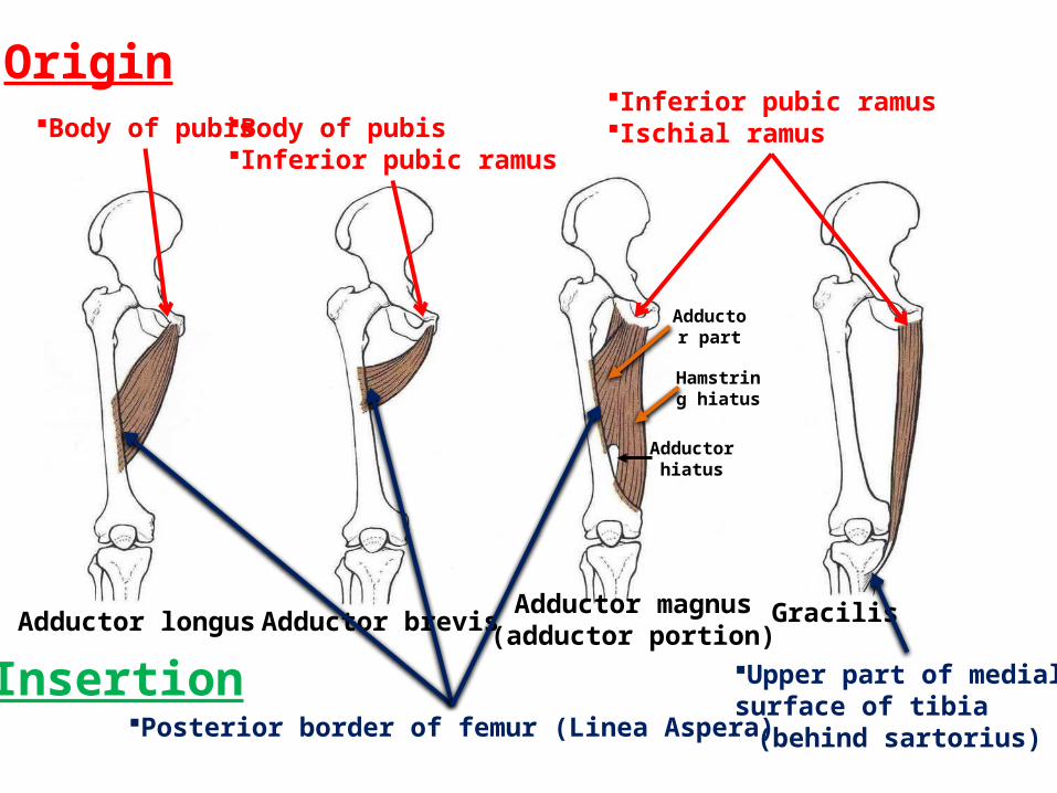
InsertionPosterior border of femur (Linea Aspera)
Upper part of medialsurface of tibia
(behind sartorius)
Adductor longus Adductor brevisAdductor magnus(adductor portion)
Gracilis
OriginBody of pubis Body of pubis
Inferior pubic ramus
Inferior pubic ramusIschial ramus
Adductor hiatus
Hamstring hiatus
Adductor part

Medial Compartment

Medial Compartment
Blood Supply:Obturator artery:Branch of internal iliac artery.

Medial Compartment
Innervation:Obturator nerve.Tibial nerve:To hamstring portion of adductor magnus.
Action:Adduction

Anterior Compartment of Thigh
Vastus Intermedius(deep to rectus femoris)
12
3
1
4
2
3
4
Contains the: Flexor of the hip:
1. Sartorius2. Pectineus3. Psoas major4. Iliacus
Extensors of knee (Quadriceps femoris): 1. Rectus femoris 2. Vastus lateralis3. Vastus medialis 4. Vastus intermedius (deep to
rectus femoris)
Nerve supply: Femoral nerve
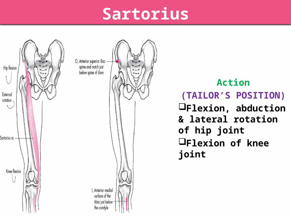
Sartorius
S
Action(TAILOR’S POSITION)
Flexion, abduction & lateral rotation of hip jointFlexion of knee joint
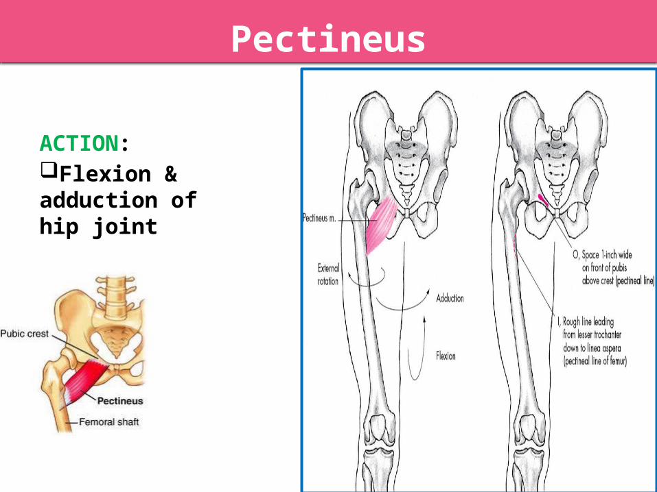
Pectineus
ACTION:Flexion & adduction of hip joint
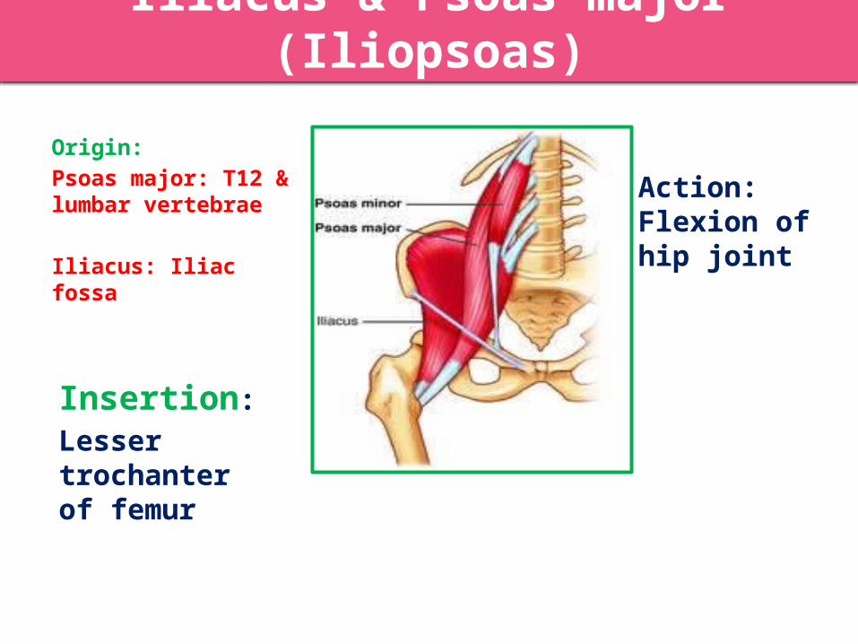
Iliacus & Psoas major (Iliopsoas)
Insertion:Lesser trochanter of femur
Action:Flexion of hip joint
Origin:Psoas major: T12 & lumbar vertebrae
Iliacus: Iliac fossa
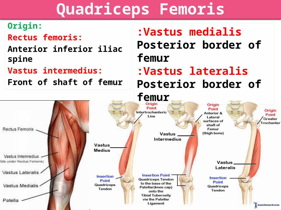
Quadriceps FemorisOrigin:Rectus femoris: Anterior inferior iliac spineVastus intermedius:Front of shaft of femur
Vastus medialis:Posterior border of femurVastus lateralis:
Posterior border of femur

INSERTION:Into PATELLA (Patella is a sesamoid bone)From patella into TUBEROSITY OF TIBIA through Ligamentum Patellae (Patellar Ligament)
ACTION:Extension of knee joint
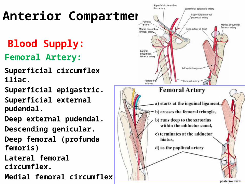
Anterior Compartment
Blood Supply:Femoral Artery:
Superficial circumflex iliac.Superficial epigastric.Superficial external
pudendal.Deep external pudendal.Descending genicular.Deep femoral (profunda femoris)Lateral femoral circumflex.Medial femoral circumflex.
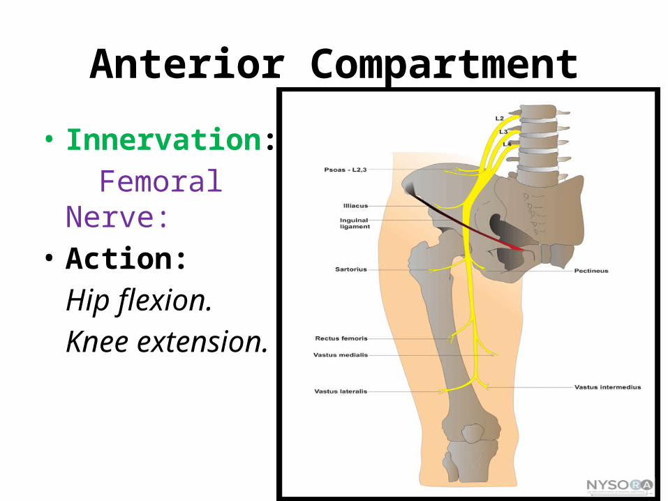
Anterior Compartment
• Innervation:Femoral Nerve:
• Action:Hip flexion.Knee extension.
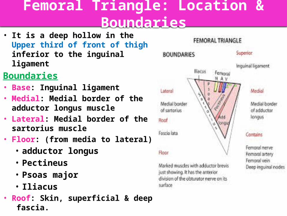
Femoral Triangle: Location & Boundaries• It is a deep hollow in the Upper third
of front of thigh inferior to the inguinal ligament
Boundaries• Base: Inguinal ligament• Medial: Medial border of the
adductor longus muscle• Lateral: Medial border of the
sartorius muscle• Floor: (from media to lateral)
• adductor longus • Pectineus • Psoas major• Iliacus
• Roof: Skin, superficial & deep fascia.
PectineusIliopsoas
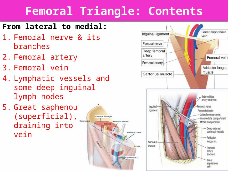
Femoral Triangle: ContentsFrom lateral to medial:1. Femoral nerve & its branches2. Femoral artery3. Femoral vein4. Lymphatic vessels and some deep
inguinal lymph nodes5. Great saphenous vein
(superficial), draining into femoral vein

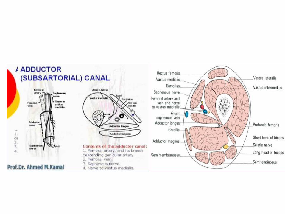
DEFINITION: an aponeurotic tunnel for femoral artery & veinSITE: In middle third of front of thigh deep to sartoriusEXTENT: From apex of femoral triangle to adductor hiatusBOUNDARIES:
Roof (Anterior): Sartorius (medially) and vastus medialis (laterally) Floor (Posterior): Adductor longus & magnus
ADDUCTOR CANAL
(Subsartorial/Hunter’s canal) Adductor hiatus
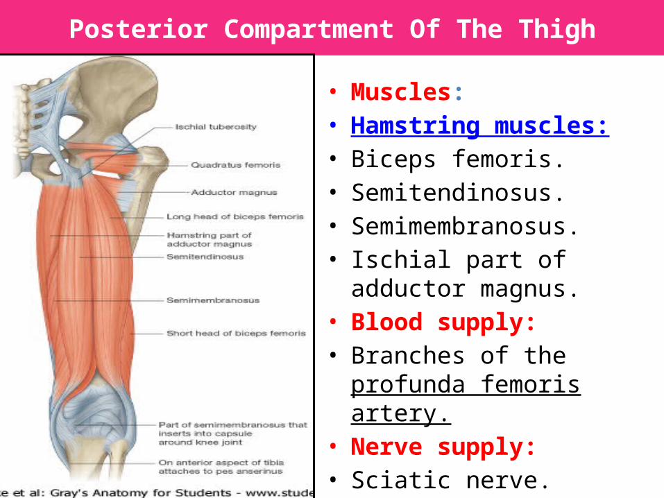
Posterior Compartment Of The Thigh
• Muscles: • Hamstring muscles:• Biceps femoris. • Semitendinosus.• Semimembranosus.• Ischial part of adductor
magnus.• Blood supply: • Branches of the profunda
femoris artery.• Nerve supply: • Sciatic nerve.

Biceps Femoris • Origin:
– The long head from the ischial tuberosity.
– The short head from the linea aspera .
• Insertion: • Into the head of the fibula.• Nerve supply: • The long head is supplied by the
tibial part of the sciatic; • the short head is supplied by the
common peroneal part of the sciatic.
• Action :• Flexion of knee.• Lateral rotation of flexed leg.• Long head: extends hip.

Semitendinosus• Origin: • Ischial tuberosity.• Insertion: • Upper part of the medial
surface of the shaft of the tibia (SGS)..
• Nerve supply: • Tibial portion of the sciatic.• Action: • Flexes and medially rotates
the leg at the knee joint; • Extends the thigh at the
hip joint.
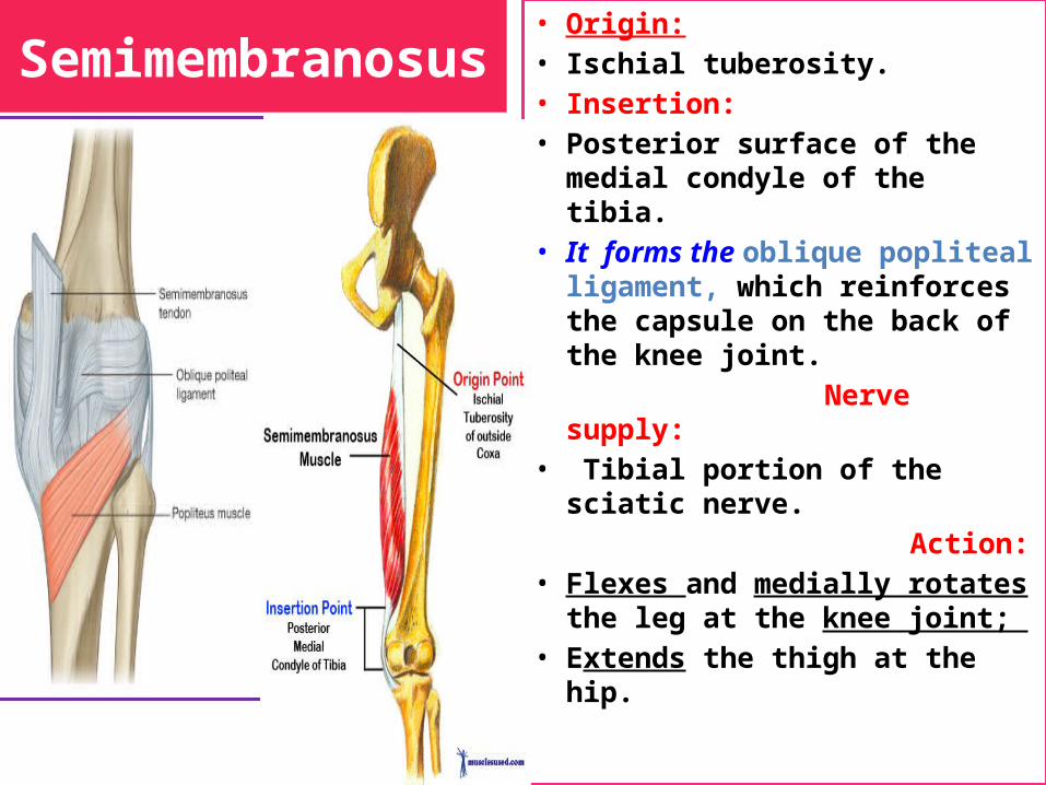
Semimembranosus• Origin: • Ischial tuberosity.• Insertion: • Posterior surface of the medial
condyle of the tibia. • It forms the oblique popliteal
ligament, which reinforces the capsule on the back of the knee joint.
Nerve supply:• Tibial portion of the sciatic
nerve. Action: • Flexes and medially rotates the
leg at the knee joint; • Extends the thigh at the hip.
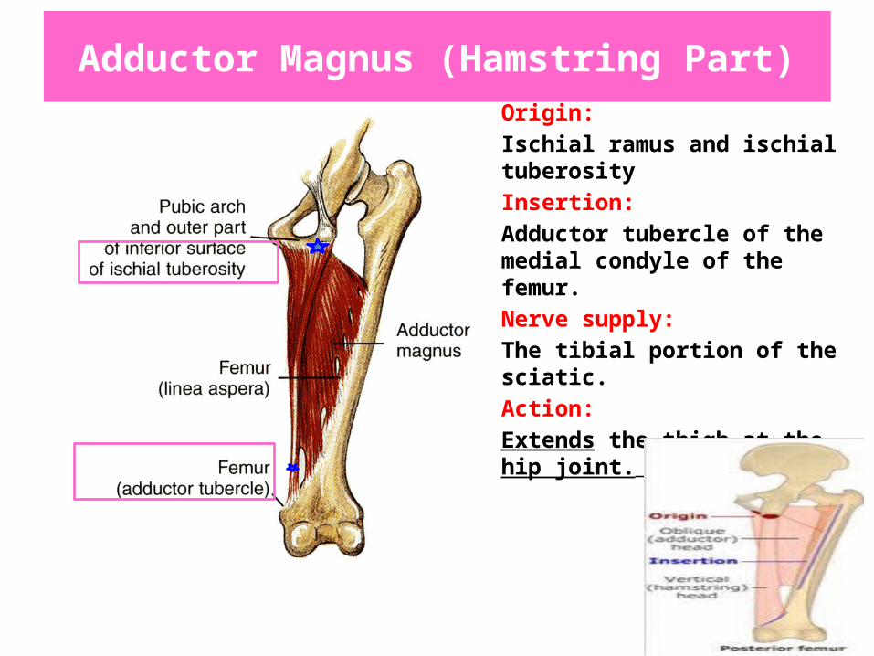
Adductor Magnus (Hamstring Part)• Origin:• Ischial ramus and ischial
tuberosity• Insertion: • Adductor tubercle of the medial
condyle of the femur.• Nerve supply: • The tibial portion of the sciatic.• Action: • Extends the thigh at the hip
joint.

Posterior compartment of the thighInnervation:
•Tibial division of sciatic nerve
•Except short head of biceps femoris: common fibular division of sciatic nerve
posterior

BLOOD SUPPLY
• The four perforating branches of the profunda femoris artery provide a rich blood supply to this compartment.
• The profunda femoris vein drains the greater part of the blood from the compartment.

NERVE SUPPLY • Sciatic Nerve• The sciatic nerve, a branch of the
sacral plexus (L4 and 5; S1, 2, and 3), leaves the gluteal region as it descends in the midline of the thigh.
• It is overlapped posteriorly by the adjacent margins of the biceps femoris and semimembranosus muscles.
• It lies on the posterior aspect of the adductor magnus.
• In the lower third of the thigh it ends by dividing into the tibial and common peroneal nerves.
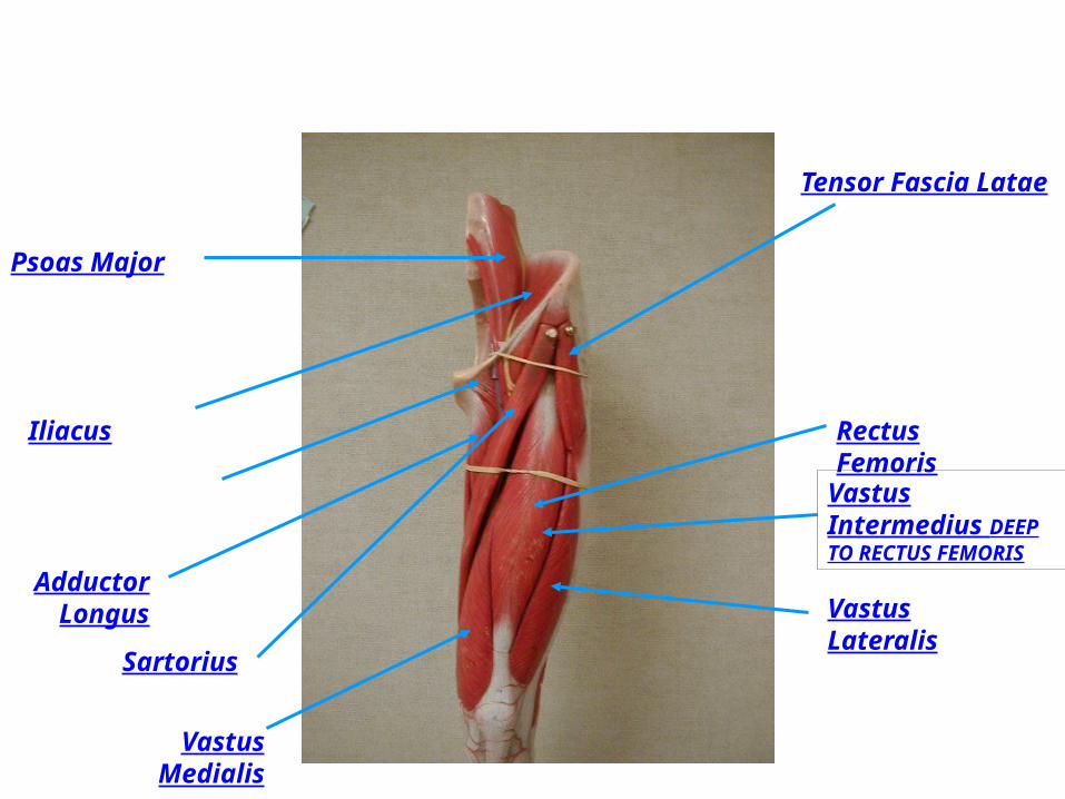
Psoas Major
Tensor Fascia Latae
Rectus Femoris
Adductor Longus
Sartorius
Vastus Medialis
Iliacus
Vastus Lateralis
Vastus Intermedius DEEP TO RECTUS FEMORIS

Top Related