


Languages
Pages
Legal


Muscle• Skeletal muscle
– Unit Cell Structure– Architecture
• Series/parallel• Force/velocity
– Stimulation• Summation/tetanus/rate-coding
– Muscle mechanics• Force-length relation • Force velocity relation
– Pre-stretch

Skeletal Muscle
• Striated and voluntary– Cardiac muscle is striated– Smooth muscle is unstriated and involuntary
• Attaches to skeleton via tendons• Most abundant tissue in the body
– 45-75% of body weight
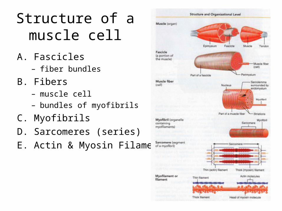
Structure of a muscle cell
A. Fascicles – fiber bundles
B. Fibers – muscle cell– bundles of myofibrils
C. MyofibrilsD. Sarcomeres (series)E. Actin & Myosin Filaments
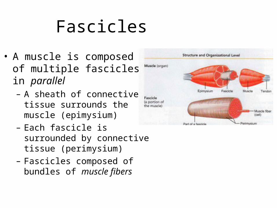
Fascicles
• A muscle is composed of multiple fascicles in parallel– A sheath of connective tissue
surrounds the muscle (epimysium)
– Each fascicle is surrounded by connective tissue (perimysium)
– Fascicles composed of bundles of muscle fibers

Muscle Fiber
• Long, cylindrical, multinucleated cells
• Between fibers are blood vessels
• Surrounded by endomysium• Composed of myofibrils

Myofibrils• Literally (muscle thread)• Contractile element of muscle• Made up of filaments• Aligned in parallel• filaments make striations
– Banding pattern
• One repeating unit is called a sarcomere
• string of sarcomeres in series

Sarcomeres• Functional unit of muscle
contraction• Literally ‘muscle segment’• Number of sarcomeres in a
fiber is very important to muscle function
• When each sarcomere shortens the same amount, the fiber with more sarcomeres will shorten more.
• Made up of myofilaments– Thick and thin filaments
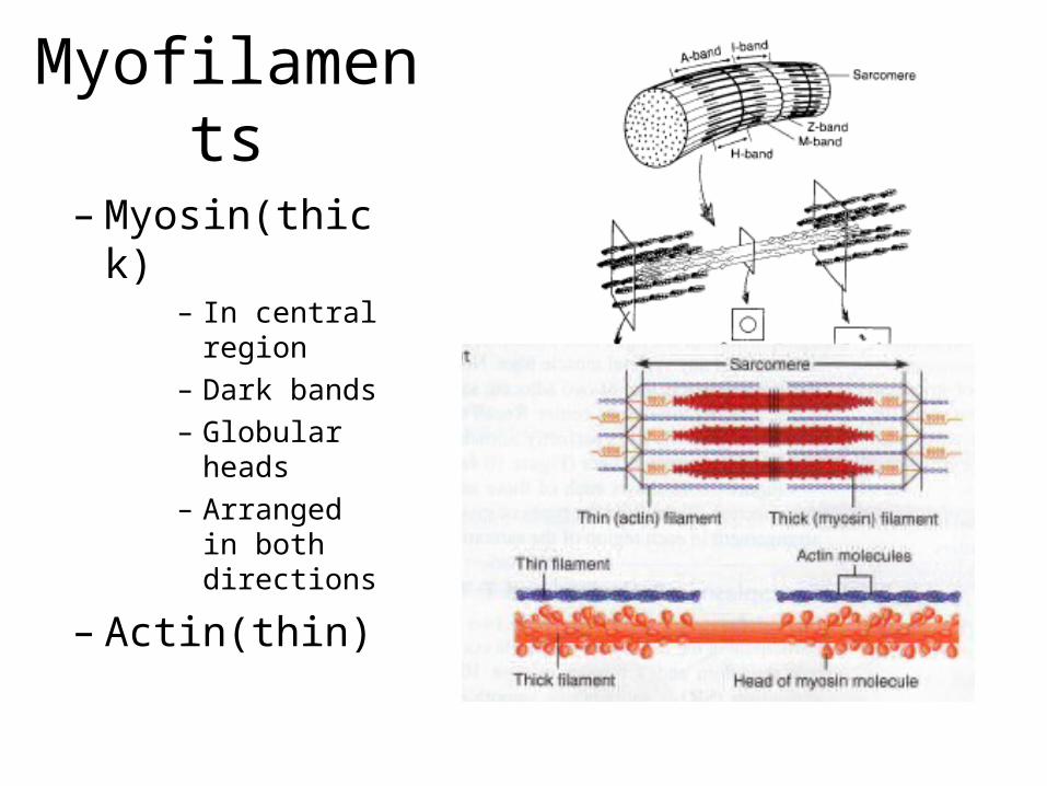
Myofilaments
– Myosin(thick)– In central
region– Dark bands– Globular heads– Arranged in
both directions
– Actin(thin)

Banding Pattern

• Based on myofilaments:– Z-Disc– I-Band– A-Band– H-zone– M-line

Z-DiscM-line
<--I-Band---><--------------------A-Band--------------->
<-H-Zone->
<--I-Band--->
Sarcomere:

Muscle contraction
• Sliding filament theory– AF Huxley and HE Huxley– Light and Electron microscopy– Both published results same time in Nature– Does not explain lengthening contractions

Sliding Filament Theory
• The exertion of force by muscle is accompanied by the sliding of thick and thin filaments past one another
• Commonly explained by cross-bridges

• cross-bridge theory:• muscle force is
proportional to the number of cross bridges attached

Sliding filament theory
• A band stay the same• I band shorten

A single functional unit in a muscle contraction is a
A) fascicleB) fiberC) myofibrilD) sarcomere

According to sliding filament theory, during a contraction the distance
between the M and Z lines
A) increasesB) decreasesC) stays the sameD) need more information

Muscle• Skeletal muscle
– Unit Cell Structure– Architecture
• Series/parallel• Force/velocity
– Stimulation• Summation/tetanus/rate-coding
– Muscle mechanics• Force-length relation • Force velocity relation
– Pre-stretch

Muscle architecture
• Organization of muscle fibers– Muscle also organized at macro level– Architecture is the arrangement of muscle fibers
relative to the axis of force generation• Muscle fibers have fairly consistent diameters among
muscle of different size, but arrangement can be very different
• So cannot tell force capacity of a muscle from a biopsy– Need number of fibers and how arranged

3 types of arrangements• Longitudinal (parallel)
– Fibers run parallel to force generating axis• Pennate
– Fibers at a single angle– shallow
• Multipennate– several angles

What are advantages/disadvantages ofa)longitudinal arrangement?b)pennate arrangement?

Muscle architecture
• Determines– Max muscle force
• Fibers in parallel• Pennation angle
– Max muscle shortening velocity• no of sarcomeres in series

Hill Muscle Model
CE: Contractile Element (active force generation)SE: Series Elastic Element
represents elasticity in: cross-bridges and myofilamentstendon and aponeuroses
PE: Parallel Elastic Elementconnective tissue surrounding muscle fibers

• Can use Hill muscle model to illustrate effects of muscle length and width on muscle’s – maximum force– maximum shortening velocity

f, l
f, l
f, l
f, l f, l
f, l
f, l
Series
Parallel

f, l
f, l f, l f, l
Series
F=?L=?
A) F = f ; L = lB) F = 3f ; L = 3lC) F = 3f ; L = lD) F = f ; L = 3lE) don’t understand

f, l
f, LL=nl
F,lF=nf
f, l
f, l
f, l f, l
f, l
f, l
Series
Parallel
A) F = f ; L = lB) F = 3f ; L = 3lC) F = 3f ; L = lD) F = f ; L = 3lE) don’t understand

Pennation Angle


Pennation Angle
• Pennation angle is a space saving strategy• Allows you to pack more fibers into a smaller space• Doesn’t hurt b/c cos0=1, cos 30=0.87 (13% force loss)

Muscle architecture
• Determines– Max muscle force
• Fibers in parallel• Pennation angle
– Max muscle shortening velocity• no of sarcomeres in series

Physiological Cross-Sectional Area
• PCSA ~ max muscle force• M=muscle mass (g)• =muscle density (g/cm3) = 1.056 g/cm3
• l=fiber length (cm)• V= Muscle volume = M/

How do we measure PCSA?


More on PCSA
• Not proportional to muscle mass• Not proportional to anatomical cross-sectional
area

Muscle architecture
• Determines– Max muscle force (~PCSA)
• Fibers in parallel• Pennation angle
– Max muscle shortening velocity• no of sarcomeres in series

Muscle fiber length
• Assumed that fiber length ~fiber velocity• Fiber length ~ no. of sarcomeres in series

Muscle architecture
• Determines– Max muscle force (~PCSA)
• Fibers in parallel• Pennation angle
– Max muscle shortening velocity (~Fiber length)• no of sarcomeres in series

What are advantages/disadvantages ofa)longitudinal arrangement?b)pennate arrangement?

Significance of Architecture
• Clever design– Same functional component can yield so many
different motors
• Muscles designed for a purpose– Perhaps this simplifies the control

ProblemImagine you have 10 sarcomeres; each generates a maximum of 1 unit
of force, and shortens with a maximum velocity of 1 unit/s. Diagram an arrangement of sarcomeres that will create a muscle fiber with the following force and velocity characteristics. Use I to represent individual sarcomeres, and draw ellipses around sarcomeres to specify fibers.
i) Fmax= 5 units; Vmax= 2 units/s ii) Fmax= 2 units; Vmax=5 units/s iii) Fmax=5cos10o units; Vmax=2cos10o units/s

Net muscle force
Enoka Fig 1.6
Vector math can illustrate the effect of coactivating different parts of the pectoralis major muscle.Suppose clavicular component exerted a force of 224N at 0.55 rad above horizontal, and the sternal portions has a magnitude of 251N at 0.35 rad below horizontal.
What is the resultant force?
A) F = 472 N, angle = 64.5 degB) F = 472 N, angle = 25.4 degC) F = 428 N, angle = 4.17 degD) F = 428 N, angle = 85.82E) I don’t understand

Enoka Fig 1.6

Muscle• Skeletal muscle
– Unit Cell Structure– Architecture
• Series/parallel• Force/velocity
– Stimulation• Summation/tetanus/rate-coding
– Muscle mechanics• Force-length relation • Force velocity relation
– Pre-stretch

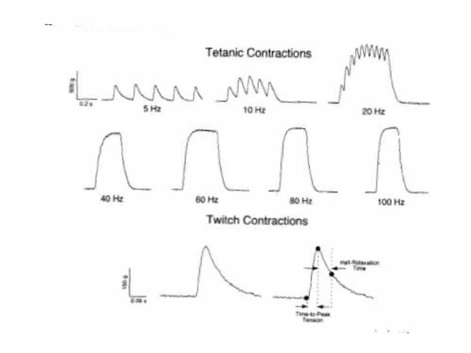
Temporal Summation• Excitation fast (~1-2ms)• Contraction/relaxation slow (100ms)
– Muscle twitch lags because slack in the elastic components must be taken up.
– Contraction time:– Relaxation time:
• Summation– If second impulse comes along before the first one has relaxed, they
sum– Get more force with multiple impulses then alone
• Tetanic Summation– maximum tension is sustained because rapidity of stimulation
outstrips the contraction-relaxation time of the muscle

TimeStimulation(Action potentials)
Single Low frequency High frequency
Twitch
FusedTetanusUnfused
TetanusFor
ce
Neural Stimulation

If the contraction-relaxation time for a muscle twitch is 100 ms, at what stimulation frequency
will we begin to see summation? NB: 1 Hz corresponds to 1 stimulus/second
A)100 Hz and greaterB)5 Hz and greaterC)10 Hz and greaterD)I don’t understand

Max Force• PCSA
– No. sarcomeres in parallel– Pennation angle
• Stimulation
Max Shortening Velocity• No. of sarcomeres in series
– Muscle fiber length

Muscle• Skeletal muscle
– Unit Cell Structure– Architecture
• Series/parallel• Force/velocity
– Stimulation• Summation/tetanus/rate-coding
– Muscle mechanics• Force-length relation • Force velocity relation
– Pre-stretch– WorkLoops

Muscle Mechanics
• Force-length• Force-velocity

Force-Length
• Isometric force varies with muscle length– Forces generation in muscle is a direct function of
the amount of overlap between actin and myosin filaments
– Po is maximum tetanic force
– Length of muscle at Po is muscle’s optimal length


0
1.0
0.6
0.8
0.4
0.2
100 120 140 1608060Rest length (%)
Relativeforce
Force-Length Relationship

0
1.0
0.6
0.8
0.4
0.2
100 120 140 1608060Rest length (%)
Relativeforce
Force-Length Relationship

0
1.0
0.6
0.8
0.4
0.2
100 120 140 1608060Rest length (%)
Relativeforce
Force-Length Relationship

0
1.0
0.6
0.8
0.4
0.2
100 120 140 1608060Rest length (%)
Relativeforce
Force-Length Relationship

0
1.0
0.6
0.8
0.4
0.2
100 120 140 1608060Rest length (%)
Relativeforce
Force-Length Relationship

Passive force production

Titin• Cross-bridge not
responsible, so what it?• Origin of passive muscle
tension within myofibrils– Researchers compared
whole muscle, single fibers, and single fibers w/membranes removed (1986)
– Huge protein responsible - titin


Force-Velocity

Muscle Actions
1. Shortening2. Isometric3. Lengthening

Force-VelocityRelative Force Velocity100% Po 0% Vmax
95% Po 1% Vmax
90% P 2.2% Vmax
75% Po 6.3% Vmax
50% Po 16.6% Vmax
25% Po 37.5% Vmax
10% Po 64.3% Vmax
5% Po 79.1% Vmax
0% Po 100% Vmax

Shortening Contractions
• Force decreases with velocity

Knee
Shank
Thigh
Knee extensor muscles in shortening contraction during knee extension

Knee
Shank
Thigh
Isometric Contractions

Isometric

KneeShank
Thigh
Active and Lengthening)

Lengthening Contractions
• Higher force (160%!)• Velocity-independent• Don’t know why• Important
– Common– Selective for soreness and
injury– Muscle strengthening greatest

How will the force-angle curves change for different muscle actions?Fo
rce
Isometric
Knee Angle

Force• PCSA
– No. sarcomeres in parallel– Pennation angle
• Stimulation• Sarcomere Length
– Filament overlap
• Velocity
Shortening Velocity• No. of sarcomeres in series
– Muscle fiber length
• Force

Summary
• Force and velocity– Structure of the unit cell– Sliding Filament Theory– Architecture– Stimulation– F-L– F-V

Put it all together
• Compare muscles w/two different pcsas– Draw F-L– Draw F-V for same fiber length
• Compare muscle w/different fiber lengths– Draw F-L, for same pcsa– Draw F-V

Muscle• Skeletal muscle
– Unit Cell Structure– Architecture
• Series/parallel• Force/velocity
– Stimulation• Summation/tetanus/rate-coding
– Muscle mechanics• Force-length relation • Force velocity relation
– Pre-stretch

Prestretch: muscle is active and stretched before beginning to shorten
Activelengthening(prestretch)
Activeshortening

Force
P0
Shortening Velocity0
0
Prestretch
Noprestretch
Frog knee flexor(semitendinosis)From Cavagna &
Citterio, 1974.
Prestretch effectlasts for a limitedtime

Data from Gregor et al. 1988., (fig. 6.36 Enoka)
Velocity (mm/s)

SSC
• Muscle can produce more power if actively stretched before it is allowed to shorten
• Can also lower metabolic cost

Immediately after being stretched
Resting length
Crossbridges (and/or titin?) act like springs: after being stretched, higher F per xbridge

Prestretch Shorten
Extensor stretch-shorten cycle in countermovement jump

Prestretch occurs in a variety of activities
• Jumping with countermovement• Running• Other examples?
Top Related