


Languages
Pages
Legal

Muscle Pain: Understanding the Mechanisms
Siegfried Mense l Robert D. GerwinEditors
Muscle Pain: Understandingthe Mechanisms
EditorsProf. Dr. Siegfried MenseUniversitat HeidelbergMedizinische Fakultat Mannheim CBTM,NeuroanatomieLudolf-Krehl-Str. 13-17, R. C61468167 [email protected]
Dr. Robert D. GerwinAssociate Professor of NeurologyJohns Hopkins UniversityBaltimoreMaryland 21287USAPain and Rehabilitation Medicine7830 Old Georgetown RoadSuite C-15Bethesda, [email protected]
ISBN 978-3-540-85020-5 e-ISBN 978-3-540-85021-2DOI 10.1007/978-3-540-85021-2Springer Heidelberg Dordrecht London New York
Library of Congress Control Number: 2010927599
# Springer-Verlag Berlin Heidelberg 2010This work is subject to copyright. All rights are reserved, whether the whole or part of the material isconcerned, specifically the rights of translation, reprinting, reuse of illustrations, recitation, broadcasting,reproduction on microfilm or in any other way, and storage in data banks. Duplication of this publicationor parts thereof is permitted only under the provisions of the German Copyright Law of September 9,1965, in its current version, and permission for use must always be obtained from Springer. Violationsare liable to prosecution under the German Copyright Law.The use of general descriptive names, registered names, trademarks, etc. in this publication does not imply,even in the absence of a specific statement, that such names are exempt from the relevant protective lawsand regulations and therefore free for general use.
Cover design: WMXDesign GmbH, Heidelberg, Germany
Printed on acid-free paper
Springer is part of Springer Science+Business Media (www.springer.com)

Foreword
This edition of the companion volumes Muscle Pain: Understanding the Mechan-isms and Muscle Pain: Diagnosis and Treatment is essential reading for those
interested in clinical approaches to acute and chronic pain conditions involving
muscle tissues and in the mechanisms underlying these conditions. The volumes
cover a very important topic in pain medicine, since muscle pain is very common
and can often be difficult to diagnose and treat effectively. Furthermore, chronic
pain involving muscle and other components of the musculoskeletal system
increases with age, such that it is a common complaint of those of us who are
middle-aged or older. Indeed, as changing population demographics in “wester-
nized” countries result in higher proportions of the population living longer and
being middle-aged and elderly, chronic muscle pain will likely become even more
of a health problem.
In the case of acute muscle pain, this can often be very intense, and in the short
term can limit or modify the use of components of the musculoskeletal system
associated with the sensitive muscle. Chronic muscle pain can also be intense, as
well as unpleasant and disabling, and it is in many cases the over-riding symptom of
most musculoskeletal disorders that are associated with long-term deleterious
changes in musculoskeletal function. This can present a challenge both to the
patient who has to live with the condition and to the clinician called upon to assist
the patient, not only because of the physical or biomechanical impediment but also
because of the presence of chronic pain reflecting persistent alterations in the
peripheral muscle tissues and/or central nervous system. Chronic pain is now
recognized as a multidimensional experience encompassing cognitive, emotional
and motivational aspects as well as the sensory or perceptual dimension. Thus, as
the editors of this work note in their preface, it can distort the patient’s life,
including work, family and social relationships, and can change the patient’s
perception of himself or herself from being an effective and independent human
being to one who is ineffective and dependent. These features apply especially to
patients with chronic muscle pain, and the range and impact of most musculoskele-
tal disorders and the pain that they manifest dictate that clinicians need to have a
v
good knowledge base about pain and adopt a broad biopsychosocial perspective in
order to provide effective management of the patient. These companion volumes
provide this knowledge base and perspective.
Although the etiology and pathogenesis of several muscle pain conditions are
still unclear, recent advances have been made in understanding muscle pain
mechanisms and in the management of the conditions. The chapters in these
books collectively provide up-to-date details of these mechanisms and management
approaches. The anatomy and neurophysiology relevant to muscle pain is covered
in Muscle Pain: Understanding the Mechanisms. It offers a solid basic science
underpinning for the more clinically oriented second volume, Muscle Pain: Diag-nosis and Treatment, which outlines present knowledge of etiologic and pathophys-iologic processes, and which also deals with current approaches to the management
of the various conditions manifesting muscle pain.
Like its predecessor, these companion volumes should prove to be an invaluable
resource not only to clinical practitioners wanting to have a basic understanding of
pain mechanisms and clinical approaches currently available to diagnose and
manage muscle pain problems, but also to basic and clinical pain scientists who
are interested in an up-to-date and comprehensive review of the diagnostic and
management approaches to muscle pain.
Toronto Barry J. Sessle
vi Foreword
Preface
Disorders of the musculoskeletal system are the leading causes of disability in
western societies. Musculoskeletal pain syndromes can be divided into two broad
categories: (1) myalgias, which include the major condition of myofascial pain
syndromes, as well as inflammatory and other myosites, and (2) articular disorders,
which include all of the arthritides. Fibromyalgia has long been considered a
chronic musculoskeletal pain syndrome, but recent research supports categorizing
fibromyalgia as a widespread chronic pain syndrome. Ergonomic and postural and
other structural dysfunctions, including pain associated with the hypermobility
syndromes, can bridge these two categories, although they tend to fall more into
the myalgic group of disorders.
A problem for the practitioner trying to understand a patient’s pain is that pain is
a subjective sensation that is colored by the patient’s personal life experience, and
ethnic and cultural background. Chronic pain is not simply a sensation, but a global
experience that involves suffering and a distortion of the patient’s role in all phases
of life, including family, work and social relationships, and can change the patient’s
self perception of him- or herself from being an independent, effective human
being, to being a dependent, ineffective person. Communication is particularly
difficult with chronic pain patients, because chronic pain is such a personal experi-
ence of global suffering, rather than a simple sensation like touch. There is a
definite effect of gender on pain perception. Therefore, when examining muscles
in patients for painful conditions, the greater sensitivity of women to painful stimuli
has to be taken into account.
Pain from muscle and skin is subjectively and objectively distinct. Muscle pain
is described as aching and cramping, diffuse and poorly localized, whereas cutane-
ous pain is described as sharp and pricking, and precisely localized. Cutaneous pain
is usually not referred to other body regions, while muscle pain is commonly
referred to other deep somatic structures like tendons and fascia or other muscles,
and viscera (viscerosomatic pain syndromes). Objective differences between
muscle and cutaneous pain exist in the processing of neuronal information at the
vii

spinal and brainstem level and continue up to the brain, where nociceptive activity
from skin and muscle terminates in different regions. Some of the established pain
terms used in this book are defined in chapter 1 of the volume “Muscle Pain:
Understanding the Mechanisms”.
Heidelberg/Mannheim S. Mense
Bethesda R.D. Gerwin
viii Preface

Contents
1 Introduction . . . . . . . . . . . . . . . . . . . . . . . . . . . . . . . . . . . . . . . . . . . . . . . . . . . . . . . . . . . . . . . . 1
Siegfried Mense and Robert D. Gerwin
2 Functional Anatomy of Muscle: Muscle, Nociceptors
and Afferent Fibers . . . . . . . . . . . . . . . . . . . . . . . . . . . . . . . . . . . . . . . . . . . . . . . . . . . . . . . 17
Siegfried Mense
3 Peripheral Mechanisms of Muscle Pain: Response Behavior
of Muscle Nociceptors and Factors Eliciting Local
Muscle Pain . . . . . . . . . . . . . . . . . . . . . . . . . . . . . . . . . . . . . . . . . . . . . . . . . . . . . . . . . . . . . . . . 49
Siegfried Mense
4 Central Nervous Mechanisms of Muscle Pain: Ascending
Pathways, Central Sensitization, and Pain-Modulating Systems . . . . . . 105
Siegfried Mense
5 Referral of Musculoskeletal Pain . . . . . . . . . . . . . . . . . . . . . . . . . . . . . . . . . . . . . . . 177
Thomas Graven-Nielsen and Siegfried Mense
6 Increased Muscle Tone as a Cause of Muscle Pain . . . . . . . . . . . . . . . . . . . . 207
Siegfried Mense and Alfonse T. Masi
7 Reorganized Motor Control Due to Muscle Pain . . . . . . . . . . . . . . . . . . . . . . 251
Thomas Graven-Nielsen and Lars Arendt-Nielsen
Glossary . . . . . . . . . . . . . . . . . . . . . . . . . . . . . . . . . . . . . . . . . . . . . . . . . . . . . . . . . . . . . . . . . . . . . . . 269
Index . . . . . . . . . . . . . . . . . . . . . . . . . . . . . . . . . . . . . . . . . . . . . . . . . . . . . . . . . . . . . . . . . . . . . . . . . . 319
ix


Contents to
Muscle Pain: Diagnosis and Treatment
1 Introduction
Siegfried Mense and Robert D. Gerwin
Part I Myofascial Pain Syndrome
2 Myofascial Pain Syndrome
Robert D. Gerwin
3 Morphology of Myofascial Trigger Points: What Does
a Trigger Point Look Like?
Siegfried Mense
Part II Fibromyalgia Syndrome
4 Fibromyalgia Syndrome: Clinical Aspects and Management
Emma Guymer and Geoffrey Littlejohn
5 Gender, Genetics, and Other Risk Factors Increasing
Vulnerability to Fibromyalgia
Geoffrey O. Littlejohn
Part III Other Muscle Pain Syndromes
6 Low Back Pain of Muscular Origin
Robert D. Gerwin
xi

7 Masticatory Muscle Pain
S. Palla and M. Farella
8 Inflammatory Myopathies
Ingrid E. Lundberg and Li Alemo Munters
9 Whiplash Injury, Muscle Pain and Motor Dysfunction
Jan Dommerholt
10 Brain Imaging of Muscle Pain
Sandra Kamping and Herta Flor
Glossary
Index
xii
Contributors
Lars Arendt-Nielsen Center for Sensory-Motor Interaction, Department of Health
Science and Technology, Aalborg University Fredrik Bajers Vej 7, Bld. D3 DK-
9220 Aalborg E, Denmark, [email protected]
Robert D. Gerwin Associate Professor of Neurology, Johns Hopkins University,
Baltimore, Maryland 21287, USA; Pain and Rehabilitation Medicine, 7830
Old Georgetown Road, Suite C-15, Bethesda, MD 20814-2432, USA, gerwin@
painpoints.com
Thomas Graven-Nielsen Center for Sensory-Motor Interaction (SMI), Laboratory
for Musculoskeletal Pain and Motor Control, Department of Health Science and
Technology, Aalborg University, Fredrik Bajers Vej 7D-3, DK-9220 Aalborg E,
Denmark, [email protected]
Alfonse T. Masi University of Illinois at Chicago, Dept. Epidemiology and
Biostatistics, 1603 W. Taylor Street, (MC 923), Chicago, Il 60612-4394, USA,
Siegfried Mense Institut fur Anatomie, Universitat Heidelberg, Medizinische
Fakultat Mannheim, CBTM, Ludolf-Krehl-Str. 13-17, R. C614, 68167 Mannheim,
Germany, [email protected]
xiii
Chapter 1
Introduction
Siegfried Mense and Robert D. Gerwin
Contents
1.1 Subjective Nature of Pain Terms . . . . . . . . . . . . . . . . . . . . . . . . . . . . . . . . . . . . . . . . . . . . . . . . . . . . . . . . . 2
1.2 Established Pain Terms (partly after Merskey
and Bogduk 1994; Loeser and Treede 2008) . . . . . . . . . . . . . . . . . . . . . . . . . . . . . . . . . . . . . . . . . . . . . 3
1.2.1 General Terms . . . . . . . . . . . . . . . . . . . . . . . . . . . . . . . . . . . . . . . . . . . . . . . . . . . . . . . . . . . . . . . . . . . . . 3
1.2.2 Increased Sensitivity . . . . . . . . . . . . . . . . . . . . . . . . . . . . . . . . . . . . . . . . . . . . . . . . . . . . . . . . . . . . . . . 4
1.2.3 Decreased Sensitivity . . . . . . . . . . . . . . . . . . . . . . . . . . . . . . . . . . . . . . . . . . . . . . . . . . . . . . . . . . . . . . 5
1.3 Established, But Often Ill-Defined, Pain Terms . . . . . . . . . . . . . . . . . . . . . . . . . . . . . . . . . . . . . . . . . . 5
1.3.1 Contracture (in the Physiological Sense) . . . . . . . . . . . . . . . . . . . . . . . . . . . . . . . . . . . . . . . . . . 5
1.3.2 Muscle Spasm . . . . . . . . . . . . . . . . . . . . . . . . . . . . . . . . . . . . . . . . . . . . . . . . . . . . . . . . . . . . . . . . . . . . . 5
1.3.3 Muscle Stiffness . . . . . . . . . . . . . . . . . . . . . . . . . . . . . . . . . . . . . . . . . . . . . . . . . . . . . . . . . . . . . . . . . . . 6
1.3.4 Muscle Tone . . . . . . . . . . . . . . . . . . . . . . . . . . . . . . . . . . . . . . . . . . . . . . . . . . . . . . . . . . . . . . . . . . . . . . . 6
1.3.5 Projected Pain . . . . . . . . . . . . . . . . . . . . . . . . . . . . . . . . . . . . . . . . . . . . . . . . . . . . . . . . . . . . . . . . . . . . . . 6
1.3.6 Referred Pain . . . . . . . . . . . . . . . . . . . . . . . . . . . . . . . . . . . . . . . . . . . . . . . . . . . . . . . . . . . . . . . . . . . . . . 6
1.3.7 Spread of Pain . . . . . . . . . . . . . . . . . . . . . . . . . . . . . . . . . . . . . . . . . . . . . . . . . . . . . . . . . . . . . . . . . . . . . 7
1.4 General Aspects of Pain and Nociceptor Function . . . . . . . . . . . . . . . . . . . . . . . . . . . . . . . . . . . . . . . . 7
1.5 Muscle Pain Versus Skin Pain . . . . . . . . . . . . . . . . . . . . . . . . . . . . . . . . . . . . . . . . . . . . . . . . . . . . . . . . . . . . 8
1.5.1 Subjective Differences . . . . . . . . . . . . . . . . . . . . . . . . . . . . . . . . . . . . . . . . . . . . . . . . . . . . . . . . . . . . . 9
1.5.2 Objective Differences . . . . . . . . . . . . . . . . . . . . . . . . . . . . . . . . . . . . . . . . . . . . . . . . . . . . . . . . . . . . 11
1.6 Descending Pain-Modulating Influences . . . . . . . . . . . . . . . . . . . . . . . . . . . . . . . . . . . . . . . . . . . . . . . . . 13
1.7 Transition from Acute to Chronic Pain . . . . . . . . . . . . . . . . . . . . . . . . . . . . . . . . . . . . . . . . . . . . . . . . . . 13
1.8 Interactions Between Psychosocial and Somatic Factors . . . . . . . . . . . . . . . . . . . . . . . . . . . . . . . . 13
References . . . . . . . . . . . . . . . . . . . . . . . . . . . . . . . . . . . . . . . . . . . . . . . . . . . . . . . . . . . . . . . . . . . . . . . . . . . . . . . . . . . . . . 14
S. Mense (*)
Universit€at Heidelberg, Mediz. Fakult€at Mannheim CBTM, Neuroanatomie, Ludolf-Krehl-Str.
13-17, R. C614, 68167, Mannheim, Germany
e-mail: [email protected]
R.D. Gerwin
Associate Professor of Neurology, Johns Hopkins University, Baltimore, Maryland 21287, USA;
Pain and Rehabilitation Medicine, 7830 Old Georgetown Road, Suite C-15, Bethesda, MD 20814-
2432, USA
e-mail: [email protected]
S. Mense and R.D. Gerwin (eds.), Muscle Pain: Understanding the Mechanisms,DOI 10.1007/978-3-540-85021-2_1, # Springer-Verlag Berlin Heidelberg 2010
1

Abstract The introduction presents the terms and concepts that underlie the
chapters that follow. Many complex and unique terms have arisen in the field of
muscle pain; they must be clearly understood when studying this phenomenon. For
example, there is a difference between nociception, the activation of the neural
matrix that responds specifically to noxious stimuli, and the sensation of pain that is
subjective and of chronic pain in particular that evokes an affective and emotional
response reflected in activation of specific areas of the brain. The terms associated
with nociception are defined, and the mechanisms of transduction and transmission
of nociceptive impulses through the nervous system cranially to activate the cortex
resulting in the perception of pain are introduced. The differences between acute
and chronic pain, and the unique features of muscle pain in contrast to cutaneous
pain, are addressed in the introduction. Peripheral and central sensitization is of
great importance in the transition from acute to chronic pain. The principles that
underlie sensistization that results in increased intensity of pain, and in the spread of
pain sensation spatially and temporally, summarized in the term referred pain, are
introduced in this section. Muscle pain is associated with muscle hardness (specifi-
cally in myofascial pain syndrome) that is associated with muscle hardness. Muscle
contracture, muscle spasm, cramp and stiffness are differentiated in the introduc-
tion. The mechanisms that underlie contracture, important in such conditions as
myofascial pain syndrome, are explained. Finally, it is recognized that nociception
does not simply follow a direct ascending path from peripheral stimulation to
activation of the dorsal horn neuron and then thalamic and cortical neurons. The
introduction lays the groundwork for the later discussion in the text of descending
pain modulation mechanisms that are essential to understanding the sensation that
we finally call pain.
1.1 Subjective Nature of Pain Terms
There is no objective standard against which to test the extent to which the
sensation which one individual describes as an aching pain is physiologically
the same as that which another individual means when describing aching pain.
The problems presented by the subjective quality of pain perception and the lack of
a precise descriptive terminology regarding it are more than semantic. The official
definition of pain by the International Association for the Study of Pain (IASP)
emphasizes this aspect:
Pain is an unpleasant sensory and emotional experience associated with actual or potentialtissue damage or described in terms of such damage
(Merskey and Bogduk 1994).
Cortical imaging studies show that individuals with pain due to the same
stimulus in the same part of the body may exhibit activations in different parts of
the brain or different degrees of activity in the same part. Pain is an individual
experience, particularly if it is chronic, and is influenced by past life experiences
and several other factors (e.g., genetic, emotion, attention, etc).
2 S. Mense and R.D. Gerwin

Chronic pain has a dimension of suffering not characteristic of acute pain.
Apparently, patients describing their chronic “pain” actually describe suffering
caused by the persistence of pain. Most practitioners do not have a comparable
experience, and therefore are likely to have difficulty understanding what the
patient is trying to communicate. Chronic pain activates brain areas that are distinct
from those activated by acute pain. This has been demonstrated by cortical imaging
studies (see May 2007 for review). Generally, acute pain mainly activates the
primary sensory cortex, whereas chronic pain elicits strong activation in the anterior
cingulate gyrus, and in other cortical areas that are associated with the affective–
motivational component of pain.
The subjective nature of pain makes it impossible to prove or disprove that the
patient is in pain. For these reasons, the examiner must accept the patients’ pain at
face value. Validation of the patient’s complaint of pain by the examiner is critical
in establishing a therapeutic relationship. If patient’s descriptions seem exagger-
ated, it may be due to the patient’s impression that the examiner is discounting what
is reported. It is also possible that peripheral and central sensitization (see Chaps. 3
and 4) results in pain complaints that may seem out of proportion to the cause of the
pain or to the physical findings.
1.2 Established Pain Terms (partly after Merskey
and Bogduk 1994; Loeser and Treede 2008)
1.2.1 General Terms
1.2.1.1 Nociception
Pain originates in the cortex; therefore, all the processing of neuronal information
about a noxious stimulus in the central nervous system (CNS) below the cortex is
called nociception. Nociception can be studied in an anesthetized individual, but
pain cannot (under anesthesia, there is nociception but no pain). Fibers conducting
the information about a noxious stimulus are called nociceptive fibers; when a
sufficiently high level of activity in these fibers reaches the cortex, pain may be
perceived.
1.2.1.2 Noxious Stimulus
An actual or potential tissue-damaging event.
1.2.1.3 Nociceptor
A sensory receptor that is capable of transducing and encoding noxious stimuli.
1 Introduction 3

1.2.1.4 Nociceptive Neuron
A central or peripheral neuron that is capable of encoding noxious stimuli. Noci-
ceptive central neurons are
1. Wide-dynamic range (WDR) neurons, which have a low threshold for activation
and reach maximum discharges during noxious stimulation, and
2. High-threshold (HT) or nociceptive specific (NS) neurons, which have a high
stimulation threshold in the noxious range. These neurons require noxious
stimuli for activation.
1.2.1.5 Pain Matrix
The term includes all structures of the brain that are activated – more or less
specifically – by painful stimuli. The matrix includes the insula, prefrontal cortex,
anterior cingulate cortex and amygdala (parts of the limbic system), and the thalamus.
1.2.1.6 Central Pain
Pain initiated or caused by a primary lesion or dysfunction in the CNS.
1.2.2 Increased Sensitivity
1.2.2.1 Allodynia
Pain due to a stimulus that does not normally evoke pain. Pain threshold is
decreased, and the stimulus and the response are of different sensory modalities
(categories). An example is pain evoked by gentle tactile stimulation.
1.2.2.2 Hyperalgesia
An increased pain response to a stimulus that is normally painful (stimulus and
response are of the same modality). Many cases of hyperalgesia have also features
of allodynia. The IASP (Loeser and Treede 2008) proposes to use hyperalgesia as
an “umbrella term” for all cases of increased pain sensitivity, because it is often
difficult to know if a stimulus is capable of activating nociceptors.
1.2.2.3 Hyperesthesia
Increased sensitivity to stimulation. The increased sensation is of the same category
as the applied stimulus.
4 S. Mense and R.D. Gerwin
1.2.2.4 Hyperpathia
A painful syndrome characterized by abnormally painful reaction to a stimulus,
especially a repetitive stimulus, as well as an increased threshold. There is an
increased threshold and increased response, and stimulus and response are of the
same category.
1.2.3 Decreased Sensitivity
1.2.3.1 Analgesia
Absence of pain in response to stimulation which would normally be painful.
1.2.3.2 Hypoalgesia
Diminished pain in response to a normally painful stimulus. There is an increased
threshold and decreased response, and stimulus and response are of the same
category.
1.3 Established, But Often Ill-Defined, Pain Terms
1.3.1 Contracture (in the Physiological Sense)
Sliding of the actin and myosin filaments of a muscle without preceding acti-
vation of the neuromuscular endplate. For instance, contractures can occur when
Ca++ is released from the sarcoplasmic reticulum (the intracellular calcium
store of the muscle cell) not by action potentials propagating along the mus-
cle fiber, but by chemical agents (e.g., high doses of caffeine; for details see
chaps. 2–4 in the companion volume by Mense and Gerwin 2010).
1.3.2 Muscle Spasm
Spasm can be defined as an involuntary, longer-lasting contraction of a muscle or a
muscle group. If the contraction is painful, it is often called cramp. Spasm and
cramp in the sense of this definition are associated with electromyograph (EMG)
activity. If chronic involuntary shortening of a muscle occurs without EMG
activity, the term contracture is more appropriate.
1 Introduction 5
1.3.3 Muscle Stiffness
The term is used to describe discomfort with movement of a joint. It is also used in
the engineering sense: with increased stiffness, greater force is required to produce
the same movement. In this sense, spastic muscles have increased stiffness.
1.3.4 Muscle Tone
The tone of a muscle is usually defined as its resting tension, clinically determined
as resistance to passive movement. Muscle tone has two main components: (1) the
viscoelastic component. It is independent of nerve activity and reflects the passive
physico-chemical properties of muscle tissue (tension of elastic fibers, osmotic
pressure of cells). (2) The contractile component. It is due to a low-frequency
activation of a small number of motor units. Its presence can be detected in the
EMG. Note that a completely relaxed muscle has only viscoelastic tone, i.e., it is
“silent” in the surface EMG. (In the needle EMG, however, there is low-level
activity in some motor units).
1.3.5 Projected Pain
Pain caused by a lesion of nerve fibers (compression or inflammation) along their
course in a peripheral nerve or dorsal root. At the site of the lesion, action potentials
are generated that reach central nervous neurons via the same afferent fibers that
normally signal the presence of a stimulus at the receptive ending. The central
neurons cannot recognize the origin of the action potentials, and interpret any
activity in a nerve fiber as coming from the receptive ending. Therefore, projected
pain is felt in the innervation territory of the damaged nerve fibers.
1.3.6 Referred Pain
Referred pain is not felt at the site of a tissue lesion but remote from it. The area of
referred pain is often discontinuous with the site of the lesion. Referred pain can
occur together with local pain (at the lesion site) or in isolation. Since pain
originating in a given muscle tends to exhibit a relatively constant pattern of
referral, it is often possible to identify the muscle from which the pain originates
if the pattern is known. If the pain is referred from one site to several remote
locations it is often described as “radiating.” Referred pain is usually – but not
always – segmental, occurring in myotomes innervated by the same nerve root or
neighboring nerve roots which innervated the original source of pain. Trigger points
6 S. Mense and R.D. Gerwin
in axial (trunk) muscles can thus refer pain through the body, so TrPs in dorsal
muscles can have a ventral body representation. Thus, referred pain is due to a
central nervous mechanism (see Chap. 5) and does not necessarily occur in the
same segment, whereas projected pain occurs exclusively in the innervation terri-
tory of a lesioned nerve or dorsal root.
1.3.7 Spread of Pain
The term used by patients for describing the expansion of a body region in which pain
is felt. In contrast to referred pain (see above) the expansion is often continuous with
the original site of pain. The mechanisms underlying spread of pain are probably
identical to those leading to referral of pain, and probably involve central sensitization.
1.4 General Aspects of Pain and Nociceptor Function
Centuries ago, pain was assumed to be an emotion like pleasure and fear. In the
nineteenth century, the hypothesis was put forward that pain is due to activation of a
set of specialized nerve endings, i.e., pain is a sensation like touch. Max von Frey
(1896) was the first to link pain to fine nerve terminals in the skin. Recently, the old
view has returned in a certain way: pain is often viewed “more like a motivational
drive than a sensation, resembling hunger, thirst or sexual drive” (Scholz andWoolf
2002). Pain is not a homogenous entity. Pain is classified by its underlying mecha-
nisms. Thus, there is nociceptive pain (due to excitation of nociceptors by external
stimuli), inflammatory pain (mediated by inflammatory mediators released by an
inflamed organ), and neuropathic pain (evoked by lesions of the central or peri-
pheral nervous system). Pain can arise from different organ systems. Thus, cutaneous,
visceral, and musculoskeletal (somatic) pain can be distinguished.
The term “nociceptor” is derived from the Latin word “noxius” for damaging or
harmful. It denotes a sensory ending that detects actual or potential tissue damage.
Usually, the stimulation threshold of a nociceptor is just below tissue-damaging
intensity. The nociceptive system is supposed not to signal tissue damage but to
have primarily a warning function: it is activated when stimuli approach tissue-
threatening intensity. However, not all pain is associated with potential tissue
damage. Distension of hollow viscera such as the bladder, and stretching of organ
capsules can give rise to nociceptive activity. Excitation of nociceptors may cause
pain in humans and pain-related behavior in animals. The information originating
in a nociceptor together with the neuronal structures carrying this information are
called nociceptive (“measuring harmful stimuli”); the stimulus exciting a nocicep-
tor is a “noxious” one.
A nociceptor can also be defined by its capability of distinguishing between an
innocuous and a noxious stimulus. This means that a nociceptor’s discharge
frequency increases with the intensity of a stimulus, not only until pain threshold
1 Introduction 7
is reached but also within the noxious range. (In contrast, the discharge of a
sensitive mechanoreceptor levels off when the stimulus reaches noxious intensi-
ties). The term “pain receptor” instead of nociceptor should be avoided, because
receptive endings are named after the stimulus they measure and not after the
sensation they can cause. Thus, pain is the result of an activation of nociceptors,
but nociceptors do not measure pain.
Excitation of nociceptors does not always evoke pain. Actually, the link between
nociceptor activation and induction of pain is relatively loose: there are situations in
which massive traumas do not cause pain (e.g., in soldiers in combat), and in other
situations there can be pain without activation of nociceptors (e.g., in patients with
phantom pain (Nikolajsen and Jensen 2006)). Moreover, there are pain-modulating
pathways in the CNS that enhance or reduce pain. Therefore, the occurrence of pain
depends on the balance between pain-promoting and pain-inhibiting influences, and
not only on the presence of a noxious stimulus or excitation of nociceptors.
Nociceptive free nerve endings are present in almost all tissues and organs of the
body. Exceptions are the brain, parenchyma of the lung, liver, and cartilage, where
nociceptors are missing. Conversely, pain is the predominant or only sensation that
can be elicited by stimulation of cornea, dura mater, and tooth pulp.
The great majority of nociceptors are connected to the CNS by unmyelinated
afferent fibers. (An afferent fiber, or “afferent” for short, conducts action potentials
from the periphery to the CNS). In morphological studies of the endings of
unmyelinated fibers, it may be difficult to distinguish postganglionic sympathetic
(efferent) fibers from unmyelinated afferent ones. The presence of neuropeptides is
an important criterion, since ¼ calcitonin gene-related peptide (CGRP), for exam-
ple, is absent from postganglionic sympathetic fibers (Ju et al. 1987), but is present
in and released from most unmyelinated nociceptive nerve endings.
Not all unmyelinated afferent fibers supply nociceptors. In cutaneous nerves,
there are thermoreceptors and low-threshold mechanoreceptors with unmyelinated
afferent fibers, and low-threshold mechanoreceptors with unmyelinated afferents
can also be found in skeletal muscle (Light and Perl 2003; Hoheisel et al. 2005).
The latter ones are assumed to mediate pressure and tension sensations from muscle
tissue (Graven-Nielsen et al. 2004). Another important function for these non-
nociceptive endings in muscle is to adjust circulation and respiration to the require-
ments of muscle work. In contrast to nociceptive fibers, these slowly-conducting
muscle afferents are activated by a physiological degree of exercise (moderate
contraction force and physiological stretch) and send their information to the
circulatory and respiratory centers in the medulla (McCloskey and Mitchell 1972).
1.5 Muscle Pain Versus Skin Pain
While painful sensations derived from the human skin are associated with brisk movements,with rise of pulse rate, and with a sense of invigoration, those derived from deeperstructures are often associated with quiescence, with slowing of the pulse, a fall of blood
8 S. Mense and R.D. Gerwin
pressure, sweating, and nausea. The last phenomenon, nausea, is responsible for thecommon designation “sickening,” which is applied to pain derived from the deeperstructures but never to cutaneous pain. This syndrome, or vasovagal response, to deeppain occurs frequently when joints are painfully stimulated; it has also been witnessed inpainful stimulation of muscle, deep fascia, and periosteum and in puncturing arteries. ......It never occurs, apparently, with cutaneous pain
Thomas Lewis 1942
1.5.1 Subjective Differences
1.5.1.1 First and Second Pain
Some of the subjective differences between muscle and skin pain are listed in
Table 1.1. First plus second pain occurs when a fast, short-lasting stimulus acts on a
cutaneous nerve or innervation territory of a skin nerve, for instance an electrical
stimulus or a sudden blow to a skin region. The first pain has as a sharp, pricking
character, whereas the second pain is felt much later, has a dull or aching character
and often includes after-sensations. The first pain in the skin is due to impulses in
the relatively fast-conducting thin myelinated fibers. In muscle, activity in these
fibers is apparently not felt, and therefore there is no first pain from muscle.
1.5.1.2 Pain Localization
In contrast to cutaneous pain, which is localized with great accuracy, muscle pain is
difficult to localize. However, in older studies on pain from other deep somatic
structures (subcutaneous tissues excluding viscera), e.g., fascia and periosteum, the
painful sensations are often described as originating in a single spot (Kellgren 1938;
Staff 1988).
From a teleological point of view, there is no need for a good localization of
muscle pain, because the typical body reaction to this type of pain is guarding or
immobilization of the painful muscle. In contrast, cutaneous pain typically exhibits
the good localization necessary for removing external noxious stimuli by well-
directed motor reflexes and voluntary body movements. Muscle pain is not avoided
or abolished by such reflexes or movements.
Table 1.1 Subjective differences between muscle and skin pain
Muscle pain Skin pain
No first and second pain upon electrical
stimulation
First and second pain upon electrical
stimulation
Ill localizable Well localizable
Tearing, cramping, pressing character Stinging, burning, cutting character
Strong tendency for pain referral No pain referral
Hard to tolerate, strong affective component Better tolerable, weak affective component
1 Introduction 9

One possible explanation why a painful stimulus to muscle is less easy to
localize than a stimulus to the skin is the lower innervation density of muscle tissue
(see also Chap. 2). However, direct quantitative comparisons between the innerva-
tion density of muscle and skin have not been published so far and are difficult to
make, because muscle tissue is three-dimensional and skin largely two-dimensional.
It is generally assumed that innervation density decreases in the order: skin, muscle,
and viscera. Nevertheless, the innervation density of muscle tissue is high enough to
ensure a multiplicity of sensory functions. In the rat gastrocnemius–soleus muscle
nerve, more than half of all fibers are afferent, i.e., they conduct impulses from
receptive nerve endings to the spinal cord (Baron et al. 1988). These data underline
the function of skeletal muscle as a sense organ.
Moreover, in addition to innervation density, many other factors determine the
capacity to localize a stimulus, e.g., the degree of convergence in the CNS.
Convergence means that many afferent fibers from different tissues contact one
central neuron. A high spatial resolution of a neuronal network requires a combi-
nation of a high innervation density of the peripheral tissue with little convergence
in the CNS.
1.5.1.3 Nature of Pain
Typically, muscle pain is described as aching and cramping, while cutaneous pain is
characterized by its sharp, pricking, and stabbing nature. For the differences in the
character of muscle and skin pain, no ready explanations are available, except the
speculation that both pains activate different areas in the cortex, which may lead to
different sensations (Svensson et al. 1997a).
1.5.1.4 Referral of Pain
Electrical stimulation of muscle nerve fascicles with a needle electrode has been
shown to elicit localized muscle pain at low intensities of stimulation; at high
intensities, the area of pain first expanded, and then pain referral to regions not
innervated by the stimulated nerve appeared (Torebjork et al. 1984). High-intensity
stimulation of a cutaneous nerve fascicle did not lead to pain referral (Marchettini
et al. 1990). The skin very rarely refers pain to other regions.
Visceral pain resembles muscle pain in that it is difficult to localize. The main
difference between these two types of pain is that visceral pain is mainly referred to
the skin, whereas muscle pain is referred generally to deep somatic structures.
Another difference is that tissue-threatening (noxious) stimulation of muscle and of
skin always elicits pain, if strong enough. However, noxious stimulation of viscera,
e.g., cutting or crushing – which are effective pain stimuli for muscle and skin –
do not produce pain, if the area of stimulation is small (Bielefeldt and Gebhart
2006). Both muscle and visceral pains are associated with autonomic symptoms
10 S. Mense and R.D. Gerwin
such as drop in blood pressure, sweating, and nausea. In cutaneous pain, these
reactions are missing.
The strong tendency of muscle pain to be referred may be due to the spinal
connectivity of nociceptive fibers from muscle, and their capacity to induce
changes in the excitability of neurons in the CNS. Nociceptive input from muscle
is known to be more effective in this regard than cutaneous input (Wall and Woolf
1984). This view is supported by the finding that temporal summation is more
pronounced with stimulation of periosteum and muscle than skin (Nie et al. 2005).
Temporal summation of pain means that the pain increases when painful stimuli of
constant intensity are repeated at short intervals (seconds).
1.5.1.5 Affective Pain Component
Muscle pain is typically harder to tolerate than cutaneous pain; the affective–
emotional (suffering) component of the pain sensation is stronger with muscle
pain (Svensson et al. 1997b). This difference may be due to the different supra-
spinal centers to which the nociceptive neurons project: the centers for muscle pain
appear to have stronger connections with the limbic system, which is responsible
for the affective–emotional component of pain.
1.5.2 Objective Differences
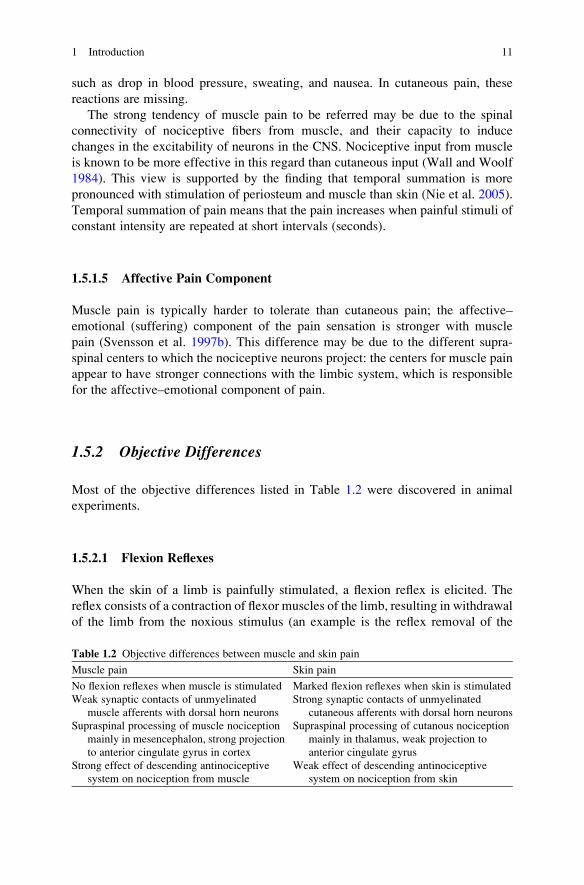
Most of the objective differences listed in Table 1.2 were discovered in animal
experiments.
1.5.2.1 Flexion Reflexes
When the skin of a limb is painfully stimulated, a flexion reflex is elicited. The
reflex consists of a contraction of flexor muscles of the limb, resulting in withdrawal
of the limb from the noxious stimulus (an example is the reflex removal of the
Table 1.2 Objective differences between muscle and skin pain
Muscle pain Skin pain
No flexion reflexes when muscle is stimulated Marked flexion reflexes when skin is stimulated
Weak synaptic contacts of unmyelinated
muscle afferents with dorsal horn neurons
Strong synaptic contacts of unmyelinated
cutaneous afferents with dorsal horn neurons
Supraspinal processing of muscle nociception
mainly in mesencephalon, strong projection
to anterior cingulate gyrus in cortex
Supraspinal processing of cutanous nociception
mainly in thalamus, weak projection to
anterior cingulate gyrus
Strong effect of descending antinociceptive
system on nociception from muscle
Weak effect of descending antinociceptive
system on nociception from skin
1 Introduction 11

hand from a flame). Teleologically, the lack of flexion reflexes elicited from
muscle makes sense, because a painful lesion in muscle is not influenced by flexing
the limb.
1.5.2.2 Differences in Synaptic Effectiveness and CNS Targets of
Afferent Fibers
In animal experiments, the weak synaptic contacts between unmyelinated muscle
afferents and dorsal horn neurons become obvious when the action potentials of dorsal
horn neurons are recorded. Approximately 10% of the dorsal horn neurons respond to
the stimulus when unmyelinated muscle afferents are stimulated electrically; the
proportion is greater than 30% to stimulation of a cutaneous nerve (sural n.) (Hoheisel
et al. 1997). One of the reasons for this difference is the tonic inhibition of nociceptive
fibers from muscle by activity in thick afferent fibers from the skin. When the thick
fibers are blocked pharmacologically, the unmyelinated fibers frommuscle havemuch
stronger synaptic effects (Lambertz et al. 2006). This means that generally the
excitatory action of nociceptive fibers from muscle in the spinal cord is small, but
their modulating effects on dorsal horn neurons are strong.
Dorsal horn cells driven by muscle nociceptors show marked convergence of
input from skin and other deep somatic tissues and probably also from viscera
(Grubb et al. 1993; Yu and Mense 1990), i.e., these neurons can be excited by
stimulation of all these tissues. This applies particularly to neurons having input
from soft tissues in the low back (Taguchi et al. 2008).
Dorsal horn cells processing information from cutaneous nociceptors show little
convergence, i.e., they cannot be driven by input from different types of tissue (e.g.,
skin, muscle, viscera). Many appear to receive input exclusively from nociceptors
in the skin. The highly convergent input from various tissues to neurons processing
information from muscle nociceptors may contribute to the diffuse nature of muscle
pain in comparison to cutaneous pain.
In the CNS, the information from muscle and cutaneous nociceptors is processed
differently. One example is that the nociceptive information carried in spinal
afferent fibers from deep tissues and the skin, respectively, terminates in different
regions of the spinal cord: noxious stimulation of skin excites neurons in all laminae
of the dorsal horn, whereas following muscle stimulation, no neurons in lamina II
(substantia gelatinosa) are activated (Ohtori et al. 2000). In the mesencephalon,
there are similar differences: Neurons responding to stimulation of muscle noci-
ceptors are located in the ventral periaqueductal gray matter (PAG), whereas cells
responding to cutaneous nociceptors are located in the lateral PAG (Keay and
Bandler 1993; Ohtori et al. 2000). Thalamic nuclei with strong spinal nociceptive
input from the skin are the ventral posterolateral (VPL) and medial intramedullary
nuclei, whereas for nociceptive input from muscle, the ventrolateral (VL) nucleus is
an important relay (Gholami et al. 2006).
In other brain areas, too, differences in the termination between nociceptive input
from muscle and skin were found. In the rat, a particularly strong input from muscle
12 S. Mense and R.D. Gerwin
nociceptors was found in the accumbens and amygdala nuclei as well as the para-
ventricular nucleus of the hypothalamus (Ohtori et al. 2000). In an fMRI study of the
cortex in humans, a stronger activation of the anterior cingulate gyrus was found
when a skeletal muscle was stimulated in comparison to stimulation of the overlying
skin. However, in this study the differences were not significant (Svensson et al.
1997a). The authors concluded that “the perceived differences between acute skin
and muscle pain are mediated by differences in the intensity and temporospatial
pattern of neuronal activity within similar sets of forebrain structures.”
1.6 Descending Pain-Modulating Influences
The PAG in the mesencephalon – one of the main supraspinal centers for the
processing of nociceptive impulses from muscle – is also the site of origin of
descending pain-modulating pathways (Fields 2004). There are not only pathways
that inhibit pain sensation (antinociceptive tracts) but also those that facilitate
(enhance) them (pronociceptive tracts). In electrophysiological experiments on
rats, a stronger action of the descending antinociceptive system on dorsal horn
neurons mediating nociception from muscle was found (Yu and Mense 1990). This
finding may relate to the observation that long-lasting physical exertion (e.g., in
sports competitions) which is known to activate the descending antinociceptive
tracts, reduces pain sensations from deep somatic tissues to an extent that lesions of
the musculoskeletal system go unnoticed.
1.7 Transition from Acute to Chronic Pain
One definition of chronic pain is that pain which persists past the normal time of
healing; in clinical practice, a period of 3–6 months is accepted as a dividing line
between acute and chronic pain (Merskey and Bogduk 1994). Muscle pain is well-
known for its tendency to become chronic.
In chronic pain patients, the pain has lost the function of a warning system; it has
become a disease of its own. The factors controlling the transition from acute to
chronic muscle pain are still under investigation. In Chap. 4, some of the mechan-
isms governing this transition are presented in more detail.
1.8 Interactions Between Psychosocial and Somatic Factors
There can be no doubt that psychosocial factors modulate pain perception. This is
particularly valid for chronic pain. Since it is likely that affective status has some
influence on pain perception, the remaining questions are by what mechanisms
and by what magnitude do psychosocial influences cause somatic dysfunctions.
1 Introduction 13

This problem may be linked to another question, namely why one patient with an
acute lesion recovers normally and another patient with the same lesion develops
chronic pain. The answer that in one patient the lesion resolved, relieving the pain,
whereas in the other the lesion did not resolve and the pain persisted, is much too
simple. There is general agreement that exactly the same lesions in two patients can
show quite different time courses, one resolving completely and the other leading
to chronic pain. Therefore, as well as somatic factors, other influences must
be involved. A possible explanation is that in different patients the descending
pain-inhibiting or -facilitating pathways have different degrees of activity. It is
conceivable that a low tonic discharge rate in neurons of the descending pain-
inhibiting pathway is associated with a propensity to develop chronic pain.
Another explanation is the influence of psychosocial factors. This does not mean
that in these cases the chronic pain is psychogenic; pain is aggravated and perpe-
tuated by psychosocial problems and dysfunctions. The neuroanatomic basis of
these influences is strong connections between the limbic system (e.g., the amyg-
dala which is involved in fear and stress reactions) and those mesencephalic nuclei
that mediate muscle pain. The psychosocial factors are often overlooked because
they are so varied, and it takes great experience to identify them. In some patients,
psychological stressors can be potent aggravating factors (for instance, in fibromy-
algia and chronic temporomandibular pain). For details, see Chaps. 4–7 in compa-
nion volume by Mense and Gerwin (2010).
References
Baron R, J€anig W, Kollmann W (1988) Sympathetic and afferent somata projecting in hindlimb
nerves and the anatomical organization of the lumbar sympathetic nervous system of the rat.
J Comp Neurol 275:460–468
Bielefeldt K, Gebhart GF (2006) Visceral pain: basic mechanisms, Chap. 48. In: McMahon SB,
KoltzenburgM (eds)Wall andMelzack´s textbook of pain. Churchill Livingstone, Philadelphia
Fields H (2004) State-dependent opioid control of pain. Nat Rev 5:565–575
Gholami S, Lambertz D, Hoheisel U et al. (2006) Effects on c-Fos expression in the PAG and
thalamus by selective input via tetrodotoxin-resistant afferent fibres from muscle and skin.
Neurosci Res 56:270–278
Graven-Nielsen T, Mense S, Arendt-Nielsen L (2004) Painful and non-painful pressure sensations
from human skeletal muscle. Exp Brain Res 59:273–283
Grubb BD, Stiller RU, Schaible HG (1993) Dynamic changes in the receptive field properties of
spinal cord neurons with ankle input in rats with chronic unilateral inflammation in the ankle
region. Exp Brain Res 92:441–452
Hoheisel U, Sander B, Mense S (1997) Myositis-induced functional reorganization of the rat
dorsal horn: effects of spinal superfusion with antagonists to neurokinin and glutamate
receptors. Pain 69:219–230
Hoheisel U, Unger T, Mense S (2005) Excitatory and modulatory effects of inflammatory
cytokines and neurotrophins on mechanosensitive group IV muscle afferents in the rat. Pain
114:168–176
Ju G, Hokfelt T, Brodin E et al. (1987) Primary sensory neurons of the rat showing calicitonin
gene-related peptide immunoreactivity and their relation to substance P, somatostatin-,
14 S. Mense and R.D. Gerwin

galanin-, vasoactive intestinal polypeptide- and cholecystokinin-immunoreactive ganglion
cells. Cell Tissue Res 247:417–431
Keay KA, Bandler R (1993) Deep and superficial noxious stimulation increases Fos-like immu-
noreactivity in different regions of the midbrain periaqueductal grey of the rat. Neurosci Lett
154:23–26
Kellgren JH (1938) Observations on referred pain arising from muscle. Clin Sci 3:175–190
Lambertz D, Hoheisel U, Mense S (2006) Distribution of synaptic field potentials induced by
TTX-resistant skin and muscle afferents in rat spinal segments L4 and L5. Neurosci Lett
409:14–18
Lewis T (1942) Pain. MacMillan, London (citation taken from facsimile edition 1981)
Light AR, Perl ER (2003) Unmyelinated afferent fibres are not only for pain anymore. J Comp
Neurol 461:137–139
Loeser JD, Treede R-D (2008) The Kyoto protocol of IASP basic pain terminology. Pain 137:
473–477
Marchettini P, Cline M, Ochoa JL (1990) Innervation territories for touch and pain afferents of
single fascicles of the human ulnar nerve. Mapping through intraneural microrecording and
microstimulation. Brain 113:1491–1500
May A (2007) Neuroimaging: visualising the brain in pain. Neurol Sci 28:101–107
McCloskey DI, Mitchell JH (1972) Reflex cardiovascular and respiratory responses originating in
exercising muscle. J Physiol 224:173–186
Mense S, Gerwin RD (eds) (2010) Muscle pain: diagnosis and treatment. Springer, Heidelberg
Merskey H, Bogduk N (1994) Classification of chronic pain: descriptions of chronic pain syn-
dromes and definitions of pain terms, 2nd edn. IASP Press, Seattle
Nie H, Arendt-Nielsen L, Andersen H et al. (2005) Temporal summation of pain evoked by
mechanical stimulation in deep and superficial tissue. J Pain 6:348–355
Nikolajsen L, Jensen, TS (2006) Phantom limb, Chap 61. In: McMahon SB, Koltzenburg M (eds)
Wall and Melzack’s textbook of pain. Churchill Livingstone, Philadelphia
Ohtori S, Takahashi K, Chiba T et al. (2000) Fos expression in the rat brain and spinal cord evoked
by noxious stimulation to low back muscle and skin. Spine 25:2425–2430
Scholz J, Woolf CJ (2002) Can we conquer pain? Nat Neurosci 5:1062–1067
Staff PH (1988) Clinical consideration in referred muscle pain and tenderness – connective tissue
reactions. Eur J Appl Physiol 57:369–372
Svensson P, Minoshima S, Beydoun A et al. (1997a) Cerebral processing of acute skin and muscle
pain in humans. J Neurophysiol 78:450–460
Svensson P, Beydoun A, Morrow TJ et al. (1997b) Human intramuscular and cutaneous pain:
psychophysical comparisons. Exp Brain Res 114:390–392
Taguchi T, Hoheisel U, Mense S (2008) Dorsal horn neurons having input from low back
structures in rats. Pain 138(1):119–129
Torebjork HE, Ochoa JL, Schady W (1984) Referred pain from intraneural stimulation of muscle
fascicles in the median nerve. Pain 18:145–156
von Frey M (1896) Untersuchung €uber die Sinnesfunktionen der menschlichen Haut. Abhandlun-
gen der mathematisch-physischen Klasse der Koniglichen S€achsischen Gesellschaft der
Wissenschaften. Koniglichen S€achsischen Gesellschaft der Wissenschaften, Leipzig
Wall PD, Woolf CJ (1984) Muscle but not cutaneous C-afferent input produces prolonged
increases in the excitability of the flexion reflex in the rat. J Physiol 356:443–458
Yu X-M, Mense S (1990) Response properties and descending control of rat dorsal horn neurons
with deep receptive fields. Neuroscience 39:823–831
1 Introduction 15
Chapter 2
Functional Anatomy of Muscle: Muscle,
Nociceptors and Afferent Fibers
S. Mense
Contents
2.1 Structure and Basic Function of Skeletal Muscle . . . . . . . . . . . . . . . . . . . . . . . . . . . . . . . . . . . . . . . . 18
2.2 Morphology of Muscle Nociceptors . . . . . . . . . . . . . . . . . . . . . . . . . . . . . . . . . . . . . . . . . . . . . . . . . . . . . 25
2.2.1 Structure of Muscle Nociceptors in the Light and Electron Microscope . . . . . . . . . 25
2.2.2 Receptor Molecules in the Membrane of Nociceptors . . . . . . . . . . . . . . . . . . . . . . . . . . . 28
2.2.3 Neuropeptide Content of Nociceptors . . . . . . . . . . . . . . . . . . . . . . . . . . . . . . . . . . . . . . . . . . . 33
2.3 The Nociceptive Afferent Fiber . . . . . . . . . . . . . . . . . . . . . . . . . . . . . . . . . . . . . . . . . . . . . . . . . . . . . . . . . . 35
2.4 Fiber Composition of a Muscle Nerve . . . . . . . . . . . . . . . . . . . . . . . . . . . . . . . . . . . . . . . . . . . . . . . . . . . 37
2.5 Muscle Receptors Other Than Nociceptors . . . . . . . . . . . . . . . . . . . . . . . . . . . . . . . . . . . . . . . . . . . . . . 39
2.6 Free Nerve Endings in Tendon . . . . . . . . . . . . . . . . . . . . . . . . . . . . . . . . . . . . . . . . . . . . . . . . . . . . . . . . . . 40
2.7 Free Nerve Endings in Fascia . . . . . . . . . . . . . . . . . . . . . . . . . . . . . . . . . . . . . . . . . . . . . . . . . . . . . . . . . . . . 41
2.8 Efferent Functions of Nociceptors . . . . . . . . . . . . . . . . . . . . . . . . . . . . . . . . . . . . . . . . . . . . . . . . . . . . . . . 41
2.8.1 Release of Neuropeptides from the Nociceptive Ending . . . . . . . . . . . . . . . . . . . . . . . . . 41
2.8.2 The Axon Reflex . . . . . . . . . . . . . . . . . . . . . . . . . . . . . . . . . . . . . . . . . . . . . . . . . . . . . . . . . . . . . . . . . 43
2.8.3 Neurogenic Inflammation . . . . . . . . . . . . . . . . . . . . . . . . . . . . . . . . . . . . . . . . . . . . . . . . . . . . . . . . 44
References . . . . . . . . . . . . . . . . . . . . . . . . . . . . . . . . . . . . . . . . . . . . . . . . . . . . . . . . . . . . . . . . . . . . . . . . . . . . . . . . . . . . . . 45
Abstract Nociceptors are free nerve endings, but not all free nerve endings in
skeletal muscle are nociceptive. Nociceptive free nerve endings are connected
to the CNS by thin myelinated fibers or unmyelinated afferent fibers. In the light
microscope, free nerve endings look like a string of beads, i.e., they consist of axonal
expansions (varicosities) connected by thin axonal segments. The neuropeptide
substance P has been reported to be present predominantly in nociceptive afferent
fibers.
In the electron microscope, a prominent feature of nociceptive nerve endings
is that they are not free in the strict sense but ensheathed by Schwann cells.
S. Mense
Universitat Heidelberg, Mediz. Fakultat Mannheim CBTM, Neuroanatomie, Ludolf-Krehl-Str.
13-17, R. C614, 68167, Mannheim, Germany
e-mail: [email protected]
S. Mense and R.D. Gerwin (eds.), Muscle Pain: Understanding the Mechanisms,DOI 10.1007/978-3-540-85021-2_2, # Springer-Verlag Berlin Heidelberg 2010
17
Top Related