


Languages
Pages
Legal


Management of Penetrating Neck Injury (PNI)
Gamal Marey SUNY Downstate Medical Center
4/17/2014
www.downstatesurgery.org

History
22 y/o man, BIBEMS to ED, ETOH (+), s/p stab wound to the left posterior neck, LOC(-).
• PMH- Denies • PSH- Denies • Meds.- None • SH- smokes 1PPD, ETOH occasionally • Allergy- NKDA
www.downstatesurgery.org

Physical Exam
Primary Survey- GCS 15, ABCD/ WNL • VS: BP132/82 T98.4 P100 RR20 O2100 Secondary Survey- • Head: no lacerations, no deformity, no crepitus • Neck: 2 cm transverse wound in the posterior
cervical triangle at the level of the angle of the mandible (at the junction of zone 2 & zone 3), small hematoma with some external bleeding, trachea central, no subcut. emphysema, no C-spine tenderness
www.downstatesurgery.org

Physical Exam
• Chest: CTAB, no point of tenderness • Abdomen: SNTND, no ecchymosis • Pelvis: symmetrical, no crepitus, no
tenderness • Back: no tenderness, no step off def. • Rectal exam: normal tone, no blood • Ext: symmetrical, no deformity, intact pulses
2+ B/L, intact motor and sensory
www.downstatesurgery.org

Labs
• CBC- 6.47/14.0/41.6/218 • BMP-139/3.1/100/24/10/0.78/103 • LFTs-7.4/4.7/39/26/86/0.2 • A/L-107/44 Lactate- 2.6 • VBG- 7.36/46/43/74/24/0.4 • Coags-10/27/0.9 ETOH 94.8 • Urinalysis- negative UTOX- negative
www.downstatesurgery.org

CXR
www.downstatesurgery.org

CTA Neck
www.downstatesurgery.org

CTA Neck
www.downstatesurgery.org

CTA Neck
www.downstatesurgery.org

CTA Neck
www.downstatesurgery.org

OR
• left anterior neck exploration • Anterior sternocleidomastoid incision • wound exploration • Control of muscular bleed • Discharged home POD#1
www.downstatesurgery.org

Q?
www.downstatesurgery.org

Overview
• History • Epidemiology • Anatomy • Diagnosis • Management • Clinical Cases • Conclusions
www.downstatesurgery.org

History
• Ambrose Paré (1510-1590)- First documented surgical intervention by ligating the carotid artery and jugular vein of a wounded French soldier.
• Homer’s Iliad –Achilles delivered a fatal lance to Hector’s neck.
• American Civil War-> 4000 cases of neck injuries • World War I- expectant management, mortality
35% • World War II/Bailey (1944)- early exploration if
deep to platysma.
www.downstatesurgery.org
History
• Fogelman & Stewart (1956) - 6% mortality in early exploration vs. 35% if delayed or no intervention. 56% negative exploration rate.
• Monson (1969)- neck zones description. • Roon & Christensen (1979)- Mandatory
exploration for zone II and angiogram for stable zone I&III.
• 1980s - Selective Surgical Management concept (identify patients who would benefit from surgical management), based on clinical exam and adjunctive tests. 86% positive exploration
www.downstatesurgery.org

Epidemiology
• PNIs defined by platysma violation • PNIs- 5 % of traumatic injuries in adults. • Stab injuries (40%)- Knife, razor blades, glass. • Projectile injuries(45%)- Handgun, Rifle,
Shotgun. • Low velocity vs high velocity injuuries • Injuries- 50% GSW vs 10-20% Stab wounds • Overall mortality- 3-10%
www.downstatesurgery.org

Epidemiology
• Most common( zone II > zone I > zone III) • Mortality is highest with zone 1. • Kinetic Energy of Projectile & Muzzle velocity ->More energy = More damage • Most vital structure in the anterior triangle • PNIs to posterior Triangle much lower chance of
significant injury. Platysma violation, mandates carful search for
aerodigestive and neurovascular injuries.
www.downstatesurgery.org

Epidemiology
Structures injured • No significant damage 40% • Major vein 15-25% • Major artery 10-15% • Digestive tract 5-15% • Respiratory tract 4-12% • Major nerves 3-8% GSW tract can transgress zone boundaries,
superficial wound may not correspond well to deeper structures injured.
www.downstatesurgery.org

Anatomy www.downstatesurgery.org

Anatomy
• Deep to platysma is deep cervical fascia
www.downstatesurgery.org
Anatomy
• Skeletal (cervical vertebrae & hyoid bone) • Nervous (spinal cord, IX,X,XI,XII CN, symp. chain ) • Respiratory (Oropharynx,larynx, trachea) • Gastrointestinal (oropharynx, esophagus) • Vascular ( common,internal and external carotid
arteries, vertebral arteries, internal and external jugular veins)
• Lymphatic (thoracic duct) • Endocrine (thyroid and parathyroid glands) • Immune (cervical extension of thymus)
www.downstatesurgery.org
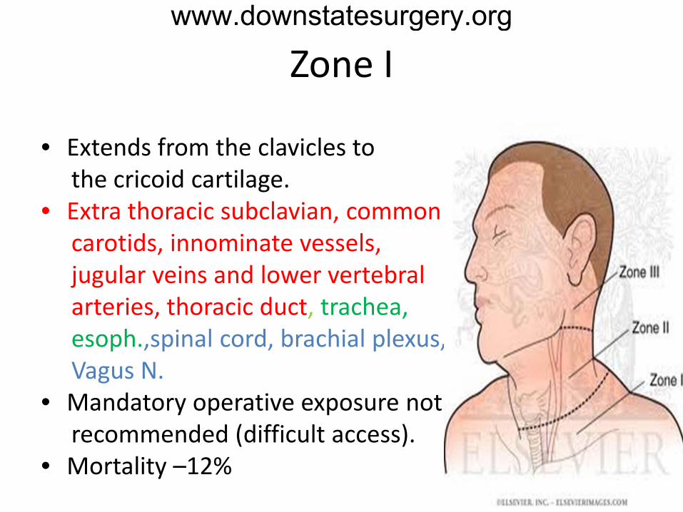
Zone I
• Extends from the clavicles to the cricoid cartilage. • Extra thoracic subclavian, common carotids, innominate vessels, jugular veins and lower vertebral arteries, thoracic duct, trachea, esoph.,spinal cord, brachial plexus, Vagus N. • Mandatory operative exposure not recommended (difficult access). • Mortality –12%
www.downstatesurgery.org

Zone II
• Most commonly involved 50-75% • Extends from the cricoid cartilage to the angle of the mandible. • common carotid and bifurcation, vertebral arteries, jugular veins, Larynx, trachea, esoph., spinal • Cord, (CN. X,XI,XII) • Operative exposure (easy access). • Selective vs Mandatory.
www.downstatesurgery.org

Zone III • Dangerous Area dt. Proximity of vasculature to skull base. • Extends from the angle of the mandible to the mastoid process. • External carotid branches, internal carotid artery, vertebral artery and internal jugular, facial veins, Pharynx, oral cavity, spinal cord, (CR. VII,IX, X,XI,XII). • Mandatory operative exposure not recommended (difficult access).
www.downstatesurgery.org

Laryngotracheal Injuries o 10% PNIs will have laryngotracheal trauma o Trachea most commonly involved (2/3), larynx (1/3) o Mortality 20% o 25% have esophageal injury • Airway compromise • Massive subcutaneous emphysema • Air bubbling through wound • Hemoptysis • Odynophagia, dysphonia
www.downstatesurgery.org

Pharyngoesophageal Injuries
o 10% PNIs will have pharyngoesophageal trauma o 30% no signs or symptoms o Leading cause of delayed M&M. o Mortality 22% • Dysphagia • Hematemesis • Subcutaneous emphysema
www.downstatesurgery.org

Vascular Injuries Soft Signs • Mild Bleeding • Nonexpanding hematoma • Paresthesia Hard signs • Severe external bleeding • Expanding hematoma • Pulsatile swelling • Bruit, thrill • Pulse deficit • Neurologic deficit
www.downstatesurgery.org

Nervous Injuries
CNS- Spinal cord. PNS- CN. VII through XII, sympathetic chain,
peripheral nerve roots, brachial plexus. • Neurogenic shock • Brown-Sequard syndrome • Horner’s syndrome • Speech/ movement of the tongue • Shoulder Shrug
www.downstatesurgery.org

Diagnosis
Clinical Exam Imaging • CTA neck • IR Angiography • Carotid Ultrasound • esophagography Endoscopy • Flexible laryngoscopy, bronchoscopy • esophagoscopy
www.downstatesurgery.org

CT Angiography
Indications: Stable patients with PNIs • A thorough physical examination is highly
sensitive (>95%) for detecting arterial vascular injury but a lower sensitivity for aerodigestive tract injuries. • CTA - highly sensitivity for detecting vacsular and
aerodigestive tract injuries • CTA- reduced the need for operative neck
exploration • Negative CTA, no symptoms Observation
www.downstatesurgery.org

IR Angiography
• Gold standard for vascular injury • Diagnostic & therapeutic • Zones I & III difficult to assess clinically and
often involve complex surgery • Cost-effective for zones I & III • Decreased surgery rates to 5% in zone I and
13% in zone III
www.downstatesurgery.org
Endoscopy
Indications: • Negative CTA imaging but concerning
trajectory • Intra-Operative, concern for aerodigestive
injury Expeditious evaluation increased morbidity with delayed esophageal repair Sensitivity 92.4%, specificity 100%
www.downstatesurgery.org

Management • Clinical exam, ACLS 1ry &2ry Survey • Secure Airway (avoid techniques not done under direct
vision) modest or moderate symptoms or signs • undergo a diagnostic evaluation(CTA, Endoscopy, IR) • Expectant and selective operative management vs IR (zone I&III) ‘‘hard signs’’ or hemodynamic instability • Life threating bleeding- direct pressure/ insert Foley • Activate massive transfusion • OR
www.downstatesurgery.org

Vascular Operative Exposure
• Zone II- anterior SCM incision or Transverse cervical collar incision for transcervical injuries • Zone I- Median sternotomy with extension to
an anterior SCM incision or supraclavicular incision with or without clavicular head resection.
• Zone III- subluxation, dislocation, or resection of the mandible for distal control.
www.downstatesurgery.org
Vascular injuries Management
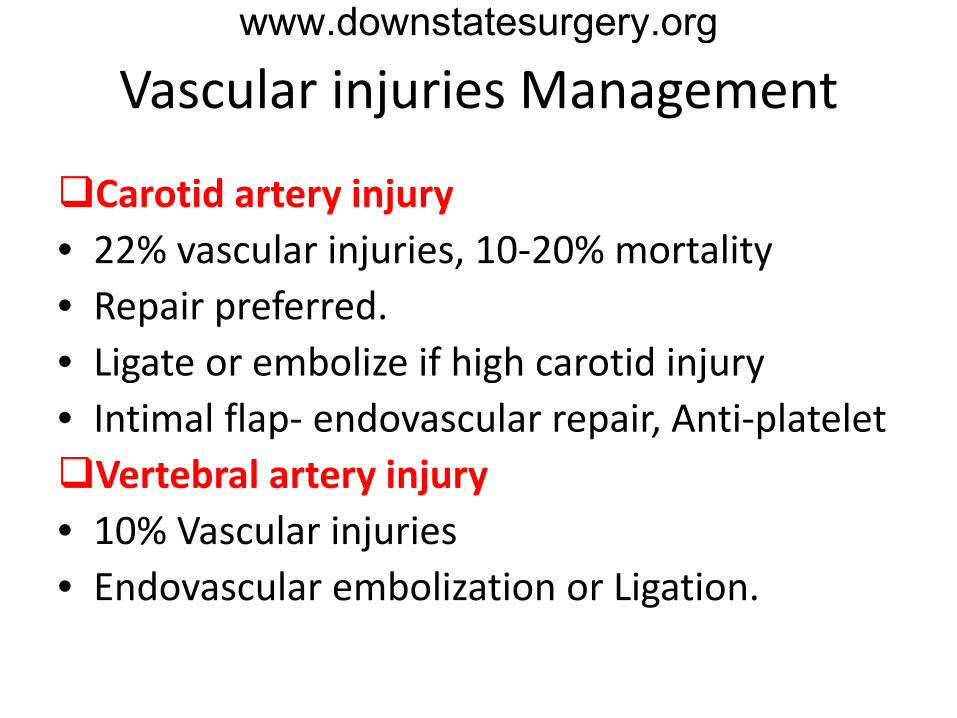
Carotid artery injury • 22% vascular injuries, 10-20% mortality • Repair preferred. • Ligate or embolize if high carotid injury • Intimal flap- endovascular repair, Anti-platelet Vertebral artery injury • 10% Vascular injuries • Endovascular embolization or Ligation.
www.downstatesurgery.org

Airway Injuries management
Stable airway: CTA, Flexible laryngoscopy, bronchoscopy Unstable airway: Be prepared for surgical
airway, tracheotomy safest option Tracheal injury: debridement, primary repair
with absorbable suture, Interposition of well-vascularized tissue between tracheal and esophageal injuries reduce fistula rate
www.downstatesurgery.org

Esophageal Injuries Management
19% mortality, 41% morbidity in delayed repair Goals: early operative management,
debridement, primary closure with buttressing and adequate drainage
Esophagoscopy: if high suspicion but studies negative24 hrs observation
Early diagnosis: primary repair Late diagnosis: drainage/resection/diversion Pharyngeal injury NPO, IV antibiotics, NGT
www.downstatesurgery.org

www.downstatesurgery.org

www.downstatesurgery.org

www.downstatesurgery.org

Conclusion • Mechanism of injury is emphasized • Thorough physical examination is key • CT Angiography, stable patients (All zones), effective and
reliable (less neg. exploration Rate), allow for less utilization of services.
• Hard signs/unstable patients, immediate surgical exploration of any zone +/- Angiogram (zone I&III)
• Esophagram/flexible esophagoscopy/laryngoscopy, if suspect or see injury on CTA early repair is a key
• Stable patients with Zone I and III injury undergo angiography and endoscopy
• Stable patients with Zone II injury CTA, Selective testing, Selective Surgical Exploration
www.downstatesurgery.org

References 1. Gracias VH, Reilly PM, PhilpottJ, et al. Computed tomography in the evaluation of penetrating neck trauma: a
preliminary study. Arch Surg2001;136:1231-5. 2. MuneraF, Danton G, Rivas LA. Multidetectorrow computed tomography in the management of penetrating neck
injuries. SeminUltrasound CT MRI 2004;30:195-204. 3. McConnell DB, Trunkey DD. Management of penetrating trauma to the neck. Adv Surg 1994; 27:97. 4. Apffelstaedt JP, Müller R. Results of mandatory exploration for penetrating neck trauma. World J Surg 1994;
18:917. 5. Asensio JA, Valenziano CP, Falcone RE, Grosh JD. Management of penetrating neck injuries. The controversy
surrounding zone II injuries. Surg Clin North Am 1991; 71:267 6. Bell RB, Osborn T, Dierks EJ, et al. Management of penetrating neck injuries: a new paradigm for civilian trauma.
J Oral Maxillofac Surg 2007; 65:691. 7. Demetriades D, Asensio JA, Velmahos G, Thal E. Complex problems in penetrating neck trauma. Surg Clin North
Am 1996; 76:661. 8. 12.Mittal VK, Paulson TJ, Colaiuta E, et al. Carotid artery injuries and their management. J Cardiovasc Surg
(Torino) 2000; 41:423. 9. 13.Asensio JA, Berne J, Demetriades D, et al. Penetrating esophageal injuries: time interval of safety for
preoperative evaluation--how long is safe? J Trauma 1997; 43:319. 10. RoonAJ, Christensen N. Evaluation and Treatment of Penetrating Cervical Injuries. J Trauma.1979;19:391-7. 11. •ZaidiSMH, Ahmad R. Penetrating neck trauma: a case for conservative approach. Am J Otolaryng.2011;32:591-
6. 12. O’Brien PJ, Cox MW. A modern approach to cervical vascular trauma. PerspectVascSurgEndovascTher2011 23:
90.
www.downstatesurgery.org

Thank You
www.downstatesurgery.org
Top Related