


Languages
Pages
Legal


Magnetic Resonance Atlas of Skeletal Development of the Knee
A Standard of Reference
Andrew T. Pennock, MDJames D. Bomar, MPH
Rady Children’s Hospital, San DiegoUniversity of California, San Diego

2©SD PedsOrtho

3©SD PedsOrtho
DisclaimerIn view of the possibility of human error or chang-es in medical science, neither the authors, nor the institutions warrant that the information contained
in this atlas is accurate or complete, and they are not responsible for any errors or omission or the results obtained from the use of such information.

4©SD PedsOrtho

5©SD PedsOrtho
Contents
Introduction
Atlas creation
Instructions for use
Anatomic and radiographic terms
Standards Female – Age 1 Male – Age 1
Validation
References
7
9
15
20
2441
59
60

6©SD PedsOrtho

7©SD PedsOrtho
Introduction
Proper assessment of skeletal development is es-sential for managing many conditions of bone and joints. Interest in this topic began nearly 100 years ago by T. Wingate Todd a Professor of Anatomy at Western Reserve University. In 1926 a series of studies in Cleveland, OH were initiated with a goal of creating radiographic standards of reference for skeletal maturation based on the hand, elbow, shoulder, hip, knee, and foot. Between 1931 and 1937, tentative developmental standards were assembled for each of these joints. Unfortunately,
1. It is based on a single left hand radiograph and the maturation of other joints (such as the knee) have been shown to be independent of the hand.
2. The original “source films” came from “care-fully selected” patients from Cleveland, OH which were largely Caucasian with higher so-cioeconomic backgrounds. This selection bias has potentially limited the applicability of this data to larger and more diverse patient popu-lations seen across many urban areas in the United States.
While widely used, there are limitations and drawbacks to the Greulich and Pyle Atlas:
History of Bone Age Assessment
Drawbacks of the Greulich and Pyle Atlas
with the untimely death of Professor Todd, most of these series were never completed. Fortunately, William Walter Greulich and S. Idell Pyle carried on the work of Professor Todd and published the Radiographic Atlas of Skeletal Development of the Hand and Wrist in 1950. Since its publication, this reference has become the “gold standard” for assessing bone age and it is widely used across multiple sub-specialties including endocrinology, pediatrics, and orthopedics.
3. The “source data” is nearing 100 years old and it is unclear whether these standards still apply to children that are now entering puberty at an earlier age.
4. Obtaining an additional left hand radiograph exposes patients to additional ionizing radi-ation, adds additional cost to the health care system, and potentially slows clinic efficiency.
8©SD PedsOrtho
In 1969, S. Idell Pyle and Normand L. Hoerr pub-lished a separate reference entitled Radiographic Atlas of Skeletal Development of the Knee. This atlas was created using an identical methodology as the Greulich and Pyle Atlas from knee radio-graphs that were performed between 1928 and 1942. Interestingly, this atlas is rarely referenced and has not been routinely used in the bone age assessment of patients with knee specific condi-tions. The reason for this in unclear, but possi-
To date, several studies within the forensic liter-ature have evaluated the utility of knee MRIs for assessing chronological age. The rationale for these studies has been to develop better tools for assessing an accurate age of asylum seekers and resolving immigration proceedings (particularly in Europe). As such, these studies have almost ex-
The aim of this project was to create a new atlas of knee MRIs across a spectrum of pediatric ages that would be comprehensive enough to discrimi-nate patient bone age during the critical pre-ado-
Alternative Knee Atlas
bilities include (1) the fact that subtle differences between certain age standards (especially in ad-olescence) can be subtle and difficult to reproduc-ibly identify, (2) compared to the hand and wrist where there are many bones and growth plates, there are less distinguishing radiographic features about the knee to help differentiate certain ages, (3) unlike the Greulich and Pyle Atlas, the knee atlas combines the male and female standards.
clusively focused on the ages of 14 to 30 years, as legal responsibility applies to individuals be-tween the ages of 14 and 22 years in most coun-tries. Therefore, the existing forensic literature is largely inadequate for conditions that require an accurate MRI bone age for patients under the age of 14 years.
lescent and adolescent years of 11 to 18 as well as to adequately cover the earlier period from 2 to 10 years.
Bone Age using Magnetic Resonance Imaging
Purpose of the Current Atlas

9©SD PedsOrtho
Atlas Creation
• The knee MRI atlas was created in a similar fashion as the Greulich and Pyle Hand Atlas and the Pyle and Hoerr Knee Atlas.
• First, a preliminary series of skeletal maturity indicators were identified by examining knee MRIs across of a spectrum of skeletal matu-rity.
• In reviewing hundreds of MRIs, several indi-cators were identified and independently eval-uated for each bone (femur, tibia, patella, and fibula).
Selection of Skeletal Maturity Indicators
• These features were found to be most identi-fiable and reproducible on the coronal T1 and sagittal T1 images.
• For each bone, a single standardized coronal and sagittal slice was identified.
• A 1.5 Tesla magnet was utilized for all MRIs in this series
The coronal slice through the center of the distal aspect of the femur at the at-tachment site of the posterior cruciate ligament on the medial femoral condyle was selected.
The sagittal slice through the center of the medial femoral condyle was select-ed
Femur Slice Standardization

10©SD PedsOrtho
The following features of the femur were identi-fied: the presence of the epiphyseal secondary (2°) ossification center, complete ossification of the epiphysis, disappearance of the laminated
Femur Specific Features
appearance of the subchondral epiphyseal carti-lage (termed the “Oreo” sign), narrowing of the physis, partial closure of the physis, and complete closure of the physis.
11©SD PedsOrtho
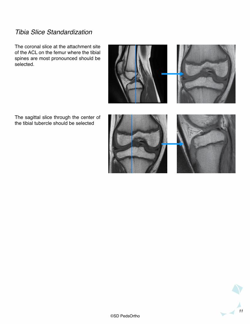
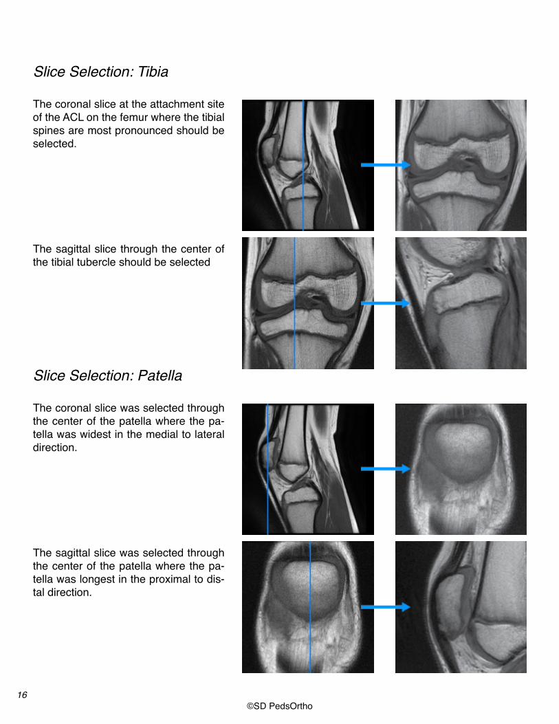
The coronal slice at the attachment site of the ACL on the femur where the tibial spines are most pronounced should be selected.
The sagittal slice through the center of the tibial tubercle should be selected
Tibia Slice Standardization
12©SD PedsOrtho
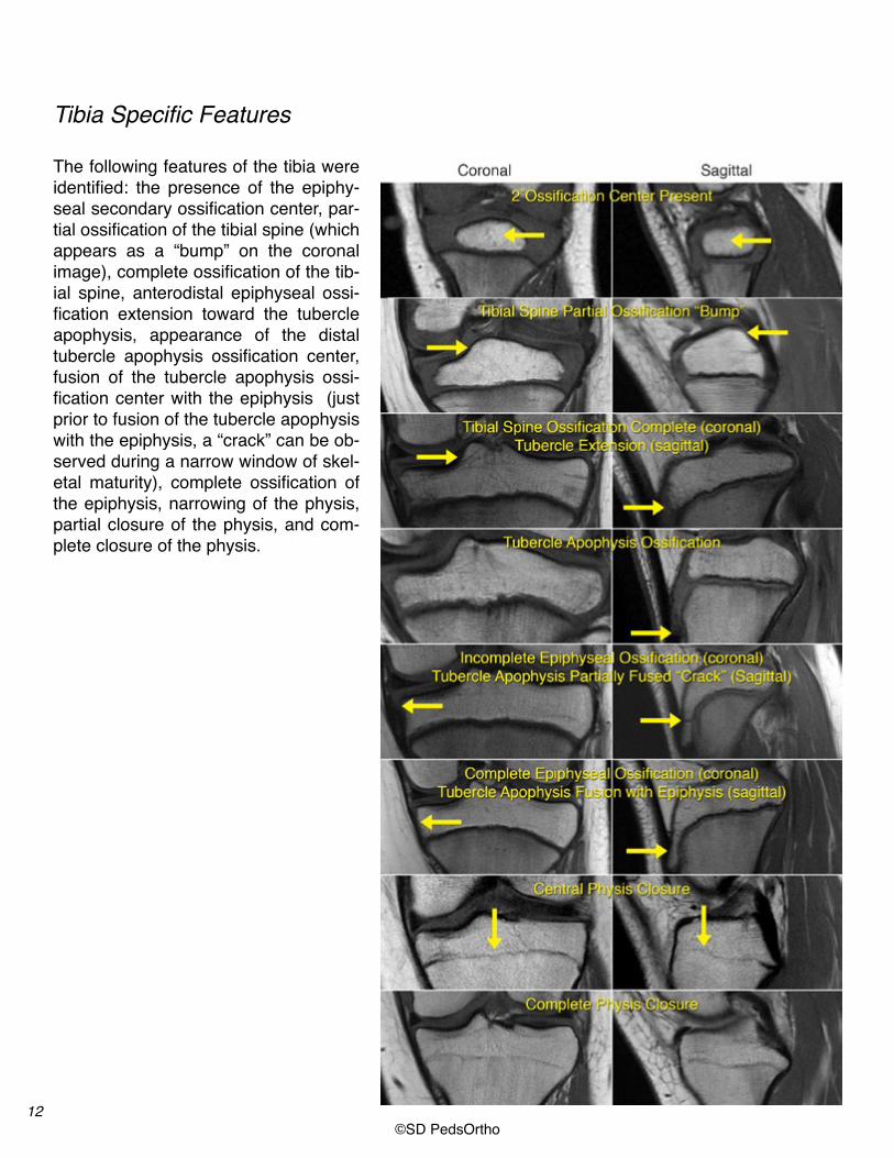
Tibia Specific Features
The following features of the tibia were identified: the presence of the epiphy-seal secondary ossification center, par-tial ossification of the tibial spine (which appears as a “bump” on the coronal image), complete ossification of the tib-ial spine, anterodistal epiphyseal ossi-fication extension toward the tubercle apophysis, appearance of the distal tubercle apophysis ossification center, fusion of the tubercle apophysis ossi-fication center with the epiphysis (just prior to fusion of the tubercle apophysis with the epiphysis, a “crack” can be ob-served during a narrow window of skel-etal maturity), complete ossification of the epiphysis, narrowing of the physis, partial closure of the physis, and com-plete closure of the physis.
13©SD PedsOrtho
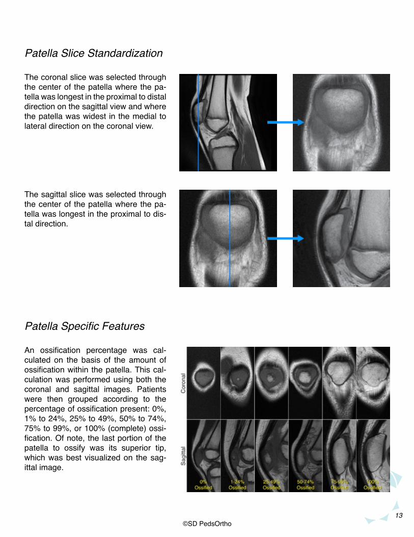
The coronal slice was selected through the center of the patella where the pa-tella was longest in the proximal to distal direction on the sagittal view and where the patella was widest in the medial to lateral direction on the coronal view.
The sagittal slice was selected through the center of the patella where the pa-tella was longest in the proximal to dis-tal direction.
Patella Slice Standardization
Patella Specific Features
An ossification percentage was cal-culated on the basis of the amount of ossification within the patella. This cal-culation was performed using both the coronal and sagittal images. Patients were then grouped according to the percentage of ossification present: 0%, 1% to 24%, 25% to 49%, 50% to 74%, 75% to 99%, or 100% (complete) ossi-fication. Of note, the last portion of the patella to ossify was its superior tip, which was best visualized on the sag-ittal image.

14©SD PedsOrtho
The coronal slice through the center of the fibular styloid was selected.
The sagittal slice through the center of the fibular styloid was selected
Fibula Slice Standardization
Fibula Specific Features
The following features of the fibula were identified: the presence of the epiphyse-al secondary ossification center, com-plete epiphyseal ossification (other than the styloid), ossification of the fibular styloid tip, partial closure of the physis, and complete closure of the physis.
To create a standard of reference for each age and each gender, we repeatedly ordered approx-imately 30 patients (when available) for each age and gender from least mature to most mature. The patient determined to be in the middle of the
Standard Reference Creation
maturity spectrum was identified as the “stan-dard”. The atlas “standard” consists of 8 images, including both coronal and sagittal images, of the femur, tibia, patella, and fibula.

15©SD PedsOrtho
Instructions for UseIf a clinician wishes to determine the skeletal ma-turity of a knee MRI compared to the standard atlas, we recommend that they proceed to the age and gender “standard” corresponding to the chronologic age of the patient. By comparing the standardized coronal and sagittal slices from each bone of their patient to this “standard” as well as to the standard immediately preceding and follow-ing the selected age, one can rapidly determine whether the patient’s skeletal age corresponds approximately with the age in this standard of ref-erence.
If the clinician does not know the patient’s age whose MRI they wish to assess, the clinician may look through the atlas until they come to the stan-dard that most closely resembles their patient. Similar to the Greulich and Pyle Hand atlas, the clinician will occasionally find that the MRI fea-tures of a patient resemble two successive stan-dards. In these situations, we recommend that that clinician use all available bones (femur, tibia, patella, and fibula) and images (coronal and sag-ittal) to best match the patient to the standard.
Slice selection for use of the atlas is performed in the identical fashion as Slice Standardization in creating the atlas, as described in pages 9-14. Many of the MRI indicators or features identified in this Atlas are subtle making appropriate image
Proper Image Selection
selection essential for each individual bone and for each image sequence (coronal and sagittal). The following slides depict which image should be selected for each bone.
The coronal slice through the center of the distal aspect of the femur at the at-tachment site of the posterior cruciate ligament on the medial femoral condyle was selected.
The sagittal slice through the center of the medial femoral condyle was select-ed
Slice Selection: Femur
16©SD PedsOrtho
The coronal slice at the attachment site of the ACL on the femur where the tibial spines are most pronounced should be selected.
The sagittal slice through the center of the tibial tubercle should be selected
Slice Selection: Tibia
The coronal slice was selected through the center of the patella where the pa-tella was widest in the medial to lateral direction.
The sagittal slice was selected through the center of the patella where the pa-tella was longest in the proximal to dis-tal direction.
Slice Selection: Patella

17©SD PedsOrtho
Femoral ossification is measured on the coronal view as a percentage of the width of the entire distal femur (both the ossified and unossified por-tions of the bone) compared to the ossified por-tion. In this case, 57% of the femur is ossified (29mm/51 mm).
Percent Ossification Measurement: Femur
The coronal slice through the center of the fibular styloid was selected.
The sagittal slice through the center of the fibular styloid was selected
Slice Selection: Fibula
18©SD PedsOrtho
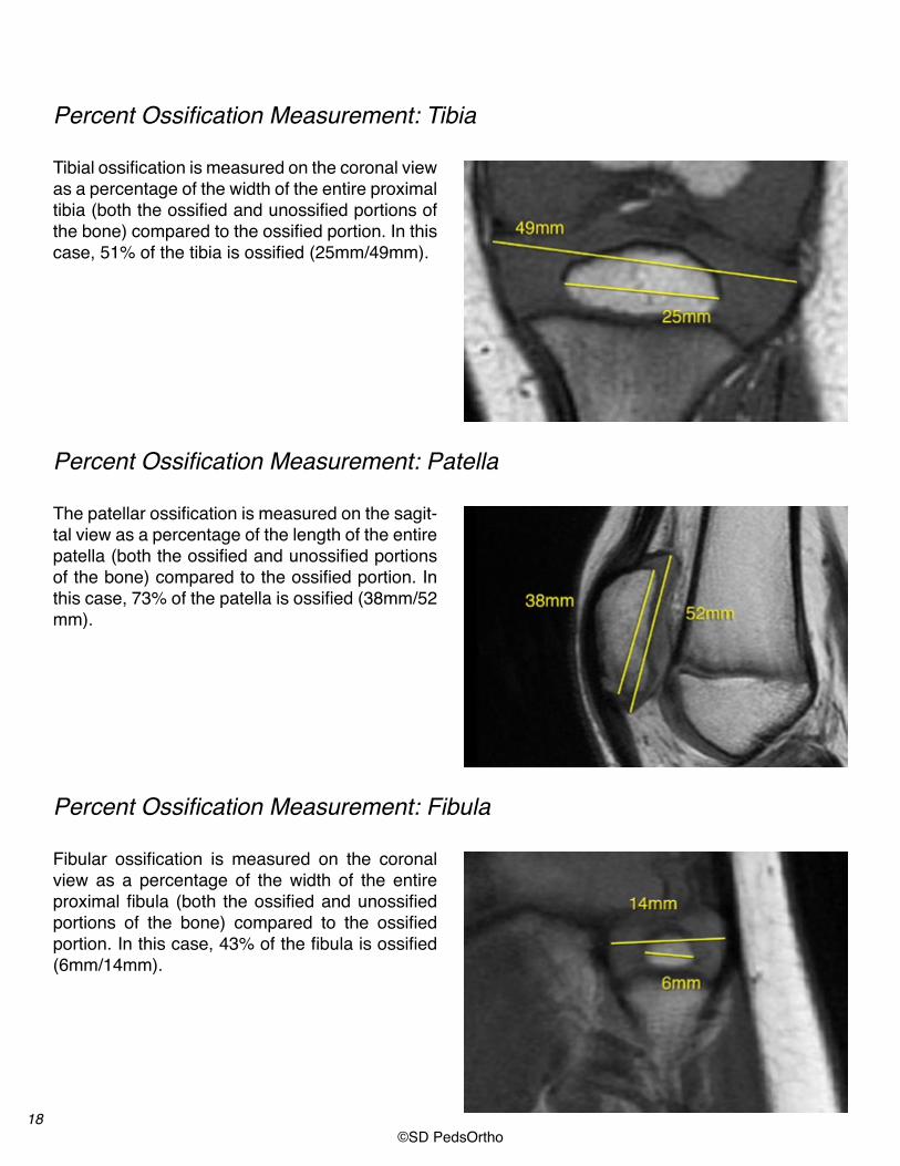
Tibial ossification is measured on the coronal view as a percentage of the width of the entire proximal tibia (both the ossified and unossified portions of the bone) compared to the ossified portion. In this case, 51% of the tibia is ossified (25mm/49mm).
The patellar ossification is measured on the sagit-tal view as a percentage of the length of the entire patella (both the ossified and unossified portions of the bone) compared to the ossified portion. In this case, 73% of the patella is ossified (38mm/52 mm).
Fibular ossification is measured on the coronal view as a percentage of the width of the entire proximal fibula (both the ossified and unossified portions of the bone) compared to the ossified portion. In this case, 43% of the fibula is ossified (6mm/14mm).
Percent Ossification Measurement: Tibia
Percent Ossification Measurement: Patella
Percent Ossification Measurement: Fibula

19©SD PedsOrtho
As a quick reference, we have identified the medi-an ages for each gender as to when specific MRI
Quick Reference
features become apparent. This table can serve as an expedited means of aging patients.

20©SD PedsOrtho
Anatomic and Radiographic TermsSeveral MRI indicators used in this atlas may be unfamiliar to clinicians. Additionally, some of the terms such as tibial tubercle “crack” and the fem-oral “oreo sign” have not been clinically used. In
the following figures, we hope to clarify these im-portant findings to make them easier to recognize and anatomically understand.
• During a relatively narrow window of skeletal development the epiphyseal cartilage immedi-ately adjacent to the articular cartilage has an oreo appearance.
• Unossified cartilage that may be appreciated on coronal imaging, immediately adjacent to the medial and lateral epicondyles, is another maturity marker on the knee.
Femoral “Oreo” Sign
Complete Ossification of the Femoral Epiphysis
• For boys this disappears at a median age of 15.6 years (range 13.4-16.9)
• For girls this disappears at a median age of 13.9 years (range 11.7-15.0)
9 year-old boy
Presence of “Oreo” sign
13.5 year-old boy
Presence of “Oreo” sign
15.5 year-old boy
Disappearance of “Oreo” sign
14 year-old boy
Incomplete Ossification
15 year-old boy
Complete Ossification
• For boys this cartilage completely ossifies at a median age of 14.2 years (range 10.1-15.5)
• For girls this cartilage completely ossifies at a median age of 11.9 years (range 9.8-12.3)
21©SD PedsOrtho
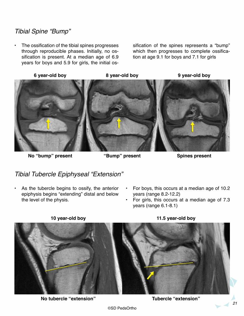
• The ossification of the tibial spines progresses through reproducible phases. Initially, no os-sification is present. At a median age of 6.9 years for boys and 5.9 for girls, the initial os-
• As the tubercle begins to ossify, the anterior epiphysis begins “extending” distal and below the level of the physis.
Tibial Spine “Bump”
Tibial Tubercle Epiphyseal “Extension”
sification of the spines represents a “bump” which then progresses to complete ossifica-tion at age 9.1 for boys and 7.1 for girls
• For boys, this occurs at a median age of 10.2 years (range 8.2-12.2)
• For girls, this occurs at a median age of 7.3 years (range 6.1-8.1)
6 year-old boy
No “bump” present
8 year-old boy
“Bump” present
9 year-old boy
Spines present
10 year-old boy
No tubercle “extension”
11.5 year-old boy
Tubercle “extension”

22©SD PedsOrtho
• As the tubercle apophysis appears (median age 11.8 years for boys and 10.2 years for girls), ossification can first be identified dis-tally. The tubercle epiphyseal extension and
• The last portions of the tibial epiphysis to ossi-fy can be seen best on coronal imaging medi-ally and sagittal imaging posteriorly.
Tibial Tubercle Ossification versus “Crack”
Complete Ossification of the Tibial Epiphysis
the apophyseal ossification then merge giving the appearance of a “crack” which occurs in boys at a median age of 12.8 years and girls at 10.7 years
• For boys this cartilage completely ossifies at a median age of 14.6 years (range 12.1-16.0)
• For girls this cartilage completely ossifies at a median age of 11.9 years (range 9.8-12.3)
12 year-old boy
Tubercle apophysisossification
13 year-old boy
“Crack”
14 year-old boy
Complete ossification
14 year-old boy 14 year-old boy
Incomplete ossification Incomplete ossification
15 year-old boy15 year-old boy
Complete ossificationComplete ossification

23©SD PedsOrtho
• The last portion of the patella to ossify is the patella tip which ossifies at a median age of 13.7 years (range 10.1-15.1) for boys and a median age of 11.9 years (range 9.8-12.3) for girls.
• The fibular styloid is the last portion of the fibu-la to ossify and occurs at a median age of 15.6 years for boys and 13.3 years for girls.
Patella Superior Tip Ossification
Fibular Styloid Ossification
14 year-old boy
16 year-old boy
Completeossification
Fibular styloidossified
12 year-old boy
14 year-old boy
Incompleteossification
Fibular styloidunossified
24©SD PedsOrtho
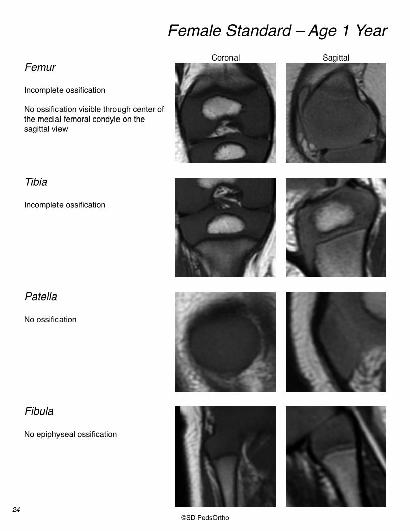
Female Standard – Age 1 Year
FemurCoronal Sagittal
Tibia
Patella
Fibula
Incomplete ossification
No ossification visible through center ofthe medial femoral condyle on the sagittal view
Incomplete ossification
No ossification
No epiphyseal ossification

25©SD PedsOrtho
Female Standard – Age 2 Years
Femur
Tibia
Patella
Fibula
Incomplete ossification(50-75%)
Incomplete ossification(50-75%)
No ossification
No epiphyseal ossification
Coronal Sagittal

26©SD PedsOrtho
Female Standard – Age 3 Years
Femur
Tibia
Patella
Fibula
Incomplete ossification(75-90%)
Incomplete ossification(50-75%)
Incomplete ossification(0-25%)
No ossification or Incomplete ossification
Coronal Sagittal

27©SD PedsOrtho
Female Standard – Age 4 Years
Femur
Tibia
Patella
Fibula
Incomplete ossification(75-90%)
Incomplete ossification(50-75%)
Incomplete ossification(25-50%)
Incomplete ossification(25-75%)
Coronal Sagittal

28©SD PedsOrtho
Female Standard – Age 5 Years
Femur
Tibia
Patella
Fibula
Incomplete ossification(75-90%)
Incomplete ossification(75-90%)Tibial spine “bump”
Incomplete ossification(50-75%)
Incomplete ossification(25-75%)
Coronal Sagittal

29©SD PedsOrtho
Female Standard – Age 6 Years
Femur
Tibia
Patella
Fibula
Incomplete ossification(>90%)Oreo sign present
Incomplete ossificationTibial spine “bump”
Incomplete ossification(>75%)Superior and inferior tips
Incomplete ossification(75-95%)
Coronal Sagittal

30©SD PedsOrtho
Female Standard – Age 7 Years
Femur
Tibia
Patella
Fibula
Incomplete ossification(>90%)Oreo sign present
Incomplete ossification(>90%)Tibial spine ossified
Incomplete ossification(Superior and inferior tips)
Incomplete ossificationFibular styloid not ossified
Coronal Sagittal

31©SD PedsOrtho
Female Standard – Age 8 Years
Femur
Tibia
Patella
Fibula
Incomplete ossification(Medially and laterally)Oreo sign present
Incomplete ossificationNo apophyseal ossificationTibial spine ossifiedTubercle epiphyseal extension
Incomplete ossification(Especially superior tip)
Incomplete epiphyseal ossificationFibular styloid not ossified
Coronal Sagittal
32©SD PedsOrtho
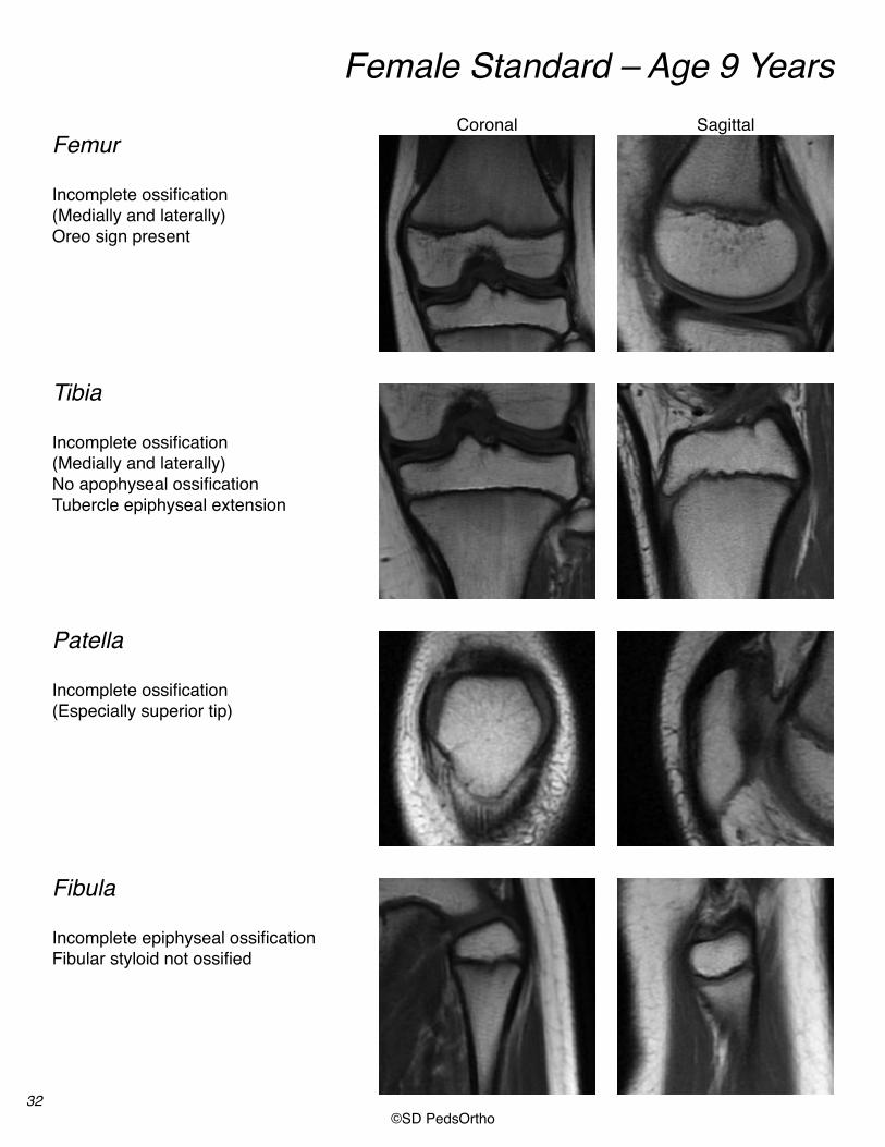
Female Standard – Age 9 Years
Femur
Tibia
Patella
Fibula
Incomplete ossification(Medially and laterally)Oreo sign present
Incomplete ossification(Medially and laterally) No apophyseal ossificationTubercle epiphyseal extension
Incomplete ossification(Especially superior tip)
Incomplete epiphyseal ossificationFibular styloid not ossified
Coronal Sagittal
33©SD PedsOrtho
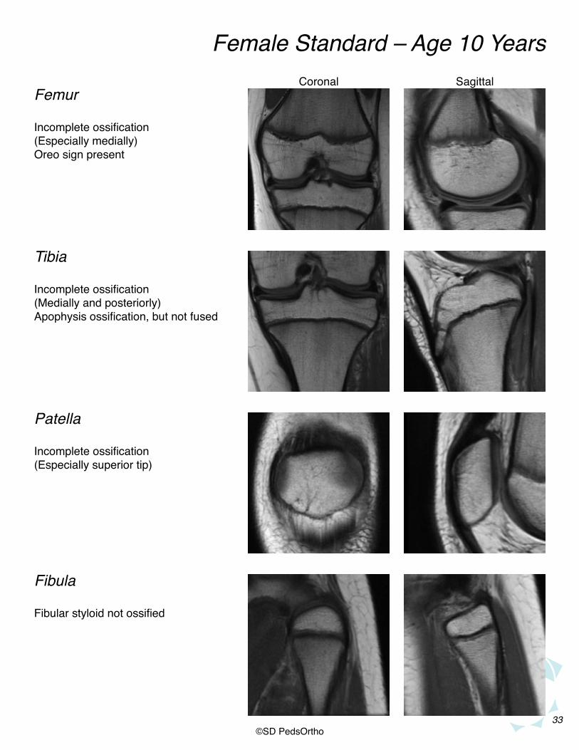
Female Standard – Age 10 Years
Femur
Tibia
Patella
Fibula
Incomplete ossification(Especially medially)Oreo sign present
Incomplete ossification(Medially and posteriorly) Apophysis ossification, but not fused
Incomplete ossification(Especially superior tip)
Fibular styloid not ossified
Coronal Sagittal
34©SD PedsOrtho
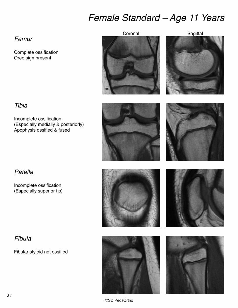
Female Standard – Age 11 Years
Femur
Tibia
Patella
Fibula
Complete ossificationOreo sign present
Incomplete ossification(Especially medially & posteriorly)Apophysis ossified & fused
Incomplete ossification(Especially superior tip)
Fibular styloid not ossified
Coronal Sagittal

35©SD PedsOrtho
Female Standard – Age 12 Years
Femur
Tibia
Patella
Fibula
Complete ossificationOreo sign present Entire physis visible
Incomplete ossification(especially medially & posteriorly)Entire physis visible
Complete ossification
Fibular styloid not ossifiedEntire physis visible
Coronal Sagittal
36©SD PedsOrtho
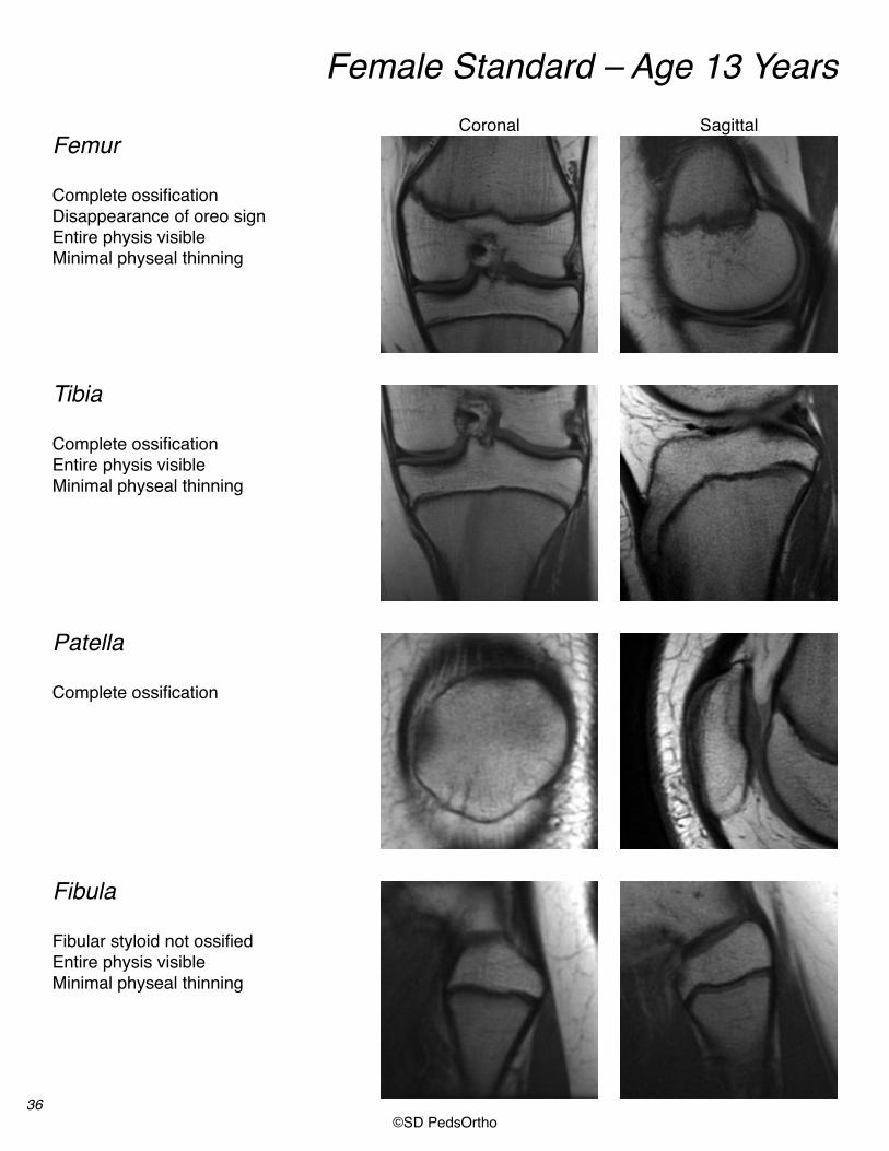
Female Standard – Age 13 Years
Femur
Tibia
Patella
Fibula
Complete ossificationDisappearance of oreo sign Entire physis visibleMinimal physeal thinning
Complete ossificationEntire physis visibleMinimal physeal thinning
Complete ossification
Fibular styloid not ossifiedEntire physis visible Minimal physeal thinning
Coronal Sagittal

37©SD PedsOrtho
Female Standard – Age 14 Years
Femur
Tibia
Patella
Fibula
Complete ossificationEntire physis visiblePhysis thinning (<2mm in height)
Complete ossificationEntire physis visiblePhysis thinning (<2mm in height)
Complete ossification
Fibular styloid ossifiedEntire physis visible Physis thinning (<2mm in height)
Coronal Sagittal

38©SD PedsOrtho
Female Standard – Age 15 Years
Femur
Tibia
Patella
Fibula
Complete ossificationEntire physis visiblePhysis thinning (<2mm in height)
Complete ossificationPartial closure of physis
Complete ossification
Fibular styloid ossifiedPartial closure of physisorEntire physis visible Physis thinning (<2mm in height)
Coronal Sagittal

39©SD PedsOrtho
Female Standard – Age 16 Years
Femur
Tibia
Patella
Fibula
Complete ossificationPartial closure of physis
Complete ossificationPartial or complete closure of physis
Complete ossification
Complete ossificationPartial or complete closure of physis
Coronal Sagittal

40©SD PedsOrtho
Female Standard – Age 17 Years
Femur
Tibia
Patella
Fibula
Complete ossificationComplete closure of physis
Complete ossificationComplete closure of physis
Complete ossification
Complete ossificationComplete closure of physis
Coronal Sagittal
41©SD PedsOrtho
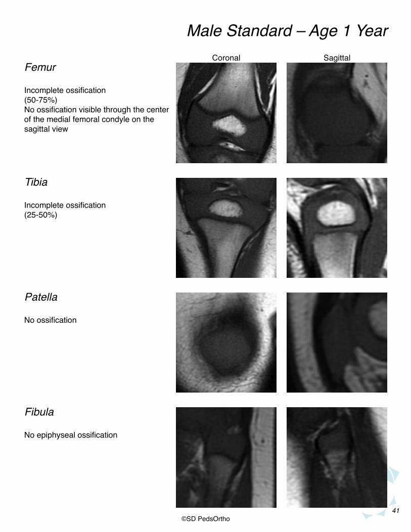
Male Standard – Age 1 Year
FemurCoronal Sagittal
Tibia
Patella
Fibula
Incomplete ossification(50-75%)No ossification visible through the centerof the medial femoral condyle on thesagittal view
Incomplete ossification(25-50%)
No ossification
No epiphyseal ossification
42©SD PedsOrtho
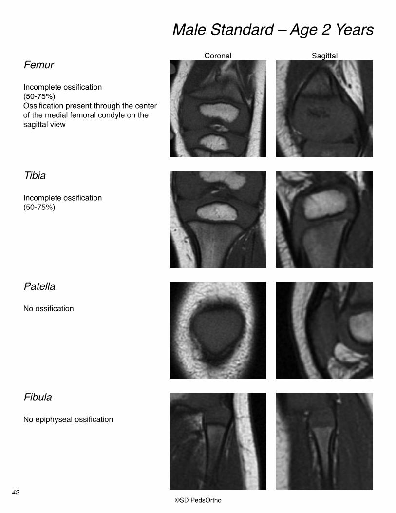
Male Standard – Age 2 Years
Femur
Tibia
Patella
Fibula
Incomplete ossification(50-75%)Ossification present through the centerof the medial femoral condyle on thesagittal view
Incomplete ossification(50-75%)
No ossification
No epiphyseal ossification
Coronal Sagittal
43©SD PedsOrtho
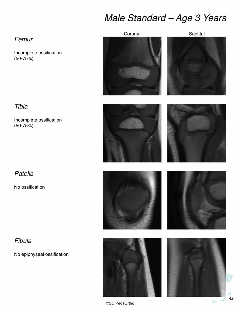
Male Standard – Age 3 Years
Femur
Tibia
Patella
Fibula
Incomplete ossification(50-75%)
Incomplete ossification(50-75%)
No ossification
No epiphyseal ossification
Coronal Sagittal
44©SD PedsOrtho
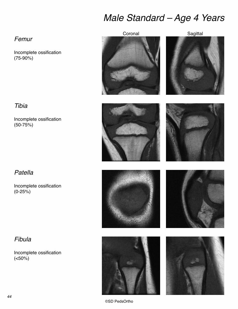
Male Standard – Age 4 Years
Femur
Tibia
Patella
Fibula
Incomplete ossification(75-90%)
Incomplete ossification(50-75%)
Incomplete ossification(0-25%)
Incomplete ossification(<50%)
Coronal Sagittal

45©SD PedsOrtho
Male Standard – Age 5 Years
Femur
Tibia
Patella
Fibula
Incomplete ossification(75-90%)
Incomplete ossification(50-75%)
Incomplete ossification(25-50%)
Incomplete ossification(25-75%)
Coronal Sagittal

46©SD PedsOrtho
Male Standard – Age 6 Years
Femur
Tibia
Patella
Fibula
Incomplete ossification(75-90%)
Incomplete ossification(50-75%)
Incomplete ossification(33-66%)
Incomplete ossification(25-75%)
Coronal Sagittal

47©SD PedsOrtho
Male Standard – Age 7 Years
Femur
Tibia
Patella
Fibula
Incomplete ossification(75-90%)
Incomplete ossification(75-90%)Tibial spine “bump”
Incomplete ossification(50-75%)
Incomplete ossification(50-75%)
Coronal Sagittal

48©SD PedsOrtho
Male Standard – Age 8 Years
Femur
Tibia
Patella
Fibula
Incomplete ossification(>90%)Oreo sign present
Incomplete ossification(75-90%) Tibial spine “bump”
Incomplete ossification(>75%)(Superior and inferior tips)
Incomplete ossification(75-95%)
Coronal Sagittal

49©SD PedsOrtho
Male Standard – Age 9 Years
Femur
Tibia
Patella
Fibula
Incomplete ossification(>90%)Oreo sign present
Incomplete ossification(75-90%)Tibial spine “bump”
Incomplete ossification(>75%)(Superior and inferior tips)
Incomplete ossification(75-95%)
Coronal Sagittal

50©SD PedsOrtho
Male Standard – Age 10 Years
Femur
Tibia
Patella
Fibula
Incomplete ossification(>90%)Oreo sign present
Incomplete ossification(>90%)Ossified tibial spines
Incomplete ossification(Superior and inferior tips)
Incomplete ossificationFibular styloid not ossified
Coronal Sagittal

51©SD PedsOrtho
Male Standard – Age 11 Years
Femur
Tibia
Patella
Fibula
Incomplete ossification(>90%)Oreo sign present
Incomplete ossification(Medially and laterally) No apophyseal ossificationTubercle epiphyseal extension
Incomplete ossification(Superior and inferior tips)
Incomplete ossificationFibular styloid not ossified
Coronal Sagittal

52©SD PedsOrtho
Male Standard – Age 12 Years
Femur
Tibia
Patella
Fibula
Incomplete ossification(Especially medially)Oreo sign present
Incomplete ossification(Especially medially and posteriorly) Apophysis ossification, but not fused
Incomplete ossification(Superior tip)
Incomplete ossificationFibular styloid not ossified
Coronal Sagittal
53©SD PedsOrtho
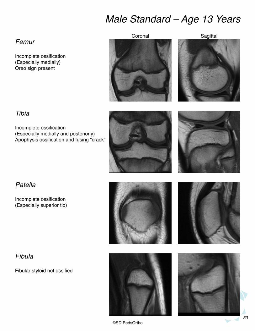
Male Standard – Age 13 Years
Femur
Tibia
Patella
Fibula
Incomplete ossification(Especially medially)Oreo sign present
Incomplete ossification(Especially medially and posteriorly) Apophysis ossification and fusing “crack”
Incomplete ossification(Especially superior tip)
Fibular styloid not ossified
Coronal Sagittal

54©SD PedsOrtho
Male Standard – Age 14 Years
Femur
Tibia
Patella
Fibula
Complete ossificationOreo sign present
Incomplete ossification(Especially medially and posteriorly) Apophysis ossification and fused
Incomplete ossification(Especially superior tip)
Fibular styloid not ossified
Coronal Sagittal

55©SD PedsOrtho
Male Standard – Age 15 Years
Femur
Tibia
Patella
Fibula
Complete ossificationDisappearance of oreo signEntire physis visible
Incomplete ossification(Especially medially and posteriorly) Entire physis visible
Complete ossification
Fibular styloid not ossifiedEntire physis visible
Coronal Sagittal

56©SD PedsOrtho
Male Standard – Age 16 Years
Femur
Tibia
Patella
Fibula
Complete ossificationEntire physis visiblePhysis thinning (<2mm in height)
Complete ossificationPartial closure of physis
Complete ossification
Fibular styloid ossifiedPhysis thinning (<2mm in height) or Partial closure of physis
Coronal Sagittal

57©SD PedsOrtho
Male Standard – Age 17 Years
Femur
Tibia
Patella
Fibula
Complete ossificationPartial closure of physis
Complete ossificationPartial or complete closure of physis
Complete ossification
Complete ossificationPartial or complete closure of physis
Coronal Sagittal

58©SD PedsOrtho
Male Standard – Age 18 Years
Femur
Tibia
Patella
Fibula
Complete ossificationComplete closure of physis
Complete ossificationComplete closure of physis
Complete ossification
Complete ossificationComplete closure of physis
Coronal Sagittal

59©SD PedsOrtho
This system of assessing bone age was validated using a cohort of 323 knee MRIs. These were separate from the knee MRIs used to create the system. Two orthope-dic surgeons were blinded to this cohort of subject’s chronological ages while they determined bone age using this new system.
Inter-observer reliability between the two surgeons was assessed using the intraclass correlation coefficient (ICC). Inter-observer reliability among our two surgeons was found to be high with an ICC of 0.957, p<0.001.
The comparison of knee MRI bone ages to chronological bone age was found to be highly correlated, with Spearman’s rho being 0.978, p<0.001.
Left hand x-rays were available on 48 of the patients in the co-hort used to validate this system. The Greulich and Pyle atlas was used to determine bone age from the wrist x-rays in this cohort. The graph to the left shows that the Greulich and Pyle bone age method and the knee MRI bone age method were similarly cor-related to chronological age, with the knee MRI method having a marginally higher correlation with chronological age.
Pennock A.T., Bomar J.D., Manning J.D.: The Cre-ation and Validation of a Knee Bone Age Atlas Uti-lizing MRI. J Bone Joint Surg Am. 2018;100(4):e20.
Validation

60©SD PedsOrtho
Pennock AT, Bomar JD, Manning JD. The Creation and Validation of a Knee Bone Age Atlas Utilizing MRI. J Bone Joint Surg Am. 2018;100:e20)1-10
Greulich WW, Pyle SI. Radiographic atlas of skeletal development of the hand and wrist. 2nd ed. Palo Alto: Stanford University Press; 1959.
Hackman L, Black S. The reliability of the Greulich and Pyle atlas when applied to a modern Scottish population. J Forensic Sci. 2013 Jan;58(1):114-9. Epub 2012 Oct 12.
Roche AF, Roberts J, Hamill PV. Skeletal maturity of children 6-11 years: racial, geographic area, and socioeconomic differentials, United States. Vital Health Stat 11. 1975 Oct;(149):1-81.
Aicardi G, Vignolo M, Milani S, Naselli A, Magliano P, Garzia P. Assessment of skeletal maturity of the hand-wrist and knee: a comparison among methods. Am J Hum Biol. 2000 Sep;12(5):610-5.
Dedouit F, Auriol J, Rousseau H, Rougé D, Crubézy E, Telmon N. Age assessment by magnetic resonance imaging of the knee: a preliminary study. Forensic Sci Int. 2012 Apr 10;217(1-3):232.e1-7. Epub 2011 Dec 5.
Pyle SI, Hoerr NL. A radiographic standard of reference for the growing knee. Springfield: Charles C. Thomas; 1969.
Schaefer M, Hackman L, Gallagher J. Variability in developmental timings of the knee in young American children as assessed through Pyle and Hoerr’s radiographic atlas. Int J Legal Med. 2016 Mar;130(2):501-9. Epub 2015 Jan 17.
Krämer JA, Schmidt S, Jürgens KU, Lentschig M, Schmeling A, Vieth V. Theuse of magnetic resonance imaging to examine ossification of the proximal tibial epiphysis for forensic age estimation in living individuals. Forensic Sci Med Pathol. 2014 Sep;10(3):306-13. Epub 2014 Apr 17.
References
Top Related