


Languages
Pages
Legal


Available online at www.jpsscientificpublications.com
Life Science Archives (LSA)
ISSN: 2454-1354
Volume – 1; Issue - 2; Year – 2015; Page: 142 - 156
©2015 Published by JPS Scientific Publications Ltd. All rights reserved
Research Article
ENDOPHYTIC FUNGAL COMMUNITIES ASSOCIATED WITH
ETHNO MEDICINAL PLANTS FROM INDIA AND THEIR POTENTIAL
PTODUCTION OF ANTICANCER DRUG CAMPTOTHECIN
N. Lakshmi1, V. Bhuvaneswari
2, G. Kathiravan
3 and B. Shanmugapriya
2
1S.D.N.B. Vaishnav College for Women, Chromepet, Chennai, Tamil Nadu, India.
2Chikkaiah Naicker College, Veerapan chitram, Erode, Tamil Nadu, India.
3Department of Biotechnology, Vels University, Pallavaram, Chennai, Tamil Nadu, India.
Abstract
Endophytic fungi are ubiquitous, ecologically specialized group and are assumed to be widely present
in virtually all land plants. The anticancer properties of several secondary metabolites from endophytes have
been investigated recently. Following, is one of the example Camptothecin (CPT) is a monoterpenoid indole
alkaloid originally isolated from Camptotheca acuminata Decne (Nyssaceaea), a deciduous tree native to
south China, that has gained great attention for its significant antitumor activities in experimental studies. In
the present investigation, extraction of Camptothecin and its analogues from novel endophytic fungal sources
isolated from two medicinal plants namely Nerium oleander and Nyctanthus arbor-tristis were studied. Of
the taxa identified in the current study, mitosporic fungi dominated the endophyte assemblages. Therefore, in
the present investigation two hyphomyceteous fungi namely, Aspergillus flavus and Aspergillus niger were
screened for the production of Camptothecin. The production of Camptothecin was confirmed and quantified by different analytical methods. The amount of Camptothecin was found to be maximum in Aspergillus
flavus (55.5 µg/L) followed by Aspergillus niger (32.5 µg/L). UV spectroscopic analysis showed
characteristic absorption peaks at 226 nm and 269 nm, which was similar to that of the authentic
Camptothecin (Sigma aldrich). Mass spectroscopy (MS) done in the two fungal samples yielded [M + H]+
ions of CPT at m/z 349. The sodium adduct of CPT was also formed and was visible in the mass spectrum at
m/z 371 [M + Na]+. Thus, these research efforts are significant for both practical and philosophical reasons.
Article History Received : 02.04.2015
Revised : 19.04.2015
Accepted : 26.04.2015
Key words: Camptothecin, Camptotheca
acuminata, antitumor, endophytic fungi,
Aspergillus flavus and Aspergillus niger.
1. Introduction
An endophyte is an endosymbiont, often a
bacterium or fungus, which lives within a plant for
at least part of its life without causing apparent
disease. Symptomless fungal endophytes have
been discovered in the aerial plant tissues of over
300 plant species, including angiosperms,
* Corresponding author: N. Lakshmi, S.D.N.B.
Vaishnav College for Women, Chromepet, Chennai,
Tamil Nadu, India
gymnosperms, marine macro algae, mosses and
ferns (Petrini and Fisher, 1990; Clay, 1991; Lodge
et al., 1996). Studies on the distribution,
biodiversity and biochemical characteristics of
endophytes are of immense importance in plant
biology to understand and to improve plant fitness.
Plant endophytic fungi are an important and novel
resource of natural bioactive compounds with their
potential applications in agriculture, medicine and
food industry. In the past two decades, many
valuable bioactive compounds with antimicrobial,

N. Lakshmi/Life Science Archives (LSA), Volume – 1, Issue – 2, Page – 142 - 156, 2015 143
©2015 Published by JPS Scientific Publications Ltd. All rights reserved
insecticidal, cytotoxic and anticancer activities
have been successfully discovered from the
endophytic fungi. During the long period of co-
evolution, a friendly relationship was formed
between each endophyte and its host plant. Some
endophytes have the ability to produce the same or
similar bioactive compounds as those originated
from their host plants.
Bioactive natural compounds produced
by endophytes have been promising potential
usefulness in safety and human health concerns,
although there is still a significant demand of drug
industry for synthetic products due to economic
and time-consuming reasons. Problems related to
human health such as the development of drug
resistance in human pathogenic bacteria, fungal
infections and life threatening virus claim for new
therapeutic agents for effective treatment of
diseases in human, plants, and animals that are
currently unmet (Mariana Recco et al., 2011).
Symbiotic relationship of host-microbes
producing new and interesting bioactive
compounds may find uses in pharmaceutical
industries. Some of the potentially important
compounds isolated from endophytic fungi
include cryptocin, cryptocandin, preussomerin,
phomosichalasin and torreyanic acid. Above all,
endophytes have been proved to be an effective
source of Taxol, a cytotoxic drug used in the
treatment of cancer (Wani et al., 1971). Also of
notice, endophytes have been found to be capable
of producing immunosuppressive compounds (Lee
et al., 1995). Apart from the medicinal
compounds fungal endophytes are the reservoirs
of more eco-friendly useful compounds to
mankind. Reports showed that they are producing
volatile antimicrobials from Muscodor albus
(Strobel et al., 2001), antifungal ambuic acid from
Pestalotiopsis spp. and Monochaetia sp. (Li et al.,
2001), isopectacin an antifungal and antioxidant
compound from Pestalotiopsis microspora
(Strobel et al., 2002), Napthalene an insect
repellant from Muscodor vitigenus (Daisy et al.,
2002; Azevedo et al., 2000).
There are some evidences that bioactive
compounds produced by endophytes could be
alternative approaches for discovery of novel
drugs, since many natural products from plants,
microorganisms, and marine sources were
identified as anticancer agents. The anticancer
properties of several secondary metabolites from
endophytes have been investigated recently.
Following, is one of the example Camptothecin
(CPT) is a monoterpenoid indole alkaloid
originally isolated from Camptotheca acuminata
Decne (Nyssaceaea), a deciduous tree native to
south China, that has gained great attention for its
significant antitumor activities in experimental
studies (Wall et al.,1966). Irinotecan (CPT-11)
(Masuda et al., 1992; Abigeres et al., 1995 &
1999) and topotecan (TPT) (Lilenbaum et al.,
1995; Romanelli et al., 1998 and Clements et al.,
1999), two water-soluble derivatives of CPT, have
gained approval by the Food and Drug
Administration of the United States of America
(FDA) for treating colorectal and ovarian cancer.
Other camptothecins - such as 9-
aminocamptothecin (9AC), 9- nitrocamptothecin
(9NC), and 7-(4-methyl piperazino-methylene)-
10,11- (GG211) have also showed remarkable
potential in the treatment of Carcinoma (Wall and
Wani, 1996; Giovanella, 1997; Jeha et al., 1998
and Stevenson et al., 1999).
Currently, it is believed that many of these
compounds act in defense of the harmful effects of
toxins, carcinogens, or mutagens found in the
plant or attack by external predators. The
compounds were isolated guided by bioassay on
various extracts and chromatographic fractions.
Their unique and hitherto unknown structures
were elucidated by nuclear magnetic resonance,
mass spectrometry, and X-ray analysis. Both
compounds have unique mechanisms of antitumor
activity; camptothecin uniquely inhibits an
enzyme, topoisomerase I, involved in DNA
replication. Camptothecin and analogues singly or
combined with cisplatin show efficacy against
solid tumors, breast, lung, and colorectal, which
hitherto have been unaffected by most cancer
chemotherapeutic agents (Wall and Wani, 1996).
Therefore, in the present investigation, extraction
of Camptothecin and its analogues from novel
endophytic fungal sources isolated from medicinal
plants were studied.

N. Lakshmi/Life Science Archives (LSA), Volume – 1, Issue – 2, Page – 142 - 156, 2015 144
©2015 Published by JPS Scientific Publications Ltd. All rights reserved
2. Materials and Methods
2.1. Collection and processing of plant samples
For isolating endophytes, two healthy
medicinal plants namely Nyctanthes arbor-tristis
L. and Nerium oliander L. were collected in sterile
polythene bags from their natural habitat. The
plants were collected from Chromepet, Chennai.
The samples were brought to the laboratory and
processed within 24 hours. The collected samples
were first washed thoroughly in running tap water.
From each plant 20 segments (approx. 0.5 cm2)
from different tissues (leaf, rachis/petiole and
stem) were screened for the presence of
endophytes. The modified Standard triple ethanol
- sodium hypochlorite - ethanol surface
sterilization techniques (Fisher et al., 1993;
Dobranic et al., 1995 and Schultz et al., 1998)
were followed throughout the present study. The
segments were surface sterilized in 70% ethanol
for 5 sec, immersed in 4% Sodium hypochlorite
(NaOCl) for 90 sec, rinsed in sterile distilled water
and then dried on sterilized filter paper. The
surface sterilized segments were placed on Potato
Dextrose Agar (PDA) medium amended with
Streptopenicillin (150 mg/L). The petriplates were
then sealed with ParafilmTM
.
2.2. Incubation and isolation of endophytes
The petriplates were incubated in a light
chamber and observations were done from the
second day onwards for a period of 3 - 4 weeks for
the fungal colonies (Bills and Pollishook, 1991).
The light regime was 12 hours light followed by
12 hours darkness. The hyphae, which grew out
from the tissues, were transferred to fresh PDA
slants. They were maintained by sub-culturing.
To prevent the rapidly growing fungi from
inhibiting the slow growing species, the former
were removed as soon as they appeared on the
plates (Bills, 1996).
2.3. Identification of the fungi
The isolated endophytic fungi from selected plants
were identified down to species level with the help
of standard monographs (Guba, 1961; Ellis, 1971;
Sutton, 1980; Onions et.al., 1981 and Nag Raj,
1993). The non-sporulating sterile forms were
separated into culture groups based on their
colony morphology, hyphal mat characteristics
(texture, zonation, sectoring), presence of sclerotia
(masses of short celled, lobed and closely packed
hyphae) and pigmentation as described by Frolich
et al. (2000). Such sterile forms were included as
'species' for the analysis of the results.
2.4. Calculation of Colonization frequency (CF)
The colonization frequency of each
endophyte species was calculated by the method
of Hata and Futai (1995).
The number of colonized segments
CF % = × 100
Total number of segments observed
2.5. Relative percentage occurrence (RPO)
Relative percentage occurrence (RPO) of
each group (viz., Ascomycetes, Hyphomycetes,
Coelomycetes and sterile morphospecies) of
fungal species in each plant species was calculated
as follows (Petrini, 1991)
Density of colonization of single group
RPO= × 100
Total density of colonization
2.6. Cultivation and extraction of test fungi for
Camptothecin production
The selected endophytic fungi namely,
Aspergillus niger and Aspergillus flavus were
cultured in three different media viz., Corn Meal
Agar, Rose Bengal Agar and Sabouraud’s
Dextrose Agar medium respectively for
Camptothecin production. The methodology for
the extraction of Camptothecin was as given by Li
et al. (2012). After incubating the culture for 2 - 3
weeks, both cell homogenate and cell-free broth
were extracted four times with equal volume of
chloroform: methanol (4:1 v/v). After stripping off
the solvent, the residue was analyzed both
chromatographically as well as spectrometrically.
Blank cultures (uninoculated sterile medium) were
also maintained along with fungal cultures. The
blank cultures were autoclaved, incubated and
processed exactly in the same manner as the
inoculated fungal cultures. The blanks were also
analyzed and tested with the fungal samples.

N. Lakshmi/Life Science Archives (LSA), Volume – 1, Issue – 2, Page – 142 - 156, 2015 145
©2015 Published by JPS Scientific Publications Ltd. All rights reserved
2.7. Chromatographic analysis of test samples
TLC analysis was carried out on Merk 1 mm (20 x
20 cm) silica gel precoated plates. The plates
were developed using chloroform: methanol (9: 1
v/v) solvent system. TLC analysis exhibited spots
which were super imposable with the standard
CPT. The spots were visualized under UV.
Camptothecin standard solutions within the range
from 1 - 100 μg/ml concentrations were prepared
for HPLC analysis. The camptothecin standard
was prepared by dissolving in a solution of DMSO
and absolute HPLC grade methanol in a ratio of 5:
50 (v/v). The standard sample solutions for HPLC
were filtered using 0.2 syringe filter before
injection. The analysis of extracts was done in
High Performance Liquid Chromatographic
system (HPLC) equipped with LC8A pump, SPD-
M 10 Apphoto array detector in combination with
class LC 10 A software (Shimadzu). The presence
of camptothecin in the samples was detected by
comparing with the retention time of the standard
sample. The area of the standard was compared
with area of the sample and the amount of
camptothecin in the extracts was calculated. The
chromatographic conditions for the analysis were
as follows: mobile phase: acetonitrile : water (60:
40), column: ODS (Octadecyl silane) C18, 5
µsize, 250 X 4.6 mm (Supelco), Detector: SPD-M
10 A Vpphoto array detector, wave length: 254
nm, flow rate: 1.0 ml/min, injection volume: 25
µl, retention time: 6.4 min.
Standard concentration ×
Total area of the sample
Camptothecin
content =
Total area of the Standard
After chromatography, the two potential
Hyphomycetes fungi were analyzed
spectroscopically for further confirmation of
Camptothecin sample were taken and dissolved in
Methanol and analyzed using Beckman DU-40
with two maximum at 226 nm and 269 nm.
2.8. Mass Spectrometry analysis of test samples
In this study, the fungal test samples were
analyzed using the mass spectrometer which was
fitted with an electrospray interface. All the
interface parameters of GC–MS/MS studies were
optimized by infusing the standard solution of
CPT. The other parameters for GC–MS analysis
were set at dry gas flow of 11 l/ min, nebulizer
pressure 35 psi and drying gas temperature 320˚C.
The isolated peak width was taken as 0.8 m/z and
fragmentation amplitude value was 2.40. A mass
spectrum of CPT in methanol: chloroform [1:3]
was recorded under ESI on a Bruker Ion Trap
(Esquire 3000) mass spectrometer in the positive
ion mode, with a mass range from 50 to 800 amu.
Bruker Daltonics Esquire 5.0 software was used to
obtain the mass spectra and Chemstation Rev.
06.03 (509) software was used to acquire the GC–
MS spectra.
3. Results and Discussion
In the recent years, the quest for isolation
of new compounds from medicinal plants has
become a fascinating area of research. Plants with
ethno-pharmaceutical importance are being
exploited because of their healing properties.
However, large scale harvesting of medicinal
plants has already become a major threat to
biodiversity. As an alternative, microbes, which
live inside such plants (endophytes), may offer
tremendous potential source of therapeutic
compounds. The present study has been aimed
towards isolation and identification of endophytic
fungi associated with selected medicinal plants
and analysis of test fungi for Camptothecin
production was carried out. The plant species
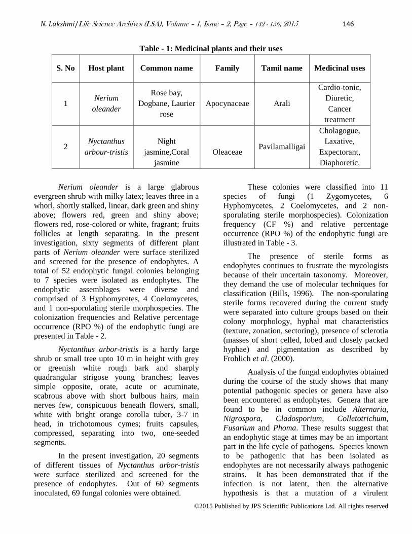
chosen for this study and their medicinal uses are
presented in Table - 1. Different plant parts viz.,
leaf (including midrib), petiole/rachis and stems of
the selected symptomless host plants were surface
sterilized and incubated as per the procedure
mentioned in materials and methods. The plants
segments were studied for the presence of
endophytic fungi.
N. Lakshmi/Life Science Archives (LSA), Volume – 1, Issue – 2, Page – 142 - 156, 2015 146
©2015 Published by JPS Scientific Publications Ltd. All rights reserved
Table - 1: Medicinal plants and their uses
S. No Host plant Common name Family Tamil name Medicinal uses
1 Nerium
oleander
Rose bay,
Dogbane, Laurier
rose
Apocynaceae Arali
Cardio-tonic,
Diuretic,
Cancer
treatment
2 Nyctanthus
arbour-tristis
Night
jasmine,Coral
jasmine
Oleaceae Pavilamalligai
Cholagogue,
Laxative,
Expectorant,
Diaphoretic,
Nerium oleander is a large glabrous
evergreen shrub with milky latex; leaves three in a
whorl, shortly stalked, linear, dark green and shiny
above; flowers red, green and shiny above;
flowers red, rose-colored or white, fragrant; fruits
follicles at length separating. In the present
investigation, sixty segments of different plant
parts of Nerium oleander were surface sterilized
and screened for the presence of endophytes. A
total of 52 endophytic fungal colonies belonging
to 7 species were isolated as endophytes. The
endophytic assemblages were diverse and
comprised of 3 Hyphomycetes, 4 Coelomycetes,
and 1 non-sporulating sterile morphospecies. The
colonization frequencies and Relative percentage
occurrence (RPO %) of the endophytic fungi are
presented in Table - 2.
Nyctanthus arbor-tristis is a hardy large
shrub or small tree upto 10 m in height with grey
or greenish white rough bark and sharply
quadrangular strigose young branches; leaves
simple opposite, orate, acute or acuminate,
scabrous above with short bulbous hairs, main
nerves few, conspicuous beneath flowers, small,
white with bright orange corolla tuber, 3-7 in
head, in trichotomous cymes; fruits capsules,
compressed, separating into two, one-seeded
segments.
In the present investigation, 20 segments
of different tissues of Nyctanthus arbor-tristis
were surface sterilized and screened for the
presence of endophytes. Out of 60 segments
inoculated, 69 fungal colonies were obtained.
These colonies were classified into 11
species of fungi (1 Zygomycetes, 6
Hyphomycetes, 2 Coelomycetes, and 2 non-
sporulating sterile morphospecies). Colonization
frequency (CF %) and relative percentage
occurrence (RPO %) of the endophytic fungi are
illustrated in Table - 3.
The presence of sterile forms as
endophytes continues to frustrate the mycologists
because of their uncertain taxonomy. Moreover,
they demand the use of molecular techniques for
classification (Bills, 1996). The non-sporulating
sterile forms recovered during the current study
were separated into culture groups based on their
colony morphology, hyphal mat characteristics
(texture, zonation, sectoring), presence of sclerotia
(masses of short celled, lobed and closely packed
hyphae) and pigmentation as described by
Frohlich et al. (2000).
Analysis of the fungal endophytes obtained
during the course of the study shows that many
potential pathogenic species or genera have also
been encountered as endophytes. Genera that are
found to be in common include Alternaria,
Nigrospora, Cladosporium, Colletotrichum,
Fusarium and Phoma. These results suggest that
an endophytic stage at times may be an important
part in the life cycle of pathogens. Species known
to be pathogenic that has been isolated as
endophytes are not necessarily always pathogenic
strains. It has been demonstrated that if the
infection is not latent, then the alternative
hypothesis is that a mutation of a virulent

N. Lakshmi/Life Science Archives (LSA), Volume – 1, Issue – 2, Page – 142 - 156, 2015 147
©2015 Published by JPS Scientific Publications Ltd. All rights reserved
pathogen has occurred and the fungus has become
a non-pathogenic strain of the pathogen (Freeman
and Rodrigues, 1993).
Saprotrophic taxa that were isolated as
endophytes in the present study include
Nigrospora sphaerica, Aspergillus niger and
Phoma sp. The above-mentioned fungi are known
to biodegrade cellulose and lignin and their
ecological role is primary in decomposing dying
plant material.
Endophytic fungi have been recognized as
a repository of novel compounds of immense
value in agriculture, industry and medicine (Naik
et al., 2009). To date, many valuable bioactive
compounds and anticancer activities have been
obtained from the endophytic fungi (Verma et al.,
2009; Aly et al., 2010; Yu et al., 2010;
Kharwar et al., 2011). On the whole, the percent
contribution of Hyphomycetes (57 %) as
endophytes was very high followed by
Coelomycetes (31 %) non-sporulating sterile
morphospecies (8%), and Zygomycetes (3 %)
(Fig-1). Among the different plant parts viz., stem,
leaf lamina and petiole studied for the presence of
endophytes, more numbers of isolates were
recovered from leaf lamina (67), followed by stem
(36) and petiole (17) (Fig 2).
Apart from trying to understand the
biology of endophytes, another motivation to
study these fungi is its ability to produce a novel
bioactive compound Camptothecin. Wani et al.
(1971) from North Carolina discovered that an
extract of the Yew tree bark has antitumor activity
and the compound was named "Paclitaxel" or
"Taxol". Interest in developing the drug increased
after the mechanism of action of tubulin
polymerization was studied by Horowitz in 1980.
It was discovered that paclitaxel promotes tubulin
polimerization and stabilizes microtubules against
depolymerization (Schiff et al., 1979; Schiff and
Horowitz, 1980). However, Taxol supply from
the original source cannot meet the rising demand
of clinical uses because of the scarcity and slow
growth of Taxus yew trees and the low Taxol
(0.02% dry weight) content in the bark (Cragg et
al., 1993). Moreover, the extraction procedure is
complex and expensive. Hence, alternative
sources were explored for anticancer drug supply
including chemical synthesis, cultural techniques
and extraction from different microbial sources.
Camptothecin, a quinoline indole alkaloid
and its analog, 10 –hydroxy Camptothecine are
potent inhibitors of eukaryotic topoisomerase I.
Because of this activity, several semi synthetic
derivatives of CPT topotecan and camptostar are
used clinically. The production of Camptothecin
from a microbial source has many advantages over
other sources. Industrial production of bioactive
compounds like Camptothecin requires
reproducible, dependable productivity. If a fungus
is the source organism, it can be grown in tank
fermenters to produce an inexhaustible supply of
Camptothecin. The added advantage is that the
fungi usually respond favorably to routine cultural
techniques. Normally tissue cultures of
Camtotheca accuminata for Camptothecin
extraction will take a longer time when compared
with the fungi. Also, tissue culture needs
specialized techniques and conditions have to be
maintained whereas in fungi one can easily alter
the cultural conditions for the production of
different bioactive compounds.
Touseef et al. (2006) isolated
ectomycorrhizal fungi from Camtotheca
accuminata which demonstrated the ability to
produce Camptothecin. The presence of
Camptothecin in this fungus was confirmed by
mass spectrometry, chromatography, and
biochemical techniques. He also suggested that
improved culture techniques, the addition of
activators and the application of genetic
engineering methods may permit the
commercialization of Camptothecin production.
Techniques previously applied in the confirmation
of Camptothecin from Camptotheca species and
fungi include Thin layer chromatography (TLC),
High performance thin layer chromatography
(HPLC), Ultraviolet (UV), Infrared (IR), and Mass
spectrometry (MS) Many other investigators have
supported these confirmatory techniques (Li et al.,
1996; Strobel et al., 1996 ; Li et al., 1998;
Baloglu, 1999; Wang et al., 2000). Hence, in the
present study TLC, HPLC, UV and MS methods
were employed for confirming the presence of
Camptothecin in the fungal sample.

N. Lakshmi/Life Science Archives (LSA), Volume – 1, Issue – 2, Page – 142 - 156, 2015 148
©2015 Published by JPS Scientific Publications Ltd. All rights reserved
Table - 2: CF % and RPO % of endophytic fungi recovered from Nerium oleander
Therefore, in the present investigation two
endophytic hyphomycetous fungi namely,
Aspergillus flavus and Aspergillus niger obtained
from the selected medicinal plants were screened
for the production of Camptothecin. The
methodology for the extraction of Camptothecin
was as given by Li et al. (2002). The selected test
fungi were grown in the medium as mentioned
earlier. After the incubation period of 16 days, the
fungal samples were extracted with methanol and
were used for further analysis. Analytical thin
layer chromatography reference standard and test
sample fractions was conducted on silica gel
GF254 analytical plate developed with
Chloroform/methanol (90:10, v/v). Blue
fluorescent spot detected by long UV were
observed at Rf values of 6.9, which indicates the
presence of Camptothecin in the fungal test
samples. Among the three different media tested
the fungi grown in SDA showed the best result.
Hence these fungi were further analyzed by using
High Performance Liquid Chromatography
(HPLC), Ultra Violet (UV) and Mass
Spectrometry (MS) analysis.
S.
No. List of fungi isolated
Colonization frequency of endophytic fungi
RPO Stem Leaf lamina Petiole
NIC CF % NIC CF % NIC CF %
Hyphomycetes - - - - - -
50 1 Alternaria alternate 6 42.8 - - - -
2 Aspergillus niger - - 8 29.6 2 18.1
3 Cladosporium cladosporioids 3 21.4 7 25.9 - -
Coelomycetes - - - -
40.3
4 Colletotrichum sp. 2 14.8 - - - -
5 Phoma herbarum - - 4 14.8 - -
6 Phoma pomorum 3 21.4 - - 4 36.3
7 Phyllosticta sp. - - 8 29.6 - -
Sterile morphospecies - - - - 9.6
8 Sterile form 1 - - - - 5 45.4
Total No. of isolates observed 14 - 27 - 11 - -
No. of species recorded 4 - 4 - 3 - -

N. Lakshmi/Life Science Archives (LSA), Volume – 1, Issue – 2, Page – 142 - 156, 2015 149
©2015 Published by JPS Scientific Publications Ltd. All rights reserved
Table - 3: CF % and RPO % of endophytic fungi recovered from Nyctanthus arbor-tristis
S.
No. List of fungi isolated
Colonization frequency of endophytic fungi
RPO Stem Leaf lamina Petiole
NIC CF % NIC CF % NIC CF %
Zygomycetes
1 Rhizopus stolonifer - - 4 10 - - 5.79
Hyphomycetes
2 Alternaria alternate 4 18.1 - - 2 28.5 -
3 Aspergillus flavus - - 8 20 - - -
4 Aspergillus ochraceous 5 22.7 - - - -
5 Cladosporium cladosporioids - - 9 22.5 3 42.8 -
6 Fusarium sp. 4 18.1 - - - - -
7 Nigrospora sphaerica - - 3 7.5 - - -
8 Penicillium citrinum 3 13.6 - - 2 28.5 62.31
Coelomycetes
9 Colletotrichum sp. - - 8 20 - - -
10 Phoma sp. 4 18.1 5 12.5 - - 24.63
Sterile morphospecies
11 Sterile form 2 - - 3 7.5 - - -
12 Sterile form 3 2 9 - - - - 7.24
Total no. of isolates observed 22 - 40 - 7 - -
No. of species recorded 6 - 7 - 3 - -

N. Lakshmi/Life Science Archives (LSA), Volume – 1, Issue – 2, Page – 142 - 156, 2015 150
©2015 Published by JPS Scientific Publications Ltd. All rights reserved
Figure – 1: RPO% of test fungi isolated from
two medicinal plants
Figure – 2: Influence of different plant parts on
distribution of Endophytes
Further the presence of Camptothecin was
confirmed and quantified using HPLC analysis.
The fungal test sample was subjected to HPLC
analysis for further quantification. The retention
time of the sample was found to be 6.1 min when
compared to that of the authentic Camptothecin
(5.1 min) (Fig - 3a and 3b). As per the formula
mentioned in Materials and Methods, the amount
of Camptothecin content in the sample was found
to be 32.5 µg/L for Aspergillus niger and 55.5
µg/L for Aspergillus flavus.
The fungal compound having
chromatographic properties comparable to
Camptothecin when subjected to Ultraviolet (UV)
spectroscopic analysis gave characteristic
absorption peaks at 226 and 269 nm. This
confirmed the presence of Camptothecin (Fig - 4a
and 4b). Mass spectrum corresponded to the [M +
H]+ ions of CPT at m/z 349 (Fig - 5a and 5b). The
sodium adduct of CPT was also formed and was
visible in the mass spectrum at m/z 371 [M +
Na]+. Therefore the chromatographic and
spectroscopic analysis of fungal samples showed
the presence of Camptothecin by comparing this
with authentic.
The fungal filtrate may contain not only
Camptothecin but also other bioactive compounds.
Analysis of all will involve tremendous time and
expenditure. In the present study particular
attention was paid to Camptothecin, an anticancer
drug. The significance of finding a fungus
capable of producing Camptothecin should not be
understated, since such a discovery will
revolutionize the search for effective
pharmaceutical agents.
4. Conclusion
Thus these research efforts are significant
for both practical and philosophical reasons. First,
they could have a profound effect on the supply
issues concerning the important anticancer
compound Camptothecin. A fungus or bacterium
capable of producing Camptothecin at a rate of
50mg/liter would represent an inexhaustible
source of the drug. From both an ecological and
an economic viewpoint, a microbial source would
supplant reliance on the yew. We would no longer
be confronted with the choice of saving lives or
saving yews. If any of the microbial sources
isolated can provide reasonable, reliable quantities
of Camptothecin, more drug would be available
for both studies and treatment regimen, at a lower
cost to patient and at no cost to the environment.
0%
10%
20%
30%
40%
50%
60%
ZygomycetesHypomycetesCoelomycetesSterile Morphospecies
3%
57%
31%
8%
Rela
tive p
erc
en
tag
e o
ccu
ren
ce
RPO% of test fungi isolated from two medicinal plants
0%
10%
20%
30%
40%
50%
60%
70%
Leaf lamina Petiole Stem
67%
17%
36%
No
. o
f co
lon
ies o
bserv
ed
Plant parts
Influence of different plant parts on the distribution of endophytes

N. Lakshmi/Life Science Archives (LSA), Volume – 1, Issue – 2, Page – 142 - 156, 2015 151
©2015 Published by JPS Scientific Publications Ltd. All rights reserved
Authentic Camptothecin
Aspergillus niger
Figure - 3a: HPLC analysis of Camptothecin
extracted from Aspergillus niger
Authentic Camptothecin
Aspergillus flavus
Figure – 3b: HPLC analysis of Camptothecin
extracted from Aspergillus niger
Authentic Camptothecin
Figure - 4: UV-Visible spectrometer analysis of
Camptothecin extracted from Aspergillus niger
and Aspergillus flavus
Fig 4a: Fungal Camptothecin: Aspergillus niger

N. Lakshmi/Life Science Archives (LSA), Volume – 1, Issue – 2, Page – 142 - 156, 2015 152
©2015 Published by JPS Scientific Publications Ltd. All rights reserved
Fig - 4b: Fungal Camptothecin: Aspergillus flavus
Authentic Camptothecin
Figure - 5a: Gas chromatography-mass spectrometry analysis of Camptothecin extracted from
Aspergillus niger

N. Lakshmi/Life Science Archives (LSA), Volume – 1, Issue – 2, Page – 142 - 156, 2015 153
©2015 Published by JPS Scientific Publications Ltd. All rights reserved
Authentic Camptothecin
Figure 5b: Gas chromatography-mass spectrometry analysis of Camptothecin extracted from
Aspergillus flavus

N. Lakshmi/Life Science Archives (LSA), Volume – 1, Issue – 2, Page – 142 - 156, 2015 154
©2015 Published by JPS Scientific Publications Ltd. All rights reserved
5. References
1) Abigerges, D., G.G. Chabot, J.P. Armand,
P. Herait, A. Gouyette, and D. Gandia. 1995.
Phase I and pharmacologic studies of the
camptothecin analog irinotecan administered
every 3 weeks in cancer patients. J. Clin.
Oncol. 13: 210-221.
2) Aly AH, Debbab A, Kjer J, Proksch P
(2010). Fungal endophytes from higher plants: a
profilic source of phytochemicals and other
bioactive natural products. Fungal Diversity. 41:1-
16.
3) Azevedo JL.,Maccheroni Junior. W.,
Pereira JO., Araújo WL.2000. Endophytic
microrganisms: a review on insect control and
recent advances on tropical plants. Electron.J.
Biotechnol. 3: 40-65.
4) Baloglu, E. and Kingston, D.G.I. 1999. A
new semisynthesis of paclitaxel from baccatin III.
J Nat Prod 62, 1068–1071.
5) Bills, G.F. 1996. Isolation and analysis of
endophytic fungal communities from woody
plants. Endophytic fungi in grass and woody
plants Systematic, ecology, and evolution. Edited
by S.C. Redliss and L.M. Carris. American
Phytopathological Society Press. St. paul. Minn.
Pp: 31-65.
6) Bills, G.F. and Polishook, lD. 1991.
Microfungi from Carpinus caroliniana. Canadian
Journal of Botany 69:1477-1482.
7) Clay, K. 1991. Fungal endophytes, grasses
and herbivores. Microbial mediation of plant –
herbivore interactions. Edited by P.Barbosa, V.S.
Krischik and E.B.G. Jones. John Wiley and Sons,
New York.
8) Clements, M.K., C.B. Jones, M. Cumming,
and S.S. Daoud. 1999. Antiangiogenic potential of
camptothecin and topotecan. Cancer Chemother.
Pharmacol. 44: 411-416.
9) Cragg, G.M., S.A. Schepartz, M. Suffness,
and M.G. Grever. 1993. The taxol supply crisis.
New NCI policies for handling the large-scale
production of novel natural product anticancer and
anti-HIV agents. J. Nat. Prod. 56:1657-1668.
10) Daisy, B., Strobel, G., Ezra, D., Castillo,
U., Baird, G. & Hess, W. M. 2002. Muscodor
vitigenus sp. nov., an endophyte from Paullinia
paullinoides. Mycotaxon 84: 39–50.
11) Dobranic, J.K., Johnson, lA. and Alikhan,
Q.R. 1995. Isolation of endophytic fungi from
eastern larch (Larix laricina) leaves from New
Brunswick, Canada. Canadian Journal of
Microbiology 41: 194-198.
12) Ellis, M.B. 1971. Dematiaceous
Hypomycetes. Common-wealth Mycology
Institute, Kew, Surrey, England. pp. 319, 413-414,
465-466, 555-556.
13) Fisher, P.J. and Petrini, O. 1990. A
comparative study of fungal endophytes in xylem
and bark of Alnus species in England and
Switzerland. Mycological Research. 94, 313-319.
14) Fisher, P.J., Petrini, O. and Sutton, B.E.
1993. A comparative study of fungal endophytes
in leaves, xylem and bark of Eucalyptus in
Australia and England. Sydowia .45: 338-345.
15) Forhlich, J., K.D. Hyde and O. Petrini.
2000. Endophytic fungi associated with palms.
Mycological Research. 104: 1202-1212.
16) Freeman S, Rodriguez RJ. 1993. Genetic
conversion of a fungal plant pathogen to a
nonpathogenic, endophytic mutualism. Science.
260: 75–78.
17) Giovanella, B.C. 1997. Topoismerase I
Inhibitors. In B.A. Teicher (ed.), Cancer
Therapeutics: Experimental and Clinical Agents,
Humana Press, Totowa, pp. 137-152.
18) Guba, E.F. 1961. Monograph of
Monochaetia and Pestalotia. Harvard University
Press. Cambridge, Masssachusetts, USA.
19) Hata, K and Futai, K. 1995. Endophytic
fungi associated with healthy pine needles and
needles infested by the pine needle gall midge,
Thecodiplosis japonensis. Canadian Journal of
Botany 73: 384-390.
20) Jeha, S., H. Kantarjian, S. O'Brien, L.
Vitek, and M. Beran. 1998. Activity of oral and
intravenous 9-aminocamptothecin in SCID mice
N. Lakshmi/Life Science Archives (LSA), Volume – 1, Issue – 2, Page – 142 - 156, 2015 155
©2015 Published by JPS Scientific Publications Ltd. All rights reserved
engrafted with human leukemia. Leuk
Lymphoma. 32: 159-164.
21) Kharwar RN, Mishra A, Gond SK, Stierle
A, Stierle D. 2011. Anticancer compounds derived
from fungal endophytes: their importance and
future challenges. Nat. Prod. Rep., 28: 1208-1228.
22) Lee, J. C., X. Yang, M. Schwartz G.
Strobel, and J. Clardy. 1995. The relationship
between an endangered North American tree and
an endophytic fungus. Chem. Biol. 2:721-727.
23) Li H, Shen M, Zhou Z, Li T, Wei Y, Lin L.
2012. Diversity and cold adaptation of endophytic
fungi from five dominant plant species collected
from the Baima Snow Mountain, Southwest
China. Fungal Diversity. 54(1):79–86.
24) Li, J. Y., and G. A. Strobel. 2001.Jesterone
and hydroxy-jesterone antioomycetcyclo -
hexenenone epoxides from the endophytic
fungus Pestalotiopsis jesteri. Phytochemistry.
57:261-265.
25) Li, J. Y., G. A. Strobel, R. Sidhu, W. M.
Hess, and E. Ford. 1996. Endophytic taxol
producing fungi from Taxodium
distichum. Microbiology. 142:2223-2226.
26) Li, J. Y., R. S. Sidhu, E. Ford, W. M. Hess,
and G. A. Strobel. 1998. The induction of taxol
production in the endophytic fungus Periconia
sp. from Torreya grandifolia. J. Ind.
Microbiol. 20:259-264.
27) Lilenbaum, R.C., M.J. Ratain, A.A. Miller,
J.B. Hargis, D.R. Hollis, G.L. Rosner, S.M.
O'Brien, L. Brewster, M.R. Green, and R.L.
Schilsky. 1995. Phase I study of paclitaxel and
topotecan in patients with advanced tumors: A
cancer and leukemia group B study. J. Clin.
Oncol. 13: 2230-2237.
28) Lodge, D.J., P.J. Fisher and B.C. Sutton.
1996. Endophytic fungi of Manilkara bidentata
leaves in Puerto Rico. Mycologia 88: 733-738.
29) Mariana Recco Pimentel, Gustavo
Molina, Ana Paula Dionísio, Mário Roberto
Maróstica Junior, and Gláucia Maria Pastore.
2011. The Use of Endophytes to Obtain Bioactive
Compounds and Their Application in
Biotransformation Process. Biotechnology
Research International. Vol 2011.Article ID
576286, 11 pages.
30) Masuda, N., M. Fukuoka, Y. Kusunoki, K.
Matsui, N. Takifuji, S. Kudoh, S. Negoro, M.
Nishioka, K. Nakagawa, and M. Takada. 1992.
CPT-11 a new derivative of camptothecin for the
treatment of refractory or relapsed small-cell lung
cancer. J. Clin. Oncol. 10: 1225-1229.
31) Nag Raj, T.R. 1993. Coelomycetous
Anamorphs with Appendage-Bearing Conidia.
Mycologue Publications, Canada.
32) Naik B.S, Sashikala J, Krishnamurthy Y.L.
2009. Study on the diversity of endophytic
communities from rice (Oryza sativa L) and their
antagonistic activities in vitro. Microbiological
Research. 164 (3):290–296.
33) Onions A, Allosopp M.S, Eggins D. 1981.
Smith’s introduction to industrial mycology, 7th
(Eds.) Arnold London p. 398.
34) Petrini, A.and P.J. Fisher. 1990.
Occurrence of fungal endophytes in twigs of Salix
fragilis and Querucus robur. Mycological
Research 94: 1077-1080.
35) Petrini, O. 1991. Fungal endophytes of tree
leaves. In: Microbial ecology of the leaves (eds.
N.J. Fokkema and 1. van den Heuvel). Cambridge
University Press, Cambridge: 185- 187.
36) Romanelli, S., P. Perego, G. Pratesi, N.
Carenini, M. Tortoreto, and F. Zunino. 1998. In
vitro and in vivo interaction between cisplatin and
topotecan in ovarian carcinoma systems. Cancer
Chemother. Pharmacol. 41: 385-390.
37) Schiff, P. B., and S. B. Horowitz. 1980.
Taxol stabilizes microtubules in mouse fibroblast
cells. Proc. Natl. Acad. Sci. USA 77:1561-1565.
38) Schiff, P. B., Fant, J. & Horowitz, 5. B.
1979. Promotion of microtubule assembly
in vitro by taxol. Nature 277: 665-667.
39) Schulz B, Guske S, Dammann U, Boyle C.
1998. Endophyte-host interactions. II. Defining
symbiosis of the endophyte-host interaction.
Symbiosis. 25: 213–227.
N. Lakshmi/Life Science Archives (LSA), Volume – 1, Issue – 2, Page – 142 - 156, 2015 156
©2015 Published by JPS Scientific Publications Ltd. All rights reserved
40) Stevenson, J.P., D. DeMaria, J. Sludden,
S.B. Kaye, L. Paz-Ares, L.B. Grochow, A.
McDonald, K. Selinger, P. Wissel, P.J. O'Dwyer,
and C. Twelves. 1999. Phase I/pharmacokinetic
study of the topoisomerase I inhibitor GG211
administered as a 21-day continuous infusion.
Ann. Oncol. 10(3): 339-344.
41) Strobel, G. A., Dirkse, E., Sears, J. &
Markworth, C. 2001. Volatile antimicrobials from
Muscodor albus, a novel endophytic fungus.
Microbiology. 147, 2943–2950.
42) Strobel, G. A., E. Ford, J. Worapong, J. K.
Harper, A. M. Arif, D. M. Grant, P. C. W. Fung,
and K. Chan. 2002. Ispoestacin, an
isobenzofuranone from Pestalotiopsis microspora,
possessing antifungal and antioxidant
activities. Phytochemistry. 60:179-183.
43) Strobel, G., X. Yang, J. Sears, R. Kramer,
R. S. Sidhu, and W. M. Hess. 1996. Taxol
from Pestalotiopsis microspora, an endophytic
fungus of Taxus wallichiana.
Microbiology. 142:435-440.
44) Sutton, B.C. 1980. The Coelomycetes,
Fungi Imperfecti with Pycnidia, Aceruli and
Stromata. Robert Mac Lechose and Co. Ltd.,
University of Glasgow. England. pp. 82, 382, 385.
45) Touseef Amma, K. Ravi. K. Khajuria, C.
Satish R. Puri. 2006. Determination and
quantification of Camptothecin in an Endophytic
fungus by liquid chromatography-positive
Electrospray ionization tandem mass
spectrometry, Current Science. Vol: 91.
46) Verma, V.C., S.K. Gond, A. Mishra, A.
Kumar, R.N. Kharwar and A.C. Gange. 2009.
Endophytic actinomycetes from Azadirachta
indica A. Juss.: Isolation, diversity and anti-
microbial activity. Microb. Ecol. 57: 749−756.
47) Wall ME, Wani MC, Cooke CE, Palmer
KH, MePhail AT, Sim GA. 1966. Plant antitumor
agents I. The isolation and structure of
camptothecin, a novel alkaloidal leukaemia and
tumor inhibitor from Camptotheca acuminata.
Journal of the American Chemical Society 88:
3888-3890.
48) Wall, M.E. and M.C. Wani. 1996.
Camptothecin: discovery to clinic. In P. Pantazis,
B. C. Giovanella, and M. L. Rothenberg (eds.),
The Camptothecins from Discovery to the Patient.
The New York Academy of Sciences, New York,
pp. 1-12.
49) Wang, J., G. Li, H. Lu, Z. Zheng, Y.
Huang, and W. Su. 2000. Taxol
fromTubercularia sp. strain TF5, an endophytic
fungus of Taxus mairei. FEMS Microbiol.
Lett.193:249-253.
50) Wani M.C., H. L. Taylor, M. E. Wall, P.
Coggon, and A. T. McPhail. 1971. “Plant
antitumor agents. VI. The isolation and structure
of taxol, a novel antileukemic and antitumor agent
from Taxus brevifolia,” Journal of the American
Chemical Society. vol. 93 (9):2325–2327.
51) Wani, M. C., H. L. Taylor, M. E. Wall, P.
Goggon, and A. T. McPhail. 1971. Plant antitumor
agents, VI. The isolation and structure of taxol,
anovel antileukemic and antitumor agent from
Taxus brevifolia. J. Am. Chem. Soc. 93:2325-
2327.
52) Yu, H., L. Zhang, L. Li, C. Zheng and L.
Guo et al., 2010. Recent developments and future
prospects of antimicrobial metabolites produced
by endophytes. Microbiological Res., 165: 437-
449.
Top Related