


Languages
Pages
Legal


Karyotypes
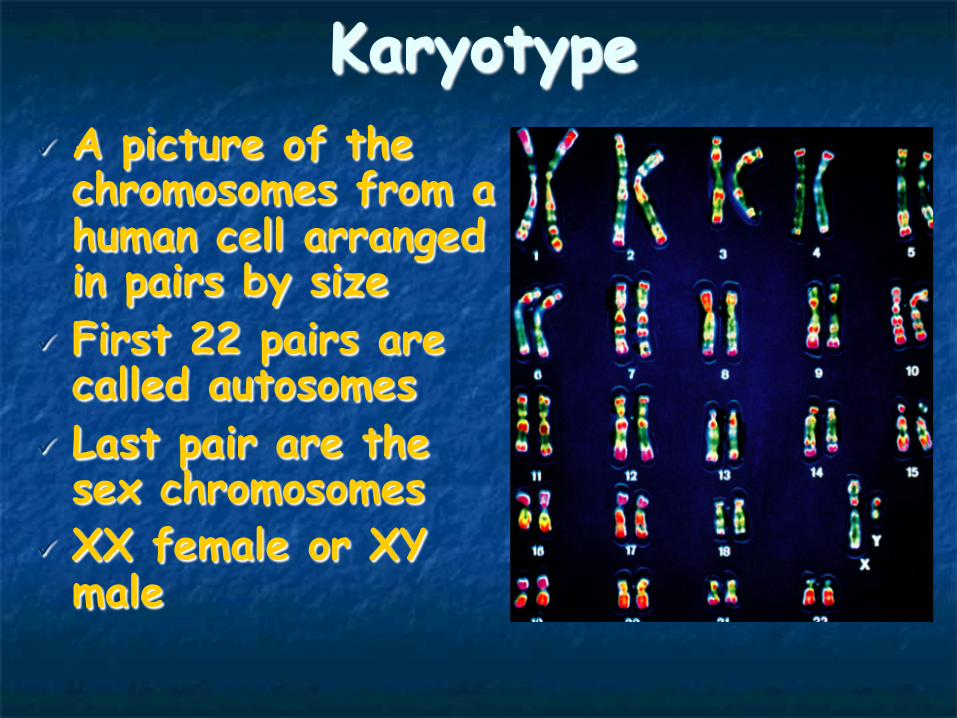
Karyotype ü A picture of the chromosomes from a human cell arranged in pairs by size
ü First 22 pairs are called autosomes
ü Last pair are the sex chromosomes
ü XX female or XY male

Karyotype Procedure n 5 ml of blood is removed from the patient. n If a fetus is being karyotyped amniotic fluid is removed from
the amniotic sac which surrounds the fetus during development. This is done with the aid of a large syringe and ultrasound picturing. There are cells which have come off the fetus in this fluid.
n The white blood cells are removed from the blood or the living cells are removed from the amniotic fluid.
n These cells are then cultured in a medium in which they undergo mitosis. Mitosis is stopped at metaphase using chemicals.
n The cells are then placed onto a slide and spread out. n They are viewed under a microscope which is specially adapted
with a camera to take a picture of the chromosomes from one of the cells.
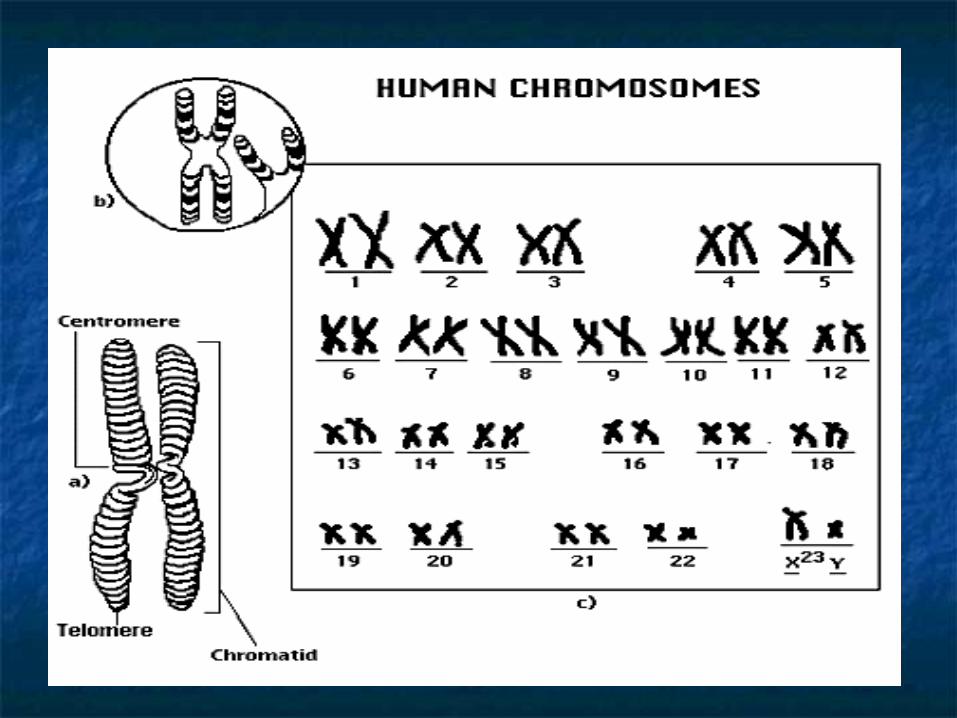
n Once the picture is taken and enlarged the chromosomes are cut out and arranged in pairs according to size and location of the centromere.

n Karyotyping is the process by which doctors and geneticists take pictures of the chromosomes while the cell are undergoing mitosis.
n The picture is then enlarged. n The picture of the chromosomes are then cut up so that
each chromosome is removed. The chromosomes are matched up and attached to a paper according to size, banding patterns, & centromere position.
n The chromosomes pairs are numbered from largest to smallest.
n There are 22 pairs of chromosomes that are aligned first & which match up exactly. These are called autosomes & will code for human body characteristics.
n Then the sex chromosomes are paired, in the female (XX) the chromosomes match and in the male (XY) the chromosomes do not match.

Boy or Girl?
Y - Chromosome
X - Chromosome
The Y Chromosome Decides

Average Male Average Female

Mutations
Mutations
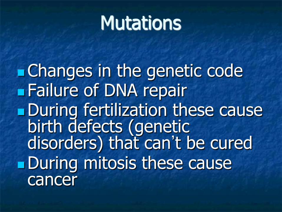
n Changes in the genetic code
n Failure of DNA repair n During fertilization these cause birth defects (genetic disorders) that can’t be cured
n During mitosis these cause cancer

Types of Mutations
Mutations
Gene Mutations
Chromosomal
Gene Mutations
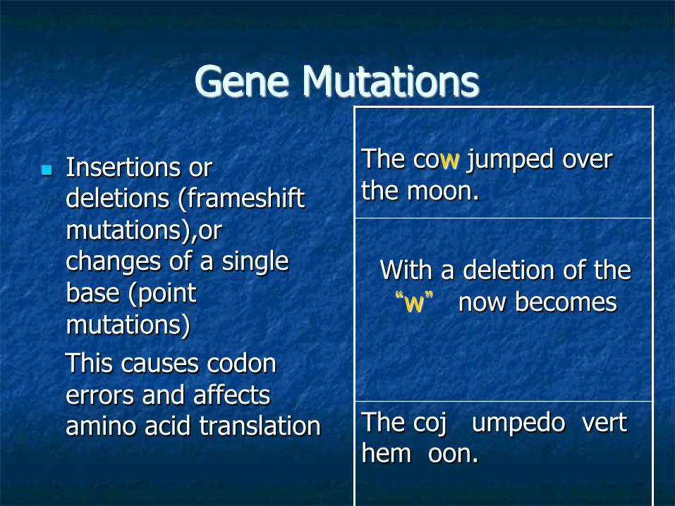
n Insertions or deletions (frameshift mutations),or changes of a single base (point mutations)
This causes codon errors and affects amino acid translation
The cow jumped over the moon.
With a deletion of the “w” now becomes
The coj umpedo vert hem oon.

Point Mutations vs. Frameshift Changing a base original: AUG CAU GGC changed: AUG CCU GGC
Deleting or Inserting a base
original: AUG CAU GGC
changed: AUG CUG GC
The codons have shifted!
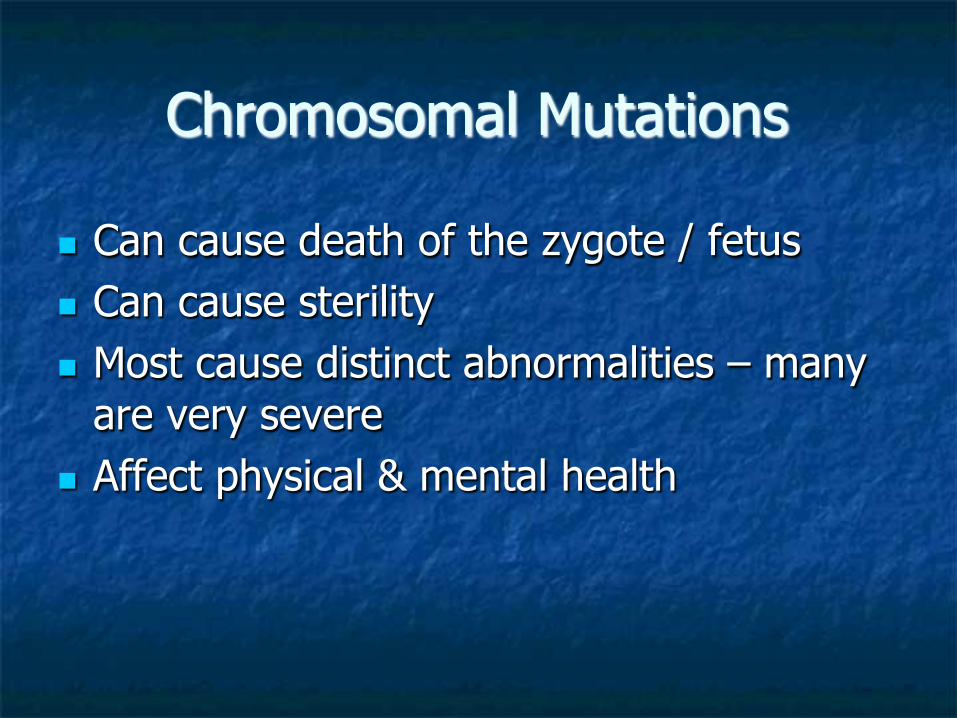
Chromosomal Mutations
n Can cause death of the zygote / fetus n Can cause sterility n Most cause distinct abnormalities – many
are very severe n Affect physical & mental health

Types of chromosomal mutations
1. Duplications 2. Translocations 3. Deletions 4. Inversions 5. Changes in the numbers of
chromosomes

Duplications
n Involves a chromosome that has a piece repeated
n Causes extra length (info) in the strand.
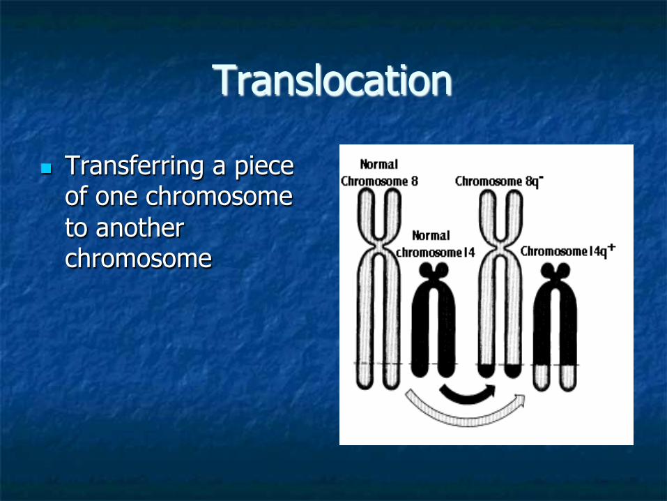
Translocation
n Transferring a piece of one chromosome to another chromosome

Deletions n Omitting or losing a
piece of a chromosome
Prader – willi syndrome

Inversion
n Attaching a piece of a chromosome backward

Changes in chromosome number
n Having more than two copies of each chromosome
n Can have from 3N to 5N of a chromosome including the sex chromosomes

Trisomy
n Having 3 copies of the chromosome instead of the pair (3N)
n Examples: 1. Down’s Syndrome – trisomy 21
2. Klinefelter’s - XXY

Edward’s Syndrome

Monosomy
n Having 1 copy of the chromosome instead of the pair
n Example: 1. Turner Syndrome

Polysomy
n Having more than 3 copies of a chromosome.
n Example: not assigned a name but normally found only in sex chromosomes XXXXY XYYYY
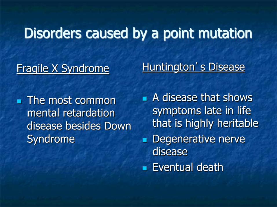
Disorders caused by a point mutation
Fragile X Syndrome
n The most common mental retardation disease besides Down Syndrome
Huntington’s Disease
n A disease that shows
symptoms late in life that is highly heritable
n Degenerative nerve disease
n Eventual death


Mutagens
n Chemicals or agents that cause copying errors during cell division
1. Exposure to radiation 2. Chemicals used in war 3. Chemicals in food preservatives 4. Viruses

Mutagens n Exposure to radiation can cause a multitude of
chromosomal mutations

Human Genetic Disorders 1. Turner’s Syndrome 2. Klinefelter’s Syndrome 3. Microcephaly 4. Marfan’s Syndrome 5. Prader-Willi Syndrome 6. Edward’s Syndrome 7. Epidermolysis Bullosa 8. Congenital Generalized
Hypertrichosis 9. Cri du Chat 10. Achondroplasia 11. Gaucher’s Disease 12. Duchenne Muscular
Dystrophy 13. Fragile X Syndrome 14. Neurofibromatosis 15. Huntington’s
31. Xeroderma Pigmentosum • Phenylketonuria (PKU) • Albinism • Tay-Sachs • Sickle Cell Anemia • Progeria • Cystic Fibrosis • Cleft Palate • Polydactyly • Colorblindness • Hemophilia • Ichthyosis • Spina Bifida • Jacob’s Syndrome • Amyloidosis • Down’s Syndrome • Gastroschisis

Albinism

Phenylketonuria Testing & diagnosed child
Cleft Palate Achondroplasia

Dwarfism
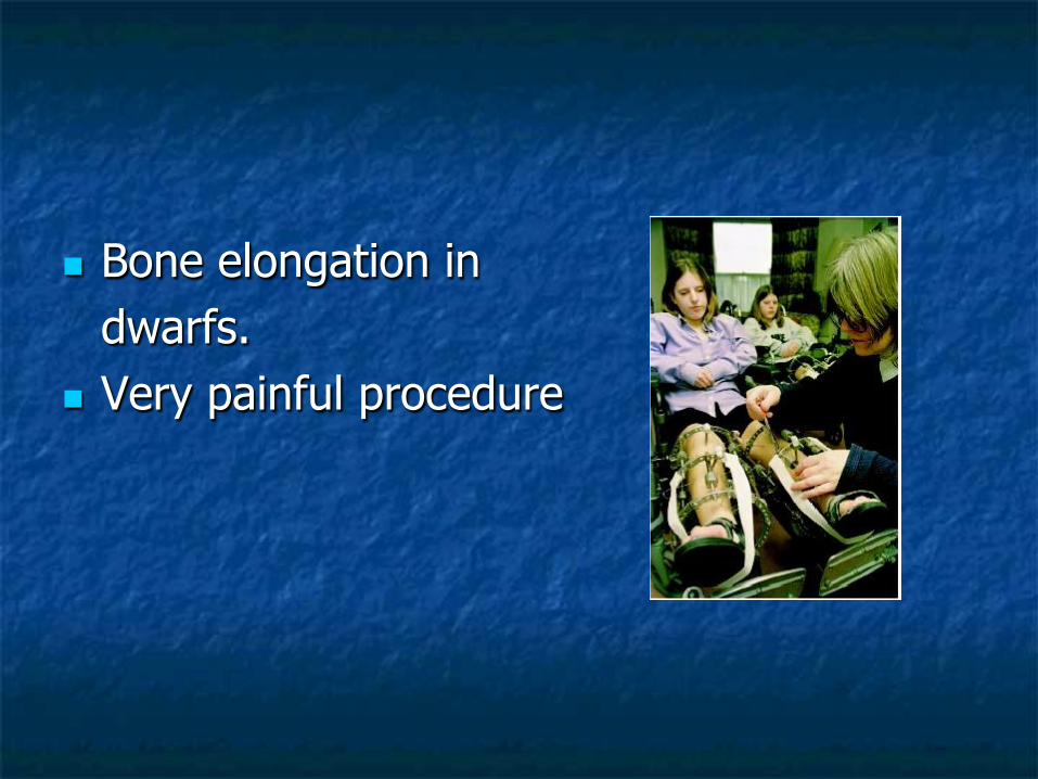
n Bone elongation in dwarfs.
n Very painful procedure

n Excess or deficits can result in obvious skeletal malproportions.
Twelve-year-old boy with pituitary
gigantism measuring 6'5" with his mother.
Not the coarse facial features and
prominent jaw.
n Picture 1. Gigantism and
acromegaly. The author with a statue of Robert Wadlow, the "Alton Giant," who was the tallest person ever recorded. He measured 8 feet 11 inches at the time of his death.

Progeria
Sickle cell
Microcephaly

CGH Duchenne Muscular Dystrophy
Polydactyly

Epidermolysis Bullosa

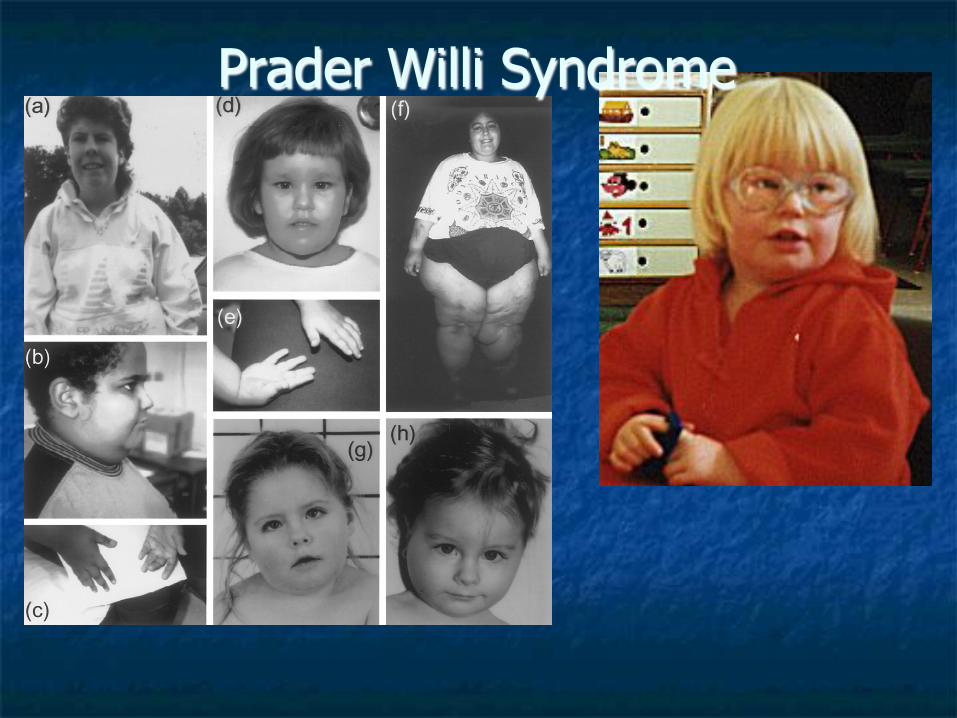
Prader Willi Syndrome

“The Cry of the Cat”
Top Related