


Languages
Pages
Legal


1
Instrumental vaginal birth
Values: The evidence was reviewed by the Women’s
Health Committee (RANZCOG), and applied to
local factors relating to Australia and New Zealand.
Validation: This statement was compared with
RCOG, ACOG and SOGC Guidelines on
instrumental vaginal birth.
Background: This statement was first developed by
Women’s Health Committee in July 2002. During
the statements review in November 2015 Rotational
Forceps (C-Obs 13) was incorporated to create one
statement on Instrumental Vaginal Birth.
Funding: The development and review of this
statement was funded by RANZCOG.
This statement has been developed and reviewed by
the Women’s Health Committee and approved by
the RANZCOG Board and Council.
A list of Women’s Health Committee Members can
be found in Appendix D.
Disclosure statements have been received from all
members of this committee.
Disclaimer This information is intended to provide
general advice to practitioners. This information
should not be relied on as a substitute for proper
assessment with respect to the particular
circumstances of each case and the needs of any
patient. This document reflects emerging clinical
and scientific advances as of the date issued and is
subject to change. The document has been
prepared having regard to general circumstances.
First endorsed by RANZCOG: July 2002
Current: March 2016
Review due: March 2019

Instrumental Vaginal Birth C-Obs 16
2
Table of contents
1. Patient summary ....................................................................................................................... 3
2. Summary of recommendations ................................................................................................... 3
3. Introduction .............................................................................................................................. 4
4. Discussion and recommendations............................................................................................... 5
4.1 Non Operative Interventions .................................................................................................... 5
4.2 Manual Rotation ..................................................................................................................... 5
4.3 Indications for instrumental birth .............................................................................................. 5
4.4 Relative contraindications and contraindications to instrumental vaginal birth .............................. 6
4.5 Conditions required for safe instrumental vaginal birth ............................................................... 6
4.6 Techniques of instrumental vaginal birth ................................................................................... 8
4.6.1 Manual rotation ............................................................................................................... 9
4.6.2 Vacuum extraction ........................................................................................................... 9
4.6.3 Rotation forceps ............................................................................................................. 10
4.7 Suggested guidelines in performing rotational forceps ............................................................. 11
4.8 Complications of instrumental birth ........................................................................................ 11
4.9 Factors affecting choice between vacuum and forceps delivery ................................................. 12
4.10 Unsuccessful instrumental vaginal birth ................................................................................. 13
4.11 Postnatal care .................................................................................................................... 13
5. Conclusion ............................................................................................................................. 15
6. References .............................................................................................................................. 16
7. Other suggested reading ......................................................................................................... 19
8. Links to other College statements ............................................................................................. 20
9. Patient information .................................................................................................................. 20
Appendices ................................................................................................................................... 21
Appendix A Techniques for Manual Rotation 7 ............................................................................... 21
Appendix B Key factors in a rotational forceps birth (The following is provided as an example of
guidance but may not cover all circumstances) .............................................................................. 22
Appendix C Women’s Health Committee Membership .................................................................. 23
Appendix D Overview of the development and review process for this statement .............................. 23
Appendix E Full Disclaimer .......................................................................................................... 25

Instrumental Vaginal Birth C-Obs 16
3
1. Patient summary
The use of instruments – a vacuum cup (ventouse) or forceps – may be required to achieve a safe vaginal
birth. Using instruments to assist birth is usually recommended when the condition of either the baby or the
mother makes it less safe to allow time for normal birth to occur. The choice of which instrument to use
depends on the clinical situation, and every birth is different. There are different types of vacuum cup and
different types of forceps, and each has different advantages and potential disadvantages. Sometimes a
caesarean section will be performed instead of, or even after an attempted instrumental delivery, however a
caesarean section when the baby’s head is deep in the pelvis and the cervix is fully dilated can be very
difficult and poses risks to mother and baby. Further, a caesarean section has important implications for the
mother’s future pregnancies. For this reason, the risks and benefits of instrumental vaginal birth need to be
weighed up in each case, and the following statement provides information for clinicians on the principles
that guide instrumental vaginal births.
2. Summary of recommendations
Recommendation 1 Grade As instrumental vaginal birth may be associated with maternal and neonatal
morbidity. Measures which safely reduce the need for instrumental birth are
recommended.
Consensus-based recommendation
Recommendation 2 Grade
Safe instrumental vaginal birth requires a careful assessment of the clinical situation and clear communication with the mother. Instrumental birth should be performed by or in the presence of, an operator with expertise in the chosen procedure and the management of any complications which may arise.
Consensus-based recommendation
Recommendation 3 When there is an increased likelihood that attempted instrumental birth may not
be successful, where feasible, the attempt should be conducted in a place
where immediate recourse to caesarean section is possible.
Consensus-based recommendation
Recommendation 4 The instrument recommended, forceps or vacuum, will appropriately depend on
the preference of the operator in that particular clinical circumstance.
Consensus-based recommendation
Recommendation 5 Where there is difficulty with operative vaginal birth, including recourse to
Caesarean section or sequential use of instruments, the doctor responsible for
the care of the baby should be advised so that appropriate surveillance and
management of the baby can be instituted.
Consensus-based recommendation
Recommendation 6 Postnatal care following operative vaginal birth requires attention to analgesia,
voiding function, bowel function, thromboembolic prophylaxis, rehabilitation of
the pelvic floor and counselling regarding the birth and future births.
Consensus-based recommendation

Instrumental Vaginal Birth C-Obs 16
4
3. Introduction
Instrumental vaginal birth retains an important role in current obstetric practice. Vacuum and forceps assisted vaginal birth account for approximately 11% of births in Australia (1990- 2012)1 and just under 10% of births in New Zealand.2 Rates have been reported to vary from 7.4-16% of all births across a spectrum of Australian and New Zealand hospitals.3 A number of reviews and guidelines have been published.4-7 When labour has progressed to full dilatation and concerns exist regarding wellbeing of the fetus, mother, or both, three options exist: (1) to allow the labour to proceed aiming for spontaneous vaginal birth; (2) to proceed to instrumental vaginal birth; or, (3) to perform a caesarean section. Each of these options carries both benefits and risks and each individual case has particular circumstances which may influence the recommendation. Emergency caesarean sections at full dilation can be technically difficult with the head sometimes deep in the maternal pelvis, with the potential for injury to both mother and fetus. When compared with a caesarean section performed in the first stage of labour, a caesarean section performed in second stage of labour is associated with significantly increased risk of maternal morbidity including, tears in relation to the uterine incision, haemorrhage, blood transfusion, bladder trauma and requirement for intensive care.8-11 There is also potential for complications in future pregnancies relating to uterine scar rupture in labour and risks associated with repeat caesarean section, which increase with each additional caesarean section required.12,
13 These issues are addressed in the College statement Vaginal birth after previous caesarean section (C-Obs 38). In general, and balanced against the risks of leaving a fetus undelivered, instrumental vaginal birth provides a safe and effective option in appropriately selected cases. Further, a vaginal birth in a first pregnancy is associated with a high (78-91%) rate of spontaneous vaginal birth in the next pregnancy.14, 15
Instrumental Vaginal Birth C-Obs 16
5
4. Discussion and recommendations
4.1 Non Operative Interventions
Several approaches to care may reduce the need for instrumental delivery. These include continuous
midwifery support during labour and the use of upright or lateral positions in the second stage of labour.16,
17 Epidural analgesia, compared with non-epidural methods of pain relief, is associated with an increased
incidence of instrumental vaginal birth.18 However, some of these observed differences may be attributable
to the complicated labour rather than the epidural itself.
Judicious use of oxytocin infusion after careful assessment and with continuous fetal monitoring may reduce
the need for operative birth. Commencement of an oxytocin infusion in the second stage of labour in
nulliparous women who have an epidural in situ has been reported to reduce the need for non-rotational
forceps.19 However commencing an oxytocin infusion in the second stage of labour for parous women
should be undertaken with extreme caution, and requires careful examination to exclude abnormal
presentation, signs of cephalo-pelvic disproportion, and other causes of secondary arrest of labour such as
uterine rupture.
A meta-analysis demonstrated that primiparous patients who received epidurals were likely to have fewer
rotational or mid-cavity interventions when pushing was delayed for up to two hours or until they had a
strong urge to push, provided there was no evidence of fetal compromise.20
4.2 Manual Rotation
The purpose of manual rotation is to turn the fetal head to the more favourable occiput anterior position.
When successful it has been shown to significantly reduce the need for caesarean section and increase the
rate of vaginal birth.21, 22
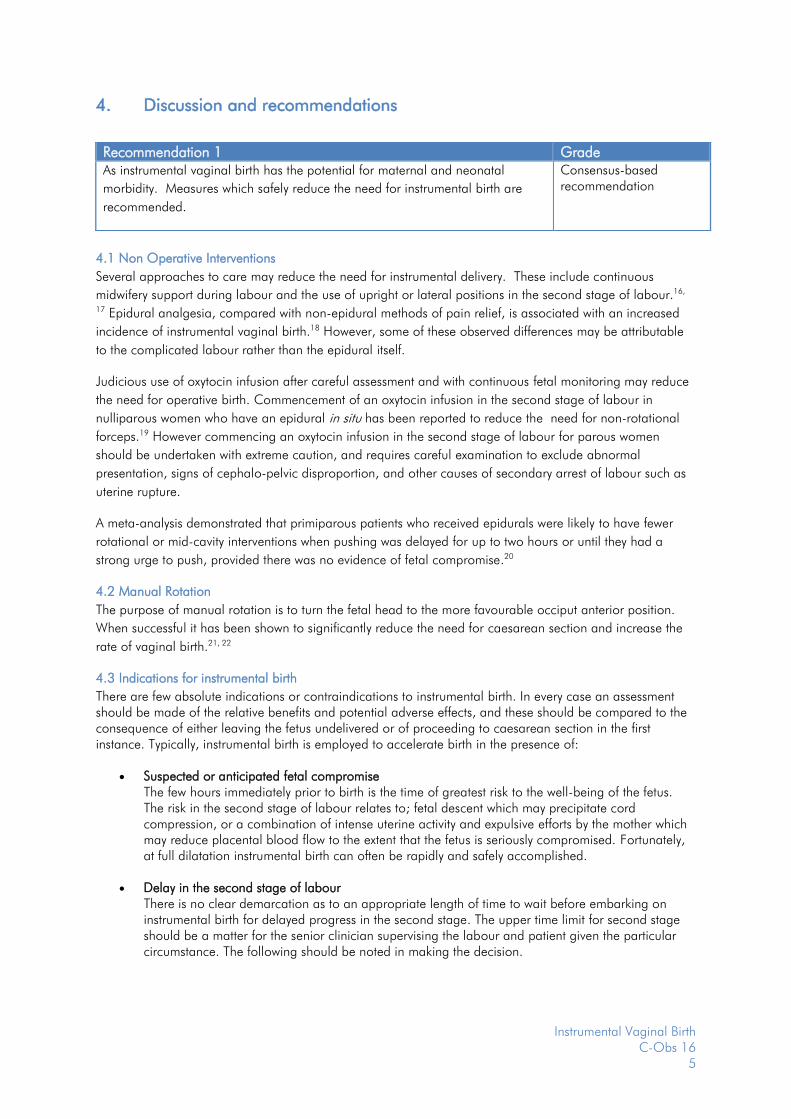
4.3 Indications for instrumental birth
There are few absolute indications or contraindications to instrumental birth. In every case an assessment should be made of the relative benefits and potential adverse effects, and these should be compared to the consequence of either leaving the fetus undelivered or of proceeding to caesarean section in the first instance. Typically, instrumental birth is employed to accelerate birth in the presence of:
Suspected or anticipated fetal compromise The few hours immediately prior to birth is the time of greatest risk to the well-being of the fetus. The risk in the second stage of labour relates to; fetal descent which may precipitate cord compression, or a combination of intense uterine activity and expulsive efforts by the mother which may reduce placental blood flow to the extent that the fetus is seriously compromised. Fortunately, at full dilatation instrumental birth can often be rapidly and safely accomplished.
Delay in the second stage of labour
There is no clear demarcation as to an appropriate length of time to wait before embarking on instrumental birth for delayed progress in the second stage. The upper time limit for second stage should be a matter for the senior clinician supervising the labour and patient given the particular circumstance. The following should be noted in making the decision.
Recommendation 1 Grade As instrumental vaginal birth has the potential for maternal and neonatal
morbidity. Measures which safely reduce the need for instrumental birth are
recommended.
Consensus-based recommendation
Instrumental Vaginal Birth C-Obs 16
6
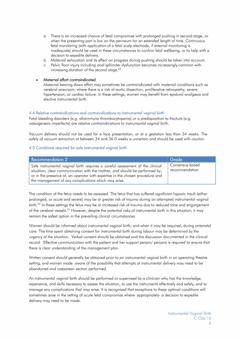
a. There is an increased chance of fetal compromise with prolonged pushing in second stage, or
when the presenting part is low on the perineum for an extended length of time. Continuous fetal monitoring (with application of a fetal scalp electrode, if external monitoring is inadequate) should be used in these circumstances to confirm fetal wellbeing, or to help with a decision to expedite delivery.
b. Maternal exhaustion and its effect on progress during pushing should be taken into account. c. Pelvic floor injury including anal sphincter dysfunction becomes increasingly common with
increasing duration of the second stage.23
Maternal effort contraindicated
Maternal bearing down effort may sometimes be contraindicated with maternal conditions such as cerebral aneurysm, where there is a risk of aortic dissection, proliferative retinopathy, severe hypertension, or cardiac failure. In these settings, women may benefit from epidural analgesia and elective instrumental birth.
4.4 Relative contraindications and contraindications to instrumental vaginal birth
Fetal bleeding disorders (e.g. alloimmune thrombocytopenia) or a predisposition to fracture (e.g. osteogenesis imperfecta) are relative contraindications to instrumental vaginal birth.
Vacuum delivery should not be used for a face presentation, or at a gestation less than 34 weeks. The safety of vacuum extraction at between 34 and 36.0 weeks is uncertain and should be used with caution.
4.5 Conditions required for safe instrumental vaginal birth
The condition of the fetus needs to be assessed. The fetus that has suffered significant hypoxic insult (either
prolonged, or acute and severe) may be at greater risk of trauma during an attempted instrumental vaginal
birth.24 In these settings the fetus may be at increased risk of trauma due to reduced tone and engorgement
of the cerebral vessels.25 However, despite the potential risks of instrumental birth in this situation, it may
remain the safest option in the prevailing clinical circumstances.
Women should be informed about instrumental vaginal birth, and when it may be required, during antenatal
care. The time spent obtaining consent for instrumental birth during labour may be determined by the
urgency of the situation. Verbal consent should be obtained and the discussion documented in the clinical
record. Effective communication with the patient and her support person/ persons is required to ensure that
there is clear understanding of the management plan.
Written consent should generally be obtained prior to an instrumental vaginal birth in an operating theatre
setting, and women made aware of the possibility that attempts at instrumental delivery may need to be
abandoned and caesarean section performed.
An instrumental vaginal birth should be performed or supervised by a clinician who has the knowledge,
experience, and skills necessary to assess the situation, to use the instruments effectively and safely, and to
manage any complications that may arise. It is recognised that exceptions to these optimal conditions will
sometimes arise in the setting of acute fetal compromise where- appropriately- a decision to expedite
delivery may need to be made.
Recommendation 2 Grade
Safe instrumental vaginal birth requires a careful assessment of the clinical situation, clear communication with the mother, and should be performed by, or in the presence of, an operator with expertise in the chosen procedure and the management of any complications which may arise.
Consensus-based recommendation

Instrumental Vaginal Birth C-Obs 16
7
Prerequisites before proceeding to an instrumental vaginal birth are shown below in Table 1
Table 1. Prerequisites for instrumental vaginal birth (vertex presentation)
Full abdominal and vaginal
examination
Less than or equal to one fifth of the head is palpable abdominally. Vertex presentation. Cervix is fully dilated and the membranes ruptured. Exact position of the fetal head can be determined so correct placement of the instrument can be achieved. Ultrasound may be helpful in determining the position of the vertex.
Assessment of caput and moulding. Pelvis is deemed adequate.
Preparation of mother Clear explanation should be given and consent obtained, appropriate to the clinical situation. Analgesia appropriate for the delivery is in place and effective.
For mid-cavity rotational births this will commonly be a regional block. A pudendal block may be appropriate, particularly in the context of urgent birth. Maternal bladder has been emptied recently. In-dwelling catheter should be removed or balloon deflated. Aseptic technique
Preparation of staff Operator must have the knowledge, experience and skill necessary, or an appropriate supervisor is present. Adequate facilities are available (appropriate equipment, bed, lighting). Back-up plan in place in case of failure to deliver. When conducting mid-cavity births, theatre staff should be immediately available to allow a caesarean section to be performed without delay (less than 30 minutes). A senior obstetrician competent in performing mid-cavity births should be present if a junior trainee is performing the birth. Anticipation of complications that may arise (e.g. shoulder dystocia, postpartum haemorrhage) Personnel present that are trained in neonatal resuscitation
Adapted from RCOG Green-top Guideline No. 265

Instrumental Vaginal Birth C-Obs 16
8
Instrumental vaginal births are classified according to the station of the vertex and whether rotation is required. Table 2. Classification for instrumental vaginal birth 5
Outlet Fetal scalp visible without separating the labia Fetal skull has reached the pelvic floor Sagittal suture is in the antero-posterior diameter or right or left occiput anterior or posterior position (rotation does not exceed 45º) Fetal head is at or on the perineum
Low Leading point of the skull (not caput) is at station plus 2 cm or more and not on the pelvic floor. Two subdivisions: - rotation of 45º or less from the occipito-anterior position - rotation of more than 45º including the occipito-posterior position
Mid Fetal head is no more than 1/5th palpable per abdomen Leading point of the skull is above station plus 2 cm but not above the ischial spines Two subdivisions: - rotation of 45º or less from the occipito-anterior position - rotation of more than 45º including the occipito-posterior position
High Not included in the classification as instrumental vaginal birth is not recommended in this situation where the head is 2/5th or more palpable abdominally and the presenting part is above the level of the ischial spines
4.6 Techniques of instrumental vaginal birth
Instrumental vaginal birth involves the use of forceps or the vacuum extractor to allow the operator to assist
the natural forces along the birth canal which are created by uterine contractions and maternal bearing
down effort. Appropriate positioning of forceps or vacuum is important for maternal and fetal safety and for
effective traction. In some cases, rotation of the fetal head may be required to make the position of the
vertex more favourable for descent.
Recognition of when it is appropriate to abandon the procedure and consider an alternative method of birth
is vital. RCOG state that ‘the bulk of malpractice litigation results from failure to abandon the procedure at
the appropriate time, particularly the failure to eschew prolonged, repeated or excessive traction efforts in
the presence of poor progress’ 5.
Higher rates of failure have been associated with:
• Maternal Body Mass Index greater than 30 Kg/m2
• Estimated fetal weight over 4 Kg.
• Occipito-posterior positions.
• Mid-cavity, or when 1/5 of the fetal head is palpable abdominally. 5
Recommendation 3 When there is an increased likelihood that attempted instrumental birth may not
be successful, where feasible, the attempt should be conducted in a place
where immediate recourse to caesarean section is possible.
Consensus-based recommendation

Instrumental Vaginal Birth C-Obs 16
9
When a higher risk of failure is suspected, instrumental vaginal delivery should be attempted in a setting
where immediate recourse to caesarean section is available.
The role of routine episiotomy for instrumental vaginal birth remains unclear and there are no large
randomised controlled trials to guide practice. In a retrospective Dutch study of 28,732 women undergoing
an instrumental birth, use of right mediolateral episiotomy was effective in reducing the risk of anal sphincter
tears in both vacuum and forceps births. Significant risk factors for anal sphincter tears were primiparity,
occipito-posterior position, and increasing fetal weight.26 Other smaller studies have not reported a
protective benefit of episiotomy against anal sphincter injury.27-29 In the absence of robust evidence to
support routine use of episiotomy in operative vaginal delivery, selective use of episiotomy based on the
judgement of the operator is supported.
4.6.1 Manual rotation
Manual rotation of the fetal head to an occipito-anterior position may be used alone, with a view to
increasing the chance of a normal birth, or in conjunction with forceps or vacuum extraction to affect a
vaginal birth. Success rates for rotation of 89% and 76% have been reported in two retrospective trials.21, 22
Success rates were less when performed in nulliparous patients, when performed before full dilation, or
when failure to progress was evident before manual rotation was attempted. 21, 22 When successful there was
a significant reduction in the caesarean section rate with an increase in both the spontaneous and
instrumental vaginal birth rates.21, 22 The complication rate of manual rotation appears to be low, although
data are sparse.22 Techniques for manual rotation are detailed in Appendix A.
4.6.2 Vacuum extraction
Indications for vacuum are similar to those of forceps. Contraindications include prematurity (gestation less
than 34 weeks because of the risk of fetal intracranial haemorrhage) face presentation bleeding diatheses,
and fetal disorders such as osteogenesis imperfecta. Relative contraindications include use between 34 and
36 weeks,( where evidence for use at these gestations is scarce) and prior scalp blood sampling.
Placement of the cup at the flexion point, which is situated 6cm from the anterior fontanelle and 3 cm from
the posterior fontanelle in the midline over the sagittal suture, enables flexion of the fetal head with traction,
improving the chance of rotation of the head if necessary.
Rigid cups are more likely to affect birth (9.5% failure rate versus 14.8% failure rate with a soft cup OR
1.65, 95% CI 1.19-2.29), but are associated with more scalp injuries (24% versus 13% OR 0.45, 95 % CI
0.15-0.60). 6
To minimise the risk of subgaleal haemorrhage, shearing forces on the scalp should be minimised (eg avoid ‘rocking’). Cup placement should be:
i. Placed evenly across the sagittal suture, rather than being applied to one or other parietal bone to avoid asynclitism with traction.
ii. The edge of the cup should be placed at least 3 cm from the anterior fontanelle to avoid extension of the fetal head during traction (assuming a standard 6cm cup is being used).
iii. Appropriate cup placement may be impossible if there is significant deflexion or asynclitism of the head and a “large soft-stemmed” device is being used, because it cannot be placed sufficiently posteriorly.
iv. It is important to check there is no maternal tissue under the vacuum cup both before and after the application of suction.
Vacuum suction pressures of 500-600mm Hg are recommended, and establishment of negative pressure without delay reduces procedure time without compromising effectiveness or safety.30
Instrumental Vaginal Birth C-Obs 16
10
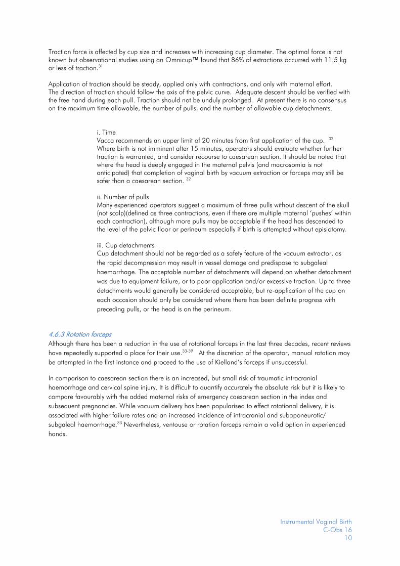
Traction force is affected by cup size and increases with increasing cup diameter. The optimal force is not known but observational studies using an Omnicup™ found that 86% of extractions occurred with 11.5 kg or less of traction.31 Application of traction should be steady, applied only with contractions, and only with maternal effort. The direction of traction should follow the axis of the pelvic curve. Adequate descent should be verified with the free hand during each pull. Traction should not be unduly prolonged. At present there is no consensus on the maximum time allowable, the number of pulls, and the number of allowable cup detachments.
i. Time Vacca recommends an upper limit of 20 minutes from first application of the cup. 32 Where birth is not imminent after 15 minutes, operators should evaluate whether further traction is warranted, and consider recourse to caesarean section. It should be noted that where the head is deeply engaged in the maternal pelvis (and macrosomia is not anticipated) that completion of vaginal birth by vacuum extraction or forceps may still be safer than a caesarean section. 32 ii. Number of pulls Many experienced operators suggest a maximum of three pulls without descent of the skull (not scalp)(defined as three contractions, even if there are multiple maternal ‘pushes’ within each contraction), although more pulls may be acceptable if the head has descended to the level of the pelvic floor or perineum especially if birth is attempted without episiotomy. iii. Cup detachments Cup detachment should not be regarded as a safety feature of the vacuum extractor, as
the rapid decompression may result in vessel damage and predispose to subgaleal
haemorrhage. The acceptable number of detachments will depend on whether detachment
was due to equipment failure, or to poor application and/or excessive traction. Up to three
detachments would generally be considered acceptable, but re-application of the cup on
each occasion should only be considered where there has been definite progress with
preceding pulls, or the head is on the perineum.
4.6.3 Rotation forceps
Although there has been a reduction in the use of rotational forceps in the last three decades, recent reviews
have repeatedly supported a place for their use.33-39 At the discretion of the operator, manual rotation may
be attempted in the first instance and proceed to the use of Kielland’s forceps if unsuccessful.
In comparison to caesarean section there is an increased, but small risk of traumatic intracranial
haemorrhage and cervical spine injury. It is difficult to quantify accurately the absolute risk but it is likely to
compare favourably with the added maternal risks of emergency caesarean section in the index and
subsequent pregnancies. While vacuum delivery has been popularised to effect rotational delivery, it is
associated with higher failure rates and an increased incidence of intracranial and subaponeurotic/
subgaleal haemorrhage.33 Nevertheless, ventouse or rotation forceps remain a valid option in experienced
hands.
Instrumental Vaginal Birth C-Obs 16
11
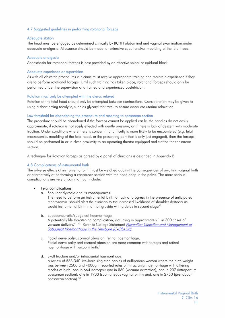
4.7 Suggested guidelines in performing rotational forceps
Adequate station
The head must be engaged as determined clinically by BOTH abdominal and vaginal examination under
adequate analgesia. Allowance should be made for extensive caput and/or moulding of the fetal head.
Adequate analgesia
Anaesthesia for rotational forceps is best provided by an effective spinal or epidural block.
Adequate experience or supervision
As with all obstetric procedures clinicians must receive appropriate training and maintain experience if they
are to perform rotational forceps. Until such training has taken place, rotational forceps should only be
performed under the supervision of a trained and experienced obstetrician.
Rotation must only be attempted with the uterus relaxed
Rotation of the fetal head should only be attempted between contractions. Consideration may be given to
using a short acting tocolytic, such as glyceryl trinitrate, to ensure adequate uterine relaxation.
Low threshold for abandoning the procedure and resorting to caesarean section
The procedure should be abandoned if the forceps cannot be applied easily, the handles do not easily
approximate, if rotation is not easily effected with gentle pressure, or if there is lack of descent with moderate
traction. Under conditions where there is concern that difficulty is more likely to be encountered (e.g. fetal
macrosomia, moulding of the fetal head, or the presenting part that is only just engaged), then the forceps
should be performed in or in close proximity to an operating theatre equipped and staffed for caesarean
section.
A technique for Rotation forceps as agreed by a panel of clinicians is described in Appendix B.
4.8 Complications of instrumental birth
The adverse effects of instrumental birth must be weighed against the consequences of awaiting vaginal birth or alternatively of performing a caesarean section with the head deep in the pelvis. The more serious complications are very uncommon but include:
Fetal complications a. Shoulder dystocia and its consequences.
The need to perform an instrumental birth for lack of progress in the presence of anticipated macrosomia should alert the clinician to the increased likelihood of shoulder dystocia as would instrumental birth in a multigravida with a delay in second stage40
b. Subaponeurotic/subgaleal haemorrhage.
A potentially life threatening complication, occurring in approximately 1 in 300 cases of vacuum delivery.41, 42 Refer to College Statement Prevention Detection and Management of Subgaleal Haemorrhage in the Newborn (C-Obs 28)
c. Facial nerve palsy, corneal abrasion, retinal haemorrhage. Facial nerve palsy and corneal abrasion are more common with forceps and retinal haemorrhage with vacuum birth.6
d. Skull fracture and/or intracranial haemorrhage. A review of 583,340 live-born singleton babies of nulliparous women where the birth weight was between 2500 and 4000gm reported rates of intracranial haemorrhage with differing modes of birth: one in 664 (forceps); one in 860 (vacuum extraction); one in 907 (intrapartum caesarean section); one in 1900 (spontaneous vaginal birth); and, one in 2750 (pre-labour caesarean section).43

Instrumental Vaginal Birth C-Obs 16
12
e. Cervical spine injury. Injury to the fetal cervical spinal cord is rare but the absolute rate is difficult to define. In a series from Canada it was estimated the risk may be 0.7/1000 rotation forceps births.44 This injury may occur less frequently with vacuum delivery. The risk may be minimised by ensuring uterine relaxation prior to attempting rotation. These complications may require admission to a neonatal unit and may be associated with feeding and bonding difficulties.
Maternal complications
Maternal complications which may occur include vaginal trauma, postpartum haemorrhage, urinary tract injury, and damage to pelvic floor and anal sphincter (details below).6
4.9 Factors affecting choice between vacuum and forceps delivery
Each instrument has a different profile of complications.6 Vaginal birth is more likely to be achieved with forceps than vacuum and will occur over a shorter time interval.45 The clinician should select the instrument based on clinical experience and the individual clinical circumstances. A Cochrane review of 32 studies including 6597 women requiring instrumental vaginal birth compared any kind of forceps birth (rotational and non-rotational) with any type of vacuum extraction (rotational or non-rotational).6 Compared to the vacuum extractor forceps were found to:
▪ be less likely to fail to achieve a vaginal birth (RR 0.65, 95% CI 0.45-0.94)
▪ have a trend toward fewer cases of cephalhaematoma (RR 0.64, 95% CI 0.37-1.11)
▪ have a trend toward fewer cases of fetal retinal haemorrhage (RR 0.6, 95% CI 0.43-1.06)
▪ have a trend toward fewer cases of neonatal jaundice (RR 0.79, 95% CI 0.59-1.06)
▪ have a trend toward fewer cases of shoulder dystocia (RR 0.4, 95% CI 0.16-1.04) Compared to vacuum delivery, use of forceps was associated with a higher incidence of:
▪ third or fourth degree tears of the anal sphincter (RR 1.89, 95% CI 1.51-2.37)
▪ any type of vaginal trauma (RR 2.48, 95% CI 1.59-3.87)
▪ incontinence/altered continence (RR 1.77, 95% 1.19-2.62) There was no significant difference between instruments in the risk of:
▪ any neonatal injury
▪ low Apgar score (<7) at 5 minutes
▪ low pH (<7.2) in umbilical artery at birth. A meta-analysis of 23 studies of rotational assisted instrumental births reported that Kielland’s forceps were less likely to fail (RR 0.32, 95% CI 0.14-0.76) and less likely to cause neonatal trauma (RR 0.62, 95% CI 0.46-0.85) when compared to rotational ventouse birth. 33 A study of 3753 women who were contacted at 12 years after their first birth found that symptoms of faecal incontinence were reported by 11.5% of respondents who had only unassisted vaginal births, 10.5% following only planned caesarean sections with no vaginal births, 16.7% with any forceps birth and 10.9% following any vacuum assisted (but no forceps) birth. Urinary incontinence rates reported by the same groups of women defined by mode of birth were 54.7%, 38.7%. 51.4% and 56% respectively.46
Recommendation 4 The choice of either vacuum or forceps for instrumental vaginal delivery will
depend on the judgement of the operator and the individual clinical
circumstances.
Consensus-based recommendation

Instrumental Vaginal Birth C-Obs 16
13
4.10 Unsuccessful instrumental vaginal birth
Unsuccessful attempts at instrumental birth may be associated with adverse outcome.47 If an initial attempt does not affect delivery ,or there is lack of progress of rotation ,if required, and/or descent of the vertex, then assessment should be made as to whether alternate instrumental birth should be attempted (ie sequential use of instruments) or a caesarean section performed without further attempt at instrumental birth.
Sequential Use of Instruments
The use of sequential instruments has been associated with an increased risk of trauma to the fetus and
mother when compared to the use of forceps or vacuum alone.48 Nevertheless, these findings need to be
interpreted cautiously, given the increasing use of ventouse as the primary instrument in contemporary
obstetrics. This change in practice has followed increasing evidence suggesting that ventouse (compared to
forceps) reduces maternal obstetric anal sphincter injury 49, levator any muscle avulsion,50 urinary
incontinence and pelvic organ prolapse. Yet ventouse is also associated with an increased risk of non-
completion of delivery with cohort studies suggesting a 30% chance that vacuum devices such as the kiwi
cup will fail, requiring completion of delivery by forceps, increasing to 40% for rotational delivery. 51 Hence,
the use of sequential instruments may be considered an inevitable consequence of the increasing use of the
ventouse.
Caesarean section following attempted instrumental delivery
The alternative to sequential use of instruments is caesarean section following an attempted instrumental birth. These complex caesarean sections, with the fetal head deep within the pelvis, are associated with an increase in maternal morbidity (major postpartum haemorrhage,8 transfusion, lower segment tear, cystotomy, hysterectomy, ICU admission) and fetal morbidity (neonatal acidosis, intracranial haemorrhage, need for resuscitation). 8, 52 In some cases the adverse outcomes associated with difficult instrumental birth may reflect the indication for which instrumental birth was being attempted (e.g. severe fetal compromise) rather than a direct effect of attempts at instrumental birth. The threshold for abandoning an attempted instrumental birth as well as the decision between either choosing an alternative instrument or performing a caesarean section will differ according to the clinician’s training, experience, and the clinical setting.
4.11 Postnatal care
Analgesia
Regular paracetamol and nonsteroidal anti-inflammatory agents should be offered when there is no
contraindication. Pain not relieved by these measures should prompt clinical assessment to exclude
complications such as haematoma formation or infection.
Recommendation 5 Where there is difficulty with operative vaginal birth, including recourse to
Caesarean section or sequential use of instruments, the doctor responsible for
the care of the baby should be advised so that appropriate surveillance and
management of the baby can be instituted.
Consensus-based recommendation
Recommendation 6 Postnatal care following instrumental vaginal birth requires attention to
analgesia, voiding function, thromboembolic prophylaxis, rehabilitation of the
pelvic floor, and counselling regarding the index birth and future births.
Consensus-based recommendation

Instrumental Vaginal Birth C-Obs 16
14
Voiding function
The risk of urinary retention after birth is increased after instrumental vaginal birth, particularly if spinal or
epidural anaesthesia has been employed for the birth. RCOG recommends an indwelling catheter for 12
hours following instrumental vaginal birth if a spinal or epidural top up has been used for anaesthesia and a
trial of instrumental vaginal birth has been planned. 5 Careful observation of postpartum voiding function
and the insertion of an indwelling catheter may be required to prevent bladder over-distention and long term
bladder dysfunction. It is appropriate for obstetric units to have protocols aimed to prevent this complication.
Antibiotic and Thromboembolic prophylaxis.
At present there is insufficient evidence to make any recommendations regarding the use of antibiotic
prophylaxis for instrumental vaginal birth.53
Following an instrumental vaginal birth women should be assessed for their risk profile for venous
thromboembolism and, if appropriate, thromboprophylaxis measures should be employed. Instrumental
vaginal birth may be associated with risk factors for venous thromboembolism such as prolonged labour,
BMI>30, pre-eclampsia and postpartum haemorrhage of greater than 1000ml. Consideration of local
hospital or published guidelines is appropriate. 54
Pelvic floor rehabilitation.
Appropriately conducted pelvic floor exercises in the postnatal period should be encouraged. There is
evidence that physiotherapist-led intervention reduces urinary incontinence in women who had had an
instrumental vaginal birth and/or a baby over 4000g.55
Postnatal discussion regarding birth.
Women should be given the opportunity to discuss the reason for operative birth, the management of any
complications, and the prognosis for future pregnancies. This discussion should occur in the early postnatal
period if possible, and ideally should be led by the clinician who performed the birth.
Instrumental vaginal delivery has been associated with a fear of subsequent birth, and in a severe form has
manifested as post-traumatic stress-type syndrome.56-59 A cohort study of women who had begun labour but
were delivered in the second stage of labour in an operating theatre (either instrumental vaginal birth or
caesarean section) and were questioned three years after their birth, reported that 32% wished to avoid
further pregnancy, and for almost half of these women fear of childbirth was the main reason for this wish.60
After an instrumental vaginal birth in a first labour the success rates for achieving a spontaneous vaginal
birth in the next pregnancy have been reported between 78% and 91%.14, 60
Women who have sustained a third or fourth degree tear will need individual counselling with regard to their
outcome and plans for future delivery based on evaluation of anal sphincter, their symptoms, and
preferences.61

Instrumental Vaginal Birth C-Obs 16
15
5. Conclusion
Instrumental vaginal birth has an important place in obstetric practice. In many cases, the decision to
perform an instrumental delivery will represent a balance between potential risks of leaving the fetus
undelivered and the additional risks of performing a caesarean section in the second stage. Each instrument
has both advantages and disadvantages. Before instrumental delivery is attempted careful assessment of the
whole clinical situation must be undertaken, and discussion with the woman carried out. Those performing
instrumental delivery must have appropriate training and experience, or be supervised closely by somebody
who does have these skills. Attention to techniques specific for the type of instrument will minimise risks and
maximise the chance of successful delivery. Care in the postnatal period is important to minimise the risk of
adverse outcomes in the short and long term.

Instrumental Vaginal Birth C-Obs 16
16
6. References
1. Australian Institute of Health and Welfare (AIHW). Australia’s Mothers & Babies Report 2012. Available from: http://www.aihw.gov.au/WorkArea/DownloadAsset.aspx?id=60129550054.
2. New Zealand Minister of Health Publications. Reports on maternity (1999-2002, 2004,2012) Available from: http://www.health.govt.nz/publications/births
3. Women’s Hospitals Australasia. Clinical Forum on Caesarean Sections Briefing Kit 2001:4-5, 47. 4. ACOG Committee on Practice Bulletins. Obstetrics. ACOG Practice Bulletin. 2000 (reaffirmed
2012). 5. Royal College of Obstetricians and Gynaecologists Guidelines (RCOG). Clinical Green Top
Guidelines.Instrumental Vaginal Delivery No.26. 2011. 6. O’Mahony F HGJ, Menon V. Choice of Instruments for Assisted Vaginal Delivery (Review), The
Cochrane Library 2010 (11). 7. SOGC Clinical Practice Guidelines Guidelines for Operative Vaginal Birth. 2004. 8. Pergialiotis V, Vlachos DG, Rodolakis A, Haidopoulos D, Thomakos N, Vlachos GD. First versus
second stage C/S maternal and neonatal morbidity: a systematic review and meta-analysis, Eur J Obstet Gynecol Reprod Biol. 2014;175:15-24.
9. Allen VM, O'Connell CM, Baskett TF. Maternal and perinatal morbidity of caesarean delivery at full cervical dilatation compared with caesarean delivery in the first stage of labour, BJOG. 2005;112(7):986-90.
10. Selo-Ojeme D, Sathiyathasan S, Fayyaz M. Caesarean delivery at full cervical dilatation versus caesarean delivery in the first stage of labour: comparison of maternal and perinatal morbidity, Arch Gynecol Obstet. 2008;278(3):245-9.
11. Alexander JM, Leveno KJ, Rouse DJ, Landon MB, Gilbert S, Spong CY, et al. Comparison of maternal and infant outcomes from primary cesarean delivery during the second compared with first stage of labor, Obstet Gynecol. 2007;109(4):917-21.
12. Royal Australian and New Zealand College of Obstetricians and Gynaecologists. Birth after previous caesarean section (C-Obs 38). 2015.
13. Silver RM, Landon MB, Rouse DJ, Leveno KJ, Spong CY, Thom EA, et al. Maternal morbidity associated with multiple repeat cesarean deliveries, Obstet Gynecol. 2006;107(6):1226-32.
14. Mawdsley SD, Baskett TF. Outcome of the next labour in women who had a vaginal delivery in their first pregnancy, BJOG. 2000;107(7):932-4.
15. Bahl R, Strachan B, Murphy DJ. Outcome of subsequent pregnancy three years after previous operative delivery in the second stage of labour: cohort study, BMJ. 2004;328(7435):311.
16. Hodnett ED, Gates S, Hofmeyr GJ, Sakala C. Continuous support for women during childbirth, Cochrane Database Syst Rev. 2007(3):CD003766.
17. Gupta JK, Hofmeyr GJ, Shehmar M. Position in the second stage of labour for women without epidural anaesthesia, Cochrane Database Syst Rev. 2012;5:CD002006.
18. Anim-Somuah M, Smyth R, Howell C. Epidural versus non-epidural or no analgesia in labour, Cochrane Database Syst Rev. 2005(4):CD000331.
19. Saunders NJ, Spiby H, Gilbert L, Fraser RB, Hall JM, Mutton PM, et al. Oxytocin infusion during second stage of labour in primiparous women using epidural analgesia: a randomised double blind placebo controlled trial, BMJ. 1989;299(6713):1423-6.
20. Roberts D, Neilson JP, Kilby MD, Gates S. Interventions for the treatment of twin-twin transfusion syndrome, Cochrane Database Syst Rev. 2014;1:CD002073.
21. Shaffer BL, Cheng YW, Vargas JE, Laros RK, Jr., Caughey AB. Manual rotation of the fetal occiput: predictors of success and delivery, Am J Obstet Gynecol. 2006;194(5):e7-9.
22. Le Ray C, Serres P, Schmitz T, Cabrol D, Goffinet F. Manual rotation in occiput posterior or transverse positions: risk factors and consequences on the cesarean delivery rate, Obstet Gynecol. 2007;110(4):873-9.
23. Zetterstrom JP, Lopez A, Anzen B, Dolk A, Norman M, Mellgren A. Anal incontinence after vaginal delivery: a prospective study in primiparous women, Br J Obstet Gynaecol. 1999;106(4):324-30.
24. O'Mahony F, Settatree R, Platt C, Johanson R. Review of singleton fetal and neonatal deaths associated with cranial trauma and cephalic delivery during a national intrapartum-related
Instrumental Vaginal Birth C-Obs 16
17
confidential enquiry, BJOG: An International Journal of Obstetrics & Gynaecology. 2005;112(5):619-26.
25. Wigglesworth J. Pathology of intrapartum and early neonatal death in the normally formed infant. In: Wigglesworth JS, DB, editor. Textboof of fetal and perinatal pathology. London: Blackwell Science; 1998.
26. de Leeuw JW, de Wit C, Kuijken JP, Bruinse HW. Mediolateral episiotomy reduces the risk for anal sphincter injury during operative vaginal delivery, BJOG. 2008;115(1):104-8.
27. Macleod M, Strachan B, Bahl R, Howarth L, Goyder K, Van de Venne M, et al. A prospective cohort study of maternal and neonatal morbidity in relation to use of episiotomy at operative vaginal delivery, BJOG. 2008;115(13):1688-94.
28. Murphy DJ, Macleod M, Bahl R, Goyder K, Howarth L, Strachan B. A randomised controlled trial of routine versus restrictive use of episiotomy at operative vaginal delivery: a multicentre pilot study, BJOG. 2008;115(13):1695-702; discussion 702-3.
29. Robinson JN, Norwitz ER, Cohen AP, McElrath TF, Lieberman ES. Episiotomy, operative vaginal delivery, and significant perinatal trauma in nulliparous women, Am J Obstet Gynecol. 1999;181(5 Pt 1):1180-4.
30. Suwannachat B, Lumbiganon P, Laopaiboon M. Rapid versus stepwise negative pressure application for vacuum extraction assisted vaginal delivery, Cochrane Database Syst Rev. 2012;8:CD006636.
31. Baskett TF, Fanning CA, Young DC. A prospective observational study of 1000 vacuum assisted deliveries with the OmniCup device, J Obstet Gynaecol Can. 2008;30(7):573-80.
32. Vacca A. Handbook of vacuum delivery in obstetric practice, Vacca Research Brisbane Australia. 2003.
33. Al Wattar BH, Wattar BA, Gallos I, Pirie AM. Rotational vaginal delivery with Kielland's forceps: a systematic review and meta-analysis of effectiveness and safety outcomes, Curr Opin Obstet Gynecol. 2015;27(6):438-44.
34. CAMERON M. KIELLAND'S FORCEPS: PAST, PRESENT AND FUTURE, Fetal and Maternal Medicine Review. 2012;23(01):32-51.
35. Tempest N, Hart A, Walkinshaw S, Hapangama DK. A re-evaluation of the role of rotational forceps: retrospective comparison of maternal and perinatal outcomes following different methods of birth for malposition in the second stage of labour, BJOG. 2013;120(10):1277-84.
36. Burke N, Field K, Mujahid F, Morrison JJ. Use and safety of Kielland's forceps in current obstetric practice, Obstet Gynecol. 2012;120(4):766-70.
37. Stock SJ, Josephs K, Farquharson S, Love C, Cooper SE, Kissack C, et al. Maternal and neonatal outcomes of successful Kielland's rotational forceps delivery, Obstet Gynecol. 2013;121(5):1032-9.
38. Bahl R, Van de Venne M, Macleod M, Strachan B, Murphy DJ. Maternal and neonatal morbidity in relation to the instrument used for mid-cavity rotational operative vaginal delivery: a prospective cohort study, BJOG. 2013;120(12):1526-32.
39. Al-Suhel R, Gill S, Robson S, Shadbolt B. Kjelland's forceps in the new millennium. Maternal and neonatal outcomes of attempted rotational forceps delivery, Aust N Z J Obstet Gynaecol. 2009;49(5):510-4.
40. Kolderup LB, Laros RK, Jr., Musci TJ. Incidence of persistent birth injury in macrosomic infants: association with mode of delivery, Am J Obstet Gynecol. 1997;177(1):37-41.
41. Fortune PM TR. Sub-aponeurotic haemorrhage: a rare but life-threatening neonatal complication associated with ventouse delivery, Br J Obstet Gynaecol 1999(177):37-41.
42. Ng PC SY, Lewindon PJ. Subaponeurotic haemorrhage in the 1990s: a 3 year surveillance., Acta Paediatr 1995(84):1065-9.
43. Towner D, Castro MA, Eby-Wilkens E, Gilbert WM. Effect of mode of delivery in nulliparous women on neonatal intracranial injury, N Engl J Med. 1999;341(23):1709-14.
44. Menticoglou SM, Perlman M, Manning FA. High cervical spinal cord injury in neonates delivered with forceps: report of 15 cases, Obstet Gynecol. 1995;86(4 Pt 1):589-94.
45. Okunwobi-Smith Y CI, MacKenzie IZ. Decision to delivery intervals for assisted vaginal vertex delivery., Br J Obstet Gynaecol 2000(107):467-71.
46. MacArthur C, Glazener C, Lancashire R, Herbison P, Wilson D, ProLong study g. Exclusive caesarean section delivery and subsequent urinary and faecal incontinence: a 12-year longitudinal study, BJOG. 2011;118(8):1001-7.

Instrumental Vaginal Birth C-Obs 16
18
47. LC E. Failed instrumental delivery: How safe is the use of a second instrument? , J Obstet Gynaecol 1999(19):460-2.
48. Gardella C, Taylor M, Benedetti T, Hitti J, Critchlow C. The effect of sequential use of vacuum and forceps for assisted vaginal delivery on neonatal and maternal outcomes, Am J Obstet Gynecol. 2001;185(4):896-902.
49. Ampt AJ, Ford JB, Roberts CL, Morris JM. Trends in obstetric anal sphincter injuries and associated risk factors for vaginal singleton term births in New South Wales 2001-2009, Aust N Z J Obstet Gynaecol. 2013;53(1):9-16.
50. van Veelen GA, Schweitzer KJ, van Delft K, Kluivers KB, Weemhoff M, van der Vaart CH. Diagnosing levator avulsions after first delivery by tomographic ultrasound: reliability between observers from different centers, Int Urogynecol J. 2014;25(11):1501-6.
51. Groom KM, Jones BA, Miller N, Paterson-Brown S. A prospective randomised controlled trial of the Kiwi Omnicup versus conventional ventouse cups for vacuum-assisted vaginal delivery, BJOG. 2006;113(2):183-9.
52. Smith J, Alexander RJ. First do no harm: addressing respiratory morbidity in the newborn and child following elective caesarian section before 39 weeks' gestation, S Afr Med J. 2009;99(12):866-8.
53. Liabsuetrakul T, Choobun T, Peeyananjarassri K, Islam M. Antibiotic prophylaxis for operative vaginal delivery, Cochrane Database Syst Rev. 2004(3):CD004455.
54. McLintock C, Brighton T, Chunilal S, Dekker G, McDonnell N, McRae S, et al. Recommendations for the prevention of pregnancy-associated venous thromboembolism, Aust N Z J Obstet Gynaecol. 2012;52(1):3-13.
55. Chiarelli P, Cockburn J. Promoting urinary continence in women after delivery: randomised controlled trial, BMJ. 2002;324(7348):1241.
56. Murphy DJ, Pope C, Frost J, Liebling RE. Women's views on the impact of operative delivery in the second stage of labour: qualitative interview study, BMJ. 2003;327(7424):1132.
57. Menage J. Post-traumatic stress disorder after childbirth: the phenomenon of traumatic birth, CMAJ. 1997;156:831-5.
58. Fisher J, Astbury J, Smith A. Adverse psychological impact of operative obstetric interventions: a prospective longitudinal study, Aust N Z J Psychiatry. 1997;31(5):728-38.
59. Creedy DK, Shochet IM, Horsfall J. Childbirth and the development of acute trauma symptoms: incidence and contributing factors, Birth. 2000;27(2):104-11.
60. Bahl R, Strachan BK. Mode of delivery in the next pregnancy in women who had a vaginal delivery in their first pregnancy, J Obstet Gynaecol. 2004;24(3):272-3.
61. Royal College of Obstetricians and Gynaecologists. The Management of Third- and Fourth-degree Perineal Tears. Greentop Guideline No 29. 2015.
62. Simpson AN, Hodges R, Snelgrove J, Gurau D, Secter M, Mocarski E, et al. Learning From Experience: Qualitative Analysis to Develop a Cognitive Task List for Kielland Forceps Deliveries, J Obstet Gynaecol Can. 2015;37(5):397-404.
Instrumental Vaginal Birth C-Obs 16
19
7. Other suggested reading
Bashore RA, Phillips WH, Brinkman CR. A comparison of the morbidity of midforceps and . Am J Obstet Gynecol 1990; 162: 1428-35.
Cardoza LD, Gibb DM, Studd JW, Cooper DJ. Should we abandon Kielland’s forceps? Br Med J 1983; 287: 315-7.
Chinnock M, Robson S. An anonymous survey of registrar training in the use of Kjelland's forceps in Australia. Aust N Z J Obstet Gynaecol 2009 Oct; 49 (5): 515-6.
Chow SLS, Johnson CM, Anderson TD, Hughes JH. Rotational with Kielland’s forceps Med J Aust 1987; 146: 616-9.
Drife JO. Kielland or Caesar? BMJ 1983; 287: 309-10.
Healey DL, Quinn MA, Pepperell RJ. Rotational of the fetus: Kielland’s forceps and two other methods compared. Br J Obstet Gynecol 1982; 89: 501-6.
Herabutya Y O, Praasertsawat P, Boonrangsimant P. Kielland’s forceps or ventouse-a comparison. Br J Obstet Gynecol 1988; 95: 483-7.
Krivak TC, Drewes P, Horowitz GM. Kielland vs. Nonrotational Forceps for the Second Stage of Labour. J Reprod Med 1999; 44: 511-7.
Liliford RJ, Van Coeverden de Groot HA, Moore PJ, Bingham P. The relative risks of caesarean section (intrapartum and elective) and vaginal a detailed analysis to exclude the effects of medical disorders and other acute pre-existing physiological disturbances. Br J Obstet Gynecol 1990; 97: 883-92. Need J. Is “the trial of forceps” over? Med J Aust 1987; 146: 613.
Nielsen TF, Hokegard KH. Cesarean section and intraoperative surgical complications. Acta Obstet Gynecol Scand 1984; 63: 103-8.
Rodrigues C, Singh PM, Gupta AN. Kielland’s forceps for deep transverse arrest. Asia-Oceania J Obstet Gynecol 1983; 9: 160-1.
Svigos JM, Cave DG, Vigneswaran R, Resch A, Christiansen J. Silastic cup vacuum extractor or forceps: a comparative study. Asia-Oceania J Obstet Gynecol 1990; 16: 323-7.
Traub AI, Morrow RJ, Ritchie JWK, Dornan KJ A continuing use for Kielland’s forceps? Br J Obstet Gynecol 1984; 91: 894-8.
Vacca A. Birth by vacuum extraction: Neonatal outcome. J Paediatr Child Health 1996; 32: 204-206.

Instrumental Vaginal Birth C-Obs 16
20
8. Links to other College statements Breech deliveries at term (C-Obs 11) Prevention, detection and management of Subgaleal Haemorrhage in the newborn (C-Obs 28) Birth after previous caesarean section (C-Obs 38) Consent and provision of information to patients in Australia regarding proposed treatment (C-Gen 02a) Consent and provision of information to patients in New Zealand regarding proposed treatment (C-Gen 2b) RCOG The Management of Third- and Fourth-degree Perineal Tears. RCOG Greentop Guidline No 29, 2015. https://www.rcog.org.uk/globalassets/documents/guidelines/gtg-29.pdf Evidence-based Medicine, Obstetrics and Gynaecology (C-Gen 15)
9. Patient information
A range of RANZCOG Patient Information Pamphlets can be ordered via:
https://www.ranzcog.edu.au/Womens-Health/Patient-Information-Guides/Patient-Information-Pamphlets

Instrumental Vaginal Birth C-Obs 16
21
Appendices
Appendix A Techniques for Manual Rotation 7
Technique No.1 1. The entire hand is placed in the woman’s vagina with the palm up. 2. The fetal head is flexed and slightly dislodged. 3. The occiput is rotated anteriorly by pronation or supination of the forearm. 4. The fetal head may need to be held in place for a few contractions or until the application of a vacuum or forceps is completed.
Technique No.2 1. The fingers may be placed along the lambdoid sutures. 2. Using mild pressure and a dialling motion, the fetal head can be rotated to an occiput anterior position. 3. The fetal head may need to be held in place for a few contractions or until the application of a vacuum or forceps is completed.

Instrumental Vaginal Birth C-Obs 16
22
Appendix B Key factors in a rotational forceps birth
(The following is provided as an example of guidance but may not cover all circumstances)
Agreed technique published by a panel of clinicians (Simpson 2015) 62
1.Prevention of fetal malposition - Early assessment and documentation of fetal position - Judicious use of oxytocin - Consider manual rotation - Consider the indication for forceps-assisted birth carefully
2. Assessment to determine suitability - Careful second stage assessment - Adequate analgesia - Empty maternal bladder - Red flag: assessment of position difficult due to moulding/caput
3. Communication and consent - Maternal complications: perineal/cervical trauma (similar to other operative births) - Fetal complications: intracranial haemorrhage, cervical spine injury, entrapment of umbilical cord (similar to other operative births) - Discuss reason for prolonged labour - Discuss timing of procedure - Recommend episiotomy
4. Assemble the multidisciplinary team - Nurse/midwife to palpate contractions - Paediatrician/respiratory technician/anaesthetist - Birth in operating room - Position patient, check equipment
5. Phantom application - Reconfirm fetal position
6. Application by the “wandering technique” - Apply between contractions - Correct asynclitism between contractions - Red flag: difficult application
7. Rotation -Between contractions -Force of one hand only; other hand on maternal abdomen -Ensure OA position after rotation -Red flag: difficult rotation
8. Application of traction -During a contraction -Force of one hand only -Episiotomy -Red flag: force of more than one hand required
9. Birth of fetus - Remove blades after birth of head - Prepare for shoulder dystocia and/or postpartum haemorrhage
10. Debrief and document - Examine cervix/vagina/perineum - Examine the baby - Document indications, discussion, timing
Instrumental Vaginal Birth C-Obs 16
23
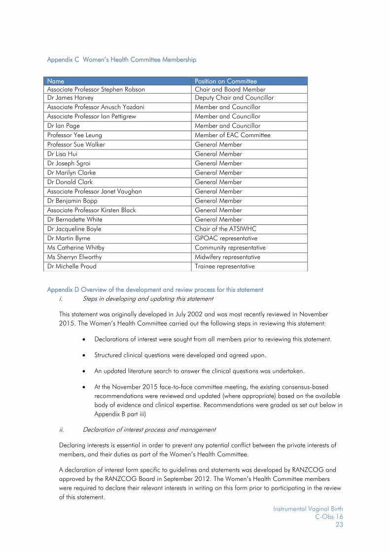
Appendix C Women’s Health Committee Membership
Appendix D Overview of the development and review process for this statement
i. Steps in developing and updating this statement
This statement was originally developed in July 2002 and was most recently reviewed in November
2015. The Women’s Health Committee carried out the following steps in reviewing this statement:
Declarations of interest were sought from all members prior to reviewing this statement.
Structured clinical questions were developed and agreed upon.
An updated literature search to answer the clinical questions was undertaken.
At the November 2015 face-to-face committee meeting, the existing consensus-based
recommendations were reviewed and updated (where appropriate) based on the available
body of evidence and clinical expertise. Recommendations were graded as set out below in
Appendix B part iii)
ii. Declaration of interest process and management
Declaring interests is essential in order to prevent any potential conflict between the private interests of
members, and their duties as part of the Women’s Health Committee.
A declaration of interest form specific to guidelines and statements was developed by RANZCOG and
approved by the RANZCOG Board in September 2012. The Women’s Health Committee members
were required to declare their relevant interests in writing on this form prior to participating in the review
of this statement.
Name Position on Committee
Associate Professor Stephen Robson Chair and Board Member
Dr James Harvey Deputy Chair and Councillor
Associate Professor Anusch Yazdani Member and Councillor
Associate Professor Ian Pettigrew Member and Councillor
Dr Ian Page Member and Councillor
Professor Yee Leung Member of EAC Committee
Professor Sue Walker General Member
Dr Lisa Hui General Member
Dr Joseph Sgroi General Member
Dr Marilyn Clarke General Member
Dr Donald Clark General Member
Associate Professor Janet Vaughan General Member
Dr Benjamin Bopp General Member
Associate Professor Kirsten Black General Member
Dr Bernadette White General Member
Dr Jacqueline Boyle Chair of the ATSIWHC
Dr Martin Byrne GPOAC representative
Ms Catherine Whitby Community representative
Ms Sherryn Elworthy Midwifery representative
Dr Michelle Proud Trainee representative
Instrumental Vaginal Birth C-Obs 16
24
Members were required to update their information as soon as they become aware of any changes to
their interests and there was also a standing agenda item at each meeting where declarations of interest
were called for and recorded as part of the meeting minutes.
There were no significant real or perceived conflicts of interest that required management during the
process of updating this statement.
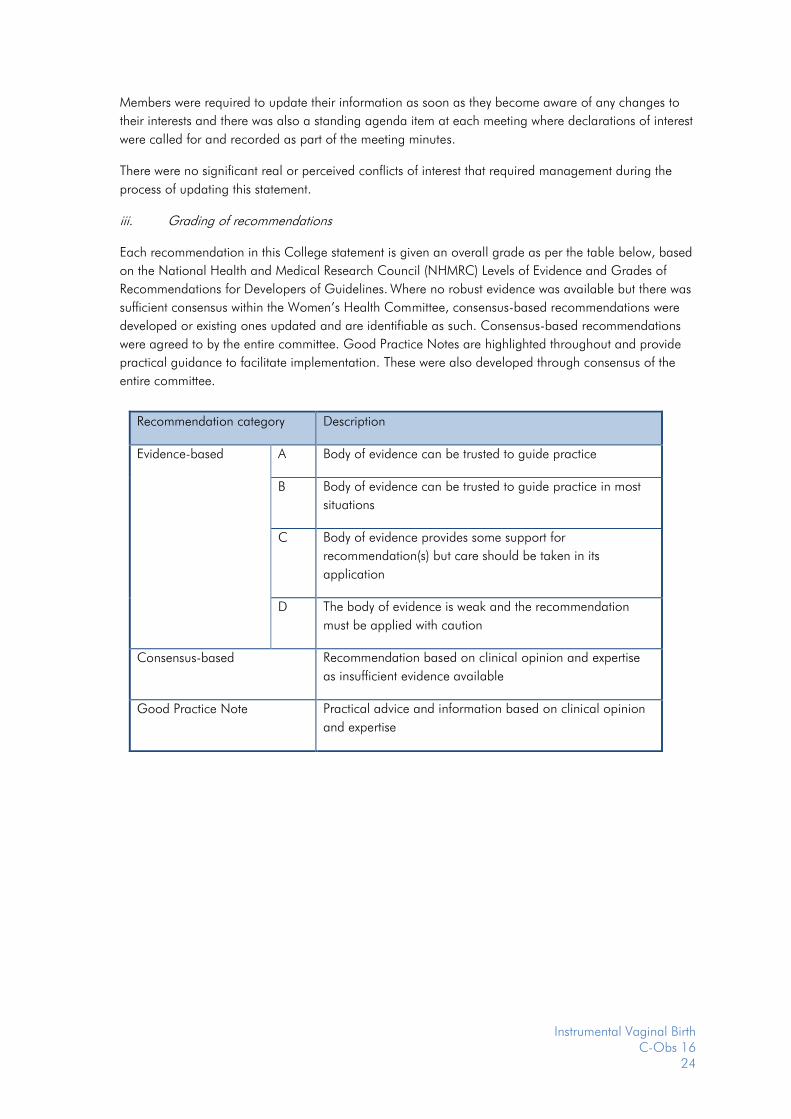
iii. Grading of recommendations
Each recommendation in this College statement is given an overall grade as per the table below, based
on the National Health and Medical Research Council (NHMRC) Levels of Evidence and Grades of
Recommendations for Developers of Guidelines. Where no robust evidence was available but there was
sufficient consensus within the Women’s Health Committee, consensus-based recommendations were
developed or existing ones updated and are identifiable as such. Consensus-based recommendations
were agreed to by the entire committee. Good Practice Notes are highlighted throughout and provide
practical guidance to facilitate implementation. These were also developed through consensus of the
entire committee.
Recommendation category Description
Evidence-based A Body of evidence can be trusted to guide practice
B Body of evidence can be trusted to guide practice in most
situations
C Body of evidence provides some support for
recommendation(s) but care should be taken in its
application
D The body of evidence is weak and the recommendation
must be applied with caution
Consensus-based Recommendation based on clinical opinion and expertise
as insufficient evidence available
Good Practice Note Practical advice and information based on clinical opinion
and expertise

Instrumental Vaginal Birth C-Obs 16
25
Appendix E Full Disclaimer
This information is intended to provide general advice to practitioners, and should not be relied on as a
substitute for proper assessment with respect to the particular circumstances of each case and the needs of
any patient.
This information has been prepared having regard to general circumstances. It is the responsibility of each
practitioner to have regard to the particular circumstances of each case. Clinical management should be
responsive to the needs of the individual patient and the particular circumstances of each case.
This information has been prepared having regard to the information available at the time of its preparation,
and each practitioner should have regard to relevant information, research or material which may have
been published or become available subsequently.
Whilst the College endeavours to ensure that information is accurate and current at the time of preparation,
it takes no responsibility for matters arising from changed circumstances or information or material that may
have become subsequently available.
Top Related