


Languages
Pages
Legal


i
Impact of H9c2 Cardiomyoblast Differentiation on
Isoproterenol Toxicity: Different Modulation of Signaling
Pathways
Universidade de Coimbra
Ana Filipa Roque Branco
2012

iii
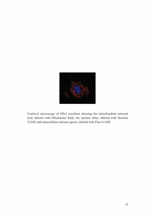
Confocal microscopy of H9c2 myoblast showing the mitochondrial network (red, labeled with Mitotracker Red), the nucleus (blue, labeled with Hoechst 33342) and intracellular calcium (green, labeled with Fluo-4-AM)


v
Impact of H9c2 Cardiomyoblast Differentiation on Isoproterenol Toxicity: Different Modulation of Signaling Pathways
Influência do Estado de Diferenciação de Cardiomioblastos H9c2 na Toxicidade Induzida pelo Isoproterenol: Diferente Modulação de Vias de Sinalização.

vii
Dissertation presented to the Faculty of Sciences and Technology of the University of Coimbra in partial fulfillment of the requirements for Doctor of Philosophy (PhD) graduation in Biology, Specialization in Cellular Biology.
Dissertação apresentada á Faculdade de Ciências e Tecnologia da Universidade de Coimbra, com vista á obtenção do grau de Doutor em Biologia, especialidade em Biologia Celular.


ix
This work was conducted at the Center for Neurosciences and Cell Biology, University of Coimbra, under the supervision of Dr. Paulo Oliveira and Dr. Maria Sancha Santos; at the Department of Anatomy, Microbiology and Pathology, University of Minnesota Medical School, Duluth, MN, USA under the local guidance of Dr. Jon Holy; at the Nencki Institute of Experimental Biology, Warsaw, Poland under the local guidance of Dr. Mariusz Wieckowski and in the Spanish National Center for Cardiovascular Research (CNIC), Madrid, Spain under the local overview of Dr. Susana Gónzalez López. This thesis was supported by a Ph.D. fellowship from the Portuguese Foundation for Science and Technology (SFRH/BD/41384/2007), co-funded by Fundo Social Europeu (FSE) in the scope of Programa Operacional Potencial Humano (POPH) of QREN, and a research grant from FCT to Dr. Paulo Oliveira (project PTDC/QUI/64358/2006)
Este trabalho foi realizado no Centro de Neurociências e Biologia Celular da Universidade de Coimbra, sob a orientação do Dr. Paulo Oliveira; no Departamento de Anatomia, Microbiologia e Patologia da Faculdade de Medicina da Universidade de Minnesota, Duluth, MN, USA no laboratório do Dr. Jon Holy; no Instituto de Biologia Experimental Nencki em Varsóvia, Polónia no laboratório do Dr. Mariusz Wieckowski e no Centro Nacional de Investigação Cardiovascular em Madrid, Espanha no laboratório da Dr. Susana Gónzalez López. Este trabalho foi realizado ao abrigo de uma bolsa de doutoramento atribuída pela Fundação para a Ciência e a Tecnologia (SFRH/BD/41384/2007), co-financiado pelo Fundo Social Europeu (FSE) no âmbito do Programa Operacional Potencial Humano (POPH) do QREN e também financiado pelo projecto PTDC/QUI/64358/2006 concedido pela FCT ao Dr. Paulo Oliveira.


xi
“If we knew what it was we were doing,
it would not be called research,
would it?" Albert Einstein


xiii
Scientific papers
Most of the work presented in this dissertation is published in international peer-reviewed scientific journals, as follows:
- Branco AF, Sampaio SF, Moreira AC, Holy J, Wallace KB, Baldeiras I, Oliveira PJ, Sardão VA. Differentiation-Dependent Doxorubicin Toxicity on H9c2 Cardiomyoblasts. Cardiovascular Toxicology, 2012 Jun 29.
- Branco AF, Pereira SL, Moreira AC, Holy J, Sardão VA, Oliveira PJ. Isoproterenol Cytotoxicity is Dependent on the Differentiation State of the Cardiomyoblast H9c2 Cell Line. Cardiovascular Toxicology, 2011 Sep;11(3):191-203.
I certify that I have obtained a permission from the co-authors of the present publications to include the above material in my thesis.


xv
Acknowledgements/ Agradecimentos
Este trabalho não poderia ter sido realizado sem a ajuda de várias pessoas e entidades, algumas das quais, pela sua tão grande importância não poderia deixar de destacar.
Agradeço à Fundação Portuguesa para a Ciência e a Tecnologia pelo financiamento concedido, incluindo o financiamento de um projecto no âmbito deste estudo.
Ao Dr. Paulo Oliveira pela orientação deste trabalho, pelas críticas, correcções e sugestões e pela competência científica. Foi por ter acreditado em mim e por todo o seu incentivo que me candidatei a doutoramento e foi através da sua ajuda incansável que o estou a terminar.
Ao Centro de Neurociências e Biologia Celular bem como à Universidade de Coimbra por me terem acolhido como estudante de Doutoramento e disponibilizado os recursos técnicos para a realização das experiências.
I am thankful to Dr. Jon Holy, to Dr. Mariusz Wieckowski, and to Dr. Susana Gónzalez for the guidance and all the support during my stay abroad, for the discussions and shared knowledge that made those times exceptional experiences.
À Dr. Sancha. A constante amabilidade e disponibilidade como inicia e termina cada dia é tão inexplicável como fundamental. Obrigada por todos os ensinamentos.
Ao Professor Moreno, pela cedência de compostos e por ser uma fonte de inspiração.
À Carolina, por todos os momentos que nos uniram. Há poucas pessoas que me conhecem bem e ainda menos as com quem partilho tudo o que sou. Obrigada por fazeres parte desse grupo, por nunca te cansares de me ajudar e de te preocupar. Obrigada por estares sempre presente e por resolveres sempre os meus pequenos/grandes stresses. É um orgulho ter-te como amiga.
À Teresa Serafim. Quando, durante 3 meses, nada no laboratório resulta e ainda assim temos a certeza que o tempo não foi passado em vão.. As palavras não chegariam para mostrar o quão grata sou por ter partilhado contigo tão bons momentos. A saudade ao recordar as aventuras, as longas conversas, o despique de músicas. Obrigada por me conheceres tão bem, por te preocupares tanto e até saberes que não passava sem os cheerios depois de tomar o xarope.
À Vilma por ser a primeira pessoa que penso quando surge qualquer dúvida. Há sempre uma explicação para uma técnica nos correr sempre bem e gostarmos tanto de fazê-la, e sem dúvida que no meu caso isso se deveu á maneira como
xvi
os ensinamentos me foram passados por ti. Obrigada pela imensa paciência e por saber que posso sempre contar contigo seja a distância de que tamanho for.
À Susana Sampaio. A minha última companhia durante os 4 anos. Ainda bem que alguém pensou que eu te poderia ensinar alguma coisa no laboratório. Obrigada por seres um exemplo, por seres incansável e preocupada. Obrigada pelos bons momentos.
À Susana Pereira. Mal eu sabia que aquela viagem que começou com meia hora de atraso e 6 doses de xarope no estômago ia criar este laço. Obrigada pelos momentos divertidos, pela amizade e pelos conselhos.
À Sónia Fiúza. A nossa primeira experiência no laboratório nunca é esquecida muito menos a pessoa que nos acompanhou. É às excelentes recordações desse tempo que devo o gosto, apesar de bizarro, pelo cheiro a lixívia da bacia da lavagem do material de laboratório.
A todos os meus colegas do laboratório, agradeço por todo o apoio prestado nas experiências e no estabelecimento de protocolos, os ensinamentos, as discussões mas também (e nunca menos importante) as proveitosas risadas.
À minha família. Especialmente aos meus Pais, por me terem mostrado, com o seu exemplo, como ser uma pessoa responsável e íntegra, por me terem mostrado tudo o que interessa saber sobre a vida e por terem deixado que fosse a vida a mostrar-me aquilo que nem sempre interessa saber.
Um agradecimento especial à minha irmã Sara, por ter “esperado” por mim no 10º ano. Obrigada por teres estado sempre comigo, pela protecção e pelos momentos divertidos que só tu sabes criar. É muito fácil partilhar a nossa vida com pessoas que têm os mesmos princípios que nós.
Agradeço aos meus avós pelo carinho e apoio durante toda a minha vida. É a vocês que dedico este trabalho por terem mais orgulho em mim do que aquilo que eu realmente represento.
Ao Hélder, podia agradecer o apoio, as formatações da tese e a compreensão da apatia em que imergi enquanto fazia a tese, mas tudo isso me faria ficar longe de tudo aquilo que representa. Por tudo aquilo que não se explica, não se escreve mas se vive, obrigada.
1
List of contents
LIST OF FIGURES ....................................................................................................... 5
LIST OF TABLES ......................................................................................................... 9
LIST OF ABBREVIATIONS ..................................................................................... 11
ABSTRACT ................................................................................................................. 15
SUMÁRIO .................................................................................................................... 17
GENERAL INTRODUCTION .................................................................................. 19
1. GENERAL INTRODUCTION .......................................................................... 21
1.1 CARDIOVASCULAR DISEASE AS A PUBLIC HEALTH PROBLEM ........................ 21 1.2 CATECHOLAMINE SYNTHESIS AND SECRETION AS A STRESS RESPONSE ......... 24 1.3 CARDIOVASCULAR EFFECTS OF CATECHOLAMINES - CARDIOMYOCYTE
APOPTOSIS TRIGGERED BY OVERSTIMULATION OF Β-ADRENERGIC RECEPTORS .......... 26 1.3.1 Cardiomyocyte structure and behavior ................................................... 27 1.3.2 Cell death mechanisms in cardiomyocyte degeneration .......................... 28 1.3.3 β-Adrenergic Receptors – Characterization of signaling pathways ........ 36 1.3.4 Beta-blockers – Improving the failing heart ............................................ 39 1.3.5 Beta-adrenergic signaling in the developing heart ................................. 41
1.4 IN VITRO MODELS TO INVESTIGATE CARDIAC Β-ADRENERGIC SIGNALING AND
CARDIOVASCULAR TOXICOLOGY................................................................................ 43 1.4.1 Neonatal Ventricular Cardiomyocytes .................................................... 43 1.4.2 HL-1 cells ................................................................................................ 44 1.4.3 P19-derived cardiomyocytes ................................................................... 45 1.4.4 ESCs-derived cardiomyocytes ................................................................. 46 1.4.5 H9c2 (2-1) cell line .................................................................................. 46
1.5 GENERAL AIMS ............................................................................................. 49
MATERIALS AND METHODS ................................................................................ 51
2. MATERIAL AND METHODS ......................................................................... 53
2.1 COMMON REAGENTS .................................................................................... 53 2.2 CELL CULTURE ............................................................................................. 54
2.2.1 H9c2 Cell Culture and Differentiation Process ....................................... 54 2.2.2 Culture of Mouse Neonatal Ventricular Cardiomyocytes ........................ 56
2
2.2.3 HL-1 Cellular Culture ............................................................................. 58 2.3 COMMON METHODS ..................................................................................... 58
2.3.1 Analysis of Drug Cytotoxicity .................................................................. 58 2.3.2 Western Blotting ...................................................................................... 59 2.3.3 Vital fluorescence of H9c2 cells .............................................................. 61 2.3.3.1 Tetramethylrhodamine methyl ester (TMRM) Hoechst 33342 and
Calcein-AM ........................................................................................................... 61 2.3.3.2 Mitotracker Red and DAPI ................................................................. 62 2.3.4 Flow cytometry analysis of intracellular calcium, mitochondrial
transmembrane potential and mitochondrial superoxide anion ........................... 62 2.3.5 Caspase-3-like colorimetric activity assay .............................................. 63 2.3.6 Determination of intracellular cAMP content ......................................... 64 2.3.7 Total RNA Extraction .............................................................................. 65 2.3.8 Microarray Gene Expression Analysis .................................................... 65 2.3.9 Statistical analysis ................................................................................... 66
RESULTS ..................................................................................................................... 67
3. RESULTS ............................................................................................................ 69
3.1 CHARACTERIZATION OF H9C2 MYOBLAST DIFFERENTIATION ...................... 69 3.1.1. Abstract ............................................................................................... 69 3.1.2. Background and Objective .................................................................. 70 3.1.3. Results ................................................................................................. 71 3.1.3.1. Morphological, Proliferative and Molecular Characterization of
Undifferentiated and Differentiated H9c2 cells .................................................... 71 3.1.3.2. Alterations on signaling protein content during H9c2 differentiation 74 3.1.3.3. Morphological and transcriptional comparison of H9c2 cells with
neonatal cardiomyocytes and HL-1 cells. ............................................................. 77 3.1.4. Discussion ........................................................................................... 85
3.2 ROLE OF Β-ADRENERGIC SIGNALING STRESS RESPONSES ON ISOPROTERENOL
CYTOTOXICITY ON H9C2 CELLS IN DIFFERENT DIFFERENTIATION STAGES ............... 92 3.2.1 Abstract.................................................................................................... 92 3.2.2 Background and Objective ...................................................................... 93 3.2.3 Results ..................................................................................................... 95 3.2.3.1 Isoproterenol Affect Differently H9c2 Cell Line Depending on the
Differentiation State of the Cells .......................................................................... 95
3
3.2.3.2 Isoproterenol-induced signaling pathway alterations ....................... 102 3.2.3.3 Calcium Overload in Differentiated H9c2 Cells after ISO Treatment ....
........................................................................................................... 105 3.2.3.4 Isoproterenol toxicity involves loss of mitochondrial respiratory
complexes, depolarizes mitochondria and increases mitochondrial superoxide
anion in RA-differentiated cells .......................................................................... 107 3.2.3.5 Cell Differentiation Impacts Isoproterenol–induced Alterations in
Survival/Death Signaling Pathways ................................................................... 111 3.2.3.6 p53 Inhibition and Antioxidant Supplementation Have Different Effects
on ISO Cytotoxicity on Undifferentiated vs. Differentiated H9c2 Cells ............. 113 3.2.3.7 Inhibition of PI3K/Akt pathway increases ISO cytotoxicity in all
differentiation and non-differentiation groups ................................................... 114 3.2.3.8 Activation of Caspase 3 During ISO-induced H9c2 Cell Death. ...... 117 3.2.4 Discussion.............................................................................................. 119
CONCLUSIONS ........................................................................................................ 129
4. CONCLUSIONS ............................................................................................... 131
4.1 FINAL REMARKS ......................................................................................... 133
BIBLIOGRAPHY ..................................................................................................... 139
5. BIBLIOGRAPHY ............................................................................................. 141

5
List of Figures
Figure 1 - Representative scheme of morphologic alterations induced by a
stressor stimulus in the heart. ............................................................................ 23
Figure 2 - Schematic representation of the components of HPA and SAM
pathways triggered by stressor stimulation ....................................................... 25
Figure 3 - Representation of apoptotic events and the main components
involved. ............................................................................................................ 35
Figure 4 - Representation of some of the signaling pathways triggered by β-
adrenergic receptors. ......................................................................................... 38
Figure 5 - Calcium transients by activation of PKA. ........................................ 40
Figure 6 - Chemical structure of Isoproterenol ................................................. 49
Figure 7 - Schematic representation of the differentiated process of H9c2
myoblasts........................................................................................................... 56
Figure 8 - Effect of serum withdrawal on H9c2 cells morphology ................... 72
Figure 9 - Alteration on proliferative rate by modulating the percentage of
serum in the medium. ........................................................................................ 73
Figure 10 - Cellular content on specific cardiac markers in different cell
populations. ....................................................................................................... 73
Figure 11 - Determination of specific proteins content in undifferentiated and
differentiated H9c2 cells. .................................................................................. 77
Figure 12 - Representative images of differentiated H9c2 cells, adult HL-1 cells
and neonatal cardiomyocytes. ........................................................................... 78
Figure 13 - Treemap diagram of gene expression in RA-differentiated cells
comparing with undifferentiated H9c2 cells. .................................................... 79
Figure 14 - Treemap diagram of gene expression of isolated neonatal
ventricular cardiomyocytes compared with HL-1 cell line. .............................. 83

6
Figure 15 - Toxicity of ISO on H9c2 myoblasts grown in high-serum media
(undifferentiated) and low serum media (generating differentiated adult muscle
cells). ................................................................................................................. 97
Figure 16 - Cellular alterations observed in H9c2 cells after exposure to ISO for
48 hours (A, B and C). .................................................................................... 100
Figure 17 - Undifferentiated H9c2 cells present morphological markers of
mitochondrial fragmentation. .......................................................................... 101
Figure 18 - Possible effects of ISO binding to serum proteins on ISO toxicity
outcome. .......................................................................................................... 102
Figure 19 - Effects of 48 h ISO treatment on cell content in Bcl-2 (A), Bax (B),
phosphorylated p38-MAPK (C), calcineurin (D), cardiac Troponin T (E) and
full length PARP protein (F). .......................................................................... 104
Figure 20 - Alteration of cytosolic calcium and cAMP after Isoproterenol
incubation. ....................................................................................................... 106
Figure 21 - Evaluation of mitochondrial alterations after ISO treatment ........ 108
Figure 22 - Evaluation of mitochondria polarization and superoxide anion in
H9c2 cells. ....................................................................................................... 109
Figure 23 - Fluorescent analysis of intracellular oxidative stress. .................. 110
Figure 24 - Isoproterenol induces stress responses in H9c2 cells. .................. 113
Figure 25 - p53 and oxidative stress involvement during ISO toxicity on H9c2
cells. ................................................................................................................ 114
Figure 26 - PI3K/Akt pathway role on ISO-induced toxicity. ........................ 117
Figure 27 - Caspase 3-like activity in H9c2 cells exposed to ISO and effect of
caspase inhibition. ........................................................................................... 118
Figure 28 - Schematic representation of β-AR downstream pathways altered by
ISO in undifferentiated H9c2 cells. ................................................................. 135

7
Figure 29 - Schematic representation of β-AR downstream pathways altered by
ISO in differentiated H9c2 cells by reducing the concentration of serum in the
medium. ........................................................................................................... 136
Figure 30 - Schematic representation of β-AR downstream pathways altered by
ISO in RA-differentiated H9c2 cells. .............................................................. 137


9
List of Tables
Table 1 - Schematic representation of molecular, functional and culture
behavior between neonatal cardiomyocytes, HL-1 cell line, P19, ESC and H9c2
(2-1) cell line. .................................................................................................... 48
Table 2 - Volume of enzyme solution and the respective time exposure used at
each digestion step in isolation of neonatal cardiomyocytes procedure. ........... 57
Table 3 – Primary and secondary antibodies used in Western Blotting assay. . 60
Table 4 - Groups of gene expression explored in H9c2 undifferentiated
myoblasts and RA-differentiated H9c2 cells. .................................................... 81
Table 5 - Groups of gene investigated in isolated neonatal cardiomyocytes and
HL-1 cells. ......................................................................................................... 84

11
List of Abbreviations
A AC: adenylate cyclase AIF: Apoptosis-inducing factor AKT/PKB: Protein Kinase B -AM: Acetoxymethyl ester ANOVA: Analysis of variance ANT: Adenine nucleotide translocase Apaf-1: Apoptotic protease-activating factor 1 ARC: Apoptosis repressor with caspase recruitment domain ATCC: American Type Culture Collection ATP: Adenosine triphosphate B β-AR: β-adrenergic receptor Bad: Bcl-2-associated death promoter Bax: Bcl-2-associated X protein Bid: BH3 interacting domain death agonist Bcl-2: B-cell lymphoma 2 BrdU: Bromodeoxyuridine BSA: Bovine serum albumin C Ca2+: Calcium cAMP: Cycle adenosine monophosphate CHAPS: 3-[(3-Cholamidopropyl)dimethylammonio]-1-propanesulfonate CypF: Cyclophilin-F Cytc: Cytochrome c CM-H2DCFDA: Chloromethyl derivate of dichlorodihydrofluorescein diacetate
D DAPI: 4',6-diamidino-2-phenylindole DHE: Dihydroethidium DISC: Death-inducing signaling complex DMEM: Dulbecco´s modified Eagle´s medium DMSO: Dimethyl sulfoxide DNA: Deoxyribonucleic DTT: Dithiothreitol Drp-1: Dynamin-related protein 1 E ECF: Eosinophil chemotactic factor EDTA: Ethylenediaminetetraacetic acid Endo G: Endonuclease G ERK: Extracellular signal–regulated kinase ETC: Electron transport chain F FADD: Fas-associated death domain protein FBS: Fetal bovine serum FL1: flow cytometer green fluorescence detector FL2: flow cytometer orange fluorescence detector G Gi: inhibitory G protein G protein: guanosine trisphosphate protein Gs: stimulatory G protein GSH: Reduced glutathione

12
H HCL: Hydrochloric acid HEPES: 4-(2-hydroxyethyl)-1-piperazineethanesulfonic acid I IAP: Inhibitor of apoptosis protein IgG: Immunoglobulin G ISO: Isoproterenol J JNK: c-Jun N-terminal kinase K kDa: kilodalton M MAPK: Mitogen-Activated Protein (MAP) Kinase MFN1: Mitofusin 1 MFN2: Mitofusin 2 MIM: Mitochondrial Inner Membrane MnSOD: Manganese Superoxide Dismutase MOM: Mitochondrial Outer Membrane mPTP: mitochondrial Permeability Transition Pore mRNA: messenger RNA N NAC: N-Acetylcysteine NDUFB8: NADH dehydrogenase [ubiquinone] 1 beta subcomplex subunit 8 O Opa1: Optic atrophy 1
P P53: Tumor protein 53 PARP: Poly(ADP-ribose) polymerase-1 PBS: Phosphate Buffered Saline PBS-T: Phosphate Buffered Saline with 0.1% Tween PI3K: Phosphoinositide 3-kinase PKA: protein kinase A PMSF: Phenylmethylsulfonyl fluoride p-NA: p-Nitroanilide PVDF: Polyvinylidene fluoride Q QIAshredder: homogenizes cell or tissue lysates to reduce viscosity R RA: All-trans retinoic acid RLT: buffer for lysis of cells and tissues before RNA isolation RNA: Ribonucleic Acid ROS: Reactive Oxygen Species RyR: Ryanodine Receptor S Smac/Diablo: Second mitochondria derived activator of caspases/direct IAP binding protein with low pI SDS: Sodium dodecyl sulfate SEM: Standard error of the mean SERCA: Sarco/Endoplasmic-reticulum-Ca2+-ATPase SRB: Sulforhodamine B SV40 large T: Simian Vacuolating Virus 40 T

13
T tBid: Truncated Bid TBST: Tris-Buffered Saline Tween-20 TMRM: Tetramethyl Rhodamine Methyl Ester TNFα: Tumor necrosis factor-α TNF: Tumor Necrosis Factor U UQCRC2: Ubiquinol-cytochrome c reductase core protein 2
V VDAC: Voltage-Dependent Anion Channel X XIAP: X-chromossome linked IAP Z z-vad-fmk: benzyloxycarbonyl-valine-alanine-aspartate fluoromethylketone
UCP: Uncoupling protein


15
Abstract
The chronic exposure of cardiomyocytes to catecholamines is associated with pathologic alterations. Stress conditions and tissue insults trigger the activation of neurohormonal systems and catecholamine synthesis is consequently increased. The binding of circulating catecholamines to adrenergic receptors normally incurs in physiological responses that comprises increasing contractile force, heart rate, metabolic activity, among other effects that affect cardiac performance. However, hyperadrenergic stimulation, resulting from sustained activation of β-adrenergic receptors (β-AR), is thought to be directly involved in cardiomyocyte apoptosis. Over-activation of specific signaling pathways results in several cellular processes which lead to progressive myocardium deterioration and contribute toward decompensated heart failure.
In this regard, the present work addresses the mechanisms by which cardiac cells respond to an overstimulation of β-AR according to their differentiation state (i. e. embryonic/adult). For this objective, we used the rat myoblastic H9c2 cell line which can be differentiated in adult skeletal or cardiac muscle cells. The synthetic catecholamine isoproterenol (ISO) was used to stimulate β-AR since its specificity represents an advantage when compared with endogenous catecholamines.
Our hypothesis is that β-AR over-stimulation results in different cell fates depending on the differentiation stage of the cell. This approach is relevant since most studies are conducted in H9c2 cells in their undifferentiated state (embryonic) raising questions on its applicability to cardiotoxicity studies.
Initial characterization of H9c2 differentiation was performed concerning differences in terms of intrinsic cell defenses and signaling pathways which may render cells more or less susceptible to ISO. The alteration in cellular physiology during differentiation was strengthened by gene array analyses showing up-regulation of mitochondrial metabolism genes and genes encoding calcium transporters and differentiation markers, in retinoic acid-differentiated H9c2 cells.
In what concern ISO-induced toxicity, the present work demonstrates that undifferentiated cells have increased resistance to ISO, which can be
16
explained by increased anti-apoptotic proteins including Bcl-2 and Bcl-xL and intrinsic defense capacity such as MnSOD. Several stress responses were increased in differentiated cells, including p66Shc, p53 and Bax, at the same with higher mitochondrial degeneration. This was particularly seen by mitochondrial depolarization, loss of respiratory complexes and oxidative stress, which was mostly observed in differentiated H9c2 cells. Moreover, after ISO treatment, differentiated H9c2 cells showed increased cAMP, cytosolic calcium accumulation, cleavage of PARP and activation of caspase 3. Alterations in calcineurin and p38-MAPK in undifferentiated and differentiated cells can also account for the larger toxicity of ISO in the latter. Moreover, the increase in pro-apoptotic proteins was not accompanied in increased anti-apoptotic counterparts, as occurred in undifferentiated myoblasts (Bcl-xL, Bcl-2). We conclude that H9c2 cells present differential regulation of stress responses during their differentiation which impact the toxicity of several agents.
In conclusion, the present thesis demonstrates that undifferentiated and differentiated H9c2 cells possess different susceptibility to the β-AR agonist ISO in which the balance between pro- and anti-apoptotic pathways are involved. The results are relevant from a basic toxicology point of view, since this cell line is very used as a model for cardiac cells. Furthermore, the data supplies evidence that the H9c2 cell line can be used as a framework to investigate developmental cardiotoxicity.
Keywords: isoproterenol, β-adrenergic receptors, apoptotic pathways, mitochondria, H9c2 myoblasts, differentiation.

17
Sumário
A exposição crónica de cardiomiócitos a catecolaminas está associada a alterações patológicas.
Condições de stress e lesões no tecido cardíaco activam sistemas neurohormonais que induzem a consequente síntese de catecolaminas. A interacção de catecolaminas com receptores adrenérgicos induz respostas fisiológicas como o aumento da força de contracção muscular, aumento da actividade metabólica, entre outros efeitos que afectam o desempenho cardíaco.
Contudo, a estimulação adrenérgica resultante de uma activação prolongada de receptores β-adrenérgicos (β-AR) pode estar directamente envolvida na apoptose de cardiomiócitos contribuindo para insuficiência cardíaca descompensada. A sobre-activação de vias de sinalização específicas resulta na estimulação de vários processos celulares que conduzem á deterioração progressiva do miocardio. Neste sentido, o presente trabalho pretende investigar os mecanismos pelos quais células cardíacas respondem à sobre-activação de β-AR segundo o seu estado de diferenciação (embrionário/adulto). Com este objectivo, usamos a linha celular mioblastica de rato H9c2 que tem a capacidade de ser diferenciada em células musculares esqueléticas e cardíacas. A catecolamina sintética isoproterenol (ISO) foi usada no sentido de estimular β-AR já que a sua especificidade representa uma vantagem quando comparada com o uso de catecolaminas endógenas.
A nossa hipótese de trabalho é que a sobre-activação de β-AR resulta em diferentes respostas dependendo do estado de diferenciação celular. Dado que a maior parte dos estudos conduzidos em células H9c2 usa esta linha celular no seu estado indiferenciado (embrionário), questões relativas á sua aplicabilidade em estudos cardiotóxicos levantam algumas dúvidas.
A caracterização inicial do processo de diferenciação de H9c2 foi realizada tendo em conta diferenças em termos de defesas celulares intrínsecas e vias de sinalização que poderão induzir maior ou menor sensibilidade das células ao ISO. A alteração de processos fisiológicos nas células foi ainda investigada através de análise genética mostrando que genes envolvidos no metabolismo mitocondrial, na differenciação

18
celular e na codificação de transportadores de cálcio, estão sobre-expressos em células H9c2 diferenciadas em presença de ácido retinóico.
O presente trabalho demonstra que células indiferenciadas apresentam maior resistência ao ISO, o que pode ser explicado pelo aumento de proteinas anti-apoptóticas tais como Bcl-2 e Bcl-xL, e na capacidade de defesa intrínseca tais como MnSOD. Múltiplas respostas a condições de stress encontram-se aumentadas em células diferenciadas, incluíndo proteinas p66Shc, p53 e Bax bem como degeneração mitocondrial. Esta última, identificada através da despolarização mitocondrial, perda de complexos respiratórios e stress oxidativo, que foi maioritariamente observado em celulas H9c2 diferenciadas. Após tratamento com ISO, em células H9c2 diferenciadas verifica-se um aumento de cAMP, acumulação de cálcio no citosol, clivagem da PARP e activação de caspase-3. As alterações nas proteínas calcineurina e p38-MAPK nas células indiferenciadas e diferenciadas podem também contribuir para uma maior toxicidade ao ISO nas últimas. O aumento de proteínas pro-apoptóticas não foi acompanhado por um concumitante aumento de próteinas anti-apoptóticas, como verificado em células indiferenciadas (Bcl-xL e Bcl-2). Concluímos que as células H9c2 apresentam uma regulação diferencial em resposta a condições de stress durante a sua diferenciação o que influencia a toxicidade de agentes cardiotóxicos.
Em conclusão, o presente trabalho demonstra que células H9c2 indiferenciadas e diferenciadas possuem diferente susceptibilidade ao agonista de β-AR, ISO, sendo neste processo, o balanço entre proteínas pro- e anti-apoptóticas de grande importância. Os resultados são relevantes do ponto de vista toxicológico básico já que esta linha celular é normalmente utilizada como modelo para células cardíacas. Os resultados obtidos evidenciam que a linha celular H9c2 poderá ser utilizada em estudos que visam investigar a resposta cardiotóxica a fármacos, no entanto, há que ter presente que diferentes estados de diferenciação poderão influenciar diferentes susceptibilidades a vários agentes. Além disso, os resultados evidenciam que a linha H9c2 pode ser usada (como ferramenta) para investigar a cardiotoxicidade durante o desenvolvimento.
Palavras-chave: isoproterenol, receptores β-adrenérgicos, vias de sinalização apoptóticas, mitocôndria, mioblastos H9c2, diferenciação.

General Introduction


General Introduction
21
1. General Introduction
1.1 Cardiovascular disease as a public health problem According to the World Health Organization (WHO), heart disease
remains the number one cause of death and disability worldwide
accounting to about one-third of all human mortality [1].
Cardiovascular disease (CVD) is a generic term to designate all
pathologic alterations that affect the heart and blood vessels. The term
cardiovascular disease collectively includes diverse disorders such as
coronary heart disease (commonly known as heart attack),
cerebrovascular disease (stroke), hypertensive heart disease,
atherosclerosis (accumulation of fat, including cholesterol deposits on
blood vessels walls), congenital heart disease and congestive heart
failure. Usually, the latter is the culmination of long-standing diseases
such as hypertension and ischemia resulting from atherosclerosis [2].
Heart failure represents one of the fastest-growing diseases and has been
extensively studied as well. Heart failure is caused by any event, such as
damage or overload, which compromises heart function. Interestingly,
all these diseases not only have in common the fact that they occur in the
cardiovascular system but also that can act together as a cause or
consequence of each other as shown by sustained overload-induced heart
failure.
Cardiac risk factors may include lifestyle habits such as unhealthy diet,
lack of regular physical activity, tobacco smoking, excessive alcohol
consumption, high blood pressure and high serum cholesterol levels as
well as uncontrolled risk factors such as age, gender and genetic
background [1].
Due to lifestyle habits, blood vessels can get hardened reducing both the
wall diameter and the flow rate. To compensate for the decrease in blood

Ana Branco PhD Thesis|2012
22
flow, increased sympathetic nervous system (SNS) activity can, initially,
contribute to increase ventricle contractility in order to sustain ejection
performance. However, over time, the neuro-hormonal stimulation of
heart beating rate initiates a process of dynamic and morphological
alterations [3, 4]. Often, the left ventricle develops hypertrophy
associated with a complex set of alterations in the expression of cellular
proteins. Also, several alterations lead to the progression of apoptotic
cell death and loss of cardiomyocytes [5] (Fig. 1). If a part of the heart
muscle becomes starved of oxygen or nutrients, development of
localized lesions can occur.
Before the industrial revolution, the incidence of death resulting from
heart diseases was lower. Modern technologies brought a sedentary
lifestyle and later, processed dairy foods, further increased that tendency
[6]. Living in a modern world implies also increasing deadlines and
worries. In fact, stressor factors can differently interact with each
individual, depending on their vulnerability to adversity which difficult
the full understanding of the link between stress and heart disease.
Psychological stress is well recognized by the scientific community as
having an important role in cardiovascular diseases by acting as a trigger
to potentiate acute heart problems [7, 8]. Several published works
suggest that the release of epinephrine or norepinephrine to the
bloodstream may contribute to the increase of fatty acids in the blood
which can then deposit in the arteries, contributing to the development of
atherosclerosis [9, 10]. The blockage of an artery can lead to stroke or
myocardial infarction [11].

General Introduction
23
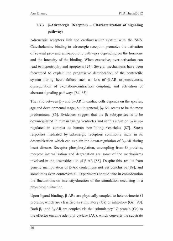
Figure 1 - Representative scheme of morphologic alterations induced by a stressor stimulus in the heart. Terminally differentiated cardiomyocytes adapt to increased work or hemodynamic stressors through an increase in heart rate. To compensate for the new cardiac output demand, neurohormonal and cellular signaling cascades are activated, resulting in ventricular remodeling as increased myocardial mass and ventricular walls thickness. Ultimately, myocardial hypertrophy leads to a progressive loss of cardiomyocyte which is the causal role of the transition to an end-stage of the disease. (Figure adapted from [12, 13]).
Evidences concerning the mechanisms by which cardiac myocytes
undergo apoptotic cell death show that mechanical conditions and
elevated levels of neurohumoral factors are stronger contributors [12]. In
an experimental model of mechanical overload performed on cardiac
papillary muscle, an increase of apoptotic cardiomyocytes were
measured, demonstrating the link between ventricular stretch and
apoptosis [14]. More details on the mechanisms of cardiac cell apoptosis
will be given in chapter 1.3.2.

Ana Branco PhD Thesis|2012
24
Fortunately, pharmacological treatments have improved in the last
decades and are actively counteracting the progression of the disease
due, in part, to extensive investigation on antihypertensive therapies and
high blood cholesterol lowering agents [15].
1.2 Catecholamine synthesis and secretion as a stress response Catecholamines are a group of organic compounds that include the
hormones dopamine, norepinephrine and epinephrine which participate
in different types of behavior, physiological conditions and diseases.
The scientific community has long investigated this family of
compounds, especially due to the fast and relatively easy way to measure
endogenous hormones in the fluids as well as their metabolites.
Throughout scientific history, many Nobel Prizes in Physiology or
Medicine resulted, at least in part, from research on the
catecholaminergic system as evidenced by studies on the
neurotransmission of nerve impulses, hypothalamus influence on
emotional behaviors, neurotransmitters storage, release and reuptake
mechanisms, among others [16].
In response to a stressful event, two main systems are activated: the
hypothalamic–pituitary–adrenocortical (HPA) system, for long term
responses, and the sympatho-adrenomedullary (SAM) pathway, for short
term responses [17, 18]. When a stress reaction is triggered, the HPA
and SAM systems are activated and, from the paraventricular nucleus of
the hypothalamus, signals aimed at stimulating the pituitary gland to
produce adrenocorticotropic hormone (ACTH) [18]. Sympathetic
preganglionic neurons release ACTH towards the adrenal gland which,
in turn, modulates the secretion of cortisol (from the adrenal cortex) and
catecholamines (from the adrenal medulla) to maintain body homeostasis
General Introduction
25
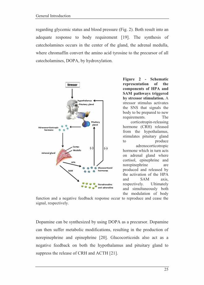
regarding glycemic status and blood pressure (Fig. 2). Both result into an
adequate response to body requirement [19]. The synthesis of
catecholamines occurs in the center of the gland, the adrenal medulla,
where chromaffin convert the amino acid tyrosine to the precursor of all
catecholamines, DOPA, by hydroxylation.
Figure 2 - Schematic representation of the components of HPA and SAM pathways triggered by stressor stimulation. A stressor stimulus activates the SNS that signals the body to be prepared to new requirements. The
corticotropin-releasing hormone (CRH) released from the hypothalamus, stimulates pituitary gland to produce
adrenocorticotropic hormone which in turn acts on adrenal gland where cortisol, epinephrine and norepinephrine are produced and released by the activation of the HPA and SAM axis, respectively. Ultimately and simultaneously both the modulation of body
function and a negative feedback response occur to reproduce and cease the signal, respectively.
Dopamine can be synthesized by using DOPA as a precursor. Dopamine
can then suffer metabolic modifications, resulting in the production of
norepinephrine and epinephrine [20]. Glucocorticoids also act as a
negative feedback on both the hypothalamus and pituitary gland to
suppress the release of CRH and ACTH [21].

Ana Branco PhD Thesis|2012
26
As referred before, a stressor condition can result from many different
events and usually, under experimental conditions, social stressors can
be distinguished from physical stressors. However, both types activate
the HPA and SAM axis and increase cortisol and catecholamine
secretion to modify heart rate and mobilize energy. There is evidence
that positive social interactions increase levels of hypothalamus-released
oxytocin that, in turn, depress HPA activity and contribute to
physiological functions such as wound healing [22, 23] . There is also
evidence that in patients with heart failure, SAM activity is increased
which is confirmed by the increased epinephrine serum concentration
[24].
1.3 Cardiovascular effects of catecholamines - Cardiomyocyte apoptosis triggered by overstimulation of β-adrenergic receptors
When cardiac performance undergoes an underloading condition,
mechanisms are readily activated to stimulate the heart to fill new
workload demands. Myocardial function is then highly stimulated by the
SNS and released catecholamines have important effects on
cardiomyocytes. During heart failure, the release of catecholamines
occurs in an attempt to restore homeostasis; however, when persistent,
this act as an insult driving stress to the heart. Commonly, catecholamine
over-signaling originating from β-adrenergic receptors (β-AR)
stimulation leads to cardiomyocyte death [25]. Stimulation of cardiac β-
AR by norepinephrine or its synthetic analog isoproterenol induces
apoptosis in vivo and in vitro [26]. During congestive heart failure,
norepinephrine´s increase in the plasma does not only result from
decreased re-uptake but also from an increase in hormone release [24].
General Introduction
27
1.3.1 Cardiomyocyte structure and behavior
The majority of the volume of the adult mammalian heart is occupied by
cardiomyocytes. During fetal life, progenitor cells and early
cardiomyocytes proliferate fast. Although a last round of DNA synthesis
and nuclear mitosis without cytokinesis occurs after birth,
cardiomyocytes soon exit the cell cycle [27]. In rats, the majority of
cardiomyocytes are binucleated, while in humans this number is
estimated to be around 57% [28]. Although not well studied, it has been
suggested that the generation of twice the RNA for protein synthesis can
be advantageous in metabolic active cells as muscles [29].
Cardiomyocytes are terminally differentiated cells which are presumed
not to reenter the cell cycle even in response to mitogen or physiological
stress activators. Despite this, when exposed to growth stimulus such as
hormones, neuroendocrine factors or mechanical load, adult
cardiomyocytes can increase their size without a concomitant cellular
division undergoing hypertrophic growth.
Cardiomyocytes have three major components that drive the contractile
force: a) the sarcolemma and T-tubules for impulse conduction; b) the
sarcoplasmic reticulum that stores and releases calcium needed for
activation of contractile proteins; and c) contractile elements including
actin and myosin [30].
In cardiomyocytes, mitochondria constitute about 30% of cell mass and
are strategically located due to their importance in ATP production
critical for cell homeostasis and contraction reflecting the dependence of
cardiac muscle on aerobic metabolism [31]. Mitochondria are the nexus
of oxidative phosphorylation machinery aimed at ATP production.
Besides the well-known function of mitochondria regarding the
generation of energy, these organelles have other important roles
including being the site of multiple metabolic pathways such as the β-
Ana Branco PhD Thesis|2012
28
oxidation of fatty acids, the tricarboxylic acid (TCA) and the urea cycle
[32]. Mitochondria also regulate reactive oxygen species (ROS)
production, oxygen sensing, calcium homeostasis and thermogenesis
[32].
Mitochondria are a complex and dynamic network. Apart from
regulating mitochondrial function during cellular death, some proteins
play a critical role on remodeling events in the mitochondrial membrane
[33]. Altered mitochondrial morphology and fragmentation of
mitochondrial network upon an apoptotic insult have been observed in
heart disease [34, 35]. At the molecular levels, these processes have been
reported to be mediated by three dynamin-family GTPases (GTPases),
namely mitofusins 1 and 2 (Mfn1 and Mfn2) and the optic atrophy type
1 (OPA1), involved in fusion processes; and the dynamin-related protein
1 (DRP-1) and FIS1, which regulate mitochondrial fission processes
[36].
1.3.2 Cell death mechanisms in cardiomyocyte degeneration
All cell death mechanisms, apoptosis, necrosis, and autophagy occur in
cardiac myocytes during heart disease [37]. Evidences demonstrate that
the inhibition of each mechanism improves cardiac function in heart
disease [38-40].
Briefly, autophagy is a quality control process where dysfunctional
organelles and long-lived proteins are sequestered to the interior of
double-membrane vesicles, called autophagosomes, where they are
degraded upon fusion with lysosomes. In this lysosomal pathway, cell
constituents can be used as energy substrates. Although the mechanisms
are not fully understood, excessive digestion of cellular components can
also trigger cell death [41].

General Introduction
29
Although the borderline is sometimes blurry, it is considered that
apoptosis is distinct of necrosis in several aspects. Necrosis is an
accidental form of cell death and results in distinct morphological
alterations including plasma membrane rupture, depletion of intracellular
ATP stores and organelle swelling. The release of cytoplasmic content to
the extracellular space evokes a detrimental inflammatory response [42].
The cardiac tissue has limited regenerative capacity which explains why
cell death is not abundant. However, when the heart is challenged by a
stressful condition, the incidence of apoptosis increases [5]. In fact,
apoptosis has an important role in stress-induced pathogenic adaptations
in several cardiovascular diseases, since it leads to the loss of terminally
differentiated myocytes. A connection between chronic heart failure and
apoptosis has been established. Patients with chronic heart failure have
higher percentage of myocytes apoptosis than normal subjects, as
determined by TUNEL-positive cells [43, 44]. In this context,
cardiomyocyte apoptosis is a promising therapeutic target and has been
extensively studied in clinical and experimental heart failure [45, 46].
Physiologically, apoptosis occurs as an essential and highly regulated
mechanism, triggering a self-renewal progress and controlling the
proliferation of aberrant cells. The cell death program can be originated
by several stimuli that can include cytokines, oxidative stress and DNA
damage [47]. The recognition of an apoptotic signal is a very controlled
mechanism and results ultimately in the recognition of the apoptotic cell.
Cellular membranes are asymmetrically composed by phospholipids and
proteins. The exposure of the phospholipid phosphatidylserine on the
cell surface is recognized by specific receptors in phagocytes as a signal
to initiate degradation of the target and removal of cellular remnants
[48].
Ana Branco PhD Thesis|2012
30
In 1972, John Kerr made use, for the first time, of the term apoptosis to
describe a morphologically different type of cellular death in which
membrane blebbing, chromatin condensation and nuclear fragmentation
is observed [49]. Later evidences suggested an essential role of a family
of proteases termed caspases. Because proteolytic activation of the
caspase cascade is a key element of the apoptotic signaling pathway,
therapies that prevent caspase activation are usually noteworthy.
Caspases are cysteine-specific proteases and can be subdivided in two
groups: the inflammatory ones (e.g. caspase-1, 4 and 13) and caspases
involved in programmed cell death, as referred above [50]. The activity
of caspases can be suppressed by a group of inhibitory proteins, called
X-linked inhibitor of apoptosis protein (XIAP) [51]. Downregulation of
the unspecific caspase inhibitors, XIAP, in the failing myocardium
contributes to increased cardiac myocyte apoptosis [52]. Therefore,
understanding the mechanisms that initiate proteolytic activation of
caspases is a crucial step in defining targets that allow for the modulation
of apoptotic cell death.
Two distinct apoptosis signaling pathways exist in vertebrates. Both are
reported to occur during heart failure: the extrinsic and intrinsic
pathways. The extrinsic pathway or “death receptor pathway” is initiated
by extracellular ligands, which bind to membrane receptors at the cell
surface, such as the tumor necrosis factor (TNF) superfamily receptors.
The interaction ligand-receptor leads to the recruitment of adapter
proteins (e.g. Fas-associated death domain (FADD)) that triggers the
assembly of the death-inducing signaling complex (DISC). Evidences
demonstrate that apoptotic signaling induced by Fas Ligand (FasL) and
TNF-α is dependent on caspase activation [53]. Upon proteolytic
activation, caspase 8 is then released from the DISC to the cytosol,
where it cleaves other effector caspases such as caspase 3 and mediates

General Introduction
31
the cleavage of Bid to its truncated form (tBid), triggering the intrinsic
pathway (Fig. 3) [54]. Multiple studies with mice over-expressing TNF-
α demonstrated that the activation of the TNF receptor can trigger
congestive heart failure [47], while others suggest that the levels of
circulating TNF-α are intimately related with the severity of cardiac
disease [55].
The intrinsic (or mitochondrial) pathway of apoptosis is initiated upon
an internal apoptotic stimulus that leads to permeabilization of the outer
mitochondrial membrane (OMM). The permeabilization of the OMM
can originate from two distinct events: a) opening of the mitochondrial
permeability transition pore (mPTP) in the inner mitochondrial
membrane (IMM) or b) by the translocation to the OMM of pro-
apoptotic proteins [56]. The opening of the mPTP can be potentiated by
augmented calcium and oxidative stress and can lead to membrane
potential loss and matrix swelling that eventually causes rupture of the
OMM [57]. Outer mitochondrial membrane permeabilization can also
result from DNA damage or multiple cellular signals that initiate the
translocation of pro-apoptotic Bcl-2 family components, such as Bax and
Bak proteins to mitochondria, which allows, without alteration of
membrane potential, the release of pro-apoptotic factors from the
intermembrane space to the cytoplasm [58, 59]. Mitochondrial released
pro-apoptotic factors may include cytochrome c, the apoptosis-inducing
factor (AIF), Smac/diablo and Endonuclease G (Endo G) (Fig. 3). The
AIF is biologically required for mitochondrial homeostasis and for the
stabilization of respiratory complex I [60] while the nuclear-encoded
mitochondrial Endo G is determinant for mitochondrial biogenesis,
mitochondrial gene transcription and also in maladaptive cardiac
hypertrophy [61]. While AIF and Endo G have the capacity to
translocate to the nucleus and mediate chromatin condensation and DNA

Ana Branco PhD Thesis|2012
32
fragmentation by a caspase-independent cell death, cytochrome c
participates in the formation of the apoptossome complex, by binding to
Apaf-1, which recruits pro-caspase 9, making it active, and initiates the
caspase cascade [62]. The release of Smac /Diablo enhances caspase
activity by antagonizing endogenous inhibitors of apoptosis, such as the
XIAP, as mentioned above. Since apoptosis is involved in organ
homeostasis, this process is highly controlled and mitochondria are a
central component in apoptosis regulation [63]. These organelles have
life-preserving and death-inducing components in their membranes,
ready to be released upon membrane permeabilization. Once disruption
of OMM occurs, mitochondrial function will be irreversibly impaired
and the viability of the cell without functional mitochondria is
energetically not possible.
As described above, the intrinsic pathway can be further activated by the
extrinsic pathway, depending on the proteolysis of Bid to t-Bid by
caspase 8, which further mediates the recruitment of Bax that
permeabilizes OMM [64]. Bax belongs to the Bcl-2 family of proteins,
which is divided in anti-apoptotic (e.g. Bcl-2 and Bcl-xL) and pro-
apoptotic members (e.g. Bax, Bak, Bcl-xS, Bad and Bid). While Bak is
located at the OMM, Bax is located in the cytosol undergoing a
conformation change that induces its translocation to the mitochondria
[65]. Although both have the ability to form pores in the mitochondrial
membrane. The translocation of Bax to mitochondria can also be related
with increased p53 expression, which transcriptionally regulates Bax.
The p53 tumor-suppressor gene acts as a linkage between DNA damage
detection and apoptosis initiation [66]. Bcl-2 regulates proteins
translocation from the mitochondrial intermembrane space to the
cytosol, counteracting the effect of pro-apoptotic proteins, as
exemplified by the blockage of cytochrome c release in a Bcl-2-
General Introduction
33
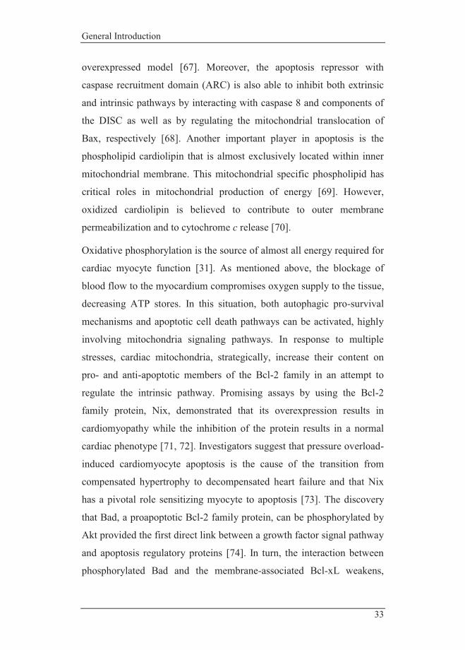
overexpressed model [67]. Moreover, the apoptosis repressor with
caspase recruitment domain (ARC) is also able to inhibit both extrinsic
and intrinsic pathways by interacting with caspase 8 and components of
the DISC as well as by regulating the mitochondrial translocation of
Bax, respectively [68]. Another important player in apoptosis is the
phospholipid cardiolipin that is almost exclusively located within inner
mitochondrial membrane. This mitochondrial specific phospholipid has
critical roles in mitochondrial production of energy [69]. However,
oxidized cardiolipin is believed to contribute to outer membrane
permeabilization and to cytochrome c release [70].
Oxidative phosphorylation is the source of almost all energy required for
cardiac myocyte function [31]. As mentioned above, the blockage of
blood flow to the myocardium compromises oxygen supply to the tissue,
decreasing ATP stores. In this situation, both autophagic pro-survival
mechanisms and apoptotic cell death pathways can be activated, highly
involving mitochondria signaling pathways. In response to multiple
stresses, cardiac mitochondria, strategically, increase their content on
pro- and anti-apoptotic members of the Bcl-2 family in an attempt to
regulate the intrinsic pathway. Promising assays by using the Bcl-2
family protein, Nix, demonstrated that its overexpression results in
cardiomyopathy while the inhibition of the protein results in a normal
cardiac phenotype [71, 72]. Investigators suggest that pressure overload-
induced cardiomyocyte apoptosis is the cause of the transition from
compensated hypertrophy to decompensated heart failure and that Nix
has a pivotal role sensitizing myocyte to apoptosis [73]. The discovery
that Bad, a proapoptotic Bcl-2 family protein, can be phosphorylated by
Akt provided the first direct link between a growth factor signal pathway
and apoptosis regulatory proteins [74]. In turn, the interaction between
phosphorylated Bad and the membrane-associated Bcl-xL weakens,

Ana Branco PhD Thesis|2012
34
potentiating the translocation of Bad from the intracellular membrane
fraction to the cytosol [75].
Several studies are focused on cellular checkpoints that provide ‘rescue’
opportunities for cardiomyocytes. The control of pro-apoptotic
regulatory mechanisms protects cells from complete execution of the
apoptotic program. Studies in experimental animals have shown that
Bcl-2 provided strong anti-apoptotic action and overexpression of this
protein provided cardioprotection [76]. Similar results were also
obtained in studies using the addition of exogenous oxidants scavengers,
demonstrating that oxidative stress may be, at least partially, involved
[77]. Calcium and ROS are known to be important mediators in
cytochrome c release from mitochondria since they potentiate the
opening of the mPTP [78]. Heart mitochondria have the capacity to store
large amounts of calcium without triggering of the mPTP [79]. However,
when heart tissue suffers an insult such as during cardiac reperfusion,
oxidative stress largely increase and lower mitochondrial calcium
concentrations are now needed to disrupt mitochondria. During
mitochondrial energy production, a small amount of leaked electrons
bind to oxygen and produce ROS [80]. As a by-product of metabolism,
mitochondria-derived ROS have important signaling functions in cardiac
cells since it mediates critical growth and differentiation processes,
among others. Matsuyama et al. demonstrated that by lowering ROS
levels during fetal-neonatal transition, the differentiation development of
cardiac cells is impaired, with commitment of binucleated mammalian
cardiomyocytes formation [81]. Also ROS-induced p38-MAPK
activation is essential for the arrest of proliferation and formation of
binucleated cells [81]. However, once detoxification through
endogenous antioxidant defenses is overwhelmed by increased rate of
ROS production, deleterious effects on cellular structures and
General Introduction
35
mitochondrial dysfunction occur [82]. Studies confirm that
overstimulation of β1-AR increases ROS formation while down-
regulating cytosolic superoxide dismutase [83].
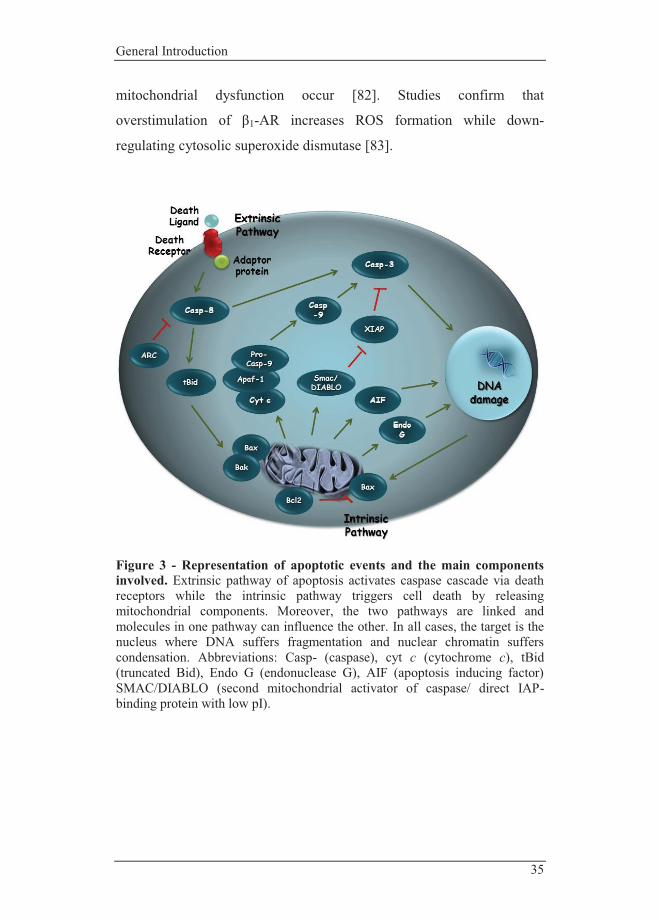
Figure 3 - Representation of apoptotic events and the main components involved. Extrinsic pathway of apoptosis activates caspase cascade via death receptors while the intrinsic pathway triggers cell death by releasing mitochondrial components. Moreover, the two pathways are linked and molecules in one pathway can influence the other. In all cases, the target is the nucleus where DNA suffers fragmentation and nuclear chromatin suffers condensation. Abbreviations: Casp- (caspase), cyt c (cytochrome c), tBid (truncated Bid), Endo G (endonuclease G), AIF (apoptosis inducing factor) SMAC/DIABLO (second mitochondrial activator of caspase/ direct IAP-binding protein with low pI).
Ana Branco PhD Thesis|2012
36
1.3.3 β-Adrenergic Receptors – Characterization of signaling
pathways
Adrenergic receptors link the cardiovascular system with the SNS.
Catecholamine binding to adrenergic receptors promotes the activation
of several pro- and anti-apoptotic pathways depending on the hormone
and the intensity of the binding. When excessive, over-activation can
lead to hypertrophy and apoptosis [24]. Several mechanisms have been
forwarded to explain the progressive deterioration of the contractile
system during heart failure such as loss of β-AR responsiveness,
dysregulation of excitation-contraction coupling, and activation of
aberrant signaling pathways [84, 85].
The ratio between β1- and β2-AR in cardiac cells depends on the species,
age and developmental stage, but in general, β1-AR seems to be the most
predominant [86]. Evidences suggest that the β1 subtype seems to be
downregulated in human failing ventricles and in this situation β2 is up-
regulated in contrast to human non-failing ventricles [87]. Stress
responses mediated by adrenergic receptors commonly incur in its
desensitization which can explain the down-regulation of β1-AR during
heart disease. Receptor phosphorylation, uncoupling from G proteins,
receptor internalization and degradation are some of the mechanisms
involved in the desensitization of β-AR [88]. Despite this, results from
genetic manipulation of β-AR content are not yet conclusive [89], and
sometimes even controversial. Experiments should take in consideration
the fluctuations on intensity/duration of the stimulation occurring in a
physiologic situation.
Upon ligand binding, β-ARs are physically coupled to heterotrimeric G
proteins, which are classified as stimulatory (Gs) or inhibitory (Gi) [90].
Both β1- and β2-AR are coupled via the “stimulatory” G protein (Gs) to
the effector enzyme adenylyl cyclase (AC), which converts the substrate
General Introduction
37
Mg-ATP to the second messenger cAMP (Fig. 4). In studies carried out
with transgenic mice and rat with cardiac overexpression of β1 receptors
Gαs, cardiomyopathic phenotype as well as increased basal rates of
cardiomyocyte apoptosis were observed [91].
It was observed in heart failure rats that the combination of β1-AR
antagonist and β2-AR agonist further improved the cardiac function and
prevented cardiomyocyte apoptosis than the administration of a β1-AR
antagonist alone. The authors suggest that the observed downregulation
of bax and upregulation of bcl-2/bax expressions might contribute to the
beneficial therapy effects [92]. Several studies have proposed the anti-
apoptotic effects of β2-AR receptors via coupling to the “inhibitory” G
protein (Gi) which is able to inhibit the signaling axis AC-cAMP-PKA
[87, 93, 94]. Moreover, studies have reported also that this survival
signaling pathway via β2-AR involves activation of phosphatidylinositol
3-kinase (PI3K) and Akt [95] (Fig. 4). Overexpression of an activated
form of PI3K catalytic subunit resulted in cardiac hypertrophy, while
forced expression of a dominant negative PI3K produced smaller hearts
and individual fibers [96]. It has been also reported that activation of this
pro-survival signaling pathway attenuates myocardial cell death after
ischemia/reperfusion injury [97]. Evidences demonstrated also that
although commonly involved in pro-apoptotic pathways [98], a number
of studies have shown that activation of p38-MAPK via β2 adrenergic
signaling promotes cell survival. This fact confirms that downstream
effects of β-adrenergic signaling cannot be considered completely
beneficial or prejudicial, due to the complex mechanisms of molecular
interaction. In fact, β-AR stimulation activates the three subgroups of
MAPKs phosphorylation cascades from which the p38-MAPK, the c-Jun
NH2-terminal kinases (JNK) and the extracellular signal regulated
kinases (ERK) are components [99]. A critical role of p38-MAPK in

Ana Branco PhD Thesis|2012
38
negatively regulating the mammalian cardiomyocyte proliferation and in
the potentiation of a terminally differentiation state has been
demonstrated [100]. Studies confirm that the human heart also express
β3-AR which exerts its effects by Gi-induced signaling pathways as well
[101].
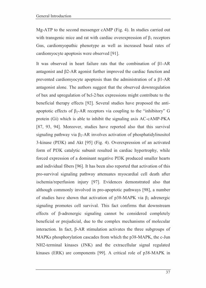
Figure 4 - Representation of some of the signaling pathways triggered by β-adrenergic receptors. β1- and β2-AR modulate cellular behavior via several signaling pathways. In this highly regulated system, maintenance of physiologic calcium levels and prevention of up-regulated pro-apoptotic elements is critical for cell death control. β1-AR is reported as a death sensor as it is coupled to a stimulatory G protein that activates PKA via adenylate cyclase and cAMP. While β2-AR signals via both the stimulatory and the inhibitory G protein, the latter one, related to anti-apoptotic pathways such as PI3K/Akt, alters the levels of pro-apoptotic proteins. Abbreviations: ROS (reactive oxygen species), PI3-K (phosphoinositide 3-kinase), PKC (protein kinase C), ATP (adenosine triphosphate), Gi (guanine nucleotide-binding protein inhibitory subunit), Gs (guanine nucleotide-binding protein stimulatory subunit), AC (adenylate cyclase), cAMP (cyclic adenosine monophosphate), CaMKII (Ca2+/calmodulin-dependent protein kinase), Ca2+ (calcium), PKA (protein kinase A), p38 (p38 mitogen-activated protein kinases), GSK3β

General Introduction
39
(glycogen synthase kinase 3 beta), Bad (Bcl-2-associated death promoter), Bcl-2 (B-cell lymphoma 2), Bcl-xL (B-cell lymphoma-extra-large), Bax (Bcl-2-associated X protein), LTCC (L-type calcium channel), SR (sarcoplasmic reticulum).
1.3.4 Beta-blockers – Improving the failing heart
It is known that β-AR blockers improve cardiac contractility and reduce
mortality in patients with heart failure [102-104]. But it is still unclear
how blocking a pathway that increases contractility of normal hearts can
improve the function of a failing heart.
Although poorly understood, it is suggested that the competition for
norepinephrine-binding receptors may attenuate cardiomyocyte
oversignaling where calcium plays a pivotal role [103]. As described, β-
AR stimulation activates both AC-cAMP-PKA and calcium /calmodulin-
dependent protein kinase II (CaMKII) pathways [105]. As in PKA
signaling, CaMKII are upregulated in failing hearts [106]. Both PKA
and CaMKII modulate the activity of a wide range of components
involved in calcium handling such as sarcoplasmic/endoplasmic
reticulum calcium-ATPase (SERCA) and its regulator, phospholamban
(PLB), ryanodine receptor RyR2 (sarcoplasmic reticulum calcium
release channel), and sarcolemmal L-type calcium channels (LTCC)
[107].
Calcium that enters into cardiomyocytes via LTCC causes the release of
a much larger amount of calcium from the sarcoplasmic reticulum (SR)
storage compartment by activating the RyR2 and thereby causing
contraction [108] (Fig.5). The hyperactivation of RyR2 appears to be one
of the causes for impaired cardiac function in heart failure [109, 110]
since it alters the gate increasing ion flux.
Ana Branco PhD Thesis|2012
40
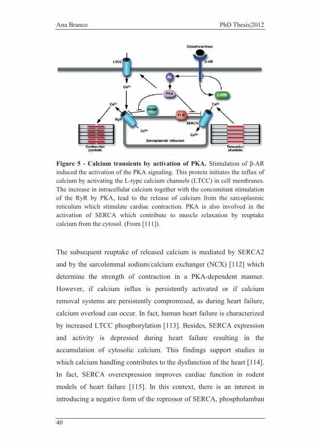
Figure 5 - Calcium transients by activation of PKA. Stimulation of β-AR induced the activation of the PKA signaling. This protein initiates the influx of calcium by activating the L-type calcium channels (LTCC) in cell membranes. The increase in intracellular calcium together with the concomitant stimulation of the RyR by PKA, lead to the release of calcium from the sarcoplasmic reticulum which stimulate cardiac contraction. PKA is also involved in the activation of SERCA which contribute to muscle relaxation by reuptake calcium from the cytosol. (From [111]).
The subsequent reuptake of released calcium is mediated by SERCA2
and by the sarcolemmal sodium/calcium exchanger (NCX) [112] which
determine the strength of contraction in a PKA-dependent manner.
However, if calcium influx is persistently activated or if calcium
removal systems are persistently compromised, as during heart failure,
calcium overload can occur. In fact, human heart failure is characterized
by increased LTCC phosphorylation [113]. Besides, SERCA expression
and activity is depressed during heart failure resulting in the
accumulation of cytosolic calcium. This findings support studies in
which calcium handling contributes to the dysfunction of the heart [114].
In fact, SERCA overexpression improves cardiac function in rodent
models of heart failure [115]. In this context, there is an interest in
introducing a negative form of the repressor of SERCA, phospholamban

General Introduction
41
[116]. Moreover, even when cardiomyocytes are isolated from the
overstimulated heart, the ability to efflux the calcium from the cytosol
during diastole is reduced [117].
By using a knockout mice for type 5 AC (AC5KO), a major cardiac
isoform, it was demonstrated that a long-term isoproterenol stress (7 to
14 days) did not result in a further increase of left ventricular ejection
fraction (LVEF) as observed in control condition, but instead resulted in
a greater degree of AC signaling downregulation, an improvement on
myocyte viability and increased Bcl-2 protein and Akt/GSK signaling,
potentially elucidating a novel approach to the therapy of heart failure
[118].
1.3.5 Beta-adrenergic signaling in the developing heart
As the embryonic heart underlies several modifications during
developmental maturation it is expected that alterations on cellular
signaling occur. The role of β-adrenergic signaling in cardiac
differentiation and in the normal development of the heart is poorly
understood.
To identify in [119] which developmental period the modulation of the
β-adrenergic system is more evident, Kudlacz et al. administered the β-
antagonist, propranolol, during fetal period and observed that a delay on
cellular development occurs at birth showing that catecholamines may
play a critical role in cardiac development [120]. In another study,
Lipshutz et al. demonstrated that 2- to 2.5 day old chicken embryos lack
β-adrenergic responsiveness to epinephrine. However, when an extract
of 11-day embryo was incubated with 2- to 2.5 day old cells in culture,
these cells developed sensitivity to epinephrine and increasing cAMP
levels [119]. As showed by the same study, basal cAMP levels of

Ana Branco PhD Thesis|2012
42
unresponsive cells with 2- to 2.5 day old are higher than older cells,
suggesting a role for cAMP in developing heart. In respect to the
contractile capacity, L-type Ca2+ channels from mouse embryos in early-
stage (day-11 to -13) lack a response to either isoproterenol or cAMP
which is reversed in later-stage (day-17 to -19) [121]. In embryonic stem
cell-derived cardiomyocytes (ESC-CMs) addition of exogenous β-
adrenergic agonists enhanced cardiac differentiation via p38-MAPK
signaling [122], while inhibition of β-adrenergic signaling reduced the
efficiency of cardiac differentiation. Retinoic acid (RA) plays an
important role mediating adrenergic action during embryonic heart
development [123]. Knockout of the dopamine β-hydroxylase results in
the loss of mouse embryos caused by heart failure where dysfunctional
retinoic acid synthesis and transport were observed [124]. Also, changes
in receptor number and signaling are known to suffer alterations during
fetal development. Importantly, mRNA and protein levels of β1-AR and
β2-AR suffer alterations during cardiac differentiation [122]. The
expression of β1-AR increases and reaches the highest level at day 14
although, β2-AR expression remain higher before and after the
differentiation process suggesting that β2-AR is predominant at early
stages, while β1-AR may be the predominant subtype for the later stages
of cardiac differentiation [122]. In mouse and human cardiac progenitor
cells (CPC) β1-AR expression is induced by differentiation stimuli while
β2-AR is present from an early stage promoting proliferation and
survival [125].
Interestingly, an elegant study demonstrated that repeated administration
of ISO to neonatal rats did not result in cardiac hypertrophy, whereas the
same treatment did produce hypertrophy in adult rats [126], suggesting
that ISO downstream signaling pathways in the neonatal rat are different
from the older, adult animal. Catecholamine-induced cardiac toxicity
General Introduction
43
during heart development was previously demonstrated by Iwasaki et al.
[127], which demonstrated that ISO administration to pregnant rats
resulted into disproportionate septal hypertrophy and frequent inter- and
intra-cellular disarray in the offspring. This result clearly indicates that
β-adrenergic over-stimulation during rapid cardiac development in the
womb can result into later profound cardiac alterations in the offspring,
which can be explained by signaling remodeling during cell
differentiation.
1.4 In vitro models to investigate cardiac β-adrenergic signaling and cardiovascular toxicology
One important limitation in investigating signaling pathways and drug-
induced toxicity responses has been the lack of adequate models to truly
reproduce in vitro what is observed in the heart in different stages of
development. In this respect, some cellular experimentally models that
can be used are below described and we point out the advantages and
disadvantages inherent to each model.
1.4.1 Neonatal Ventricular Cardiomyocytes
Neonatal primary cardiomyocytes have been one of the most widely
used models to study morphologic and biochemical features of
cardiomyocytes as well as hypertrophic mechanisms in vitro. Most of the
biochemical and gene transcription data have largely been obtained by
the use of primary cultures of 1-3 days-old mice neonatal
cardiomyocytes [128]. However, these cells are neither fully
differentiated adult cells neither maintain infinite proliferative capacity
in culture [129]. Primary cultures can be maintained in culture for days
or weeks and neonatal isolated cardiomyocytes phenotype is well
Ana Branco PhD Thesis|2012
44
conserved in culture [130]. However, on the other hand, cardiomyocyte
isolation techniques are not yet entirely established, in particular those
concerned to the purification of cardiomyocyte population. In
comparison with adult cardiomyocytes, neonatal have the advantage of
being easily isolated from the heart since it does not require aorta
cannulation and perfusion and, unlike adult cardiomyocytes, they are
less sensitive to calcium alterations in the medium during the isolation
procedure [131, 132]. Furthermore, both techniques require the sacrifice
of laboratory animals, which has become a concern. Versus animal
models, isolated cells constitute also an advantage for experiments aimed
at visualizing molecular responses as imaging techniques are often
limited to thick tissue. By preserving the in vivo integrity of
cardiomyocytes, data obtained by primary cultures may be combined
with experimental findings from living organisms due to the proximity
of this model to an intact tissue.
1.4.2 HL-1 cells
Mouse HL-1 cells are derived from a primary established cell line, the
AT-1, and were cultivated specifically to allow the maintenance of
specific characteristics. The most prominent features of this cell line are
related to the presence of spontaneous contractile activity and the
maintenance of several characteristics of an adult cardiomyocyte
phenotype such as alpha-MHC, alpha-cardiac actin and connexin43
[133]. As an immortalized cell line, HL-1 cells have some features of
such unlimited proliferation and also the capacity to be conserved and
recovered from liquid nitrogen storage [133]. HL-1 cells are widely used
in studies aimed at investigating cardiac biology such as myocardial
growth and hypertrophy.

General Introduction
45
AT-1 cells are derived from an atrial tumor that was formed in a
transgenic mouse by the specific expression of the oncogenic virus SV40
Large T protein. The SV40 Large T protein gene [134] was specifically
designed to be regulated by the atrial natriuretic factor (ANF) promoter,
an atrial specific protein, to induce a tumor growth in that tissue [133,
135]. Genome codification of SV40 Large T protein avoids the normal
function of retinoblastoma and p53 proteins disrupting the normal
operation of the cell cycle.
One disadvantage is that HL-1 cells have an atrial origin, hence their use
as ventricle cell model is not entirely accurate.
1.4.3 P19-derived cardiomyocytes
P19 are mouse embryonal carcinoma cells that have a pluripotent
behavior [136]. From these cells, a wide spectrum of adult cells such as
cardiomyocytes can be obtained by exposing them to DMSO or neurons
by using retinoic acid [137, 138]. Because preparations of differentiated
cells are far from be homogenous, special attention should be applied.
However, these cells are still derived from a tumoral cell line and
although differentiated, they are not considered completely adult cells.
Cultured cells grow exponentially and, similarly to the HL-1 cell line,
P19 cells are also able to have contractile activity under specific
conditions [139, 140]. P19 cells are commonly used to investigate
regulatory differentiation mechanisms of pluripotent cells that determine
the cell fate. In respect to our study, it is worth to note that these cells
present β-AR which respond to catecholamines [140]. To this regard,
several cardiac markers and L-type calcium channels are present in
cardiomyocyte-rich culture [141].
Ana Branco PhD Thesis|2012
46
1.4.4 ESCs-derived cardiomyocytes
Embryonic stem cells (ESCs) are derived from the inner cell mass of
pre-implantation embryos [142]. Cells proliferate in vitro and are easily
differentiated in a wide range of cell types including cardiomyocytes
expressing several markers of cardiac cells [143]. For this reason,
engraftment of ESCs constitutes a well appreciated therapeutic
intervention in failing hearts [144].
Several studies evidence the formation of teratomas followed by ESC
injection into the myocardium [145]. However, a number of other
studies have also delivered undifferentiated ESCs and failed to report
teratoma formation [146, 147]. Animals receiving ESCs or ESC-derived
cardiomyocytes following experimental cardiac injury exhibited superior
heart function [146]. Thus, the formation of teratomas and the
engraftment of ESC into the heart are technical problems that will be
solved with more research.
Collectively, the studies show evidences that ESC-derived cells
constitute a promised model for the treatment of heart disease, and
specifically for the replacement of lost cardiomyocytes, plus being a
promising in vitro tool for different toxicological studies.
1.4.5 H9c2 (2-1) cell line
The H9c2 (2-1) cell line was isolated from the ventricular part of a
BDIX rat heart [148]. Thirteen days after fecundation, cells were isolated
and, by a complex procedure, immortalized. In this stage, cells are not
fully differentiated into adult cardiomyocytes but are already
predestinated, which confer already the appearance of several
cardiomyocytes markers. By a selective serial passage, the different
adhesion kinetics of the heterogeneous isolated fraction was used to
General Introduction
47
separate the components in the culture dish. It is important to state that
these cells do not present contractile activity, even when differentiated
[149]. However, it is also worth to note that H9c2 cells and isolated
neonatal cardiomyocytes respond similarly to hypertrophy [150].
The H9c2 cell line is not considered as a pure and homogeneous sample
as it includes both embryonic skeletal (with slowly L-type calcium
channels) and cardiac (with fast L-type calcium channels) muscle cells
[149, 151]. Menard et al. confirmed that the H9c2 cell line is not only
able to differentiated into adult cells (skeletal and cardiac muscle) by
withdrawing the serum from the culture medium but also can be
differentiated into a predominantly cardiac phenotype, characterized by
the overexpression of the alpha-1 subunit of the L-type calcium
channels, by adding RA daily to the culture media [149]. Embryonic
myoblasts fuse to originate multinucleated differentiated myotubes.
Cells get an elongated shape and positioned in a parallel way [152]. One
limitation to the in vitro differentiation process is that it drives further
stress to the cells [153, 154]. Some studies document that the survival of
cardiomyocytes is committed by its culture in the absence of serum
[155]. But it should also be taken in consideration that stress-induced
cardiac differentiation is an in vivo feature and, as described, an essential
mechanism to trigger cell differentiation to an adult stage.

Ana Branco PhD Thesis|2012
48
Table 1 - Schematic representation of molecular, functional and culture behavior between neonatal cardiomyocytes, HL-1 cell line, P19, ESC and H9c2 (2-1) cell line. Abbreviations: Dif. (already differentiated); MHC (Myosin heavy chain), MLC2v (Myosin light chain 2 ventricular), BNP (brain natriuretic peptide), cTpC (cardiac troponin C), ANF (atrial natriuretic factor), TNNT2 (cardiac troponin T 2) [139, 141, 156-161]

General Introduction
49
1.5 General Aims In order to investigate further details into the mechanisms linking β-
adrenergic overstimulation to cell death, the cardiomyoblastic cell line
H9c2 was used in this study.
Since this cell line has the advantage of being differentiated to a more
adult phenotype [149] this feature was used to investigate whether the
differentiation status of the cell interfere with the final outcome of β-
adrenergic stimulation. Also, H9c2 have functional β-AR [162], which is
an essential condition to be used in the present study.
Norepinephrine signals via its interaction with α- and β- adrenergic
receptors. However, several studies document that its cytotoxicity seems
to be mediated by only β-adrenergic receptors. For that reason, in our
study, isoproterenol (ISO; Fig. 6), a synthetic catecholamine which is a
nonselective agonist of β-AR [163] was used to investigate signaling
pathways involved in cardiac cell death.
In fact, ISO has been recognized as a golden-standard agent that causes
cardiac injury [164, 165], which usually involves cardiac hypertrophy
[166].
Figure 6 - Chemical structure of Isoproterenol. Systematic name: 4-[1-hydroxy-2-(propan-2-ylamino) ethyl] benzene-1,2-diol
The present thesis investigates cell and mitochondrial stress responses
mediating the link between β-AR overstimulation and cell death.

Ana Branco PhD Thesis|2012
50
Different specific aims were addressed in the present study, including:
a) Are H9c2 cells differently susceptible to the toxicity of β-AR
overstimulation by ISO when in different stages of
differentiation?
b) If so, is this due to altered β-adrenergic signaling or to different
maturation of anti-oxidant / pro and anti-apoptotic pathways? In
this context, alterations in cytosolic and mitochondrial stress
responses to ISO were studied.
Regarding the second specific aim, we measured not only alterations of
stress/toxicity responses to ISO, but also basal differences existing in the
different groups of cells (undifferentiated vs. differentiated) which may
render them differently susceptible to ISO cytotoxicity.

Materials and Methods

Material and Methods
53
2. Material and Methods
2.1 Common Reagents Bovine serum albumin (BSA), all-trans retinoic acid (RA), ammonium
persulfate (APS), Bradford reagent, brilliant blue G, bromodeoxyuridine
(BrdU), calcium chloride (CaCl2), dimethyl sulfoxide (DMSO), DL-
Dithiothreitol (DTT), Dulbecco's-modified eagle's medium (DMEM),
ethylenediaminetetraacetic acid (EDTA), ethylene glycol-bis (2-
aminoethylether)-N,N,N´,N-tetraacetic acid (EGTA), glycerol, glycine,
heat-inactivated horse serum, isoproterenol, L-glutamine, β-
mercaptoethanol 98%, metaprolol, mouse anti-β-Actin, M199 medium,
N-acetylcysteine, pancreatin, pan-caspase inhibitor Z-Val-Ala-dl-Asp-
fluoromethylketone (z-vad-fmk), phenylmethylsulfonyl fluoride
(PMSF), protease inhibitor cocktail (containing 1mg/ml of leupeptin,
antipain, chymostatin and pepstatin A), sucrose, sodium azide,
sodiumchloride (NaCl), sodium dodecyl sulfate (SDS), sulforhodamine
B sodium salt (SRB), trizma-base, tris pH 8.8, tris pH 6.8 and trypan-
blue solution were obtained from Sigma (St. Louis, MO, USA).
Collagenase and Claycomb medium were purchased from Worthington
Biochemical Corporation (Lakewood, NJ, USA) and from SAFC
Biosciences, respectively. Acetic acid, ethanol, hydrochloric acid (HCl),
magnesium chloride (MgCl2), methanol, perchloric acid, potassium
chloride (KCl), potassium phosphate monobasic (KH2PO4), sodium
hydrogenocarbonate (NaHCO3), sodium sulfate (NaSO4) and sodium
hydroxide (NaOH) were obtained from Merck (Whitehouse Station, NJ,
USA). Hank´s Balanced Salt Solution (HBSS), penicillin, streptomycin,
fetal bovine serum (FBS) and Trypsin-EDTA were purchased from
Gibco-Invitrogen (Grand Island, NY). Acrylamide, Laemmli buffer,
polyvinylidene difluouride (PVDF) membranes, Ponceau solution and
N,N,N´,N´-tetramethylethylenediamine (TEMED) were obtained from

Ana Branco PhD Thesis|2012
54
BioRad (Hercules, CA, USA). The ECF detection system was obtained
from Healthcare Life Sciences (Buckingamshire, UK). The fluorescent
probes Hoechst 33342, 4',6-diamidino-2-phenylindole (DAPI),
dihydroethidium (DHE), tetramethylrhodamine methyl ester (TMRM),
5-(and 6)-chloromethyl- 2', 7'-dichlorodihydrofluorescein diacetate (CM
H2DCFDA), fluo4-AM, Mitotracker Red CMXROS, MitoSOX, calcein-
AM and the anti-fading reagent SlowFade® Gold were obtained from
Invitrogen-Molecular Probes (Eugene, OR, USA). PI3 kinase inhibitor
LY294002 was purchased from Cell Signaling (Beverly, MA, USA).
Caspase 3 substrate, fibronectin, pifithrin-alpha and p-nitroanilide (p-
NA) were purchased from Calbiochem (San Diego, CA, USA). Cyclic
AMP XPTM assay kit was purchased from Cell Signaling Technology
(Danvers, MA, USA). All reagents used in this work were of the greatest
degree of purity commercially available. To prepare solutions, ultrapure
distilled water filtered by the Milli Q from a Millipore system was used
in order to minimize contamination with metal ions.
2.2 Cell Culture
2.2.1 H9c2 Cell Culture and Differentiation Process
The H9c2 cell line purchased from America Tissue Type Collection
(Manassas, VA; catalog # CRL – 1446) was originally derived from
embryonic rat heart tissue using selective serial passages and were
cultured in DMEM medium supplemented with 1.5 g/L sodium
bicarbonate, 10% fetal bovine serum, 100 U/ml of penicillin and 100
μg/ml of streptomycin in 75 cm2 tissue culture flasks at 37°C in a
humidified atmosphere of 5% CO2. Cells were fed every 2 – 3 days, and
sub-cultured once they reached 70 – 80% confluence in order to prevent
the loss of differentiation potential.
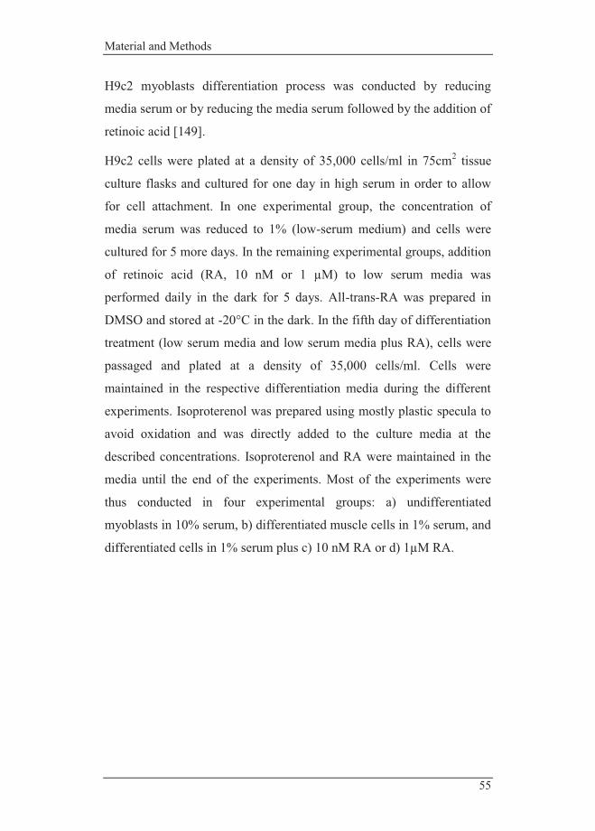
Material and Methods
55
H9c2 myoblasts differentiation process was conducted by reducing
media serum or by reducing the media serum followed by the addition of
retinoic acid [149].
H9c2 cells were plated at a density of 35,000 cells/ml in 75cm2 tissue
culture flasks and cultured for one day in high serum in order to allow
for cell attachment. In one experimental group, the concentration of
media serum was reduced to 1% (low-serum medium) and cells were
cultured for 5 more days. In the remaining experimental groups, addition
of retinoic acid (RA, 10 nM or 1 µM) to low serum media was
performed daily in the dark for 5 days. All-trans-RA was prepared in
DMSO and stored at -20°C in the dark. In the fifth day of differentiation
treatment (low serum media and low serum media plus RA), cells were
passaged and plated at a density of 35,000 cells/ml. Cells were
maintained in the respective differentiation media during the different
experiments. Isoproterenol was prepared using mostly plastic specula to
avoid oxidation and was directly added to the culture media at the
described concentrations. Isoproterenol and RA were maintained in the
media until the end of the experiments. Most of the experiments were
thus conducted in four experimental groups: a) undifferentiated
myoblasts in 10% serum, b) differentiated muscle cells in 1% serum, and
differentiated cells in 1% serum plus c) 10 nM RA or d) 1µM RA.
Ana Branco PhD Thesis|2012
56
Figure 7 - Schematic representation of the differentiated process of H9c2 myoblasts. Upon reduction of serum in the medium to 1% cells differentiate in both skeletal and cardiac muscle cells, while supplementing the same medium with RA induce the differentiation toward a mostly cardiac phenotype.
2.2.2 Culture of Mouse Neonatal Ventricular Cardiomyocytes
Ventricular part of one day-old mice neonates hearts was excised and
stored in a Ads solution (500 ml of 17g NaCl, 11.9g HEPES (Sigma
H6147), 0.3g NaH2PO4, 2.5g glucose, 1g KCl, 0.25g MgSO4, pH 7.35)
kept on ice. Ventricular myocardium was cut into small pieces (about 1-
2 mm3), and then subjected to a sequential digestion by an enzyme
solution of collagenase and pancreatin, in a horizontal shaking water
bath at 37°C, in order to digest the extracellular matrix and release cells
into the supernatant, as follow. Heart pieces were ressuspended several
times in Ads buffer, allowed to precipitate, supernatant removed
carefully and pancreatin/collagenase solution added for starting the first
digestion phase on the shaker. After 5 minutes, the supernatant was
transferred to a tube containing FBS for the inactivation of the enzymes,
centrifuged at 1000xg during 5 minutes at 25°C, mixed with 4 ml of FBS
and stored in a CO2 incubator at 37°C. The remaining tissue was then
ressuspended again with Ads buffer, the supernatant removed carefully
and pancreatin/collagenase solution added to initiate a new digestion
phase in the shaker. Afterwards, the supernatant was transferred to a
tube, mixed with FBS and centrifuged. Cells supernatants of the five
digestion steps were pooled together for a final centrifugation and the
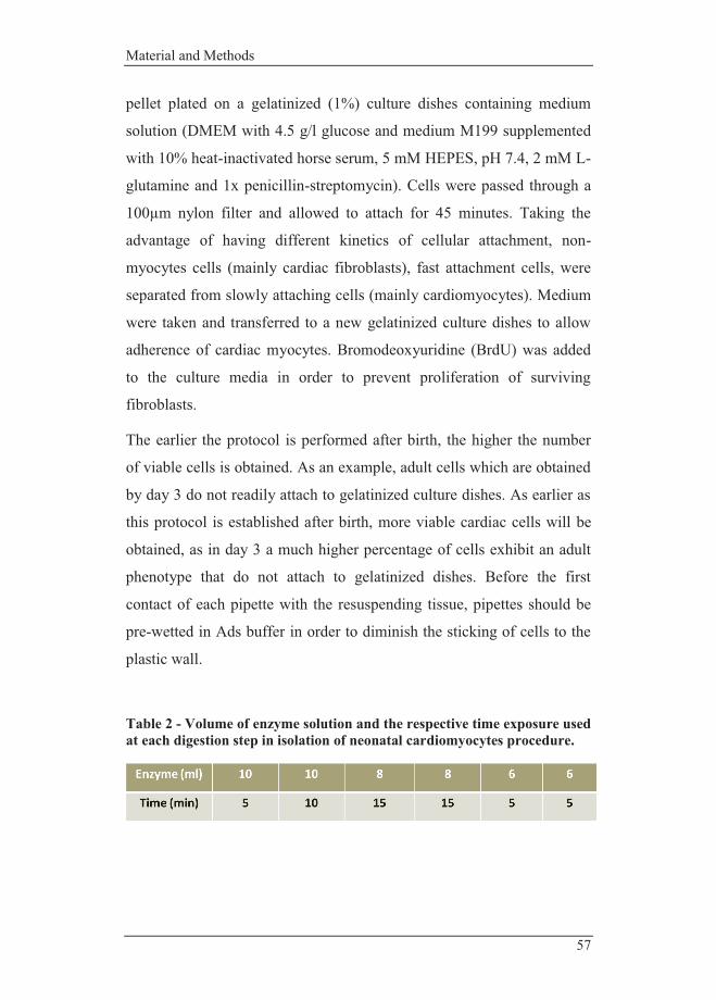
Material and Methods
57
pellet plated on a gelatinized (1%) culture dishes containing medium
solution (DMEM with 4.5 g/l glucose and medium M199 supplemented
with 10% heat-inactivated horse serum, 5 mM HEPES, pH 7.4, 2 mM L-
glutamine and 1x penicillin-streptomycin). Cells were passed through a
100µm nylon filter and allowed to attach for 45 minutes. Taking the
advantage of having different kinetics of cellular attachment, non-
myocytes cells (mainly cardiac fibroblasts), fast attachment cells, were
separated from slowly attaching cells (mainly cardiomyocytes). Medium
were taken and transferred to a new gelatinized culture dishes to allow
adherence of cardiac myocytes. Bromodeoxyuridine (BrdU) was added
to the culture media in order to prevent proliferation of surviving
fibroblasts.
The earlier the protocol is performed after birth, the higher the number
of viable cells is obtained. As an example, adult cells which are obtained
by day 3 do not readily attach to gelatinized culture dishes. As earlier as
this protocol is established after birth, more viable cardiac cells will be
obtained, as in day 3 a much higher percentage of cells exhibit an adult
phenotype that do not attach to gelatinized dishes. Before the first
contact of each pipette with the resuspending tissue, pipettes should be
pre-wetted in Ads buffer in order to diminish the sticking of cells to the
plastic wall.
Table 2 - Volume of enzyme solution and the respective time exposure used at each digestion step in isolation of neonatal cardiomyocytes procedure.
Ana Branco PhD Thesis|2012
58
2.2.3 HL-1 Cellular Culture
The HL-1 cell line (was a gentle gift from Dr. David Sanz Rosa)
originated from adult mouse atrial tissue was cultured in Claycomb
medium which is supplemented with 100 μM norepinephrine, 10% FBS,
2 mM L-glutamine and penicillin/streptomycin in 150 cm2 tissue culture
coated flasks. Flasks were coated with a salt solution supplemented with
gelatin and fibronectin and then stored at -20°C. Cells were allowed to
attach in an incubator at 37°C with a humidified atmosphere of 5% CO2.
Cells were sub-cultured once they reached 90% confluence.
2.3 Common Methods
2.3.1 Analysis of Drug Cytotoxicity
Evaluation of cell density was conducted using the sulforhodamine B
(SRB) assay in order to perform toxicity assays and observe putative
drug effects on H9c2 cell number cultured either in high- or in low-
serum medium, following established protocols [167, 168]. H9c2 cells
were seeded in 6- or 24-well plates (35000 cells/ml), allowed to attach
for 1 day and pre-incubated individually with the specific compounds:
30 minutes with 30 µM of pifithrin-alpha, 1 hour with 50 µM of
LY294002, 1 hour with 50 µM of z-vad-fmk or 2 hours with 100 µM
NAC, always prior to ISO treatment (24, 48 or 96 hours). The
concentration of the compounds used was the maximum amount that did
not cause toxicity in preliminary toxicity assessment. Vehicle controls
were also performed.
After treatment, the incubation media were removed, cells were rinsed
with PBS and fixed in 1% acetic acid in ice-cold methanol for at least 30
min. Cells were then incubated with 0.5% (wt/vol) SRB in 1% acetic
acid for 1 hour at 37°C. Unbound dye was removed by washing with 1%

Material and Methods
59
acetic acid. SRB labeled to cell proteins was extracted with 10 mM Tris
base solution, pH 10, and the optical density of the solution was
determined in a spectrophotometer at 540 nm. The amount of released
dye is proportional to the number of cells present in the sample.
2.3.2 Western Blotting
In order to obtain total cellular extracts, H9c2 cells were harvested by
trypsinization and washed once with PBS. Floating cells were also
collected and combined with adherent cells by performing two
centrifugation steps for 5 minutes at 1000 xg. The cell pellet was
resuspended in lysis buffer (20 mM HEPES/NaOH, pH 7.5, 250 mM
sucrose, 10 mM KCl, 2mM MgCl2, 1 mM EDTA) supplemented with
2mM DTT, 100mM PMSF and a protease inhibitor cocktail. Extracts
were sonicated and stored at -80°C until used. Protein contents were
determined by the Bradford method using bovine serum albumin as
standard. After denaturation at 95°C for 5 min in laemmli buffer,
equivalent amounts of total protein (25 µg) were separated by
electrophoresis in a 8, 12 or 14% SDS-polyacrylamide gels (SDS-
PAGE) and electrophoretically transferred to a polyvinylidene difluoride
(PVDF) membrane. After blocking with 5% milk or BSA, depending on
antibody information, in TBST (50mM Tris-HCl, pH 8; 154mM NaCl
and 0.1% tween 20) for 2 hours at room temperature, membranes were
incubated overnight at 4°C with the specific antibodies (listed in Table
3).

Ana Branco PhD Thesis|2012
60
Table 3 – Primary and secondary antibodies used in Western Blotting assay. Abbreviations: poly(ADP-ribose) polymerase (PARP), cardiac myosin light chain-2 (Mlc2v), superoxide dismutase 2 (SOD2), voltage-dependent anion channel (VDAC), phosphorylated (P-).
Name Dilution Host Species Brand PARP 1:1000 Rabbit Santa Cruz Akt 1:1000 Rabbit Cell Signaling Bax 1:6000 Rabbit Cell Signaling Bcl-2 1:1000 Rabbit Cell Signaling Bcl-xL 1:1000 Rabbit Cell Signaling β-Actin 1:8000 Mouse Sigma β1-AR 1:200 Rabbit Santa Cruz β2-AR 1:200 Rabbit Santa Cruz β3-AR 1:200 Goat Santa Cruz Calcineurin 1:500 Rabbit Cell Signaling Cardiac troponin T 1:1000 Mouse Abcam Caspase 3 1:1000 Rabbit Cell Signaling Cleaved-caspase 3 1:1000 Rabbit Cell Signaling Cyclophilin F 1:1000 Mouse Abcam Mlc2v OXPHOS
1:1000 1:1000
Mouse Mouse
Synaptic Systems MitoSciences
P-p38 MAPK 1:500 Rabbit Cell Signaling P-p66Shc 1:1000 Mouse Calbiochem p66Shc 1:1000 Rabbit BD Biosciences P-Akt (ser473) 1:1000 Rabbit Cell Signaling SOD2 1:1000 Rabbit Cell Signaling P-troponin I 1:1000 Rabbit Cell Signaling VDAC Rabbit anti-goat& Goat anti-rabbit& Goat anti-mouse&
1:500 1:5000 1:5000 1:5000
Rabbit - - -
Abcam Jackson IR Lab Jackson IR Lab Jackson IR Lab
* Mouse monoclonal OXPHOS cocktail comprises 5 different antibodies: CI
subunit NDUFB8, CII-30kDa SDHB, CIII-Core protein 2 UQCRC2, CIV
subunit I MTCO1 and CV alpha subunit ATP5A.
& Secondary alkaline phosphatase-conjugated antibodies
Material and Methods
61
Membranes were then washed with TBST and incubated with respective
secondary alkaline phosphatase-conjugated antibodies for 2 hours at
room temperature.
Membranes were incubated with the ECF detection system and
visualized with the Versa Doc imaging system (Bio-Rad, Barcelona,
Spain). Densities of each band were calculated with Quantity One
Software (Bio-Rad). Membranes were stained with Ponceau reagent
before blocking to confirm equal protein loading in each lane. This
experimental strategy was preferred over the use of housekeeping
proteins as the ones normally found in the literature can be theoretically
affected by the differentiation process.
2.3.3 Vital fluorescence of H9c2 cells
2.3.3.1 Tetramethylrhodamine methyl ester (TMRM)
Hoechst 33342 and Calcein-AM
After the differentiation process, H9c2 cells grown on glass-bottom
dishes (35,000 cells/ml) were treated with isoproterenol (ISO) for the
desired time exposure and then, incubated with TMRM (100 nM),
Hoechst 33342 (1 mg/ml) or calcein-AM (300 nM) at 37°C in the dark,
45 minutes prior the end of ISO incubation time. Media was then
replaced by Krebs buffer (1 mM CaCl2; 132 mM NaCl; 4 mM KCl; 1.2
mM Na2HPO4; 1.4 mM MgCl2; 6 mM glucose; 10 mM HEPES, pH 7.4)
supplemented with 10% FBS. Epifluorescence was obtained using a
Nikon Eclipse TE2000U microscope and were acquired using
Metamorph software (Universal Imaging, Downingtoen, PA). Confocal
microscopy images were collected using a Zeiss LSM 510Meta confocal
microscope and analyzed using the vendor software. Images were
obtained by using LSM Image Browser.

Ana Branco PhD Thesis|2012
62
2.3.3.2 Mitotracker Red and DAPI
H9c2 cells were seeded in 6-well plates with a glass coverslip per well at
a density of 35,000cells/ml. After 1 day of cell attachment, cells were
incubated with 50 µM of LY294002 (phosphoinositide 3-kinases-PI3Ks
inhibitor) for 1 hour prior to the incubation with 50 or 100 µM of ISO
for 24 (in the case of differentiated H9c2 cells) or 48 hour (in the case of
undifferentiated cells). 30 minutes prior the end of the exposure, cells
were incubated with the probe Mitotracker Red (125 nM) at 37°C in the
dark, washed with cold PBS, fixed with ice-cold absolute methanol and
stored at -20°C until use. A mounting solution (the anti-fading reagent
conjugated with 4',6-diamidino-2-phenylindole (DAPI) was used in
order to label nuclei and to detect apoptotic chromatin condensation.
Cells were observed by epifluorecence microscopy using a Nikon
Eclipse TE2000U microscope. The UV filter was used for DAPI
imaging and the rhodamine filter for Mitotracker Red fluorescence
imaging. Images were obtained using Metamorph software (Universal
Imaging, Downington, PA).
2.3.4 Flow cytometry analysis of intracellular calcium,
mitochondrial transmembrane potential and
mitochondrial superoxide anion
Subconfluent cells originated from the 3 groups of H9c2 used in this
study and cellular preparations after ISO treatment were harvested by
trypsinization and centrifuge. The pellet was ressuspended in 500μl of a
buffer solution (120mM NaCl; 3.5mM KCl; 0.4mM KH2PO4; 20mM
HEPES; 5mM NaHCO3; 1.2mM NaSO4 and 10mM pyruvate at pH 7.4,
1.2 mM MgCl2, 1.3mM CaCl2 and 5% FBS) plus the respective probe (2
μM Fluo-4 AM, 100 nM TMRM or 5μM MitoSOX Red) in order to
Material and Methods
63
evaluate intracellular calcium concentration, mitochondrial
transmembrane potential (ΔΨm) and mitochondrial superoxide anion
respectively. Cells were incubated at 37°C in the dark for 30 minutes
before the end of treatment with ISO. Cells were then analyzed in a
FACScalibur Flow cytometer, using the FL1 or the FL2 filter sets
(Mitosox e TMRM) and counted using the cytometer Cell Quest
software package. Generation of mitochondrial superoxide anion was
increased by incubating cells with the complex I inhibitor rotenone
during 1 hour, with this group used as a positive control.
2.3.5 Measurement of cytosolic superoxide and H2O2
production by using the probes DHE and CM-H2DCFDA
Undifferentiated and differentiated H9c2 cells seeded in 12-well plates
and incubated 48 hours with ISO were incubated for 20 min at 37°C in
the presence of 0.5 μM dihydroethidium (DHE) in PBS containing 5 mM
glucose. The cells were washed twice with PBS, and the fluorescence
was recorded in a microplate reader at 535 nm excitation and 635 nm
emission wavelengths. To detect H2O2 production, 2 μM CM-
H2DCFDA were incubated with cells and results obtained by using a
microplate reader during 30 minutes at 495 nm excitation and 520 nm
emission wavelengths.
2.3.6 Caspase-3-like colorimetric activity assay
Total cellular extracts were collected by trypsinization and centrifuged
twice at 1,000 xg, 4°C during 5 minutes. Floating cells were also
collected. The pellet was resuspended in collecting buffer (20 mM
HEPES/NaOH pH 7.5, 250 mM sucrose, 10 mM KCl, 2 mM MgCl2, 1
mM EDTA) supplemented with 2 mM DTT, 100 µM PMSF and a

Ana Branco PhD Thesis|2012
64
protease inhibitor cocktail. Protein concentration was determined by the
Bradford assay. To measure caspase 3-like activity, aliquots of cell
extracts containing 25μg of protein were incubated in the reaction buffer,
containing 25 mM HEPES (pH=7.5), 10% sucrose, 10 mM DTT, 0.1%
CHAPS and 100μM of caspase substrate Ac-DEDV-pNA, for 2h at
37°C. Caspase 3-like activity was determined by following the detection
of the chromophore p-nitroanilide (p-Na) after cleavage from labeled
substrate Ac-DEDV. The method was calibrated with known
concentrations of p-nitroanilide (p-NA). The optical density of the
solution was determined in a spectrophotometer at 405 nm.
2.3.7 Determination of intracellular cAMP content
Cyclic AMP content was measured by using a commercial kit (cyclic
AMP XPTM assay kit) and by following vendor´s instructions (cell
signaling). Cells were seeded in 96-well plates at a density of 40,000
cells/well and allowed to attach for one day. Cells were then incubated
with 50 or 100 µM of ISO during 48h. 1 hour prior the end of the
treatment, cells were incubated with 20 µM of forskolin in order to
increase cAMP levels (alasbahi 2012). Cells were washed twice with
cold PBS and incubated 10 minutes on ice with a lysis buffer solution
(supplemented with PMSF), provided inside the kit. 50 µl of the lysed
sample plus 50 µl of the HRP-linked cAMP solution were added to a
coated 96-well plate that contained already an anti-cAMP XPTM mAb
and incubated for 3 hours in a horizontal shaker. Cells were washed 4
times with washing buffer provided by the kit and incubated with 100
µM of TMB substrate for 30 minutes. The stop solution was added to
each well and optical density was measured at 450nm in a
spectrophotometer. cAMP intracellular levels were determined by
following the detection of a gradient of known concentrations of cAMP.
Material and Methods
65
2.3.8 Total RNA Extraction
Collection of total RNA extracts was performed following the vendor’s
instructions (RNeasy Mini Handbook from QIAGEN). About 1x10⁶ cells
were collected, and disrupted in 350μl of Buffer RLT from QIAGEN
(RNeasy Protect Mini Kit) supplemented with 3.5µl β-mercaptoethanol.
The suspension was homogenized, added to QIAshredder Spin Columns
and centrifuged for 2 min at the highest velocity. The supernatant was
ressuspend with 350μl of ethanol 70% and transferred to a RNeasy Mini
Spin Columns. A sequence of centrifugations was performed after
following washing steps. Total RNA bound to the membrane and was
then eluted in RNase-free water. Concentration and purity of the sample
were verified using Agilent Technologies, Eukaryote Total RNA Nano
assay and degradation was analyzed performing an agarose gel.
2.3.9 Microarray Gene Expression Analysis
H9c2 cells, HL-1 cells and mouse ventricular neonatal cardiomyocytes
were collected as described previously. Experiments were done in
triplicate. After RNA extraction and concentration determined, statistical
outliers criteria were performed to further analyze sample purification.
The microarray hybridization was carried at the Genomic Department of
the National Center for Cardiovascular Investigation (CNIC) in Madrid,
Spain, and further analyzed in the Bioinformatic Unit from the same
Institution. Based on the Bioconductor package “ArrayQualityMetrics”
there was no evidence of any outlier among the samples. Total rat and
mouse transcriptome, with about 42,000 transcripts from the Agilent
4x44K chip were identified using Agilent Rattus norvegicus / Mus
musculus genome microarrays of and, respectively. Raw signals were

Ana Branco PhD Thesis|2012
66
thresholded to 1 and quantiles normalization [169] was performed using
the software GeneSpring.
Data were considered in the log2 scale. Default flags were considered as
absent, except saturated points that were flagged as marginal.
2.3.10 Statistical analysis
Data analysis was performed by using GraphPad Prism 4.0 program
(GraphPad Software, Inc.) and data were expressed as mean±SEM for
the number of experiments indicated in the legends of the figures.
Multiple comparisons were performed using one-way analysis of
variance (ANOVA) followed by Bonferroni multiple comparison post-
hoc test and the t-test in proliferation studies to compare two groups.
Significance was accepted when p value < 0.05.

Results

Results
69
3. Results
3.1 Characterization of H9c2 Myoblast Differentiation 3.1.1. Abstract
Despite the wide use of the H9c2 cell line in the undifferentiated state, a
cardiac-like phenotype can be obtained after differentiation in a low FBS
(1%) and retinoic acid (RA)-supplemented media. Because the adult
heart tissue is composed by differentiated cardiomyocytes, toxicological
studies can have different outcomes depending on the developmental
state of the biological model used. In this chapter, we aim to characterize
the H9c2 differentiation process to obtain a cardiomyocyte-like
phenotype, which will help understanding future results involving the
possible differential toxicity of isoproterenol on H9c2 cells in different
differentiation stages. Alterations in H9c2 cells were found during the
differentiation process towards a cardiac-like phenotype: myoblasts fuse
to a multinucleated and larger cells, proliferative activity is reduced, the
content in specific markers of cardiomyocytes (e.g. troponin T, troponin
I and Mlc2v) is increased and, finally, alterations in some proteins
involved in pathways of the β-adrenergic signaling and in stress
responses/cell protection were found (e.g. increased P-p38MAPK,
VDAC, Bcl-xL, SOD2 and decreased calcineurin, β1- and β3-AR). Also,
differences in the transcriptional map were obtained when comparing
undifferentiated and differentiated H9c2 cells. The results hint at a
higher content in protective mechanisms in undifferentiated H9c2 cells,
which may impact the toxicity of cardiotoxicants. Finally, we also
compared the transcriptional map of the adult HL-1 cell line with
isolated neonatal cardiomyocytes, which are also widely used as models
for cardiac cell lines.
Ana Branco PhD Thesis|2012
70
3.1.2. Background and Objective
Cardiac tissue contains mostly terminally differentiated cells without
proliferative activity, although cardiac muscle progenitors in specific
niches have been found [170, 171]. Despite the fact that these
undifferentiated cells are a clear minority in the tissue, toxicology
studies should address the different susceptibility of terminally
differentiated vs. undifferentiated cardiac cells to cardiotoxicants. The
H9c2 myoblast cell line, originated from rat ventricular tissue, has been
used in vitro as a model for skeletal and cardiac muscle due their
morphological features and electrical/hormonal signaling properties
[148, 151]. One important feature of this embryonic cell line is its ability
to differentiate from mono-nucleated myoblasts to myotubes when
cultured in a low serum concentration media. In the differentiation
process, cells obtain mostly a skeletal muscle phenotype, as evidenced
by specific markers of skeletal muscle differentiation such as myogenin,
troponin T and MyoD [149]. Ménard et al. demonstrated that daily
additions of all-trans retinoic acid (RA) in a low-serum media lead to
increased content of cells with an adult cardiac muscle phenotype. As
described, the great majority of studies involving H9c2 myoblasts as a
model to evaluate the cardiotoxicity of different agents are performed
with cells in the undifferentiated state, raising questions on the relevance
of the results when compared to the use of adult cardiomyocytes.
The differentiation potential of H9c2 myoblasts makes this cell line an
attractive tool for investigating the cardiotoxicity of several agents.
Concerning this, only a few investigations have applied this knowledge
in experimental studies [172-175]. Since one major question in this
thesis is whether the differentiation stage of H9c2 cells impacts ISO
toxicity, we aimed in this chapter to better characterize the different cell
populations obtained in each differentiation protocol.
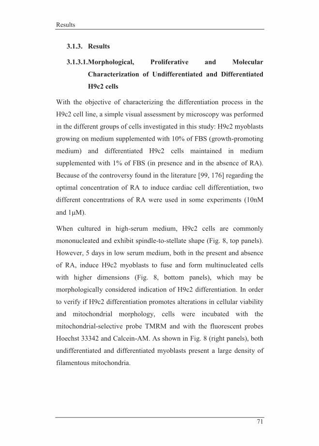
Results
71
3.1.3. Results
3.1.3.1.Morphological, Proliferative and Molecular
Characterization of Undifferentiated and Differentiated
H9c2 cells
With the objective of characterizing the differentiation process in the
H9c2 cell line, a simple visual assessment by microscopy was performed
in the different groups of cells investigated in this study: H9c2 myoblasts
growing on medium supplemented with 10% of FBS (growth-promoting
medium) and differentiated H9c2 cells maintained in medium
supplemented with 1% of FBS (in presence and in the absence of RA).
Because of the controversy found in the literature [99, 176] regarding the
optimal concentration of RA to induce cardiac cell differentiation, two
different concentrations of RA were used in some experiments (10nM
and 1�M).
When cultured in high-serum medium, H9c2 cells are commonly
mononucleated and exhibit spindle-to-stellate shape (Fig. 8, top panels).
However, 5 days in low serum medium, both in the present and absence
of RA, induce H9c2 myoblasts to fuse and form multinucleated cells
with higher dimensions (Fig. 8, bottom panels), which may be
morphologically considered indication of H9c2 differentiation. In order
to verify if H9c2 differentiation promotes alterations in cellular viability
and mitochondrial morphology, cells were incubated with the
mitochondrial-selective probe TMRM and with the fluorescent probes
Hoechst 33342 and Calcein-AM. As shown in Fig. 8 (right panels), both
undifferentiated and differentiated myoblasts present a large density of
filamentous mitochondria.

Ana Branco PhD Thesis|2012
72
Figure 8 - Effect of serum withdrawal on H9c2 cells morphology. Morphologic alterations were observed using the fluorescent probes Hoechst 33342 (nucleus, blue), Calcein-AM (cell viability, green) and TMRM (mitochondria, red). H9c2 cells cultured in 10% of FBS in the medium (top panels) showed a stellar shape, in the most cases, with one nucleus per cell. Upon culture in a medium containing 1% of FBS (bottom panels) daily supplemented with RA differentiated H9c2 cells showed an elongated shape with several nuclei. Epifluorescent microscopy images are representative of 4 different cell preparations. White bar represents 30µm.
To assess the effect of serum withdrawal on H9c2 cells proliferation, we
measured the cell mass increase during 6 days in culture by using the
colorimetric SRB method. Results obtained demonstrate a reduction in
the cell proliferation rate when H9c2 cells were cultured in low serum
medium being the difference evident in the exponential phase of the
growth (Fig. 9A).
An additional study using the same method investigated whether
previously differentiated H9c2 cells were able to re-start proliferation
when transferred to a high-serum medium (Fig. 9B). Results demonstrate
that differentiated H9c2 cells increase gradually the proliferation activity
when exposed again to a growth-promoting medium.
Hoechst 33342 2 Calcein-AM TMRM
10%
FBS
1%
FBS
+
RA
Phase Contrast

Results
73
Figure 9 - Alteration on proliferative rate by modulating the percentage of serum in the medium. H9c2 cells were seeded at a density of 5,000 cells/well and allowed to proliferate during 6 days. Results were analyzed by using the SRB assay. A) Undifferentiated H9c2 cells incubated in a high-serum medium proliferate fast, while incubation in a low-serum medium during 5 days promotes cell cycle arrest. B) Studies were conducted after 5 days in a low serum media to allow cell differentiation. When differentiated cells are exposed to a proliferation-promoting medium, H9c2 cells re-entry the cell cycle, initiating proliferative activity. Statistical analysis: * p < 0.05 vs 10% FBS.
We then investigated whether RA-driven cell differentiation results in the increase of specific markers for cardiac cells [173, 177]. Higher protein content in phosphorylated-troponin I, cardiac troponin T and ventricular myosin light chain were observed when H9c2 cells were differentiated in a low serum media supplemented with RA (Fig. 10A-C), confirming that RA treatment drives H9c2 cells to a cardiac-like phenotype.
Figure 10 - Cellular content on specific cardiac markers in different cell populations. The content of cardiac troponin T increases as cells are differentiated in a low serum media in the presence of 1 μM RA (left panel). By using Western Blotting, it was also possible to confirm that RA-differentiated
B A B
Ponceau
A B C Troponin T (35.4kDa)
MLC2v (19kDa)
P-troponin I (cardiac) (28kDa)
Ponceau Ponceau

Ana Branco PhD Thesis|2012
74
cells have increased cardiac myosin light chain-2 (Mlc2v) (middle panel) and cardiac phosphorylated-troponin I (P-troponin I) (right panel), two recognized cardiac markers. The images are representative of 3 different experimental studies which developed the same result. Control loading was performed by using Ponceau labeling.
3.1.3.2. Alterations on signaling protein content during H9c2
differentiation
Since cardiomyocyte regulation of cell death by β-adrenergic stimulation
is associated with specific signaling pathways [178], several proteins
were analyzed in the context of apoptotic signaling alterations during
H9c2 differentiation to identify whether the results would impact how
the different groups of cells react to the toxicity caused by ISO over-
stimulation. Evaluation by Western Blotting of the three subtypes of β-
adrenergic receptors (AR) (Fig. 11D-F) was performed. While
differences regarding β2-AR between cells in different states of the
differentiation process were not found, β1-AR and β3-AR were
significantly decreased in H9c2 cells that were differentiated in the
presence of RA. To investigate downstream mechanisms by which
stimulated β-AR by ISO may signal to induce cell death, alterations in
the cellular content of calcineurin and phosphorylated p38-MAPK in
H9c2 cells subjected to the different differentiation processes were
performed. Both proteins have been demonstrated to be involved in β-
adrenergic regulation of cardiomyocyte death [179, 180]. While
calcineurin decreased in H9c2 RA-differentiated cells (Fig. 11J),
phosphorylated p38-MAPK was increased in the same group (Fig. 11K).
Basal levels of the pro-apoptotic protein Bax and the anti-apoptotic
protein Bcl-2 were not altered during the differentiation process
(Fig.11H and 11I). The mitochondrial voltage-dependent anion channel
(VDAC) was also analyzed, since this protein has also been described as

Results
75
being involved in the mitochondrial apoptotic pathway [181]. H9c2 cells
differentiated in the presence of RA have increased VDAC content
compared with the other experimental groups (Fig. 11G). Basal content
of some important proteins involved in intrinsic apoptotic signaling
pathway such as Bcl-xL and cyclophilin F were evaluated by Western
Blotting (Fig. 11A and 11B). We found the same content in terms of
cyclophilin F, a mPTP component [179], in the four groups of H9c2
studied. A higher content in Bcl-xL, a protector of mitochondrial
integrity [182], was found on both groups of differentiated cells.
Mitochondrial superoxide dismutase 2 (SOD2) protein was also
evaluated due its role in mitochondrial anti-oxidant defense [178, 183].
Differentiated H9c2 cells in present of RA were found to have
significantly higher content of this ROS scavenging enzyme than
undifferentiated cells (Fig. 11C).
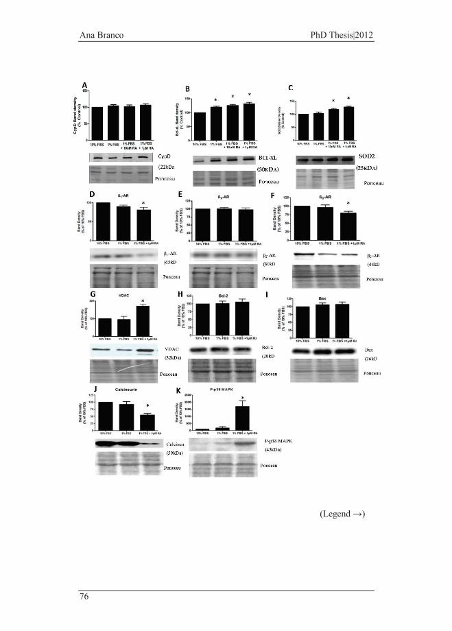
Ana Branco PhD Thesis|2012
76
(Legend →)

Results
77
Figure 11 - Determination of specific proteins content in undifferentiated and differentiated H9c2 cells. The content of cyclophilin F (A), Bcl-xL (B), SOD2 (C), β1-AR (D), β2-AR (E), β3-AR (F), VDAC (G), Bax (H), Bcl-2 (I), calcineurin (K), and phosphorylated p38-MAPK (J) in total cellular extracts was identified by Western Blotting analysis as bands with the corresponding molecular weight. Blots are representative from different cell preparations. Also shown is the respective Ponceau labeling. Graphics are expressed as mean±SEM of 7 (Bax, Bcl-2), 6 (β1-AR, β2-AR, β3-AR) and 4 (cyclophilin F, Bcl-xL, SOD2, calcineurin, P-p38MAPK and VDAC) separate experiments. p<0.05 compared with 10% FBS.
3.1.3.3.Morphological and transcriptional comparison of H9c2
cells with neonatal cardiomyocytes and HL-1 cells.
Several cell line models have been used in cardiovascular studies instead
of primary cultures due to the several limitations. Thus, in this work we
further analyzed morphological differences between the HL-1 cell line,
H9c2-differentiated cells and primary cultures of cardiomyocytes from
neonatal mice.
Differentiated H9c2 cells are morphologically more elongated and
present a fusiform shape comparing with the other groups (Fig. 12).
However, similarly to preparations of HL-1, mostly differentiated H9c2
cells present several nuclei per cell, which is a characteristic from adult
cardiomyocytes. Neonatal cardiomyocytes are mononucleated,
presenting a smaller size. Similar to HL-1 cells, neonatal cardiomyocytes
also present contractile activity under special culture conditions [128]. It
is worth to note that H9c2 cells are isolated from rat and HL-1 and
neonatal cardiomyocytes were collected from mice, although in different
ages.

Ana Branco PhD Thesis|2012
78
A B C D
Figure 12 - Representative images of differentiated H9c2 cells, adult HL-1 cells and neonatal cardiomyocytes. Microscopy images show morphological differences between groups of cells. (A) H9c2 differentiated cells present an elongated shape and are mostly multinucleated, (B) mostly adult HL-1 cells present several nucleus per cell, (C) isolated cardiomyocytes from neonatal mice are smaller and are mostly mononucleated cells. A, B and C images represent a magnification of 200x. D corresponds to a magnification of 400x of (A) to better identification of multinucleated cells.
To obtain an alternative snapshot of the alterations suffered by H9c2
cells during the differentiation process as well as differences between
HL-1 cells and isolated neonatal cardiomyocytes, we performed a
comparative gene expression analysis to identify differences in gene
transcription. After correction of the data and identification of significant
differences on gene regulation (based on the adjusted p value < 0.05), we
found several transcriptional alterations. The mentioned genes are
intrinsically related and are involved in differentiated processes,
proliferative activity and apoptosis signaling.
Genes present in the treemap diagram of the Fig. 13 were chosen based
on the selectivity to the tissue, e.g. whether they represent a specific
protein that would be mostly found on the correspondent model. The
results from microarrays expression suggest that differentiation of H9c2
cells to a cardiac phenotype led to an expected increase in cardiac
specific mRNAs.
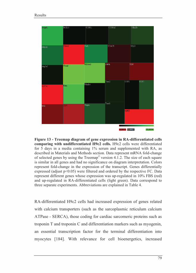
Results
79
Figure 13 - Treemap diagram of gene expression in RA-differentiated cells comparing with undifferentiated H9c2 cells. H9c2 cells were differentiated for 5 days in a media containing 1% serum and supplemented with RA, as described in Materials and Methods section. Data represent mRNA fold-change of selected genes by using the Treemap© version 4.1.2. The size of each square is similar in all genes and had no significance on diagram interpretation. Colors represent fold-change in the expression of the transcript. Genes differentially expressed (adjust p<0.05) were filtered and ordered by the respective FC. Data represent different genes whose expression was up-regulated in 10% FBS (red) and up-regulated in RA-differentiated cells (light green). Data correspond to three separate experiments. Abbreviations are explained in Table 4.
RA-differentiated H9c2 cells had increased expression of genes related
with calcium transporters (such as the sarcoplasmic reticulum calcium
ATPase - SERCA), those coding for cardiac sarcomeric proteins such as
troponin T and troponin C and differentiation markers such as myogenin,
an essential transcription factor for the terminal differentiation into
myocytes [184]. With relevance for cell bioenergetics, increased
Ana Branco PhD Thesis|2012
80
expression of genes relevant for mitochondrial energy production
including mitochondrial creatine kinase, cytochrome c oxidase subunits
COX6a2, COX8b and COX11, uncoupling proteins 2 and 3 and
Complex I subunit NADH dehydrogenase (ubiquinone) Fe-S protein 4,
was identified in RA-differentiated cells. The expression of genes
involved in signaling pathways for cellular death such as FasL, cyclin-
dependent kinase inhibitor 2B and Bcl2l11, which are involved in the
inhibition of cell cycle progress and on apoptosis, were also augmented
in RA-differentiated H9c2 cells. Interesting, we also found that RA-
differentiated cells have increased oxidative stress growth inhibitor 1
(Osgin1) gene expression. Undifferentiated myoblasts show increased
gene expression of pro-survival proteins such as Bcl-2, increased
transcripts for the mitochondrial inner membrane translocase (TIM).
Also, genes playing important roles in the regulation of cell cycle,
including gene encoding cyclin A, cell cycle regulator required for the
onset of DNA replication, and encoded or related genes were also up-
regulated in undifferentiated myoblasts. Regarding related genes of the
retinoblastoma-like 1, undifferentiated myoblasts had also increased
expression on E2F transcription factor 1, HDAC1, BRCA1 associated
RING domain 1, prohibitin-2 and mybl2. Moreover, the
phosphodiesterase 4A (PDE4A), cAMP-specific, the mitogen-activated
protein kinase, the B-cell CLL/lymphoma 2 (Bcl2) are also up-regulated
in undifferentiated cells.
The results indicate that the differentiation of H9c2 cells with serum
regulation and RA addition led to a transcriptional up-regulation of
genes involved in cardiac differentiation, which again demonstrates the
validity of using this cell system.

Results
81
Table 4 - Groups of gene expression explored in H9c2 undifferentiated myoblasts and RA-differentiated H9c2 cells. H9c2 cells were differentiated for 5 days in a low serum concentrated medium daily supplemented with RA. Both groups, undifferentiated and RA-differentiated cells, were collected and analyzed as described in Material and Methods section. FC>2 corresponds to, at least, two-fold up-regulation of the gene, FC<2 corresponds to, at least, two-fold down-regulation of the gene. Data correspond to three separate experiments and differences are also represented in Fig. 13.
1% FBS + RA vs 10% FBS Gene Involvement FC Chrna1 Cholinergic receptor, nicotinic (muscle) 33.66 Myom2 Myomesin 2 - sarcomeric protein 32.62 Ckmt Creatine kinase - important in tissues that consume ATP faster 27.50 Ampd1 Adenosine monophosphate deaminase-decrease cAMP levels 22.61 Atp2a1 ATPase, Ca2+ transporting, cardiac muscle (SERCA1) 14.43 Faslg Fas ligand - trigger apoptosis 17.01 Osgin1 Oxidative stress induced growth inhibitor 12.12 UCP3 Uncoupling protein 3 (mitochondrial, proton carrier) 10.97 Cdkn2b Cyclin-dependent kinase inhibitor 9.58 Myog Myogenin - essential for development of skeletal muscle 9.45 Cox6a2 Cytochrome c oxidase assembly protein 6 6.41 Tnnc2 Troponin C (fast) 4.60 Tnnt2 Troponin T (cardiac specific) 4.48 Pln Phospholamban – inhibitot of SERCA2 3.63 Myl2 Light myosin specific for cardiac muscle; triggered
by calcium to induce contraction 2.87
Atp2a2 Ca2+ transporting, cardiac muscle (SERCA2) 2.76 Camk2b Calcium/calmodulin-dependent protein kinase II 2.54 Cox11 Cytochrome c oxidase assembly protein 11 2.23 Cpt1a Carnitine palmitoyltransferase 1a 2.14 Ndufs4 NADH dehydrogenase (ubiquinone) Fe-S protein 4 2.10 Bcl2l11 BCL2-like 11 - apoptosis facilitator 2.07 UCP2 Uncoupling protein 2 (mitochondrial, proton carrier) 2.06 Hdac1 Histone deacetylase 1 0.50 Pde4a Phosphodiesterase 4A, hydrolyzes the cAMP 0.45 Gata2 Promotes cellular proliferation 0.45 Rbbp8 Retinoblastoma binding protein 0.43 Timm17a Translocase of inner mitochondrial membrane 0.38 Bcl2 Anti-apoptotic protein 0.36

Ana Branco PhD Thesis|2012
82
Nfatc2 Nuclear factor activated by calcineurin 0.34 E2Ftf1 E2F transcription factor-mediate both cell
proliferation and apoptosis 0.26
Gpx1 Glutathione peroxidase 1 0.20
Top2a Topoisomerase (DNA) 0.15 Adcy10 Adenylate cyclase 10 0.12 Wisp2 WNT1 inducible signaling pathway 0.12 Bard1 BRCA1 - regulate cell growth 0.10 Chrm2 Cholinergic receptor, muscarinic 0.10 Mybl2 Myeloblastosis oncogene-involved in cell cycle progression 0.09 Ccna2 Cyclin A 0.08
We also performed genotyping studies comparing isolated neonatal
cardiomyocytes with the HL-1 cell line. Neonatal cardiomyocytes
presented up-regulation of non-differentiated cell markers such as the
GATA6, a gene that is required for a proper maturation of cardiac
mesoderm, but also in BMP4 which level expression are maintained by
GATA6 [2, 185]. Overexpression of GATA6 in cardiac cells in the
declined period of it expression (before terminally differentiated cells)
results in arrest of cardiac differentiation [2].
Caveolin-2 and caveolin-3 are up-regulated in isolated cardiomyocytes.
However, evidences demonstrated that these proteins increase with age
but also when isolated cardiomyocytes are cultured in vitro [2], which
was the case in this study.
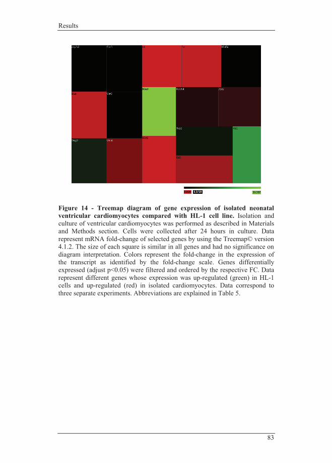
Results
83
Figure 14 - Treemap diagram of gene expression of isolated neonatal ventricular cardiomyocytes compared with HL-1 cell line. Isolation and culture of ventricular cardiomyocytes was performed as described in Materials and Methods section. Cells were collected after 24 hours in culture. Data represent mRNA fold-change of selected genes by using the Treemap© version 4.1.2. The size of each square is similar in all genes and had no significance on diagram interpretation. Colors represent the fold-change in the expression of the transcript as identified by the fold-change scale. Genes differentially expressed (adjust p<0.05) were filtered and ordered by the respective FC. Data represent different genes whose expression was up-regulated (green) in HL-1 cells and up-regulated (red) in isolated cardiomyocytes. Data correspond to three separate experiments. Abbreviations are explained in Table 5.

Ana Branco PhD Thesis|2012
84
Table 5 - Groups of gene investigated in isolated neonatal cardiomyocytes and HL-1 cells. Cellular preparations were collected and analyzed as described in Material and Methods section. FC>2 corresponds to, at least, two-fold up-regulation of the gene on HL-1 cells, FC<2 corresponds to, at least, two-fold up-regulation of the gene on neonatal cardiomyocytes. Gene corresponds to those represents in Fig. 14. Data correspond to three separate experiments.
Neonatal Cardiomyocytes vs. HL-1 cell line Gene Involvement FC Mns1 Meiosis-specific nuclear structural protein 30.267 Wif1 Wnt inhibitory factor 17.744 Casp3 Effector caspase 3.923 Terf2 Telomeric repeat binding factor 2.315 Cav3 Caveolin 3 0.473 Cav2 Caveolin 2 0.448 Mef2d Required for cardiac development 0.453 Atp2a2 ATPase, Ca2+ transporting, cardiac muscle 0.382 Notch4 Involved in cell fate decisions 0.22 Ryr2 Ryanodine receptor 2, cardiac 0.196 Gata6 Related with embryonic cardiac development 0.108 Bmp Involved in heart development 0.022
Irx Transcription factor required for cardiac development 0.018
Utf1 Undifferentiated embryonic cell transcription factor 0.065
Myl2 Myosin, light polypeptide 2, cardiac 0.006 Id1 Inhibitor of DNA binding 0.0063

Results
85
3.1.4. Discussion
The rat H9c2 cell line is an established in vitro cellular model for cardiac
cells in a wide variety of experimental designs [2, 31]. It has been
described that H9c2 cells are an excellent model to investigate
mechanisms and outcomes of cardiac hypertrophy signaling, which
increases the interest in this cell model [150]. Despite the ability of H9c2
myoblasts to differentiate in adult muscle cells, all toxicological studies
involving this cell line are performed with undifferentiated myoblasts. In
fact, the traditional use of this cell line in its undifferentiated state may
not be the best approach to use as a cardiac cell surrogate. This raises an
important question: does the differentiation state of H9c2 cells interfere
with their sensitivity to cardiotoxicants? In this regard, at least some
discussion should be carried out on the validity of this model. The
important finding that H9c2 myoblasts have the capacity to differentiate
into skeletal and cardiac muscle cells increases the uses of this cell
model for developmental toxicology studies, including those using
cardiac stem cells. For example, a similar approach, although with a
different model, demonstrated that doxorubicin cardiotoxicity involves
the depletion of the cardiac stem cell pool [43].
Muscle cell differentiation, in vivo, is commonly accompanied by
specific processes, including cell cycle arrest and morphological features
such as formation of multinucleated myotubes. In this context,
morphological alterations observed in H9c2 cells, after the induction of
differentiation by serum withdrawal were initially examined by
epifluorescence microscopy. The results obtained in this study confirm
that upon reduction of serum in the culture media to 1%, H9c2
myoblasts fuse and form multinucleated cells (Fig. 8), as previously
described [44]. A decrease in cell proliferation was another consequence
of decreasing serum media, which is more evident in the exponential
Ana Branco PhD Thesis|2012
86
phase of the growth (Fig. 9A). It has been previously shown that the
differentiation process initiates a cell cycle arrest, which decreases
cellular proliferation [60]. Cellular proliferation depends on the
existence of growth factors in the culture media, hence it is expected that
the reduction in the concentration of those factors will induce cell
growth arrest. According to the literature [66, 70] the addition of retinoic
acid (RA) drives the differentiation towards a cardiac phenotype.
Interestingly, the fact that proliferation re-starts after moving cells back
to a high-serum media (Fig. 9B) show that cell cycle arrest is reversible.
Although the differentiation protocols yield heterogeneous populations
[31], Fig. 10 shows that RA-differentiated cells present higher content in
troponin T, phosphorylated troponin I and Mlc2v, markers of cardiac
differentiation [82] although it must be stressed that the relative yield of
differentiation into a population showing cardiac markers appears to be
very dependent on the initial cell confluence. One open question in this
regard is if data from Western Blotting reflect large differences in
protein expression in a small subset of cells that differentiate, or instead
smaller changes in protein expression in a large majority of cells that
suffer differentiation. Answering this question, evaluation of gene arrays
of the entire cell genome showed that expression of multiple markers of
cardiomyogenesis occurs, including the gene encoding for the cardiac
troponin T protein.
During muscle cell differentiation, specific signaling pathways are
activated and muscle protein synthesis patterns are altered. Modulation
of signaling proteins during cell differentiation can cause a higher
susceptibility of a specific developmental stage of the tissue to a
cardiotoxic agent. Of particular importance, we found several proteins
that may be responsible for a possible different cellular response to a
toxicant. One of the objectives of the present work was to verify if

Results
87
alterations in cellular levels of β-adrenergic receptors (β-AR) were
observed during the differentiation process as overstimulation of
different β-AR subtypes results in different outcomes: β1- subtype is
commonly associated to a pro-apoptotic signaling pathway whereas the
β2-subtype is reported to be involved in an anti-apoptotic process [83].
The present study demonstrates that β1-AR content in undifferentiated
H9c2 cells is higher than in differentiated cells (Fig. 11D), being
significantly different when compared with differentiated cells in
presence of RA. In fact, not only β-ARs are involved in a large variety of
vital processes including intracellular oxygen availability [86], but also
β1-AR can become desensitized after prolonged stimuli [89]. Similar
results were obtained with β3-AR where differentiated cells in presence
of RA have lower levels of this receptor. The physiological significance
of this fact is not immediately clear. β3-ARs have different properties
from the two other cardiac subtypes. β3-AR up-regulation during disease
processes and resistance to desensitization suggests an important role for
this subtype of receptors, especially because it has been described that
β3-AR can protect the myocardium against adverse effects of excessive
catecholamine stimulation [88]. Also, the present work does not
distinguished between membrane/active receptors and receptors that may
exist in other intra-cellular compartments.
Also, proteins involved in intracellular pro and anti-apoptotic proteins
were evaluated. No alterations were found in the basal content in Bax
and Bcl-2, showing that cell differentiation does not alter the status of
these two important proteins (Fig. 11H and 11I). Interestingly, the gene
array analysis confirmed increased expression of Bcl-2, a pro-survival
protein, on undifferentiated cells (Fig. 13). Both Bcl-xL and SOD2
protein content is increased in differentiated cells which can constitute a
higher degree of protection of this cellular group. Interestingly, the

Ana Branco PhD Thesis|2012
88
mitochondrial voltage-dependent anion channel (VDAC), which has
been suggested to form/regulate the mPTP and to be involved in
apoptosis [87] is increased in cells that were differentiated in presence of
RA (Fig. 11G). The increased amount of VDAC in this experimental
group can be important to explain differences on the susceptibility of
cells to mitochondrial-mediated cell death.
The decrease in calcineurin, a calcium/calmodulin-dependent
phosphatase that is activated by sustained elevations in intracellular
calcium [94] and is inactivated by oxidative stress [87], and the increase
in p38-MAPK during the differentiation process can contribute to the
increased susceptibility of differentiated H9c2 cells to a cardiotoxicant
agent. This is a particularly interesting observation since it has been
observed that transfection of H9c2 cells with calcineurin adenovirus
resulted in increased oxidative stress [94]. The decrease of calcineurin in
RA-differentiated cells may act to suppress oxidative stress resulting
from increased mitochondrial activation during differentiation [87]. In
fact, also both decreased mitochondrial membrane potential and
mitochondrial dysfunction were observed after calcineurin up-regulation
[94], two results that were not verified in our study after H9c2
differentiation, suggesting that a decrease in this protein may contributes
to the regular mitochondrial function toward to a mature energetic
efficiency state. Moreover, we also found that the gene encoding for the
nuclear transcription factor NFAT, which is regulated by calcineurin and
calcium, was found increased in undifferentiated cells. Once activated,
NFAT translocate to the nucleus where modulate the expression of
multiple other genes [93]. Differences observed in the content of
phosphorylated p38-MAPK are interestingly opposite to what was
observed with calcineurin, with RA-differentiated cells having a very
large increase in that protein (Fig. 11). As previously described by Lim
Results
89
et al., p38-MAPK stimulates H9c2 cellular differentiation even in
presence of a proliferative medium [93]. A few studies also document
the key role of p38-MAPK during cardiomyogenesis involving both
MyoD and myocyte enhancer binding factor 2 (MEF2) [93, 94].
Moreover, p38 MAPK has an opposite influence on NFAT/calcineurin-
activated nuclear function by inducing NFAT export back to cytoplasm
which suggest that the amount of calcineurin/p38MAPK during
differentiated process, which is opposite, although not well understood,
must be supported for an unknown reason [93].
Differentiated markers specific for cardiac myocytes were found
increased upon differentiation of H9c2 cells confirming results obtained
by Western Blotting. In this regard, also the genes Myl2, Myog, and
Myom2 detail a clear transition to a more mature and adult muscle.
Furthermore, this transcriptional analysis reveals that RA-differentiated
myoblasts have increased expression of genes related with mitochondrial
function, including respiratory chain complexes and uncoupling proteins,
confirming our previous results that H9c2 cell differentiation with RA
towards a more cardiac-like phenotype involves remodeling and up-
regulation of mitochondrial function [94]. Higher expression of
uncoupling proteins 2 and 3 in RA-differentiated H9c2 cells may be a
compensatory response to an increase in mitochondrial activity after cell
differentiation. As previously described, uncoupling proteins have
important roles in fatty acid β-oxidation and mitochondrial oxidative
stress [87]. In fact, when fatty acid delivery to mitochondria exceeds
oxidative capacity, the expression of UCP3 increases leading to reduced
storage of non-esterified fatty acid anions [87]. Interestingly, the Cpt1
gene that encodes for carnitine palmitoyltransferase 1, was also
increased in RA-differentiated cells. This shuttle system transports fatty
acyl-CoA esters from the cytosol into the mitochondria, where they

Ana Branco PhD Thesis|2012
90
undergo β-oxidation metabolism [93]. Still concerning energy
production, creatine kinase, Ckmt, plays a pivotal role in muscle
function since it works to transfer a “high energy phosphate” from
mitochondria to the cytosolic carrier, creatine. Therefore, this enzyme is
highly expressed in tissues that have a highly energy demand. In our
study we found that RA-differentiated cells highly expressed this
mitochondrial enzyme when compared with undifferentiated cells.
Reticoic acid-differentiated H9c2 cells had also increased expression of
genes encoding calcium transporters, such as the sarcoplasmic reticulum
calcium ATPase – SERCA1 and 2, as well as cytochrome c oxidase
subunits COX6a2, COX8b and COX11 and complex I subunit NADH
dehydrogenase (ubiquinone) Fe-S protein 4.
Cyclic nucleotide phosphodiesterase (PDE) constitute also a mechanism
of great importance that safeguard the specificity of GPCR/cAMP
signaling in the control of cAMP transients via its degradation. The
PDE4 is responsible for cAMP hydrolysis and its inhibition increase
intracellular cAMP levels and L-type Ca2+ current increasing Ca2+
release at baseline during β-adrenergic stimulation. This mechanism can
contribute to sensitize differentiated cells to β-adrenergic stimulation
dependent of cellular Ca2+ influx [186].
Genes involved in cell cycle arrest and in death signaling pathways such
as FasL, the cyclin-dependent kinase inhibitor 2B and the Bcl2l11,
which is able to induce apoptosis, were also increased in RA-
differentiated H9c2 cells (Fig. 13). As expected, genes encoding for
proliferative mechanisms were highly expressed in undifferentiated cells
as well as some genes related with pro-survival signaling pathways.
By working with different cellular models such as HL-1, H9c2 and
isolated neonatal cardiomyocytes, especially from a toxicology
perspective, different results are expected. This work confirms the use of

Results
91
H9c2 cell line as a proper model to investigate developmental cardiac
toxicity (myoblasts vs adult cells) or to perform a basic assessment of
cardiac toxicity.

Ana Branco PhD Thesis|2012
92
3.2 Role of β-adrenergic Signaling Stress Responses on Isoproterenol Cytotoxicity on H9c2 Cells in Different Differentiation Stages
3.2.1 Abstract
H9c2 cells are used as a surrogate for cardiac cells in several
toxicological studies, which are usually performed with cells in their
undifferentiated state, raising questions on the applicability of the results
to adult cardiomyocytes. Since H9c2 myoblasts have the capacity to
differentiate into skeletal and cardiac muscle cells under different
conditions, the hypothesis of the present work was that cells in different
differentiation states differ in their susceptibility to toxicants. In order to
test the hypothesis, the effects of the cardiotoxicant isoproterenol (ISO)
were investigated. The present work demonstrates that differentiated
H9c2 cells are more susceptible to ISO toxicity. After ISO treatment, the
pro-apoptotic protein Bax increase in all experimental groups, although
only undifferentiated myoblasts up-regulate the anti-apoptotic proteins
Bcl-2 and Bcl-xL. Mitochondrial SOD2 is also increased in the
undifferentiated groups after ISO treatment. Isoproterenol-induced
toxicity results in cellular calcium overload, loss of mitochondrial
respiratory complexes, mitochondrial membrane depolarization and
increased superoxide anion in differentiated cells. Isoproterenol also
caused increased activation of the p66Shc pathway in differentiated
H9c2 cell, as well as an increase in p53 protein, caspase-3 activation and
PARP cleavage. Calcineurin is decreased in differentiated H9c2 cells,
which suggests an important role against ISO-induced cell death.
The results show that differentiation of H9c2 cardiomyoblasts impact
stress responses to cardiotoxicants, which can be relevant in the context
of developmental cardiotoxicity.

Results
93
3.2.2 Background and Objective
The release of catecholamines by the sympathetic nervous system under
stress conditions leads to increased heart rate through its action on β-AR.
The magnitude of β-adrenergic stimulation plays an important role in
cardiomyocyte stress responses, which may develop to apoptosis through
the activation of several signaling pathways. Indeed, the control of
myocyte cell loss through the suppression of cell death pathways
represents an ideal strategy to prevent many cardiodegenerative
conditions. Beta-receptor activation on cardiac cells links with several
signaling pathways such as mitogen-activated protein kinases (MAPK)
[99], adenylyl cyclase (AC)/cAMP/ protein kinase A (PKA) [176] or
phosphatidylinositol 3-kinase (PI3K)/Akt [187]. Excessive activation of
these pathways originating upstream from β-adrenergic over-stimulation,
leads to increased cytosolic calcium overload and production of reactive
oxygen species (ROS) [178] and often to activation of apoptotic
signaling [180].
Mitochondria are key regulators of cellular metabolism. However, once
mitochondrial function is affected, critical ATP-dependent mechanisms
are compromised and mitochondria-induced apoptotic cell death is
activated [179]. During oxidative phosphorylation, the reduction of O2 to
H2O is also accompanied by side reactive oxygen species production
[181]. Reactive oxygen species are continuously produced in eukaryotic
cells and play an important role in the regulation of multiple stress and
toxicity responses [182]. Assessing stress responses in the
cardiomyocyte is critical in the understanding of the mechanisms of
heart degeneration during different pathologies and to design proper
therapies. Different in vitro cell systems have been used to explore
mechanisms of cellular stress responses to different toxicants, including
P19-derived cardiomyocytes [183], HL-1 cells [178, 188], neonatal and
Ana Branco PhD Thesis|2012
94
adult primary cardiomyocytes [184, 185] and H9c2 cardiomyoblasts
[189]. The latter cell line has been used for several in vitro cardiotoxicity
studies. However, the vast majority of studies have only been conducted
with cells in their undifferentiated and proliferative state [190]. The fact
that this cell line has the ability to differentiate into adult skeletal and
cardiac muscle cells [149] make it a more attractive tool for investigating
toxic effects of several agents during heart development. Since
differences are likely to exist regarding metabolic state [191] and
signaling pathway fluxes, as described in 3.1, the differentiation state of
the cells presumably impacts the outcome of xenobiotic toxicity. H9c2
cells have been previously shown to express β-adrenergic receptors in
their undifferentiated state [162], which provides validity to the possible
use of H9c2 cells to study the role of β-adrenergic over-stimulation on
cardiomyocyte dysfunction and heart failure [192, 193]. Isoproterenol
(ISO), a synthetic catecholamine, has been used to address the
cytotoxicity associated with hyperadrenergic states in different studies
[194]. We investigated here the susceptibility of H9c2 cells in various
states of differentiation to the toxicity of supra-physiological
concentrations of the β-adrenergic agonist ISO and correlated the
toxicity with alterations in pro-apoptotic and β-adrenergic signaling
pathways, as well as with cellular and mitochondrial stress responses.
Supra-physiological ISO concentrations induce cardiomyocyte cell
death, with β1-adrenergic receptors signaling a predominantly pro-
apoptotic role, while β2-adrenergic receptor activation appears to mostly
inhibit cell death [105]. The hypothesis of the present work is that H9c2
myoblasts cultured under different differentiating conditions are
differently affected by ISO toxicity, which is explained by different
modulation of stress responses. The objective of this work is to change
the paradigm that H9c2 cells can be used irrespective of their
differentiation state to investigate the toxicity of several agents; a
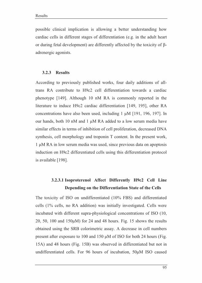
Results
95
possible clinical implication is allowing a better understanding how
cardiac cells in different stages of differentiation (e.g. in the adult heart
or during fetal development) are differently affected by the toxicity of β-
adrenergic agonists.
3.2.3 Results
According to previously published works, four daily additions of all-
trans RA contribute to H9c2 cell differentiation towards a cardiac
phenotype [149]. Although 10 nM RA is commonly reported in the
literature to induce H9c2 cardiac differentiation [149, 195], other RA
concentrations have also been used, including 1 μM [191, 196, 197]. In
our hands, both 10 nM and 1 μM RA added to a low serum media have
similar effects in terms of inhibition of cell proliferation, decreased DNA
synthesis, cell morphology and troponin T content. In the present work,
1 μM RA in low serum media was used, since previous data on apoptosis
induction on H9c2 differentiated cells using this differentiation protocol
is available [198].
3.2.3.1 Isoproterenol Affect Differently H9c2 Cell Line
Depending on the Differentiation State of the Cells
The toxicity of ISO on undifferentiated (10% FBS) and differentiated
cells (1% cells, no RA addition) was initially investigated. Cells were
incubated with different supra-physiological concentrations of ISO (10,
20, 50, 100 and 150μM) for 24 and 48 hours. Fig. 15 shows the results
obtained using the SRB colorimetric assay. A decrease in cell numbers
present after exposure to 100 and 150 μM of ISO for both 24 hours (Fig.
15A) and 48 hours (Fig. 15B) was observed in differentiated but not in
undifferentiated cells. For 96 hours of incubation, 50μM ISO caused

Ana Branco PhD Thesis|2012
96
approximately the same amount of cell loss than 100μM for 48 hours
(Fig. 15C). At least for up to 300 μM ISO, no effects on undifferentiated
cells were observed. The next question regarded the introduction of RA
in the differentiation process and its relevance for ISO toxicity. For this
experiment, one time point (24 hours) was chosen to evaluate the effects
of cell differentiation achieved with two RA concentrations. Despite the
fact that RA addition to low serum media drives the differentiation of
H9c2 cells towards a cardiac-like phenotype [149], our data indicates
that the toxicity of ISO for 24 hours was equivalent in the presence and
absence of both 10 nM and 1 μM RA (Fig. 15C), which indicates that the
three differentiated cell populations are equally susceptible to ISO, as
opposed to undifferentiated cells (Fig. 15D). A longer exposure (48
hours) to the same ISO concentrations yielded similar results (data not
shown). Since the toxicity of ISO was independent of the RA
concentration used to differentiate H9c2 cells, further experiments were
all performed with the highest concentration of RA used.

Results
97
Figure 15 - Toxicity of ISO on H9c2 myoblasts grown in high-serum media (undifferentiated) and low serum media (generating differentiated adult muscle cells). Undifferentiated control cells were maintained in 10% serum medium as described in the materials and methods section. The differentiation of H9c2 myoblasts was promoted by reducing the media serum concentration from 10% to 1%. Cells were incubated with 10, 20, 50, 100 and 150μM of ISO for 24 (A), 48 (B) and 96 hours (C). Cells number was analyzed by using the SRB colorimetric assay. Data represent the mean ±SEM of 3-9 different cell preparations. Statistical analysis: * p < 0.05, 1% FBS vs. 10% FBS for the same ISO concentration. D) Differentiation of H9c2 cells in the presence of 10nM or 1μM RA does not alter the susceptibility of differentiated cells to ISO. H9c2 cells in the several differentiation states were incubated with 10, 20, 50, 100 and 150μM of ISO during 24 hours. The results were very similar for 48h (not shown). Results were analyzed by SRB assay as described in methods section. Data represent the mean±SEM of 3 different cell preparations. No
E
48h
1% FBS 10% FBS0
25
50
75
100
125
Cel
l num
ber
(% C
ontr
ol)
*
*
A B
C D
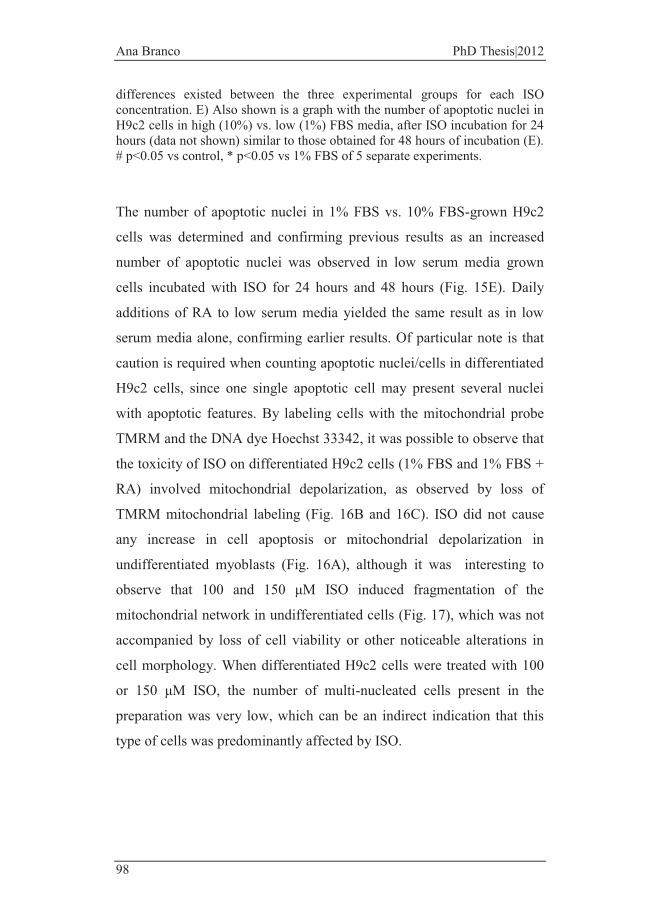
Ana Branco PhD Thesis|2012
98
differences existed between the three experimental groups for each ISO concentration. E) Also shown is a graph with the number of apoptotic nuclei in H9c2 cells in high (10%) vs. low (1%) FBS media, after ISO incubation for 24 hours (data not shown) similar to those obtained for 48 hours of incubation (E). # p<0.05 vs control, * p<0.05 vs 1% FBS of 5 separate experiments.
The number of apoptotic nuclei in 1% FBS vs. 10% FBS-grown H9c2
cells was determined and confirming previous results as an increased
number of apoptotic nuclei was observed in low serum media grown
cells incubated with ISO for 24 hours and 48 hours (Fig. 15E). Daily
additions of RA to low serum media yielded the same result as in low
serum media alone, confirming earlier results. Of particular note is that
caution is required when counting apoptotic nuclei/cells in differentiated
H9c2 cells, since one single apoptotic cell may present several nuclei
with apoptotic features. By labeling cells with the mitochondrial probe
TMRM and the DNA dye Hoechst 33342, it was possible to observe that
the toxicity of ISO on differentiated H9c2 cells (1% FBS and 1% FBS +
RA) involved mitochondrial depolarization, as observed by loss of
TMRM mitochondrial labeling (Fig. 16B and 16C). ISO did not cause
any increase in cell apoptosis or mitochondrial depolarization in
undifferentiated myoblasts (Fig. 16A), although it was interesting to
observe that 100 and 150 μM ISO induced fragmentation of the
mitochondrial network in undifferentiated cells (Fig. 17), which was not
accompanied by loss of cell viability or other noticeable alterations in
cell morphology. When differentiated H9c2 cells were treated with 100
or 150 μM ISO, the number of multi-nucleated cells present in the
preparation was very low, which can be an indirect indication that this
type of cells was predominantly affected by ISO.

Results
99
(Legend →)

Ana Branco PhD Thesis|2012
100
Figure 16 - Cellular alterations observed in H9c2 cells after exposure to ISO for 48 hours (A, B and C). H9c2 cells were cultured in high-serum media (10% FBS), low serum media (1% FBS) and low serum media daily supplemented with RA (1% FBS + 1μM RA). H9c2 cells were incubated with 10, 20, 50, 100 and 150 μM of ISO and labeled with Hoechst 33342 (nuclei, blue fluorescence) and TMRM (mitochondria, red fluorescence). Epifluorescence microscopy images are representative from 3 different cell preparations. White arrows indicate cells with apoptotic nuclei (Hoechst column) or cells with depolarized mitochondria (TMRM column).
One source of experimental artifact could be a possible binding of ISO to
serum proteins, thus reducing the effective amount of the compound
acting on undifferentiated myoblasts, which are maintained in higher
serum levels. In order to test this possibility, undifferentiated H9c2 cells
were incubated with ISO in a media with 10% or 1% FBS, while cells
differentiated in 1% FBS were incubated with ISO in a high (10%) and
low (1%) serum media. After 48h of ISO treatment, the results suggest
that ISO binding to serum proteins is not the major cause for the
different susceptibility of undifferentiated vs. differentiated H9c2 cells,
although there appears to be a small effect caused by altering serum
concentration (Fig. 18). Interestingly, H9c2 differentiated cells exposed
to a proliferation-promoting media undergo changes typical of an
undifferentiated state, including an increase on cell proliferation rate
(Fig. 9).

Results
101
Figure 17 - Undifferentiated H9c2 cells present morphological markers of mitochondrial fragmentation. Although mitochondria are not dysfunctional, undifferentiated cells show fragmentation of the mitochondria membrane network as seen by confocal microscopy (arrow). Cells were incubated with ISO for 24 hours and labeled with the probes as described in methods section. White bar represents 30µm.
Cont
rol
50 �
M IS
O
100 �M
ISO
TMRM Hoechst Calcein Overlap
TMA
Control 50 ��M ISO 100 ��M ISO B
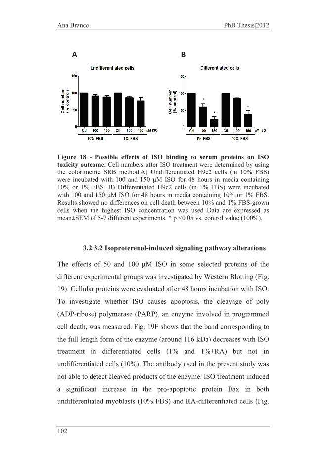
Ana Branco PhD Thesis|2012
102
Figure 18 - Possible effects of ISO binding to serum proteins on ISO toxicity outcome. Cell numbers after ISO treatment were determined by using the colorimetric SRB method.A) Undifferentiated H9c2 cells (in 10% FBS) were incubated with 100 and 150 μM ISO for 48 hours in media containing 10% or 1% FBS. B) Differentiated H9c2 cells (in 1% FBS) were incubated with 100 and 150 μM ISO for 48 hours in media containing 10% or 1% FBS. Results showed no differences on cell death between 10% and 1% FBS-grown cells when the highest ISO concentration was used Data are expressed as mean±SEM of 5-7 different experiments. * p <0.05 vs. control value (100%).
3.2.3.2 Isoproterenol-induced signaling pathway alterations
The effects of 50 and 100 μM ISO in some selected proteins of the
different experimental groups was investigated by Western Blotting (Fig.
19). Cellular proteins were evaluated after 48 hours incubation with ISO.
To investigate whether ISO causes apoptosis, the cleavage of poly
(ADP-ribose) polymerase (PARP), an enzyme involved in programmed
cell death, was measured. Fig. 19F shows that the band corresponding to
the full length form of the enzyme (around 116 kDa) decreases with ISO
treatment in differentiated cells (1% and 1%+RA) but not in
undifferentiated cells (10%). The antibody used in the present study was
not able to detect cleaved products of the enzyme. ISO treatment induced
a significant increase in the pro-apoptotic protein Bax in both
undifferentiated myoblasts (10% FBS) and RA-differentiated cells (Fig.
A B
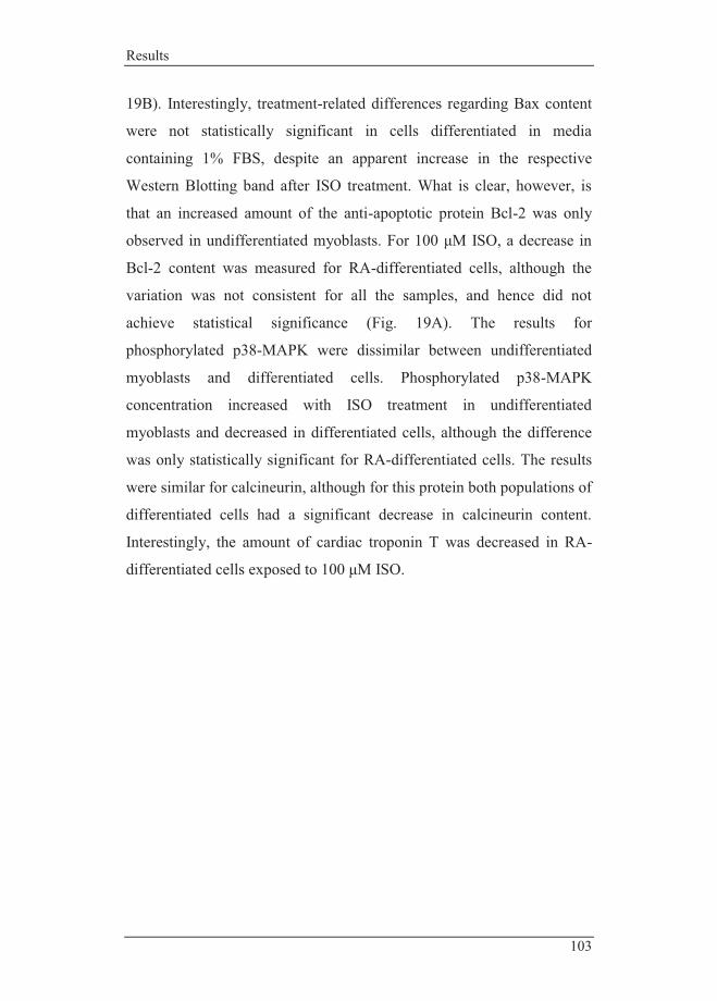
Results
103
19B). Interestingly, treatment-related differences regarding Bax content
were not statistically significant in cells differentiated in media
containing 1% FBS, despite an apparent increase in the respective
Western Blotting band after ISO treatment. What is clear, however, is
that an increased amount of the anti-apoptotic protein Bcl-2 was only
observed in undifferentiated myoblasts. For 100 μM ISO, a decrease in
Bcl-2 content was measured for RA-differentiated cells, although the
variation was not consistent for all the samples, and hence did not
achieve statistical significance (Fig. 19A). The results for
phosphorylated p38-MAPK were dissimilar between undifferentiated
myoblasts and differentiated cells. Phosphorylated p38-MAPK
concentration increased with ISO treatment in undifferentiated
myoblasts and decreased in differentiated cells, although the difference
was only statistically significant for RA-differentiated cells. The results
were similar for calcineurin, although for this protein both populations of
differentiated cells had a significant decrease in calcineurin content.
Interestingly, the amount of cardiac troponin T was decreased in RA-
differentiated cells exposed to 100 μM ISO.

Ana Branco PhD Thesis|2012
104
Figure 19 - Effects of 48 h ISO treatment on cell content in Bcl-2 (A), Bax (B), phosphorylated p38-MAPK (C), calcineurin (D), cardiac Troponin T (E) and full length PARP protein (F). Protein content in total cellular extracts were identified by Western Blotting analysis as bands with 26, 20, 43, 59, 35 and 116 kDa, respectively. Total cellular extracts from the three different groups of H9c2 cells were collected after treatment with 50 and 100 μM ISO during 48 hours, as described in methods section. Also shown is the respective Ponceau labeling for each Western Blotting as a loading control. The graphs represent densitometric analysis of bands, expressed as % of each differentiation control. Data are expressed as mean±SEM of four different
F
A B
C D
E
Results
105
experiments for each protein. Statistical analysis: * p < 0.05 compared with respective control.
3.2.3.3 Calcium Overload in Differentiated H9c2 Cells after
ISO Treatment
One of the most important mediators of ISO-induced cell cytotoxicity is
the activation of calcium channels, leading to an overload of cytosolic
calcium [199]. One possibility is that the cytosolic calcium levels is
distinct in untreated undifferentiated vs. differentiated cells, which
would create an already differential threshold for calcium-induced
signaling. To verify this, we measured intracellular calcium by flow
cytometry using the fluorescent probe Fluo-4 AM in the 3 experimental
groups studied under basal conditions: undifferentiated H9c2 myoblasts,
differentiated H9c2 cells in low-serum media (1%) and differentiated
H9c2 cells in low-serum media daily supplemented with 1 µM of RA.
We observed that the differentiation process of H9c2 myoblasts is
accompanied by an increase in cytosolic calcium, with a statistically
significant difference found for cells differentiated in the presence of RA
(Fig. 20A, left), which was previously confirmed by our group as
expressing cardiomyocyte-specific markers [191]. Since basal calcium
levels were already different, the next experimental question was
whether incubation with ISO also resulted in dissimilar cytosolic
calcium accumulation. After incubation of the 3 cellular groups with two
concentrations of ISO (50 and 100 µM) during 48 hours, we observed
statistically significant higher amount of calcium in the two groups of
differentiated H9c2 cells when incubated with the highest concentration
of ISO. No differences regarding cytosolic calcium were found on non-
differentiated cells incubated with ISO.
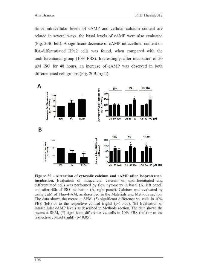
Ana Branco PhD Thesis|2012
106
Since intracellular levels of cAMP and cellular calcium content are
related in several ways, the basal levels of cAMP were also evaluated
(Fig. 20B, left). A significant decrease of cAMP intracellular content on
RA-differentiated H9c2 cells was found, when compared with the
undifferentiated group (10% FBS). Interestingly, after incubation of 50
µM ISO for 48 hours, an increase of cAMP was observed in both
differentiated cell groups (Fig. 20B, right).
Figure 20 - Alteration of cytosolic calcium and cAMP after Isoproterenol incubation. Evaluation of intracellular calcium on undifferentiated and differentiated cells was performed by flow cytometry in basal (A, left panel) and after 48h of ISO incubation (A, right panel). Calcium was evaluated by using 2µM of Fluo-4-AM, as described in the Materials and Methods section. The data shows the means ± SEM, (*) significant difference vs. cells in 10% FBS (left) or to the respective control (right) (p< 0.05). (B) Evaluation of intracellular cAMP levels as described in Methods section. The data shows the means ± SEM, (*) significant difference vs. cells in 10% FBS (left) or to the respective control (right) (p< 0.05).
A
B
Results
107
3.2.3.4 Isoproterenol toxicity involves loss of mitochondrial
respiratory complexes, depolarizes mitochondria and
increases mitochondrial superoxide anion in RA-
differentiated cells
We have identified differentiation-specific protein content related with
cell survival mechanisms including VDAC, Bax and Bcl-2 which control
the apoptotic process at the mitochondrial level [198]. The next step was
to determine if ISO toxicity also results in mitochondrial alterations in
the different groups of cells, including loss of proteins involved in
oxidative phosphorylation, mitochondrial depolarization and oxidative
stress.
To investigate this, we initially used an antibody cocktail directed at
several proteins involved in energy production including ATP synthase
subunit alpha, NADH dehydrogenase (ubiquinone) 1 beta subcomplex
subunit 8 (NDUFB8), a subunit from mitochondrial complex I and a 30
kDa subunit of mitochondrial complex II (Fig. 21). A significant
decrease in complex II 30kDa subunit and in the nuclear-encoded
NDUFB8 subunit in RA-differentiated H9c2 cells after treatment with
ISO were found (Fig. 21B and 21C). Although no differences were
found in undifferentiated cells or differentiated cells in absence of RA, it
is noteworthy that a tendency for an increase in all three subunits was
found in the former group, with the opposite being present in the latter,
except for the ATP synthase subunit investigated (Fig. 21A).

Ana Branco PhD Thesis|2012
108
Figure 21 - Evaluation of mitochondrial alterations after ISO treatment. Mitochondrial oxidative phosphorylation components were identified by Western Blotting after undifferentiated and H9c2 differentiated cells (in presence and in absence of RA) treated with 50 and 100 µM of ISO. The antibodies used detected the proteins NDUFB8 (subunit of complex I), 30kDa subunit (complex II) and ATP synthase subunit alpha. Ponceau labeling was used as loading control. The results are representative of the means ± SEM of 4 separate assays. (*) Significant different to the respective control (p< 0.05).
We next investigated mitochondrial membrane potential by using the
fluorescent probe TMRM under both basal conditions (no ISO treatment,
Fig. 22A, left) as after ISO treatment (Fig. 22A, right). No statistically
significant alterations in basal mitochondrial membrane potential were
found between groups when cell TMRM fluorescence was measured by
flow cytometry. When differentiated H9c2 cells were exposed to the
lower concentration of ISO, increased TMRM fluorescence was
measured, which may result from increased mitochondrial
transmembrane electric potential when comparing to untreated cells.
C
A
Su 1 1%
B
D

Results
109
When exposed to the higher ISO concentration, RA-differentiated cells
suffered a large decrease of mitochondrial TMRM accumulation.
Mitochondrial superoxide anion production that escapes antioxidant
scavenging was detected by using the fluorescent probe MitoSox. We
found an increase in mitochondrial superoxide anion after cellular
differentiation into a cardiac-like phenotype (Fig. 22B, left). Also, the
RA-treated group was the only one where ISO treatment caused a further
increase in MitoSox fluorescence, suggesting increased superoxide anion
(Fig. 22B, right). The increase in cell fluorescence after ISO treatment
found in the other two groups was not statistically significant. A positive
control to investigate cell loading conditions was performed by using
rotenone, which stimulates anion production.
Figure 22 - Evaluation of mitochondria polarization and superoxide anion in H9c2 cells. Quantitative evaluation of TMRM (A) and MitoSox-labeling (B), as measured by flow cytometry. Undifferentiated and differentiated cells in the presence or absence of RA were treated with ISO for 48 hours, as described in Methods section. Data are means of 3 (for TMRM-labeling assay) or 5 (for MitoSox-labeling assay) independent experiments, plus SEM. (*) p< 0.05 vs. 10% FBS cells (left panels) or respective controls (right panels).
A
B
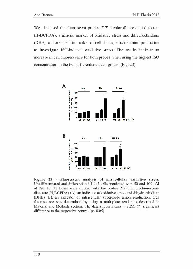
Ana Branco PhD Thesis|2012
110
We also used the fluorescent probes 2',7'-dichlorofluorescein-diacetate
(H2DCFDA), a general marker of oxidative stress and dihydroethidium
(DHE), a more specific marker of cellular superoxide anion production
to investigate ISO-induced oxidative stress. The results indicate an
increase in cell fluorescence for both probes when using the highest ISO
concentration in the two differentiated cell groups (Fig. 23)
Figure 23 - Fluorescent analysis of intracellular oxidative stress. Undifferentiated and differentiated H9c2 cells incubated with 50 and 100 µM of ISO for 48 hours were stained with the probes 2',7'-dichlorofluorescein-diacetate (H2DCFDA) (A), an indicator of oxidative stress and dihydroethidium (DHE) (B), an indicator of intracellular superoxide anion production. Cell fluorescence was determined by using a multiplate reader as described in Material and Methods section. The data shows means ± SEM, (*) significant difference to the respective control (p< 0.05).
A
B

Results
111
3.2.3.5 Cell Differentiation Impacts Isoproterenol–induced
Alterations in Survival/Death Signaling Pathways
Mitochondrial alterations, including increased macromolecular oxidative
stress can trigger cell death [200] or induce protective pathways in order
to rescue cells. We analyzed three proteins that have an important role
on mitochondrial survival/death decisions, including SOD2, the
mitochondrial manganese form of the antioxidant enzyme superoxide
dismutase, cyclophilin F, a matrix chaperone involved in the
mitochondrial permeability transition [201] and Bcl-xL, a Bcl-2 family
protein, which inhibits mitochondrial-mediated cell death [202].
By using Western Blotting, we have previously demonstrated a higher
content of both Bcl-xL and SOD2 in differentiated cells (Fig 11). We
found also that cyclophilin F content is similar in all three differentiation
groups (Fig. 11). Our present data shows that ISO (50 and 100 µM),
when incubated with H9c2 cells in different stages of differentiation
causes an up-regulation of all three proteins in the undifferentiated cell
group, while the content in Bcl-xL decreases in the RA-differentiated
group (Fig. 24 A-C).
The intracellular content of the multi-role transcription factor p53 was
also investigated after incubation with ISO (Fig. 24D), since p53 is also
involved in mitochondrial cell death signaling [203, 204]. Not
unexpectedly, the highest ISO concentration used caused a statistically
significant increase only in the differentiated cell groups. No basal
alterations existed when comparing the three cell groups.
P66Shc is an adapter protein considered as a molecular sensor for
cellular stress. Phosphorylation of p66Shc on serine 36 by protein kinase
C increases during cellular stress and results in amplifying mitochondrial
oxidative stress and apoptotic signaling [205]. With this in mind, we
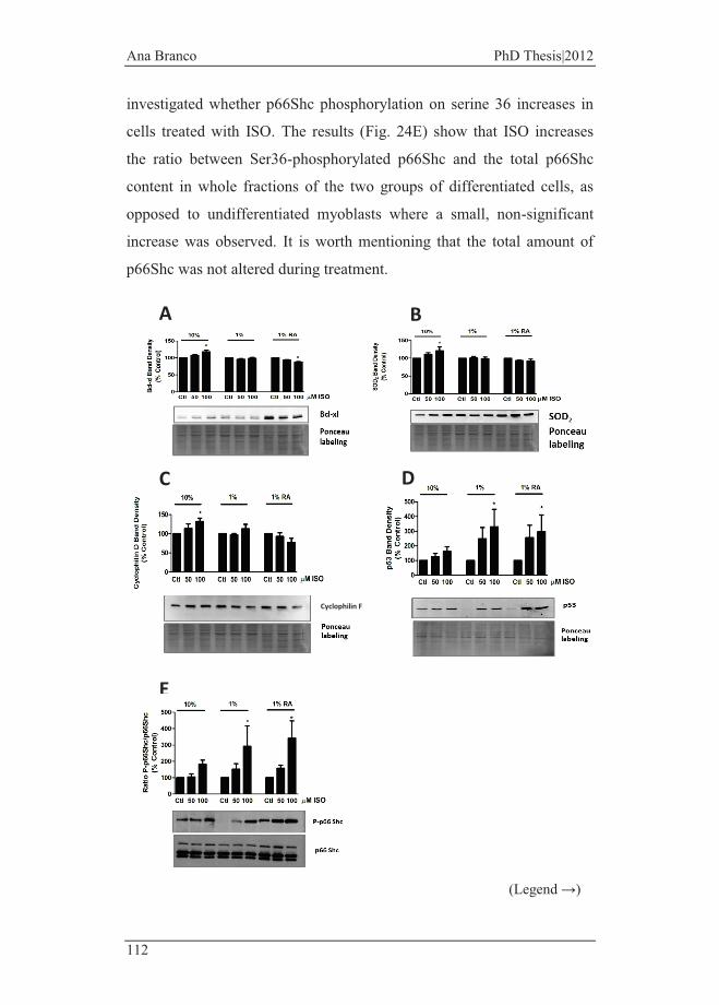
Ana Branco PhD Thesis|2012
112
investigated whether p66Shc phosphorylation on serine 36 increases in
cells treated with ISO. The results (Fig. 24E) show that ISO increases
the ratio between Ser36-phosphorylated p66Shc and the total p66Shc
content in whole fractions of the two groups of differentiated cells, as
opposed to undifferentiated myoblasts where a small, non-significant
increase was observed. It is worth mentioning that the total amount of
p66Shc was not altered during treatment.
(Legend →)
B
E
D C
A
Cyclophilin F
Results
113
Figure 24 - Isoproterenol induces stress responses in H9c2 cells. Bcl-xL (A), SOD2 (B), cyclophilin F (C), p53 (D) and total and phosphorylated-p66Shc (E) proteins content were measured by Western Blotting (see Materials and Methods) and identified as 30, 25, 22, 53 and 66 kDa bands, respectively. H9c2 cells in the different treatment groups were incubated with ISO for 48hours and total cellular extracts were collected. Ponceau labeling and non-phosphorylated p66Shc labeling represent the loading control and were used to normalize data. Data represent the mean±SEM of 5 independent experiments (*) P<0.05 versus respective control for the same differentiation group.
3.2.3.6 p53 Inhibition and Antioxidant Supplementation
Have Different Effects on ISO Cytotoxicity on
Undifferentiated vs. Differentiated H9c2 Cells
Since p53 is increased after ISO treatment in the two differentiated cell
groups (Fig. 24D), the question was whether p53 inhibition by using
pifithrin-alpha [206], prior to ISO treatment, prevented cell death. When
pre-incubated with pifithrin-alpha, ISO toxicity was prevented in the two
differentiated cell groups (Fig. 25A, middle and right panel). In order to
confirm possible pifithrin-alpha effects on ISO incubation with
undifferentiated cells, we extended ISO incubation to 72 hours in order
to increase cytoxicity. This was due because in normal conditions with
no inhibitor, no loss of cells was observed for shorter incubation time
points of incubation. Interestingly, the addition of pifithrin-alpha (pre-
incubated for 30 minutes, 30 µM) previous to ISO increased cell loss
induced by the latter.
We have also investigated the role of oxidative stress on ISO-induced
toxicity on H9c2 cells in different stages of differentiation. H9c2 cells in
the undifferentiated state were incubated with ISO for 96 hours, while
differentiated cells were incubated for 48 hours. N-acetylcysteine
(NAC), a precursor of GSH [207] was pre-incubated for 2 hours (100
µM) with cells before ISO treatment. The anti-oxidant prevented the loss
of cells on both groups of differentiated cells but not on undifferentiated

Ana Branco PhD Thesis|2012
114
myoblasts, although this last group was incubated for longer periods
(Fig. 25B).
Figure 25 - p53 and oxidative stress involvement during ISO toxicity on H9c2 cells. The sulforhodamine B assay was performed in investigate if the p53 inhibitor pifithrin-alpha (A) or the antioxidant NAC (B) prevent H9c2 cell death caused by ISO. Undifferentiated myoblasts were incubated with ISO for 3 (A) or 4 (B) days, while differentiated cells (in presence or absence of RA) were incubated with ISO for 48 hours. Cells were pre-incubated for 30 minutes with 30 µM of pifithrin alpha or for 2 hours with 100 µM NAC. Data represent the mean ± SEM of 4 (for NAC) and 5 (for pifithrin-alpha) independent experiments (*) P<0.05 undifferentiated cells vs. the same concentration used on differentiated cells.
3.2.3.7 Inhibition of PI3K/Akt pathway increases ISO
cytotoxicity in all differentiation and non-
differentiation groups
To evaluate the significance of differential PI3K/Akt signaling pathway
on ISO-induced toxicity in the three groups of H9c2 cells, intracellular
Akt content and its phosphorylated forms were evaluated by Western
Blotting. The results showed no differences on total Akt protein between
A
B
Results
115
the three experimental groups (Fig. 26A). However, the serine 473-
phosphorylated (active) form of Akt was found increased in RA-
differentiated cells when incubated with 100µM of ISO. The role of
PI3K/Akt-activated pathway on ISO-induced cell death was further
confirmed by the cytotoxicity SRB assay. We incubated the 3
experimental groups with the specific inhibitor of the PI3K/Akt
pathway, LY294002 [208] for 1 hour prior to ISO treatment. The
inhibition of PI3K/Akt pathway by LY294002 on H9c2 cells increased
the toxicity of ISO in all three groups (Fig. 26C and 26D) being evident
for lower periods of incubation (48 hours for the undifferentiated group
and 24 hours for the two differentiated groups)

Ana Branco PhD Thesis|2012
116
(Legend →)
A
C
B
D

Results
117
Figure 26 - PI3K/Akt pathway role on ISO-induced toxicity. (A) H9c2 undifferentiated and differentiated cells were incubated with ISO and samples were collected as described in Materials and Methods section. Total Akt and the phosphorylated form in serine 437 were identified by Western Blotting. Ponceau labeling was used as loading control. Data represent the mean±SEM of 4 independent experiments (*) P<0.05 versus respective control for the same differentiation state group. (C) Undifferentiated and differentiated H9c2 cells were pre-incubated with the PI3K/Akt pathway inhibitor LY294002 1 hour prior the addiction of 50 or 100µM of ISO. Differentiated cells number was analyzed 24 hours later and undifferentiated cells were analyzed 48 hours after exposure to ISO, by using the SRB assay method. Data represent the mean±SEM of 4 independent experiments (*) P<0.05 of undifferentiated cells vs. the same concentration used on differentiated cells (D) Microscopy images of differentiated H9c2 cells in the presence and in absence of RA incubated with ISO after pre-incubation with LY294002 by using the same protocol as in (C). Preparations were incubated with Mitotracker Red (to label mitochondria) and DAPI (to label nuclei) and images were obtained by confocal microscopy. White bar corresponds to 100µm.
3.2.3.8 Activation of Caspase 3 During ISO-induced H9c2
Cell Death.
The above results showed that toxic concentrations of ISO induce
poly(ADP-ribose) polymerase (PARP) cleavage which is a specific
marker of cell death (Fig. 19F). Since PARP is a target for caspase 3
[209], we confirmed the involvement of this caspase by evaluating the
content of the active form (Fig. 27A) and activity (Fig. 27B). For the
time point measured (48 hours of ISO treatment), a statistically
significant increase in caspase 3 active fragment was detected in cells
differentiated with serum reduction only. No differences were found in
the two other groups (Fig. 27A). In accordance, caspase-3-like activity
was significantly increased in the group differentiated with 1% serum
alone, although a non-significant increase was also observed in the RA-
differentiated group. Finally, we used the pan-caspase inhibitor z-vad-
fmk to inhibit the loss of cells caused by ISO. In this case, only the two
differentiated groups were used since previous results showed alterations

Ana Branco PhD Thesis|2012
118
on caspase-3 active form only in these cells. With surprise, z-vad-fmk
was not able to inhibit ISO-induced loss of cells (Fig. 27C).
Figure 27 - Caspase 3-like activity in H9c2 cells exposed to ISO and effect of caspase inhibition. (A) The full length caspase 3 and caspase 3-resulting fragments were determining by Western Blotting analysis and identified as bands with 35 and 19/17 kDa, respectively. Ponceau labeling was used as loading control and to normalize the results of cleaved-caspase 3. Data represent the mean±SEM of 3 independent experiments (*) p<0.05 versus respective control for the same differentiation state group. (B) By using a colorimetric assay, undifferentiated and differentiated H9c2 cells exposed to ISO were analyzed in order to measure caspase 3-like activity. Data represent the mean ± SEM of 4 independent experiments (*) p<0.05 versus respective control for the same differentiation state group. (C) Differentiated H9c2 cells
A
B
C

Results
119
without RA (left panel) or with RA (right panel), were pre-incubated with the pan-caspase inhibitor z-vad-fmk 1 hour prior to the treatment with 100µM of ISO and cell mass was measured by using the SRB method. The results were compared with a preparation in which no inhibitor/ISO was added. Data represent the mean±SEM of 3 independent experiments (*) P<0.05 vs. control.
3.2.4 Discussion
Several in vitro and in vivo models [163] showed that ISO-induced β-
adrenergic overload leads to cardiac cell dysfunction being a good
example of chemically-induced heart toxicity and failure. The use of the
H9c2 cell model in different differentiation stages also allows
elucidating cell signaling mechanisms that underlie possible altered
susceptibility of the developing heart to β-adrenergic agonists.
In this work, it is demonstrated that differentiated H9c2 cells are more
susceptible to the toxic effects of supra-physiological concentrations of
the β-adrenergic agonist ISO than non-differentiated myoblasts (Fig. 15).
By using 100 and 150 μM of ISO, a significant loss of differentiated
cells (with or without RA) occurred after ISO treatment while
undifferentiated myoblasts were not affected. An interesting study in
neonatal and adult rat cardiomyocytes demonstrated that stimulation
with 2 μM of NE leads to hypertrophic responses while a higher dose
(100 μM) induces an apoptotic response [210].
Isoproterenol-induced selective toxicity on differentiated cells was
further confirmed by multiple end-points including increased apoptotic
chromatin condensation (Fig. 15E) and by mitochondrial membrane
depolarization (Fig. 16). Cardiac troponin T (TnT) has been reported as
a specific biomarker for cardiac tissue, being used to detect myocardial
injury in a variety of experimental settings [211]. In vivo, degradation of
the contractile structure during cardiac pathologies also involves
degradation of cardiac troponin T, which allows it to be one possible
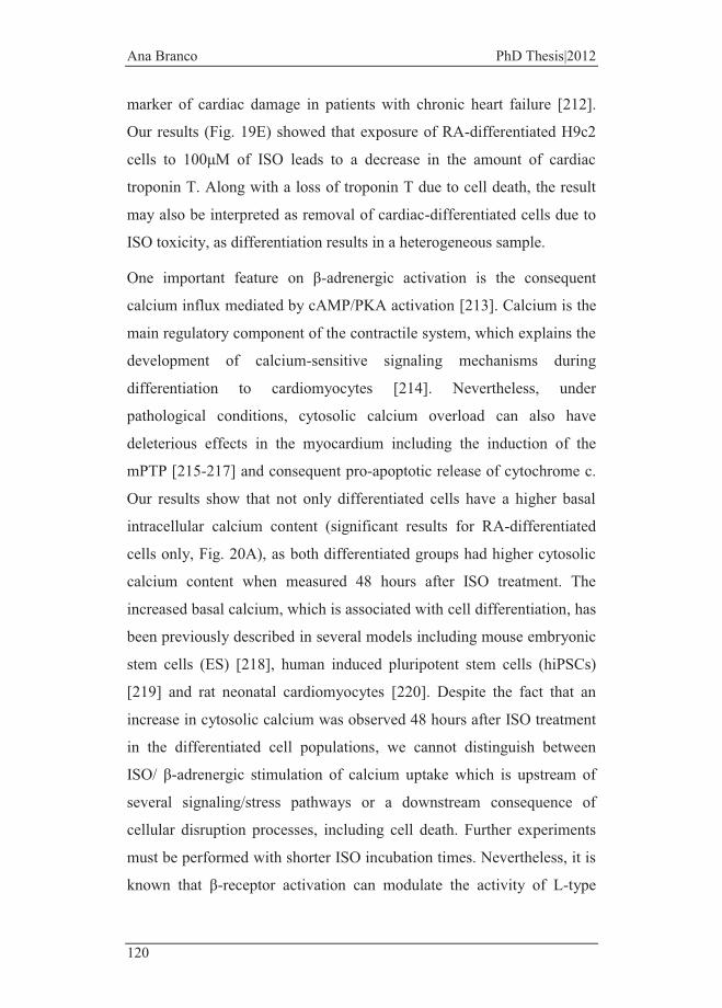
Ana Branco PhD Thesis|2012
120
marker of cardiac damage in patients with chronic heart failure [212].
Our results (Fig. 19E) showed that exposure of RA-differentiated H9c2
cells to 100μM of ISO leads to a decrease in the amount of cardiac
troponin T. Along with a loss of troponin T due to cell death, the result
may also be interpreted as removal of cardiac-differentiated cells due to
ISO toxicity, as differentiation results in a heterogeneous sample.
One important feature on β-adrenergic activation is the consequent
calcium influx mediated by cAMP/PKA activation [213]. Calcium is the
main regulatory component of the contractile system, which explains the
development of calcium-sensitive signaling mechanisms during
differentiation to cardiomyocytes [214]. Nevertheless, under
pathological conditions, cytosolic calcium overload can also have
deleterious effects in the myocardium including the induction of the
mPTP [215-217] and consequent pro-apoptotic release of cytochrome c.
Our results show that not only differentiated cells have a higher basal
intracellular calcium content (significant results for RA-differentiated
cells only, Fig. 20A), as both differentiated groups had higher cytosolic
calcium content when measured 48 hours after ISO treatment. The
increased basal calcium, which is associated with cell differentiation, has
been previously described in several models including mouse embryonic
stem cells (ES) [218], human induced pluripotent stem cells (hiPSCs)
[219] and rat neonatal cardiomyocytes [220]. Despite the fact that an
increase in cytosolic calcium was observed 48 hours after ISO treatment
in the differentiated cell populations, we cannot distinguish between
ISO/ β-adrenergic stimulation of calcium uptake which is upstream of
several signaling/stress pathways or a downstream consequence of
cellular disruption processes, including cell death. Further experiments
must be performed with shorter ISO incubation times. Nevertheless, it is
known that β-receptor activation can modulate the activity of L-type
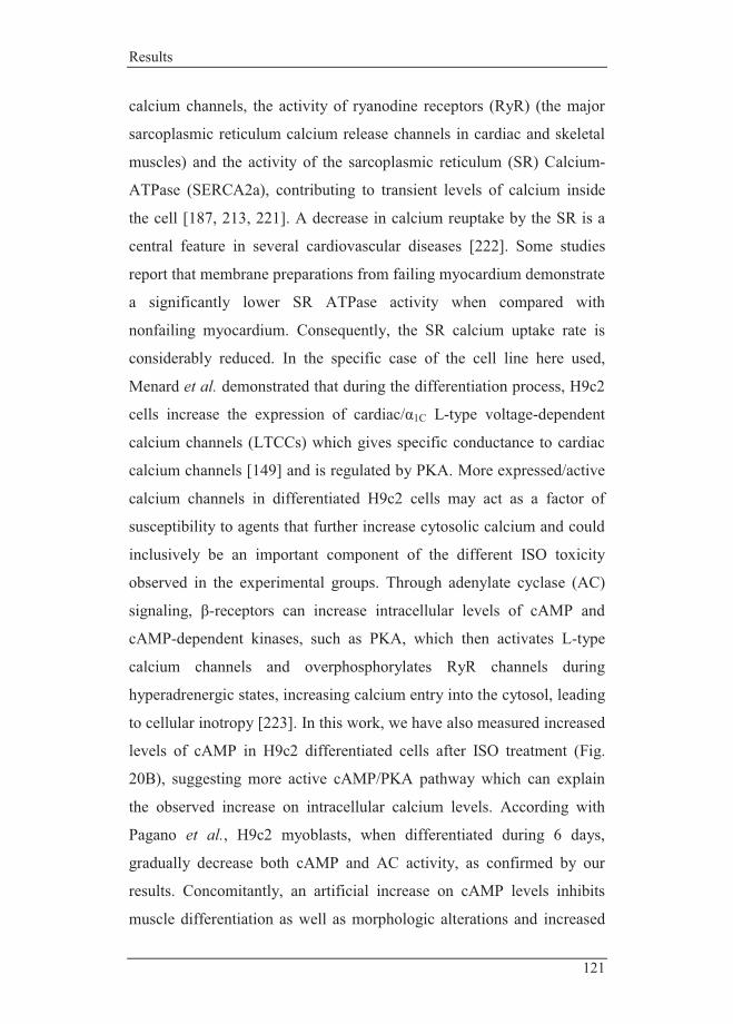
Results
121
calcium channels, the activity of ryanodine receptors (RyR) (the major
sarcoplasmic reticulum calcium release channels in cardiac and skeletal
muscles) and the activity of the sarcoplasmic reticulum (SR) Calcium-
ATPase (SERCA2a), contributing to transient levels of calcium inside
the cell [187, 213, 221]. A decrease in calcium reuptake by the SR is a
central feature in several cardiovascular diseases [222]. Some studies
report that membrane preparations from failing myocardium demonstrate
a significantly lower SR ATPase activity when compared with
nonfailing myocardium. Consequently, the SR calcium uptake rate is
considerably reduced. In the specific case of the cell line here used,
Menard et al. demonstrated that during the differentiation process, H9c2
cells increase the expression of cardiac/α1C L-type voltage-dependent
calcium channels (LTCCs) which gives specific conductance to cardiac
calcium channels [149] and is regulated by PKA. More expressed/active
calcium channels in differentiated H9c2 cells may act as a factor of
susceptibility to agents that further increase cytosolic calcium and could
inclusively be an important component of the different ISO toxicity
observed in the experimental groups. Through adenylate cyclase (AC)
signaling, β-receptors can increase intracellular levels of cAMP and
cAMP-dependent kinases, such as PKA, which then activates L-type
calcium channels and overphosphorylates RyR channels during
hyperadrenergic states, increasing calcium entry into the cytosol, leading
to cellular inotropy [223]. In this work, we have also measured increased
levels of cAMP in H9c2 differentiated cells after ISO treatment (Fig.
20B), suggesting more active cAMP/PKA pathway which can explain
the observed increase on intracellular calcium levels. According with
Pagano et al., H9c2 myoblasts, when differentiated during 6 days,
gradually decrease both cAMP and AC activity, as confirmed by our
results. Concomitantly, an artificial increase on cAMP levels inhibits
muscle differentiation as well as morphologic alterations and increased

Ana Branco PhD Thesis|2012
122
myogenin expression [224]. This phenomenon is still controversial but
some studies associate this with an increase of PKA activity (stimulated
by cAMP) which modulates MyoD effects on the transcription of
myogenin and other differentiation specific proteins [224] preventing
myoblast differentiation. [225]. Since mitochondria act as a calcium
buffer in cardiac cells sequestering rapidly that cation during cytosolic
calcium transients, cAMP/PKA hyperactivation can have deleterious
effects on its function. [226]. Under normal conditions, calcium
stimulates [227] mitochondrial metabolism leading to enhanced energy
production [227]. However, calcium overload in mitochondria leads to
organelle dysfunction, including formation of the mPTP and cell death
[228]. It is likely that increased mitochondrial superoxide anion
generation (Fig. 22B), decreased mitochondrial membrane potential (Fig.
16 and 22A) and decreased content in two oxidative phosphorylation-
related proteins (Fig. 21) can be associated with excessive calcium
accumulation after ISO treatment or be a consequence of the disruption
of different intra-cellular structures by oxidative stress, which we also
confirmed in the entire cell by using two fluorescent probes (Fig. 23). It
is noteworthy that most of these alterations were found in the RA-
differentiated group only, confirming that cell populations which tend to
have a cardiac-like phenotype are more susceptible to ISO-induced
mitochondrial alterations. Two other results are noteworthy: a) the
intermediate concentration of ISO (50 μM) results in what appears to be
mitochondrial hyperpolarization (higher TMRM accumulation) in the
two differentiated groups and b) higher mitochondrial superoxide anion
was detected in RA-differentiated cells under basal conditions. About the
former result, although we do not have evidence to support this, the
presumptive hyperpolarization can be related with mitochondrial
alterations that result in a temporary increase in mitochondrial
membrane potential during the initial stages of cell death. As for the
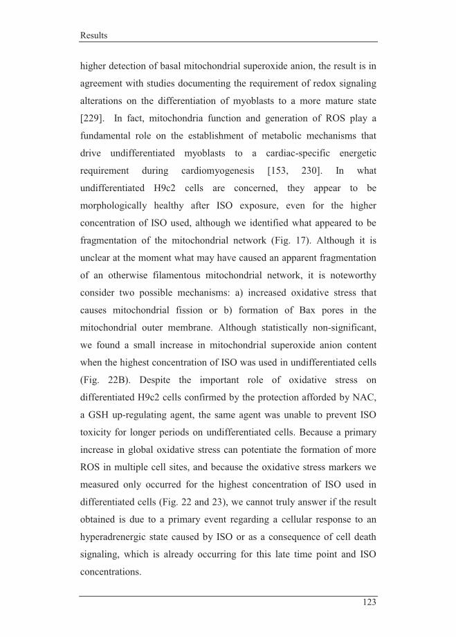
Results
123
higher detection of basal mitochondrial superoxide anion, the result is in
agreement with studies documenting the requirement of redox signaling
alterations on the differentiation of myoblasts to a more mature state
[229]. In fact, mitochondria function and generation of ROS play a
fundamental role on the establishment of metabolic mechanisms that
drive undifferentiated myoblasts to a cardiac-specific energetic
requirement during cardiomyogenesis [153, 230]. In what
undifferentiated H9c2 cells are concerned, they appear to be
morphologically healthy after ISO exposure, even for the higher
concentration of ISO used, although we identified what appeared to be
fragmentation of the mitochondrial network (Fig. 17). Although it is
unclear at the moment what may have caused an apparent fragmentation
of an otherwise filamentous mitochondrial network, it is noteworthy
consider two possible mechanisms: a) increased oxidative stress that
causes mitochondrial fission or b) formation of Bax pores in the
mitochondrial outer membrane. Although statistically non-significant,
we found a small increase in mitochondrial superoxide anion content
when the highest concentration of ISO was used in undifferentiated cells
(Fig. 22B). Despite the important role of oxidative stress on
differentiated H9c2 cells confirmed by the protection afforded by NAC,
a GSH up-regulating agent, the same agent was unable to prevent ISO
toxicity for longer periods on undifferentiated cells. Because a primary
increase in global oxidative stress can potentiate the formation of more
ROS in multiple cell sites, and because the oxidative stress markers we
measured only occurred for the highest concentration of ISO used in
differentiated cells (Fig. 22 and 23), we cannot truly answer if the result
obtained is due to a primary event regarding a cellular response to an
hyperadrenergic state caused by ISO or as a consequence of cell death
signaling, which is already occurring for this late time point and ISO
concentrations.
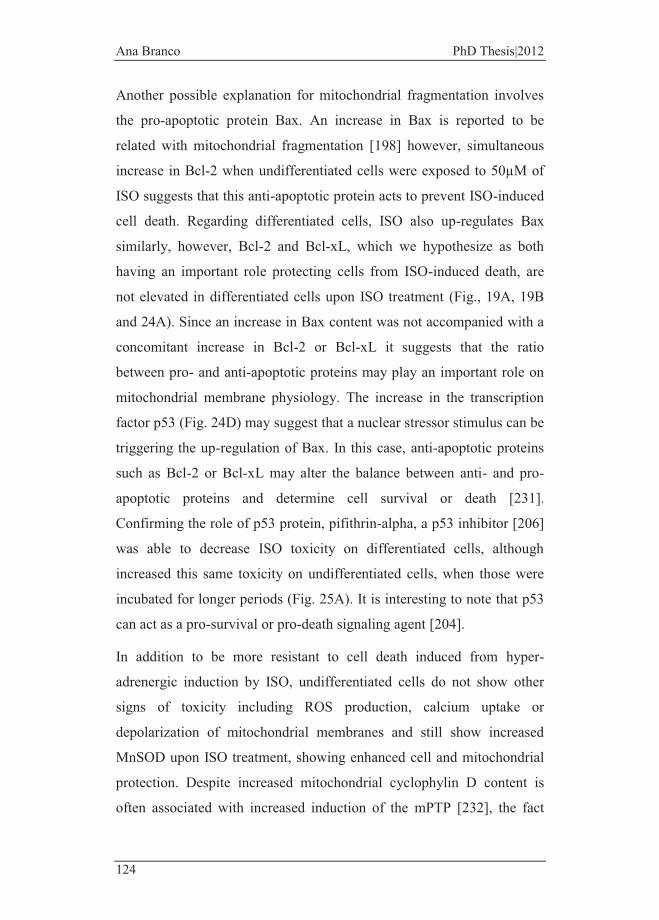
Ana Branco PhD Thesis|2012
124
Another possible explanation for mitochondrial fragmentation involves
the pro-apoptotic protein Bax. An increase in Bax is reported to be
related with mitochondrial fragmentation [198] however, simultaneous
increase in Bcl-2 when undifferentiated cells were exposed to 50µM of
ISO suggests that this anti-apoptotic protein acts to prevent ISO-induced
cell death. Regarding differentiated cells, ISO also up-regulates Bax
similarly, however, Bcl-2 and Bcl-xL, which we hypothesize as both
having an important role protecting cells from ISO-induced death, are
not elevated in differentiated cells upon ISO treatment (Fig., 19A, 19B
and 24A). Since an increase in Bax content was not accompanied with a
concomitant increase in Bcl-2 or Bcl-xL it suggests that the ratio
between pro- and anti-apoptotic proteins may play an important role on
mitochondrial membrane physiology. The increase in the transcription
factor p53 (Fig. 24D) may suggest that a nuclear stressor stimulus can be
triggering the up-regulation of Bax. In this case, anti-apoptotic proteins
such as Bcl-2 or Bcl-xL may alter the balance between anti- and pro-
apoptotic proteins and determine cell survival or death [231].
Confirming the role of p53 protein, pifithrin-alpha, a p53 inhibitor [206]
was able to decrease ISO toxicity on differentiated cells, although
increased this same toxicity on undifferentiated cells, when those were
incubated for longer periods (Fig. 25A). It is interesting to note that p53
can act as a pro-survival or pro-death signaling agent [204].
In addition to be more resistant to cell death induced from hyper-
adrenergic induction by ISO, undifferentiated cells do not show other
signs of toxicity including ROS production, calcium uptake or
depolarization of mitochondrial membranes and still show increased
MnSOD upon ISO treatment, showing enhanced cell and mitochondrial
protection. Despite increased mitochondrial cyclophylin D content is
often associated with increased induction of the mPTP [232], the fact

Results
125
that this phenomenon only occurred in undifferentiated cells suggests a
toxicity response which, together with others already described, may
have contributed to protect cells (Fig. 24C).
When incubated with ISO, both differentiated H9c2 groups show a
decrease in calcineurin while undifferentiated myoblasts show an
increase (Fig. 19D). Although calcineurin has been reported to be
implicated in a wide range of cellular processes such as cell-cycle
regulation, stress response and apoptosis [233], how calcineurin
contributes to the decision of cellular protection or commitment to cell
death is still largely unknown and debatable [234]. Nevertheless, it has
been reported that transgenic mice expressing a constitutively active
mutant of calcineurin show increased resistance to ischemia-reperfusion-
induced apoptosis [235]. These results suggest that calcineurin signaling
imparts a degree of protection against cell death in the heart, which is
consistent with the results in the present work, in which differentiated
H9c2 cells that are more susceptible to ISO present at the same time
decreased levels of calcineurin. Interestingly, it has been shown that β-
adrenoceptor stimulation produces dephosphorylation of p38-MAPK in
embryonic chick hearts [236], which is in agreement with the decrease in
phosphorylated p38-MAPK after ISO treatment in RA-differentiated
H9c2 cells (Fig. 19C). Further work is still needed to understand the link
between the decreased amount in p38-MAPK and ISO toxicity.
Another stress marker was found to be only increased in differentiated
cells after ISO treatment: p66Shc. Isoproterenol treatment resulted in
increased activation of the Ser36-phosphorylated form of p66Shc (Fig.
24E), a protein that regulates oxidative stress responses and apoptosis
[237], but only in differentiated cells. Activation of this signaling
pathway has been described for several toxicants [238] and is considered
a good end-point for intracellular stress responses. The pathway was not

Ana Branco PhD Thesis|2012
126
activated in undifferentiated cells, demonstrating once more the higher
intrinsic signaling protection or that an upstream activating pathway
(e.g., calcium overload) was not activated.
Facing the role of PI3K/Akt signaling pathways on cell survival [239],
we confirmed that inhibition of this pathway by a specific inhibitor leads
to increased cytotoxicity, including on undifferentiated cells (Fig. 26).
This pathway has clinical interest as it has also the capacity to inhibit
pro-apoptotic Bcl-2 family members such as Bax and Bad, with several
studies reporting a functional cooperation between both proteins. PI3K
inhibition on differentiated cells has deleterious effects, suggesting the
importance of the activation of this pathway on cell survival. However,
in the other hand, differentiated cells do not have increased levels of
anti-apoptotic proteins from the Bcl-2 family such as Bcl-xL and Bcl-2
which can compromise the cooperative action between both PI3K/Akt
and Bcl-xL/Bcl-2 pathways.
Finally, we measured increased caspase 3 activation in differentiated
cells after ISO treatment (Fig. 27), which demonstrates that apoptotic
pathways are activated. Furthermore, its substrate, PARP, showed a
decreased content of its full length form (Fig. 19F). Nevertheless, a pan-
caspase inhibitor had no effect on ISO toxicity, suggesting that loss of
cells after ISO treatment may occur through another pathway that
subsists even after inhibition of caspases. Cellular death is a redundant
event that includes several independent signaling pathways, which can
occur simultaneously [240]. One possibility to explain the fact that z-
vad-fmk did not protect from death in differentiated H9c2 cells is that
mitochondrial disruption may result into the release of pro-apoptotic
caspase-independent factors including the AIF. It was reported that
staurosporin-induced H9c2 death, although involving caspase activation,
was not inhibited by z-vad-fmk [241]. In another study, Youn et al.
Results
127
showed that doxorubicin-induced toxicity on H9c2 cells was not
prevented by pretreatment with the caspase inhibitor z-vad-fmk, in
contrast with cell death induced by H2O2 treatment [242].
It is noteworthy that one possibility for the results would be binding of
ISO to serum, which explain why cells in 10% FBS are less susceptible
to ISO. Data in Fig. 18 suggest that differential ISO serum binding does
not explain the results obtained, although the data may suggest that a
small effect due to ISO bonding to serum may exist. Nevertheless, one
has to have caution when analyzing this data and possible variations.
The 48 hours period in which ISO was incubated with cells, may be
enough for undifferentiated cells to start differentiating in 1% FBS
media, or to differentiated cells (in 1% FBS) to re-start proliferating
when exposed to a higher FBS concentration. In fact, data present in Fig.
9B show that 1% FBS-differentiated H9c2 cells re-started proliferating
earlier after being placed in culture media with 10% FBS, showing
biochemical alterations that may alter the susceptibility to ISO.
In conclusion, the present research describes a differential susceptibility
of H9c2 myoblasts in different differentiation states to ISO toxicity and
confirms that hyperadrenergic states have different outcomes on
undifferentiated and differentiated muscle cells. Our findings show that
the maturity of H9c2 cardiac cells is critical on their responses to β-
adrenergic agonists. Identification of the molecular mechanisms that
mediate stress/survival/death responses in muscle cells in different
developmental stages is critical to understand how cardiac resistance to
β-adrenergic stimulation varies from the womb to adulthood. In fact,
several pieces of evidence demonstrate in vivo distinct responses to
catecholamine/ISO in rats of different ages or even during heart
development in utero [126, 127].


Conclusions


Conclusions
131
4. Conclusions
Sympathetic activation has not only an important role on the
compensatory regulation of the contractility activity but is also
deleterious when chronically activated since it contributes to the
progressive deterioration of the myocardial structure and function. It is
well established that apoptosis of cardiomyocytes contributes to a
complex myocardium remodeling process [243]. Stress-activated
signaling pathways are mediated by catecholamine-induced activation of
G-protein-coupled receptors [244]. Manipulation of adrenergic signaling
using transgenic mouse models and β-blockers has also been
contributing to additional knowledge on its role in cardiac remodeling
and apoptosis [117]. The suppression of cell death pathways to control
myocyte loss represents an ideal strategy to prevent many heart diseases.
In this context, we proposed to study the involvement of β-AR signaling
on cardiac cell death during different stages of the differentiation process
in H9c2 cells.
One major objective in this thesis regards the potential use of H9c2 cells
as a surrogate for cardiac cells during toxicological studies (which
becomes more important as the use of animals in experimental research
is being limited) due to its ability to differentiate into cardiac-like cells
with molecular and morphological features of a cardiomyocyte. Further
characterization on the transcriptome of H9c2 cell upon differentiation
process showed that expression of genes related with proliferative
activity were tendentiously repressed on differentiated cells, and with
RA-differentiated cells present increased expression of genes related
with the differentiation process such as the oxidative stress growth
inhibitor 1, the cyclin-dependent kinase inhibitor 2B, the FasL, calcium
transporters (such as the sarcoplasmic reticulum calcium ATPase),
cardiac sarcomeric proteins and genes with related mitochondrial energy
Ana Branco PhD Thesis|2012
132
production such as mitochondrial creatine kinase, oxidative
phosphorylation subunits, as well as uncoupling proteins. We also
performed genotyping studies with isolated cardiomyocytes and with
HL-1 cell line in which we found neonatal cardiomyocytes upregulation
on yearly development markers such as the Gata6 gene.
The results from this thesis suggest that myoblast differentiation, for
example during fetal development, results into increased toxicity to β-
adrenergic agonists. Our work confirms that embryonic tissue appear to
be more resistant to ISO than adult cardiac cells. Some hypotheses have
been forwarded to explain the differences: one regards the intrinsic
cellular defenses and, second may involve the capacity of each cell
group to efficiently respond to a deleterious stimulus. In this context,
basal content on some anti-apoptotic proteins can represent an
advantage. Also, the fact that undifferentiated cells are metabolic more
active [191], with possibly autophagic processes being constitutively
active than in differentiated cells, may also contribute to a faster and
more positive response to an insult.
Based on the fact that ISO impacts and triggers different mechanisms in
undifferentiated and differentiated H9c2 cells we performed studies on
the β-AR signaling elements to detect downstream events that may
explain the base of such difference. In addition to supplying cellular
energy, mitochondria are also related with calcium homeostasis,
oxidative stress and apoptotic signaling. In this context, we found
mitochondria alterations that can further explain protection mechanisms
increased in undifferentiated cells. We confirmed that the mitochondrial
phosphorylative system appears to be less affected by ISO treatment in
undifferentiated cell, although we have identified early alterations of
mitochondrial network. On the other hand, calcium uptake, ROS
production, loss of the mitochondrial membrane potential, commitment

Conclusions
133
of mitochondrial complexes, decreased superoxide dismutase, decreased
Bcl-2 and Bcl-xL increased Bax, increased cAMP, release of troponin T
(a standard marker for cardiac injury), increased p53 protein and p66Shc,
activation of caspase-3 and cleavage of PARP were some of the
molecular alterations found in differentiated cells in the presence of ISO
(Fig. 28 and 29).
Although many others stress/survival pathways could have been
investigated, it appears clear that cells in an early stage of differentiation
have more intrinsic defenses, which may have evolved from the need to
protect the developing structure in utero from intrinsic and extrinsic
aggressions. It is also interesting to postulate that mitochondrial
maturation during development and remodeling towards a more
oxidative metabolism impacts how toxicants affect cellular viability.
Modulation of oxidative stress and calcium overload by intrinsic
defenses during xenobiotic toxicity is apparently also a life or death
decision to the cell.
The results obtained in this thesis are relevant from a toxicological point
of view, especially when investigating developmental cardiovascular
toxicology.
4.1 Final Remarks: Based on the results presented in this study, we propose to highlight the
following main conclusions (see Fig. 28, 29 for a summary of results):
- The differentiation of H9c2 cells led to a transcriptional up-
regulation of genes involved in cardiac differentiation,
diminished cellular proliferative rate and induced the formation
of multinucleated cells, which demonstrates the validity of using
this cell system.
Ana Branco PhD Thesis|2012
134
- Differentiated H9c2 cells exposed to ISO present increased
calcium uptake and cAMP levels, oxidative stress, depolarization
of the mitochondrial membrane potential, deterioration or loss of
mitochondrial complexes, decreased Bcl-2 and Bcl-xL, increased
Bax, released of troponin T, increased p53 protein and p66Shc,
activation of caspase-3 and PARP cleavage.
- Undifferentiated H9c2 cells exposed to ISO present
mitochondrial network fragmentation, increased Bcl-2 and Bcl-
xL proteins which counteract increased Bax content, as well as
increased superoxide dismutase 2 enzyme.

Conclusions
135
Figure 28 - Schematic representation of β-AR downstream pathways altered by ISO in undifferentiated H9c2 cells. Grey represents non-studied proteins although of importance for the signaling pathway; Green represents up-regulated proteins upon stimulation of β-AR by ISO; Red represents down-regulated proteins upon stimulation of β-AR by ISO; Brown represents protein levels that had no alterations upon stimulation of β-AR by ISO. X represents proteins that are over-expressed after the differentiation process. Abbreviations: ROS (reactive oxygen species), PI3-K (phosphoinositide 3-kinase), Gi (guanine nucleotide-binding protein inhibitory subunit), Gs (guanine nucleotide-binding protein stimulatory subunit), AC (adenylate cyclase), cAMP (cyclic adenosine monophosphate), Ca2+ (calcium), PKA (protein kinase A), p38 (p38 mitogen-activated protein kinases), Bcl-2 (B-cell lymphoma 2), Bcl-xL (B-cell lymphoma-extra-large), Bax (Bcl-2-associated X protein), SOD2 (superoxide dismutase 2), mΨ (mitochondrial membrane potential), mtComplexes (mitochondrial complexes subunits), cyD (cyclophilin F), PARP (cleaved from of poly(ADP-ribose) polymerase-1), LTCC (L-type calcium channel).

Ana Branco PhD Thesis|2012
136
Figure 29 - Schematic representation of β-AR downstream pathways altered by ISO in differentiated H9c2 cells by reducing the concentration of serum in the medium. Grey represents non-studied proteins although of importance for the signaling pathway; Green represents up-regulated proteins upon stimulation of β-AR by ISO; Red represents down-regulated proteins upon stimulation of β-AR by ISO; Brown represents protein levels that had no alterations upon stimulation of β-AR by ISO. X represents proteins that are over-expressed after the differentiation process. Abbreviations: ROS (reactive oxygen species), PI3-K (phosphoinositide 3-kinase), Gi (guanine nucleotide-binding protein inhibitory subunit), Gs (guanine nucleotide-binding protein stimulatory subunit), AC (adenylate cyclase), cAMP (cyclic adenosine monophosphate), Ca2+ (calcium), PKA (protein kinase A), p38 (p38 mitogen-activated protein kinases), Bcl-2 (B-cell lymphoma 2), Bcl-xL (B-cell lymphoma-extra-large), Bax (Bcl-2-associated X protein), SOD2 (superoxide dismutase 2), mΨ (mitochondrial membrane potential), mtComplexes (mitochondrial complexes subunits), cyD (cyclophilin F), PARP (cleaved from of poly(ADP-ribose) polymerase-1), LTCC (L-type calcium channel).
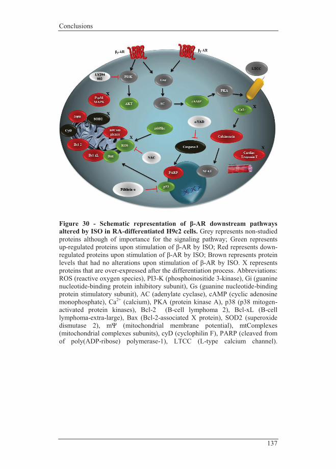
Conclusions
137
Figure 30 - Schematic representation of β-AR downstream pathways altered by ISO in RA-differentiated H9c2 cells. Grey represents non-studied proteins although of importance for the signaling pathway; Green represents up-regulated proteins upon stimulation of β-AR by ISO; Red represents down-regulated proteins upon stimulation of β-AR by ISO; Brown represents protein levels that had no alterations upon stimulation of β-AR by ISO. X represents proteins that are over-expressed after the differentiation process. Abbreviations: ROS (reactive oxygen species), PI3-K (phosphoinositide 3-kinase), Gi (guanine nucleotide-binding protein inhibitory subunit), Gs (guanine nucleotide-binding protein stimulatory subunit), AC (adenylate cyclase), cAMP (cyclic adenosine monophosphate), Ca2+ (calcium), PKA (protein kinase A), p38 (p38 mitogen-activated protein kinases), Bcl-2 (B-cell lymphoma 2), Bcl-xL (B-cell lymphoma-extra-large), Bax (Bcl-2-associated X protein), SOD2 (superoxide dismutase 2), mΨ (mitochondrial membrane potential), mtComplexes (mitochondrial complexes subunits), cyD (cyclophilin F), PARP (cleaved from of poly(ADP-ribose) polymerase-1), LTCC (L-type calcium channel).


Bibliography
g p y

Bibliography
141
5. Bibliography
1. August 30]; Available from: http://www.who.int/mediacentre/factsheets/fs317/en/.
2. Silva, G.V., S. Litovsky, J.A. Assad, A.L. Sousa, B.J. Martin, D. Vela, S.C. Coulter, J. Lin, J. Ober, W.K. Vaughn, R.V. Branco, E.M. Oliveira, R. He, Y.J. Geng, J.T. Willerson, and E.C. Perin, Mesenchymal stem cells differentiate into an endothelial phenotype, enhance vascular density, and improve heart function in a canine chronic ischemia model. Circulation, 2005. 111(2): p. 150-6.
3. Briest, W., A. Holzl, B. Rassler, A. Deten, M. Leicht, H.A. Baba, and H.G. Zimmer, Cardiac remodeling after long term norepinephrine treatment in rats. Cardiovasc Res, 2001. 52(2): p. 265-73.
4. Ren, R., R.H. Oakley, D. Cruz-Topete, and J.A. Cidlowski, Dual role for glucocorticoids in cardiomyocyte hypertrophy and apoptosis. Endocrinology, 2012. 153(11): p. 5346-60.
5. Li, X.M., Y.T. Ma, Y.N. Yang, F. Liu, B.D. Chen, W. Han, J.F. Zhang, and X.M. Gao, Downregulation of survival signalling pathways and increased apoptosis in the transition of pressure overload-induced cardiac hypertrophy to heart failure. Clin Exp Pharmacol Physiol, 2009. 36(11): p. 1054-61.
6. Baune, B.T., Y. Aljeesh, and R. Bender, Factors of non-compliance with the therapeutic regimen among hypertensive men and women: a case-control study to investigate risk factors of stroke. Eur J Epidemiol, 2005. 20(5): p. 411-9.
7. Egido, J.A., O. Castillo, B. Roig, I. Sanz, M.R. Herrero, M.T. Garay, A.M. Garcia, M. Fuentes, and C. Fernandez, Is psycho-physical stress a risk factor for stroke? A case-control study. J Neurol Neurosurg Psychiatry, 2012. 83(11): p. 1104-11.
8. Bunker, S.J., D.M. Colquhoun, M.D. Esler, I.B. Hickie, D. Hunt, V.M. Jelinek, B.F. Oldenburg, H.G. Peach, D. Ruth, C.C. Tennant, and A.M. Tonkin, "Stress" and coronary heart disease: psychosocial risk factors. Med J Aust, 2003. 178(6): p. 272-6.
9. Bachman, E.S., H. Dhillon, C.Y. Zhang, S. Cinti, A.C. Bianco, B.K. Kobilka, and B.B. Lowell, betaAR signaling required for diet-induced thermogenesis and obesity resistance. Science, 2002. 297(5582): p. 843-5.
10. Barrett, A.M., The mobilization of free fatty acids in response to isoprenaline in the rat. Br J Pharmacol Chemother, 1965. 25(2): p. 545-56.
11. Wang, J.C., S.L. Normand, L. Mauri, and R.E. Kuntz, Coronary artery spatial distribution of acute myocardial infarction occlusions. Circulation, 2004. 110(3): p. 278-84.

Ana Branco PhD Thesis|2012
142
12. Diwan, A. and G.W. Dorn, 2nd, Decompensation of cardiac hypertrophy: cellular mechanisms and novel therapeutic targets. Physiology (Bethesda), 2007. 22: p. 56-64.
13. Albert, N.M., C.A. Eastwood, and M.L. Edwards, Evidence-based practice for acute decompensated heart failure. Crit Care Nurse, 2004. 24(6): p. 14-6, 18-24, 26-9; quiz 30-1.
14. Cheng, W., B. Li, J. Kajstura, P. Li, M.S. Wolin, E.H. Sonnenblick, T.H. Hintze, G. Olivetti, and P. Anversa, Stretch-induced programmed myocyte cell death. J Clin Invest, 1995. 96(5): p. 2247-59.
15. Burke, T.A., M.C. Sturkenboom, S.E. Lu, C.E. Wentworth, Y. Lin, and G.G. Rhoads, Discontinuation of antihypertensive drugs among newly diagnosed hypertensive patients in UK general practice. J Hypertens, 2006. 24(6): p. 1193-200.
16. mccarty, J.A.f.m.m.A.j.c.d.g.g.e.r., Catecholamines: Bridging Basic Science with Clinical Medicine, A.i. Pharmacology, Editor 1997.
17. Maruyama, Y., A. Kawano, S. Okamoto, T. Ando, Y. Ishitobi, Y. Tanaka, A. Inoue, J. Imanaga, M. Kanehisa, H. Higuma, T. Ninomiya, J. Tsuru, H. Hanada, and J. Akiyoshi, Differences in salivary alpha-amylase and cortisol responsiveness following exposure to electrical stimulation versus the Trier Social Stress Tests. PLoS One, 2012. 7(7): p. e39375.
18. Ulrich-Lai, Y.M. and J.P. Herman, Neural regulation of endocrine and autonomic stress responses. Nat Rev Neurosci, 2009. 10(6): p. 397-409.
19. Ayala, I., N.F. Martos, G. Silvan, C. Gutierrez-Panizo, J.G. Clavel, and J.C. Illera, Cortisol, adrenocorticotropic hormone, serotonin, adrenaline and noradrenaline serum concentrations in relation to disease and stress in the horse. Res Vet Sci, 2012. 93(1): p. 103-7.
20. Kvetnansky, R., I. Armando, V.K. Weise, C. Holmes, K. Fukuhara, A. Deka-Starosta, I.J. Kopin, and D.S. Goldstein, Plasma dopa responses during stress: dependence on sympathoneural activity and tyrosine hydroxylation. J Pharmacol Exp Ther, 1992. 261(3): p. 899-909.
21. Hill, M.N. and J.G. Tasker, Endocannabinoid signaling, glucocorticoid-mediated negative feedback, and regulation of the hypothalamic-pituitary-adrenal axis. Neuroscience, 2012. 204: p. 5-16.
22. Gouin, J.P., C.S. Carter, H. Pournajafi-Nazarloo, R. Glaser, W.B. Malarkey, T.J. Loving, J. Stowell, and J.K. Kiecolt-Glaser, Marital behavior, oxytocin, vasopressin, and wound healing. Psychoneuroendocrinology, 2010. 35(7): p. 1082-90.
23. Grippo, A.J., D.M. Trahanas, R.R. Zimmerman, 2nd, S.W. Porges, and C.S. Carter, Oxytocin protects against negative behavioral and autonomic consequences of long-term social isolation. Psychoneuroendocrinology, 2009. 34(10): p. 1542-53.

Bibliography
143
24. Hasking, G.J., M.D. Esler, G.L. Jennings, D. Burton, J.A. Johns, and P.I. Korner, Norepinephrine spillover to plasma in patients with congestive heart failure: evidence of increased overall and cardiorenal sympathetic nervous activity. Circulation, 1986. 73(4): p. 615-21.
25. Wang, W., H. Zhang, H. Gao, H. Kubo, R.M. Berretta, X. Chen, and S.R. Houser, {beta}1-Adrenergic receptor activation induces mouse cardiac myocyte death through both L-type calcium channel-dependent and -independent pathways. Am J Physiol Heart Circ Physiol, 2010. 299(2): p. H322-31.
26. Communal, C., K. Singh, D.R. Pimentel, and W.S. Colucci, Norepinephrine stimulates apoptosis in adult rat ventricular myocytes by activation of the beta-adrenergic pathway. Circulation, 1998. 98(13): p. 1329-34.
27. Ikenishi, A., H. Okayama, N. Iwamoto, S. Yoshitome, S. Tane, K. Nakamura, T. Obayashi, T. Hayashi, and T. Takeuchi, Cell cycle regulation in mouse heart during embryonic and postnatal stages. Dev Growth Differ, 2012. 54(8): p. 731-8.
28. Olivetti, G., E. Cigola, R. Maestri, D. Corradi, C. Lagrasta, S.R. Gambert, and P. Anversa, Aging, cardiac hypertrophy and ischemic cardiomyopathy do not affect the proportion of mononucleated and multinucleated myocytes in the human heart. J Mol Cell Cardiol, 1996. 28(7): p. 1463-77.
29. Ahuja, P., P. Sdek, and W.R. MacLellan, Cardiac myocyte cell cycle control in development, disease, and regeneration. Physiol Rev, 2007. 87(2): p. 521-44.
30. Kresh, J.Y. and A. Chopra, Intercellular and extracellular mechanotransduction in cardiac myocytes. Pflugers Arch, 2011. 462(1): p. 75-87.
31. Javadov, S., M. Karmazyn, and N. Escobales, Mitochondrial permeability transition pore opening as a promising therapeutic target in cardiac diseases. J Pharmacol Exp Ther, 2009. 330(3): p. 670-8.
32. Galluzzi, L., O. Kepp, C. Trojel-Hansen, and G. Kroemer, Mitochondrial control of cellular life, stress, and death. Circ Res, 2012. 111(9): p. 1198-207.
33. Kar, R., N. Mishra, P.K. Singha, M.A. Venkatachalam, and P. Saikumar, Mitochondrial remodeling following fission inhibition by 15d-PGJ2 involves molecular changes in mitochondrial fusion protein OPA1. Biochem Biophys Res Commun, 2010. 399(4): p. 548-54.
34. Chen, Y., Y. Liu, and G.W. Dorn, 2nd, Mitochondrial fusion is essential for organelle function and cardiac homeostasis. Circ Res, 2011. 109(12): p. 1327-31.
Ana Branco PhD Thesis|2012
144
35. Chen, L., Q. Gong, J.P. Stice, and A.A. Knowlton, Mitochondrial OPA1, apoptosis, and heart failure. Cardiovasc Res, 2009. 84(1): p. 91-9.
36. Papanicolaou, K.N., R. Kikuchi, G.A. Ngoh, K.A. Coughlan, I. Dominguez, W.C. Stanley, and K. Walsh, Mitofusins 1 and 2 are essential for postnatal metabolic remodeling in heart. Circ Res, 2012. 111(8): p. 1012-26.
37. Whelan, R.S., V. Kaplinskiy, and R.N. Kitsis, Cell death in the pathogenesis of heart disease: mechanisms and significance. Annu Rev Physiol, 2010. 72: p. 19-44.
38. Zeng, W.T., W.Y. Chen, X.Y. Leng, L.L. Tang, X.T. Sun, C.L. Li, and G. Dai, Impairment of cardiac function and remodeling induced by myocardial infarction in rats are attenuated by the nonpeptide angiotensin-(1-7) analog AVE 0991. Cardiovasc Ther, 2012. 30(3): p. 152-61.
39. Yeh, C.H., T.P. Chen, Y.C. Wang, Y.M. Lin, and S.W. Fang, Cardiomyocytic apoptosis limited by bradykinin via restoration of nitric oxide after cardioplegic arrest. J Surg Res, 2010. 163(1): p. e1-9.
40. Huang, C., W. Liu, C.N. Perry, S. Yitzhaki, Y. Lee, H. Yuan, Y.T. Tsukada, A. Hamacher-Brady, R.M. Mentzer, Jr., and R.A. Gottlieb, Autophagy and protein kinase C are required for cardioprotection by sulfaphenazole. Am J Physiol Heart Circ Physiol, 2010. 298(2): p. H570-9.
41. Raben, N., R. Baum, C. Schreiner, S. Takikita, N. Mizushima, E. Ralston, and P. Plotz, When more is less: excess and deficiency of autophagy coexist in skeletal muscle in Pompe disease. Autophagy, 2009. 5(1): p. 111-3.
42. Iyer, S.S., W.P. Pulskens, J.J. Sadler, L.M. Butter, G.J. Teske, T.K. Ulland, S.C. Eisenbarth, S. Florquin, R.A. Flavell, J.C. Leemans, and F.S. Sutterwala, Necrotic cells trigger a sterile inflammatory response through the Nlrp3 inflammasome. Proc Natl Acad Sci U S A, 2009. 106(48): p. 20388-93.
43. Sato, H., I. Shiraishi, T. Takamatsu, and K. Hamaoka, Detection of TUNEL-positive cardiomyocytes and c-kit-positive progenitor cells in children with congenital heart disease. J Mol Cell Cardiol, 2007. 43(3): p. 254-61.
44. Heinke, M.Y., M. Yao, D. Chang, R. Einstein, and C.G. dos Remedios, Apoptosis of ventricular and atrial myocytes from pacing-induced canine heart failure. Cardiovasc Res, 2001. 49(1): p. 127-34.
45. Tang, Y., J. Zheng, Y. Sun, Z. Wu, Z. Liu, and G. Huang, MicroRNA-1 regulates cardiomyocyte apoptosis by targeting Bcl-2. Int Heart J, 2009. 50(3): p. 377-87.

Bibliography
145
46. Ferrandi, C., R. Ballerio, P. Gaillard, C. Giachetti, S. Carboni, P.A. Vitte, J.P. Gotteland, and R. Cirillo, Inhibition of c-Jun N-terminal kinase decreases cardiomyocyte apoptosis and infarct size after myocardial ischemia and reperfusion in anaesthetized rats. Br J Pharmacol, 2004. 142(6): p. 953-60.
47. Kubota, T., C.F. McTiernan, C.S. Frye, S.E. Slawson, B.H. Lemster, A.P. Koretsky, A.J. Demetris, and A.M. Feldman, Dilated cardiomyopathy in transgenic mice with cardiac-specific overexpression of tumor necrosis factor-alpha. Circ Res, 1997. 81(4): p. 627-35.
48. Schutters, K., D.H. Kusters, M.L. Chatrou, T. Montero-Melendez, M. Donners, N.M. Deckers, D.V. Krysko, P. Vandenabeele, M. Perretti, L.J. Schurgers, and C.P. Reutelingsperger, Cell surface-expressed phosphatidylserine as therapeutic target to enhance phagocytosis of apoptotic cells. Cell Death Differ, 2012.
49. Kerr, J.F., A.H. Wyllie, and A.R. Currie, Apoptosis: a basic biological phenomenon with wide-ranging implications in tissue kinetics. Br J Cancer, 1972. 26(4): p. 239-57.
50. Fiandalo, M.V. and N. Kyprianou, Caspase control: protagonists of cancer cell apoptosis. Exp Oncol, 2012. 34(3): p. 165-75.
51. Bhuiyan, M.S. and K. Fukunaga, Inhibition of HtrA2/Omi ameliorates heart dysfunction following ischemia/reperfusion injury in rat heart in vivo. Eur J Pharmacol, 2007. 557(2-3): p. 168-77.
52. Scheubel, R.J., B. Bartling, A. Simm, R.E. Silber, K. Drogaris, D. Darmer, and J. Holtz, Apoptotic pathway activation from mitochondria and death receptors without caspase-3 cleavage in failing human myocardium: fragile balance of myocyte survival? J Am Coll Cardiol, 2002. 39(3): p. 481-8.
53. Longthorne, V.L. and G.T. Williams, Caspase activity is required for commitment to Fas-mediated apoptosis. EMBO J, 1997. 16(13): p. 3805-12.
54. Green, D.R., Apoptotic pathways: the roads to ruin. Cell, 1998. 94(6): p. 695-8.
55. Ferrari, R., T. Bachetti, R. Confortini, C. Opasich, O. Febo, A. Corti, G. Cassani, and O. Visioli, Tumor necrosis factor soluble receptors in patients with various degrees of congestive heart failure. Circulation, 1995. 92(6): p. 1479-86.
56. Garcia-Saez, A.J., The secrets of the Bcl-2 family. Cell Death Differ, 2012. 19(11): p. 1733-40.
57. Halestrap, A.P., G.P. McStay, and S.J. Clarke, The permeability transition pore complex: another view. Biochimie, 2002. 84(2-3): p. 153-66.

Ana Branco PhD Thesis|2012
146
58. Vaux, D.L., Apoptogenic factors released from mitochondria. Biochim Biophys Acta, 2011. 1813(4): p. 546-50.
59. Kroemer, G., L. Galluzzi, and C. Brenner, Mitochondrial membrane permeabilization in cell death. Physiol Rev, 2007. 87(1): p. 99-163.
60. Joza, N., G.Y. Oudit, D. Brown, P. Benit, Z. Kassiri, N. Vahsen, L. Benoit, M.M. Patel, K. Nowikovsky, A. Vassault, P.H. Backx, T. Wada, G. Kroemer, P. Rustin, and J.M. Penninger, Muscle-specific loss of apoptosis-inducing factor leads to mitochondrial dysfunction, skeletal muscle atrophy, and dilated cardiomyopathy. Mol Cell Biol, 2005. 25(23): p. 10261-72.
61. McDermott-Roe, C., J. Ye, R. Ahmed, X.M. Sun, A. Serafin, J. Ware, L. Bottolo, P. Muckett, X. Canas, J. Zhang, G.C. Rowe, R. Buchan, H. Lu, A. Braithwaite, M. Mancini, D. Hauton, R. Marti, E. Garcia-Arumi, N. Hubner, H. Jacob, T. Serikawa, V. Zidek, F. Papousek, F. Kolar, M. Cardona, M. Ruiz-Meana, D. Garcia-Dorado, J.X. Comella, L.E. Felkin, P.J. Barton, Z. Arany, M. Pravenec, E. Petretto, D. Sanchis, and S.A. Cook, Endonuclease G is a novel determinant of cardiac hypertrophy and mitochondrial function. Nature, 2011. 478(7367): p. 114-8.
62. Martinou, J.C. and D.R. Green, Breaking the mitochondrial barrier. Nat Rev Mol Cell Biol, 2001. 2(1): p. 63-7.
63. Wang, C. and R.J. Youle, The role of mitochondria in apoptosis*. Annu Rev Genet, 2009. 43: p. 95-118.
64. Ren, D., H.C. Tu, H. Kim, G.X. Wang, G.R. Bean, O. Takeuchi, J.R. Jeffers, G.P. Zambetti, J.J. Hsieh, and E.H. Cheng, BID, BIM, and PUMA are essential for activation of the BAX- and BAK-dependent cell death program. Science, 2010. 330(6009): p. 1390-3.
65. Smaili, S.S., Y.T. Hsu, K.M. Sanders, J.T. Russell, and R.J. Youle, Bax translocation to mitochondria subsequent to a rapid loss of mitochondrial membrane potential. Cell Death Differ, 2001. 8(9): p. 909-20.
66. Zhou, B., L.J. Wu, S. Tashiro, S. Onodera, F. Uchiumi, and T. Ikejima, Silibinin protects rat cardiac myocyte from isoproterenol-induced DNA damage independent on regulation of cell cycle. Biol Pharm Bull, 2006. 29(9): p. 1900-5.
67. Yang, J., X. Liu, K. Bhalla, C.N. Kim, A.M. Ibrado, J. Cai, T.I. Peng, D.P. Jones, and X. Wang, Prevention of apoptosis by Bcl-2: release of cytochrome c from mitochondria blocked. Science, 1997. 275(5303): p. 1129-32.
68. Nam, Y.J., K. Mani, A.W. Ashton, C.F. Peng, B. Krishnamurthy, Y. Hayakawa, P. Lee, S.J. Korsmeyer, and R.N. Kitsis, Inhibition of both the extrinsic and intrinsic death pathways through nonhomotypic death-fold interactions. Mol Cell, 2004. 15(6): p. 901-12.
Bibliography
147
69. Paradies, G., G. Petrosillo, M. Pistolese, and F.M. Ruggiero, Reactive oxygen species affect mitochondrial electron transport complex I activity through oxidative cardiolipin damage. Gene, 2002. 286(1): p. 135-41.
70. Petrosillo, G., F.M. Ruggiero, M. Pistolese, and G. Paradies, Reactive oxygen species generated from the mitochondrial electron transport chain induce cytochrome c dissociation from beef-heart submitochondrial particles via cardiolipin peroxidation. Possible role in the apoptosis. FEBS Lett, 2001. 509(3): p. 435-8.
71. Diwan, A., S.J. Matkovich, Q. Yuan, W. Zhao, A. Yatani, J.H. Brown, J.D. Molkentin, E.G. Kranias, and G.W. Dorn, 2nd, Endoplasmic reticulum-mitochondria crosstalk in NIX-mediated murine cell death. J Clin Invest, 2009. 119(1): p. 203-12.
72. Yussman, M.G., T. Toyokawa, A. Odley, R.A. Lynch, G. Wu, M.C. Colbert, B.J. Aronow, J.N. Lorenz, and G.W. Dorn, 2nd, Mitochondrial death protein Nix is induced in cardiac hypertrophy and triggers apoptotic cardiomyopathy. Nat Med, 2002. 8(7): p. 725-30.
73. Diwan, A., J. Wansapura, F.M. Syed, S.J. Matkovich, J.N. Lorenz, and G.W. Dorn, 2nd, Nix-mediated apoptosis links myocardial fibrosis, cardiac remodeling, and hypertrophy decompensation. Circulation, 2008. 117(3): p. 396-404.
74. Datta, S.R., H. Dudek, X. Tao, S. Masters, H. Fu, Y. Gotoh, and M.E. Greenberg, Akt phosphorylation of BAD couples survival signals to the cell-intrinsic death machinery. Cell, 1997. 91(2): p. 231-41.
75. Miyamoto, S., A.N. Murphy, and J.H. Brown, Akt mediated mitochondrial protection in the heart: metabolic and survival pathways to the rescue. J Bioenerg Biomembr, 2009. 41(2): p. 169-80.
76. Chen, Z., C.C. Chua, Y.S. Ho, R.C. Hamdy, and B.H. Chua, Overexpression of Bcl-2 attenuates apoptosis and protects against myocardial I/R injury in transgenic mice. Am J Physiol Heart Circ Physiol, 2001. 280(5): p. H2313-20.
77. Qin, F., J. Shite, and C.S. Liang, Antioxidants attenuate myocyte apoptosis and improve cardiac function in CHF: association with changes in MAPK pathways. Am J Physiol Heart Circ Physiol, 2003. 285(2): p. H822-32.
78. Baumgartner, H.K., J.V. Gerasimenko, C. Thorne, P. Ferdek, T. Pozzan, A.V. Tepikin, O.H. Petersen, R. Sutton, A.J. Watson, and O.V. Gerasimenko, Calcium elevation in mitochondria is the main Ca2+ requirement for mitochondrial permeability transition pore (mPTP) opening. J Biol Chem, 2009. 284(31): p. 20796-803.
79. Ganitkevich, V.Y., The role of mitochondria in cytoplasmic Ca2+ cycling. Exp Physiol, 2003. 88(1): p. 91-7.

Ana Branco PhD Thesis|2012
148
80. Murphy, M.P., How mitochondria produce reactive oxygen species. Biochem J, 2009. 417(1): p. 1-13.
81. Matsuyama, D. and K. Kawahara, Oxidative stress-induced formation of a positive-feedback loop for the sustained activation of p38 MAPK leading to the loss of cell division in cardiomyocytes soon after birth. Basic Res Cardiol, 2011. 106(5): p. 815-28.
82. Trachootham, D., W. Lu, M.A. Ogasawara, R.D. Nilsa, and P. Huang, Redox regulation of cell survival. Antioxid Redox Signal, 2008. 10(8): p. 1343-74.
83. Communal, C. and W.S. Colucci, The control of cardiomyocyte apoptosis via the beta-adrenergic signaling pathways. Arch Mal Coeur Vaiss, 2005. 98(3): p. 236-41.
84. Marks, A.R., Ryanodine receptors, FKBP12, and heart failure. Front Biosci, 2002. 7: p. d970-7.
85. Lefkowitz, R.J., H.A. Rockman, and W.J. Koch, Catecholamines, cardiac beta-adrenergic receptors, and heart failure. Circulation, 2000. 101(14): p. 1634-7.
86. Kirstein, S.L. and P.A. Insel, Autonomic nervous system pharmacogenomics: a progress report. Pharmacol Rev, 2004. 56(1): p. 31-52.
87. Zhu, W., X. Zeng, M. Zheng, and R.P. Xiao, The enigma of beta2-adrenergic receptor Gi signaling in the heart: the good, the bad, and the ugly. Circ Res, 2005. 97(6): p. 507-9.
88. Tsao, P., T. Cao, and M. von Zastrow, Role of endocytosis in mediating downregulation of G-protein-coupled receptors. Trends Pharmacol Sci, 2001. 22(2): p. 91-6.
89. El-Armouche, A. and T. Eschenhagen, Beta-adrenergic stimulation and myocardial function in the failing heart. Heart Fail Rev, 2009. 14(4): p. 225-41.
90. Singh, K., L. Xiao, A. Remondino, D.B. Sawyer, and W.S. Colucci, Adrenergic regulation of cardiac myocyte apoptosis. J Cell Physiol, 2001. 189(3): p. 257-65.
91. Arai, T., [A comparison of different toothbrushes and toothbrushing methods on plaque removal (author's transl)]. Nihon Shishubyo Gakkai Kaishi, 1976. 18(1): p. 13-31.
92. Li, W.M., R.T. Gan, X. Wang, S. Wu, J.X. Shen, and C.H. Xiu, [The effects of combined beta(1) adrenergic receptor antagonist and beta(2) adrenergic receptor agonist therapy on cardiac function and myocardial apoptosis in heart failure rats]. Zhonghua Xin Xue Guan Bing Za Zhi, 2007. 35(7): p. 615-9.
93. Ahmet, I., M. Krawczyk, P. Heller, C. Moon, E.G. Lakatta, and M.I. Talan, Beneficial effects of chronic pharmacological manipulation of

Bibliography
149
beta-adrenoreceptor subtype signaling in rodent dilated ischemic cardiomyopathy. Circulation, 2004. 110(9): p. 1083-90.
94. Patterson, A.J., W. Zhu, A. Chow, R. Agrawal, J. Kosek, R.P. Xiao, and B. Kobilka, Protecting the myocardium: a role for the beta2 adrenergic receptor in the heart. Crit Care Med, 2004. 32(4): p. 1041-8.
95. Zhu, W.Z., M. Zheng, W.J. Koch, R.J. Lefkowitz, B.K. Kobilka, and R.P. Xiao, Dual modulation of cell survival and cell death by beta(2)-adrenergic signaling in adult mouse cardiac myocytes. Proc Natl Acad Sci U S A, 2001. 98(4): p. 1607-12.
96. Condorelli, G., A. Drusco, G. Stassi, A. Bellacosa, R. Roncarati, G. Iaccarino, M.A. Russo, Y. Gu, N. Dalton, C. Chung, M.V. Latronico, C. Napoli, J. Sadoshima, C.M. Croce, and J. Ross, Jr., Akt induces enhanced myocardial contractility and cell size in vivo in transgenic mice. Proc Natl Acad Sci U S A, 2002. 99(19): p. 12333-8.
97. Bhushan, S., K. Kondo, B.L. Predmore, M. Zlatopolsky, A.L. King, C. Pearce, H. Huang, Y.X. Tao, M.E. Condit, and D.J. Lefer, Selective beta2-adrenoreceptor stimulation attenuates myocardial cell death and preserves cardiac function after ischemia-reperfusion injury. Arterioscler Thromb Vasc Biol, 2012. 32(8): p. 1865-74.
98. Ma, X.L., S. Kumar, F. Gao, C.S. Louden, B.L. Lopez, T.A. Christopher, C. Wang, J.C. Lee, G.Z. Feuerstein, and T.L. Yue, Inhibition of p38 mitogen-activated protein kinase decreases cardiomyocyte apoptosis and improves cardiac function after myocardial ischemia and reperfusion. Circulation, 1999. 99(13): p. 1685-91.
99. Communal, C., W.S. Colucci, and K. Singh, p38 mitogen-activated protein kinase pathway protects adult rat ventricular myocytes against beta -adrenergic receptor-stimulated apoptosis. Evidence for Gi-dependent activation. J Biol Chem, 2000. 275(25): p. 19395-400.
100. Engel, F.B., M. Schebesta, M.T. Duong, G. Lu, S. Ren, J.B. Madwed, H. Jiang, Y. Wang, and M.T. Keating, p38 MAP kinase inhibition enables proliferation of adult mammalian cardiomyocytes. Genes Dev, 2005. 19(10): p. 1175-87.
101. Kulandavelu, S. and J.M. Hare, Alterations in beta3-adrenergic cardiac innervation and nitric oxide signaling in heart failure. J Am Coll Cardiol, 2012. 59(22): p. 1988-90.
102. Ni, L., C. Zhou, Q. Duan, J. Lv, X. Fu, Y. Xia, and D.W. Wang, beta-AR blockers suppresses ER stress in cardiac hypertrophy and heart failure. PLoS One, 2011. 6(11): p. e27294.
103. Reiken, S., X.H. Wehrens, J.A. Vest, A. Barbone, S. Klotz, D. Mancini, D. Burkhoff, and A.R. Marks, Beta-blockers restore calcium

Ana Branco PhD Thesis|2012
150
release channel function and improve cardiac muscle performance in human heart failure. Circulation, 2003. 107(19): p. 2459-66.
104. Packer, M., Current role of beta-adrenergic blockers in the management of chronic heart failure. Am J Med, 2001. 110 Suppl 7A: p. 81S-94S.
105. Yoo, B., A. Lemaire, S. Mangmool, M.J. Wolf, A. Curcio, L. Mao, and H.A. Rockman, Beta1-adrenergic receptors stimulate cardiac contractility and CaMKII activation in vivo and enhance cardiac dysfunction following myocardial infarction. Am J Physiol Heart Circ Physiol, 2009. 297(4): p. H1377-86.
106. Ai, X., J.W. Curran, T.R. Shannon, D.M. Bers, and S.M. Pogwizd, Ca2+/calmodulin-dependent protein kinase modulates cardiac ryanodine receptor phosphorylation and sarcoplasmic reticulum Ca2+ leak in heart failure. Circ Res, 2005. 97(12): p. 1314-22.
107. Kravtsov, G.M., K.W. Kam, J. Liu, S. Wu, and T.M. Wong, Altered Ca(2+) handling by ryanodine receptor and Na(+)-Ca(2+) exchange in the heart from ovariectomized rats: role of protein kinase A. Am J Physiol Cell Physiol, 2007. 292(5): p. C1625-35.
108. Xiao, B., X. Tian, W. Xie, P.P. Jones, S. Cai, X. Wang, D. Jiang, H. Kong, L. Zhang, K. Chen, M.P. Walsh, H. Cheng, and S.R. Chen, Functional consequence of protein kinase A-dependent phosphorylation of the cardiac ryanodine receptor: sensitization of store overload-induced Ca2+ release. J Biol Chem, 2007. 282(41): p. 30256-64.
109. Reiken, S., M. Gaburjakova, S. Guatimosim, A.M. Gomez, J. D'Armiento, D. Burkhoff, J. Wang, G. Vassort, W.J. Lederer, and A.R. Marks, Protein kinase A phosphorylation of the cardiac calcium release channel (ryanodine receptor) in normal and failing hearts. Role of phosphatases and response to isoproterenol. J Biol Chem, 2003. 278(1): p. 444-53.
110. Reiken, S., M. Gaburjakova, J. Gaburjakova, K.L. He Kl, A. Prieto, E. Becker, G.H. Yi Gh, J. Wang, D. Burkhoff, and A.R. Marks, beta-adrenergic receptor blockers restore cardiac calcium release channel (ryanodine receptor) structure and function in heart failure. Circulation, 2001. 104(23): p. 2843-8.
111. Olson, E.N., A decade of discoveries in cardiac biology. Nat Med, 2004. 10(5): p. 467-74.
112. Sato, M., H. Gong, C.M. Terracciano, H. Ranu, and S.E. Harding, Loss of beta-adrenoceptor response in myocytes overexpressing the Na+/Ca(2+)-exchanger. J Mol Cell Cardiol, 2004. 36(1): p. 43-8.
113. Chen, X., V. Piacentino, 3rd, S. Furukawa, B. Goldman, K.B. Margulies, and S.R. Houser, L-type Ca2+ channel density and regulation are altered in failing human ventricular myocytes and
Bibliography
151
recover after support with mechanical assist devices. Circ Res, 2002. 91(6): p. 517-24.
114. Baartscheer, A., Adenovirus gene transfer of SERCA in heart failure. A promising therapeutic approach ? Cardiovasc Res, 2001. 49(2): p. 249-52.
115. Miyamoto, M.I., F. del Monte, U. Schmidt, T.S. DiSalvo, Z.B. Kang, T. Matsui, J.L. Guerrero, J.K. Gwathmey, A. Rosenzweig, and R.J. Hajjar, Adenoviral gene transfer of SERCA2a improves left-ventricular function in aortic-banded rats in transition to heart failure. Proc Natl Acad Sci U S A, 2000. 97(2): p. 793-8.
116. Belakavadi, M., J. Saunders, N. Weisleder, P.S. Raghava, and J.D. Fondell, Repression of cardiac phospholamban gene expression is mediated by thyroid hormone receptor-{alpha}1 and involves targeted covalent histone modifications. Endocrinology, 2010. 151(6): p. 2946-56.
117. Nakayama, H., X. Chen, C.P. Baines, R. Klevitsky, X. Zhang, H. Zhang, N. Jaleel, B.H. Chua, T.E. Hewett, J. Robbins, S.R. Houser, and J.D. Molkentin, Ca2+- and mitochondrial-dependent cardiomyocyte necrosis as a primary mediator of heart failure. J Clin Invest, 2007. 117(9): p. 2431-44.
118. Okumura, S., D.E. Vatner, R. Kurotani, Y. Bai, S. Gao, Z. Yuan, K. Iwatsubo, C. Ulucan, J. Kawabe, K. Ghosh, S.F. Vatner, and Y. Ishikawa, Disruption of type 5 adenylyl cyclase enhances desensitization of cyclic adenosine monophosphate signal and increases Akt signal with chronic catecholamine stress. Circulation, 2007. 116(16): p. 1776-83.
119. Lipshultz, S., J. Shanfeld, and S. Chacko, Emergence of beta-adrenergic sensitivity in the developing chicken heart. Proc Natl Acad Sci U S A, 1981. 78(1): p. 288-92.
120. Kudlacz, E.M., H.A. Navarro, J.P. Eylers, and T.A. Slotkin, Adrenergic modulation of cardiac development in the rat: effects of prenatal exposure to propranolol via continuous maternal infusion. J Dev Physiol, 1990. 13(5): p. 243-9.
121. An, R.H., M.P. Davies, P.A. Doevendans, S.W. Kubalak, R. Bangalore, K.R. Chien, and R.S. Kass, Developmental changes in beta-adrenergic modulation of L-type Ca2+ channels in embryonic mouse heart. Circ Res, 1996. 78(3): p. 371-8.
122. Yan, L., Z. Jia, J. Cui, H. Yang, H. Yang, Y. Zhang, and C. Zhou, Beta-adrenergic signals regulate cardiac differentiation of mouse embryonic stem cells via mitogen-activated protein kinase pathways. Dev Growth Differ, 2011. 53(6): p. 772-9.
123. Pan, J. and K.M. Baker, Retinoic acid and the heart. Vitam Horm, 2007. 75: p. 257-83.
Ana Branco PhD Thesis|2012
152
124. Osuala, K., C.N. Baker, H.L. Nguyen, C. Martinez, D. Weinshenker, and S.N. Ebert, Physiological and genomic consequences of adrenergic deficiency during embryonic/fetal development in mice: impact on retinoic acid metabolism. Physiol Genomics, 2012. 44(19): p. 934-47.
125. Khan, M., S. Mohsin, D. Avitabile, S. Siddiqi, J. Nguyen, K. Wallach, P. Quijada, M. McGregor, N. Gude, R. Alvarez, D.G. Tilley, W.J. Koch, and M.A. Sussman, beta-Adrenergic regulation of cardiac progenitor cell death versus survival and proliferation. Circ Res, 2013. 112(3): p. 476-86.
126. Slotkin, T.A., S.E. Lappi, and F.J. Seidler, Beta-adrenergic control of c-fos expression in fetal and neonatal rat tissues: relationship to cell differentiation and teratogenesis. Toxicol Appl Pharmacol, 1995. 133(2): p. 188-95.
127. Iwasaki, T., Y. Takino, and T. Suzuki, Effects of isoproterenol on the developing heart in rats. Jpn Circ J, 1990. 54(1): p. 109-16.
128. Brand, N.J., E. Lara-Pezzi, N. Rosenthal, and P.J. Barton, Analysis of cardiac myocyte biology in transgenic mice: a protocol for preparation of neonatal mouse cardiac myocyte cultures. Methods Mol Biol, 2010. 633: p. 113-24.
129. Claycomb, W.C., Long-term culture and characterization of the adult ventricular and atrial cardiac muscle cell. Basic Res Cardiol, 1985. 80 Suppl 2: p. 171-4.
130. Chlopcikova, S., J. Psotova, and P. Miketova, Neonatal rat cardiomyocytes--a model for the study of morphological, biochemical and electrophysiological characteristics of the heart. Biomed Pap Med Fac Univ Palacky Olomouc Czech Repub, 2001. 145(2): p. 49-55.
131. Louch, W.E., K.A. Sheehan, and B.M. Wolska, Methods in cardiomyocyte isolation, culture, and gene transfer. J Mol Cell Cardiol, 2011. 51(3): p. 288-98.
132. Ray, M., S. Srivastava, S.C. Maitra, and M.P. Dubey, The hamster heart is resistant to calcium paradox. Pharmacol Res, 2000. 41(4): p. 475-81.
133. Claycomb, W.C., N.A. Lanson, Jr., B.S. Stallworth, D.B. Egeland, J.B. Delcarpio, A. Bahinski, and N.J. Izzo, Jr., HL-1 cells: a cardiac muscle cell line that contracts and retains phenotypic characteristics of the adult cardiomyocyte. Proc Natl Acad Sci U S A, 1998. 95(6): p. 2979-84.
134. Howe, C.C., K.B. Tan, and F. Sokol, Fate of parental simian virus 40 DNA in permissive monkey kidney cells. J Gen Virol, 1975. 27(1): p. 11-24.
135. Field, L.J., Atrial natriuretic factor-SV40 T antigen transgenes produce tumors and cardiac arrhythmias in mice. Science, 1988. 239(4843): p. 1029-33.

Bibliography
153
136. McBurney, M.W., P19 embryonal carcinoma cells. Int J Dev Biol, 1993. 37(1): p. 135-40.
137. van der Heyden, M.A. and L.H. Defize, Twenty one years of P19 cells: what an embryonal carcinoma cell line taught us about cardiomyocyte differentiation. Cardiovasc Res, 2003. 58(2): p. 292-302.
138. McBurney, M.W., E.M. Jones-Villeneuve, M.K. Edwards, and P.J. Anderson, Control of muscle and neuronal differentiation in a cultured embryonal carcinoma cell line. Nature, 1982. 299(5879): p. 165-7.
139. Habara-Ohkubo, A., Differentiation of beating cardiac muscle cells from a derivative of P19 embryonal carcinoma cells. Cell Struct Funct, 1996. 21(2): p. 101-10.
140. Wobus, A.M., T. Kleppisch, V. Maltsev, and J. Hescheler, Cardiomyocyte-like cells differentiated in vitro from embryonic carcinoma cells P19 are characterized by functional expression of adrenoceptors and Ca2+ channels. In Vitro Cell Dev Biol Anim, 1994. 30A(7): p. 425-34.
141. van der Heyden, M.A., M.J. van Kempen, Y. Tsuji, M.B. Rook, H.J. Jongsma, and T. Opthof, P19 embryonal carcinoma cells: a suitable model system for cardiac electrophysiological differentiation at the molecular and functional level. Cardiovasc Res, 2003. 58(2): p. 410-22.
142. Martin, G.R., Isolation of a pluripotent cell line from early mouse embryos cultured in medium conditioned by teratocarcinoma stem cells. Proc Natl Acad Sci U S A, 1981. 78(12): p. 7634-8.
143. Murry, C.E. and G. Keller, Differentiation of embryonic stem cells to clinically relevant populations: lessons from embryonic development. Cell, 2008. 132(4): p. 661-80.
144. Laflamme, M.A., K.Y. Chen, A.V. Naumova, V. Muskheli, J.A. Fugate, S.K. Dupras, H. Reinecke, C. Xu, M. Hassanipour, S. Police, C. O'Sullivan, L. Collins, Y. Chen, E. Minami, E.A. Gill, S. Ueno, C. Yuan, J. Gold, and C.E. Murry, Cardiomyocytes derived from human embryonic stem cells in pro-survival factors enhance function of infarcted rat hearts. Nat Biotechnol, 2007. 25(9): p. 1015-24.
145. Pomper, M.G., H. Hammond, X. Yu, Z. Ye, C.A. Foss, D.D. Lin, J.J. Fox, and L. Cheng, Serial imaging of human embryonic stem-cell engraftment and teratoma formation in live mouse models. Cell Res, 2009. 19(3): p. 370-9.
146. Lin, Q., Q. Fu, Y. Zhang, H. Wang, Z. Liu, J. Zhou, C. Duan, Y. Wang, K. Wu, and C. Wang, Tumourigenesis in the infarcted rat heart is eliminated through differentiation and enrichment of the transplanted embryonic stem cells. Eur J Heart Fail, 2010. 12(11): p. 1179-85.
Ana Branco PhD Thesis|2012
154
147. Xie, C.Q., J. Zhang, Y. Xiao, L. Zhang, Y. Mou, X. Liu, M. Akinbami, T. Cui, and Y.E. Chen, Transplantation of human undifferentiated embryonic stem cells into a myocardial infarction rat model. Stem Cells Dev, 2007. 16(1): p. 25-9.
148. Kimes, B.W. and B.L. Brandt, Properties of a clonal muscle cell line from rat heart. Exp Cell Res, 1976. 98(2): p. 367-81.
149. Menard, C., S. Pupier, D. Mornet, M. Kitzmann, J. Nargeot, and P. Lory, Modulation of L-type calcium channel expression during retinoic acid-induced differentiation of H9C2 cardiac cells. J Biol Chem, 1999. 274(41): p. 29063-70.
150. Watkins, S.J., G.M. Borthwick, and H.M. Arthur, The H9C2 cell line and primary neonatal cardiomyocyte cells show similar hypertrophic responses in vitro. In Vitro Cell Dev Biol Anim, 2011. 47(2): p. 125-31.
151. Hescheler, J., R. Meyer, S. Plant, D. Krautwurst, W. Rosenthal, and G. Schultz, Morphological, biochemical, and electrophysiological characterization of a clonal cell (H9c2) line from rat heart. Circ Res, 1991. 69(6): p. 1476-86.
152. Sardao, V.A., P.J. Oliveira, J. Holy, C.R. Oliveira, and K.B. Wallace, Vital imaging of H9c2 myoblasts exposed to tert-butylhydroperoxide--characterization of morphological features of cell death. BMC Cell Biol, 2007. 8: p. 11.
153. Chung, S., P.P. Dzeja, R.S. Faustino, C. Perez-Terzic, A. Behfar, and A. Terzic, Mitochondrial oxidative metabolism is required for the cardiac differentiation of stem cells. Nat Clin Pract Cardiovasc Med, 2007. 4 Suppl 1: p. S60-7.
154. Li, J., M. Stouffs, L. Serrander, B. Banfi, E. Bettiol, Y. Charnay, K. Steger, K.H. Krause, and M.E. Jaconi, The NADPH oxidase NOX4 drives cardiac differentiation: Role in regulating cardiac transcription factors and MAP kinase activation. Mol Biol Cell, 2006. 17(9): p. 3978-88.
155. Kim, Y.M., H.T. Chung, S.S. Kim, J.A. Han, Y.M. Yoo, K.M. Kim, G.H. Lee, H.Y. Yun, A. Green, J. Li, R.L. Simmons, and T.R. Billiar, Nitric oxide protects PC12 cells from serum deprivation-induced apoptosis by cGMP-dependent inhibition of caspase signaling. J Neurosci, 1999. 19(16): p. 6740-7.
156. Takeuchi, A., K. Shimba, M. Mori, Y. Takayama, H. Moriguchi, K. Kotani, J.K. Lee, M. Noshiro, and Y. Jimbo, Sympathetic neurons modulate the beat rate of pluripotent cell-derived cardiomyocytes in vitro. Integr Biol (Camb), 2012.
157. Jasmin, D.C. Spray, A.C. Campos de Carvalho, and R. Mendez-Otero, Chemical induction of cardiac differentiation in p19 embryonal carcinoma stem cells. Stem Cells Dev, 2010. 19(3): p. 403-12.

Bibliography
155
158. Chen, S.S., W. Fitzgerald, J. Zimmerberg, H.K. Kleinman, and L. Margolis, Cell-cell and cell-extracellular matrix interactions regulate embryonic stem cell differentiation. Stem Cells, 2007. 25(3): p. 553-61.
159. Li, W. and Y. Li, Regulation of dHAND protein expression by all-trans retinoic acid through ET-1/ETAR signaling in H9c2 cells. J Cell Biochem, 2006. 99(2): p. 478-84.
160. Xu, C., S. Police, N. Rao, and M.K. Carpenter, Characterization and enrichment of cardiomyocytes derived from human embryonic stem cells. Circ Res, 2002. 91(6): p. 501-8.
161. Paquin, J., B.A. Danalache, M. Jankowski, S.M. McCann, and J. Gutkowska, Oxytocin induces differentiation of P19 embryonic stem cells to cardiomyocytes. Proc Natl Acad Sci U S A, 2002. 99(14): p. 9550-5.
162. Dangel, V., J. Giray, D. Ratge, and H. Wisser, Regulation of beta-adrenoceptor density and mRNA levels in the rat heart cell-line H9c2. Biochem J, 1996. 317 ( Pt 3): p. 925-31.
163. Nichtova, Z., M. Novotova, E. Kralova, and T. Stankovicova, Morphological and functional characteristics of models of experimental myocardial injury induced by isoproterenol. Gen Physiol Biophys, 2012. 31(2): p. 141-51.
164. Clements, P., S. Brady, M. York, B. Berridge, I. Mikaelian, R. Nicklaus, M. Gandhi, I. Roman, C. Stamp, D. Davies, P. McGill, T. Williams, S. Pettit, D. Walker, I.H.C.T.W. Group, and J. Turton, Time course characterization of serum cardiac troponins, heart fatty acid-binding protein, and morphologic findings with isoproterenol-induced myocardial injury in the rat. Toxicol Pathol, 2010. 38(5): p. 703-14.
165. Feng, W. and W. Li, The study of ISO induced heart failure rat model. Exp Mol Pathol, 2010. 88(2): p. 299-304.
166. Heather, L.C., A.F. Catchpole, D.J. Stuckey, M.A. Cole, C.A. Carr, and K. Clarke, Isoproterenol induces in vivo functional and metabolic abnormalities: similar to those found in the infarcted rat heart. J Physiol Pharmacol, 2009. 60(3): p. 31-9.
167. Serafim, T.L., P.J. Oliveira, V.A. Sardao, E. Perkins, D. Parke, and J. Holy, Different concentrations of berberine result in distinct cellular localization patterns and cell cycle effects in a melanoma cell line. Cancer Chemother Pharmacol, 2008. 61(6): p. 1007-18.
168. Houghton, P., R. Fang, I. Techatanawat, G. Steventon, P.J. Hylands, and C.C. Lee, The sulphorhodamine (SRB) assay and other approaches to testing plant extracts and derived compounds for activities related to reputed anticancer activity. Methods, 2007. 42(4): p. 377-87.
169. Bolstad, B.M., R.A. Irizarry, M. Astrand, and T.P. Speed, A comparison of normalization methods for high density oligonucleotide

Ana Branco PhD Thesis|2012
156
array data based on variance and bias. Bioinformatics, 2003. 19(2): p. 185-93.
170. van Vliet, P., M. Roccio, A.M. Smits, A.A. van Oorschot, C.H. Metz, T.A. van Veen, J.P. Sluijter, P.A. Doevendans, and M.J. Goumans, Progenitor cells isolated from the human heart: a potential cell source for regenerative therapy. Neth Heart J, 2008. 16(5): p. 163-9.
171. Limana, F., A. Zacheo, D. Mocini, A. Mangoni, G. Borsellino, A. Diamantini, R. De Mori, L. Battistini, E. Vigna, M. Santini, V. Loiaconi, G. Pompilio, A. Germani, and M.C. Capogrossi, Identification of myocardial and vascular precursor cells in human and mouse epicardium. Circ Res, 2007. 101(12): p. 1255-65.
172. Bregant, E., G. Renzone, R. Lonigro, N. Passon, C. Di Loreto, M. Pandolfi, A. Scaloni, G. Tell, and G. Damante, Down-regulation of SM22/transgelin gene expression during H9c2 cells differentiation. Mol Cell Biochem, 2009. 327(1-2): p. 145-52.
173. Hunter, A.L., J. Zhang, S.C. Chen, X. Si, B. Wong, D. Ekhterae, H. Luo, and D.J. Granville, Apoptosis repressor with caspase recruitment domain (ARC) inhibits myogenic differentiation. FEBS Lett, 2007. 581(5): p. 879-84.
174. Hammes, A., S. Oberdorf, E.E. Strehler, T. Stauffer, E. Carafoli, H. Vetter, and L. Neyses, Differentiation-specific isoform mRNA expression of the calmodulin-dependent plasma membrane Ca(2+)-ATPase. FASEB J, 1994. 8(6): p. 428-35.
175. Schopf, G., H. Rumpold, and M.M. Muller, Alterations of purine salvage pathways during differentiation of rat heart myoblasts towards myocytes. Biochim Biophys Acta, 1986. 884(2): p. 319-25.
176. Jo, S.H., V. Leblais, P.H. Wang, M.T. Crow, and R.P. Xiao, Phosphatidylinositol 3-kinase functionally compartmentalizes the concurrent G(s) signaling during beta2-adrenergic stimulation. Circ Res, 2002. 91(1): p. 46-53.
177. Phillips, M.I., Y. Tang, K. Schmidt-Ott, K. Qian, and S. Kagiyama, Vigilant vector: heart-specific promoter in an adeno-associated virus vector for cardioprotection. Hypertension, 2002. 39(2 Pt 2): p. 651-5.
178. Zhang, G.X., S. Kimura, A. Nishiyama, T. Shokoji, M. Rahman, L. Yao, Y. Nagai, Y. Fujisawa, A. Miyatake, and Y. Abe, Cardiac oxidative stress in acute and chronic isoproterenol-infused rats. Cardiovasc Res, 2005. 65(1): p. 230-8.
179. Chiong, M., Z.V. Wang, Z. Pedrozo, D.J. Cao, R. Troncoso, M. Ibacache, A. Criollo, A. Nemchenko, J.A. Hill, and S. Lavandero, Cardiomyocyte death: mechanisms and translational implications. Cell Death Dis, 2011. 2: p. e244.
Bibliography
157
180. Lai, K.B., J.E. Sanderson, and C.M. Yu, High dose norepinephrine-induced apoptosis in cultured rat cardiac fibroblast. Int J Cardiol, 2009. 136(1): p. 33-9.
181. Brookes, P.S., Y. Yoon, J.L. Robotham, M.W. Anders, and S.S. Sheu, Calcium, ATP, and ROS: a mitochondrial love-hate triangle. Am J Physiol Cell Physiol, 2004. 287(4): p. C817-33.
182. Afanas'ev, I., ROS and RNS signaling in heart disorders: could antioxidant treatment be successful? Oxid Med Cell Longev, 2011. 2011: p. 293769.
183. Schild, L., A. Bukowska, A. Gardemann, P. Polczyk, G. Keilhoff, M. Tager, S.C. Dudley, H.U. Klein, A. Goette, and U. Lendeckel, Rapid pacing of embryoid bodies impairs mitochondrial ATP synthesis by a calcium-dependent mechanism--a model of in vitro differentiated cardiomyocytes to study molecular effects of tachycardia. Biochim Biophys Acta, 2006. 1762(6): p. 608-15.
184. Jong, C.J., J. Azuma, and S.W. Schaffer, Role of mitochondrial permeability transition in taurine deficiency-induced apoptosis. Exp Clin Cardiol, 2011. 16(4): p. 125-8.
185. Leychenko, A., E. Konorev, M. Jijiwa, and M.L. Matter, Stretch-induced hypertrophy activates NFkB-mediated VEGF secretion in adult cardiomyocytes. PLoS One, 2011. 6(12): p. e29055.
186. Molina, C.E., J. Leroy, W. Richter, M. Xie, C. Scheitrum, I.O. Lee, C. Maack, C. Rucker-Martin, P. Donzeau-Gouge, I. Verde, A. Llach, L. Hove-Madsen, M. Conti, G. Vandecasteele, and R. Fischmeister, Cyclic adenosine monophosphate phosphodiesterase type 4 protects against atrial arrhythmias. J Am Coll Cardiol, 2012. 59(24): p. 2182-90.
187. Leblais, V., S.H. Jo, K. Chakir, V. Maltsev, M. Zheng, M.T. Crow, W. Wang, E.G. Lakatta, and R.P. Xiao, Phosphatidylinositol 3-kinase offsets cAMP-mediated positive inotropic effect via inhibiting Ca2+ influx in cardiomyocytes. Circ Res, 2004. 95(12): p. 1183-90.
188. Brioschi, M., G. Polvani, P. Fratto, A. Parolari, P. Agostoni, E. Tremoli, and C. Banfi, Redox proteomics identification of oxidatively modified myocardial proteins in human heart failure: implications for protein function. PLoS One, 2012. 7(5): p. e35841.
189. Oyama, K., K. Takahashi, and K. Sakurai, Hydrogen peroxide induces cell cycle arrest in cardiomyoblast H9c2 cells, which is related to hypertrophy. Biol Pharm Bull, 2011. 34(4): p. 501-6.
190. Lund, K.C. and K.B. Wallace, Direct effects of nucleoside reverse transcriptase inhibitors on rat cardiac mitochondrial bioenergetics. Mitochondrion, 2004. 4(2-3): p. 193-202.
191. Pereira, S.L., J. Ramalho-Santos, A.F. Branco, V.A. Sardao, P.J. Oliveira, and R.A. Carvalho, Metabolic remodeling during H9c2
Ana Branco PhD Thesis|2012
158
myoblast differentiation: relevance for in vitro toxicity studies. Cardiovasc Toxicol, 2011. 11(2): p. 180-90.
192. Colucci, W.S., D.B. Sawyer, K. Singh, and C. Communal, Adrenergic overload and apoptosis in heart failure: implications for therapy. J Card Fail, 2000. 6(2 Suppl 1): p. 1-7.
193. Singh, K., C. Communal, D.B. Sawyer, and W.S. Colucci, Adrenergic regulation of myocardial apoptosis. Cardiovasc Res, 2000. 45(3): p. 713-9.
194. Ellison, G.M., D. Torella, I. Karakikes, S. Purushothaman, A. Curcio, C. Gasparri, C. Indolfi, N.T. Cable, D.F. Goldspink, and B. Nadal-Ginard, Acute beta-adrenergic overload produces myocyte damage through calcium leakage from the ryanodine receptor 2 but spares cardiac stem cells. J Biol Chem, 2007. 282(15): p. 11397-409.
195. Kageyama, K., Y. Ihara, S. Goto, Y. Urata, G. Toda, K. Yano, and T. Kondo, Overexpression of calreticulin modulates protein kinase B/Akt signaling to promote apoptosis during cardiac differentiation of cardiomyoblast H9c2 cells. J Biol Chem, 2002. 277(22): p. 19255-64.
196. Ren, X., Y. Li, X. Ma, L. Zheng, Y. Xu, and J. Wang, Activation of p38/MEF2C pathway by all-trans retinoic acid in cardiac myoblasts. Life Sci, 2007. 81(2): p. 89-96.
197. Kim, J.Y., J.J. Lee, and K.S. Kim, Acetyl-CoA carboxylase beta expression mediated by MyoD and muscle regulatory factor 4 is differentially affected by retinoic acid receptor and retinoid X receptor. Exp Mol Med, 2003. 35(1): p. 23-9.
198. Hou, Q. and Y.T. Hsu, Bax translocates from cytosol to mitochondria in cardiac cells during apoptosis: development of a GFP-Bax-stable H9c2 cell line for apoptosis analysis. Am J Physiol Heart Circ Physiol, 2005. 289(1): p. H477-87.
199. Minakawa, M., K. Takeuchi, K. Ito, T. Tsushima, K. Fukui, S. Takaya, and I. Fukuda, Restoration of sarcoplasmic reticulum protein level by thyroid hormone contributes to partial improvement of myocardial function, but not to glucose metabolism in an early failing heart. Eur J Cardiothorac Surg, 2003. 24(4): p. 493-501.
200. Li, Q., L.Y. Zhou, G.F. Gao, J.Q. Jiao, and P.F. Li, Mitochondrial network in the heart. Protein Cell, 2012. 3(6): p. 410-8.
201. Di Lisa, F., A. Carpi, V. Giorgio, and P. Bernardi, The mitochondrial permeability transition pore and cyclophilin D in cardioprotection. Biochim Biophys Acta, 2011. 1813(7): p. 1316-22.
202. Ogata, Y. and M. Takahashi, Bcl-xL as an antiapoptotic molecule for cardiomyocytes. Drug News Perspect, 2003. 16(7): p. 446-52.
203. Fujita, T. and Y. Ishikawa, Apoptosis in heart failure. -The role of the beta-adrenergic receptor-mediated signaling pathway and p53-
Bibliography
159
mediated signaling pathway in the apoptosis of cardiomyocytes. Circ J, 2011. 75(8): p. 1811-8.
204. Zheltukhin, A.O. and P.M. Chumakov, Constitutive and induced functions of the p53 gene. Biochemistry (Mosc), 2010. 75(13): p. 1692-721.
205. Cosentino, F., P. Francia, G.G. Camici, P.G. Pelicci, T.F. Luscher, and M. Volpe, Final common molecular pathways of aging and cardiovascular disease: role of the p66Shc protein. Arterioscler Thromb Vasc Biol, 2008. 28(4): p. 622-8.
206. Zhang, Y., K. Kohler, J. Xu, D. Lu, T. Braun, A. Schlitt, M. Buerke, U. Muller-Werdan, K. Werdan, and H. Ebelt, Inhibition of p53 after acute myocardial infarction: reduction of apoptosis is counteracted by disturbed scar formation and cardiac rupture. J Mol Cell Cardiol, 2011. 50(3): p. 471-8.
207. Basha, R.H. and D.H. Priscilla, An in vivo and in vitro study on the protective effects of N-acetylcysteine on mitochondrial dysfunction in isoproterenol treated myocardial infarcted rats. Exp Toxicol Pathol, 2011.
208. Sun, H., G.Y. Oudit, R.J. Ramirez, D. Costantini, and P.H. Backx, The phosphoinositide 3-kinase inhibitor LY294002 enhances cardiac myocyte contractility via a direct inhibition of Ik,slow currents. Cardiovasc Res, 2004. 62(3): p. 509-20.
209. Chaitanya, G.V. and P.P. Babu, Differential PARP cleavage: an indication of heterogeneous forms of cell death and involvement of multiple proteases in the infarct of focal cerebral ischemia in rat. Cell Mol Neurobiol, 2009. 29(4): p. 563-73.
210. Gupta, M.K., T.V. Neelakantan, M. Sanghamitra, R.K. Tyagi, A. Dinda, S. Maulik, C.K. Mukhopadhyay, and S.K. Goswami, An assessment of the role of reactive oxygen species and redox signaling in norepinephrine-induced apoptosis and hypertrophy of H9c2 cardiac myoblasts. Antioxid Redox Signal, 2006. 8(5-6): p. 1081-93.
211. York, M., C. Scudamore, S. Brady, C. Chen, S. Wilson, M. Curtis, G. Evans, W. Griffiths, M. Whayman, T. Williams, and J. Turton, Characterization of troponin responses in isoproterenol-induced cardiac injury in the Hanover Wistar rat. Toxicol Pathol, 2007. 35(4): p. 606-17.
212. Del Carlo, C.H., A.C. Pereira-Barretto, C.M. Cassaro-Strunz, R. Latorre Mdo, M.T. Oliveira Junior, and J.A. Ramires, Cardiac troponin T for risk stratification in decompensated chronic heart failure. Arq Bras Cardiol, 2009. 92(5): p. 372-80, 389-97, 404-12.
213. Zhou, P., Y.T. Zhao, Y.B. Guo, S.M. Xu, S.H. Bai, E.G. Lakatta, H. Cheng, X.M. Hao, and S.Q. Wang, Beta-adrenergic signaling accelerates and synchronizes cardiac ryanodine receptor response to a

Ana Branco PhD Thesis|2012
160
single L-type Ca2+ channel. Proc Natl Acad Sci U S A, 2009. 106(42): p. 18028-33.
214. Fu, J.D., H.M. Yu, R. Wang, J. Liang, and H.T. Yang, Developmental regulation of intracellular calcium transients during cardiomyocyte differentiation of mouse embryonic stem cells. Acta Pharmacol Sin, 2006. 27(7): p. 901-10.
215. Davidson, S.M., D.M. Yellon, M.P. Murphy, and M.R. Duchen, Slow calcium waves and redox changes precede mitochondrial permeability transition pore opening in the intact heart during hypoxia and reoxygenation. Cardiovasc Res, 2012. 93(3): p. 445-53.
216. Griffiths, E.J. and G.A. Rutter, Mitochondrial calcium as a key regulator of mitochondrial ATP production in mammalian cells. Biochim Biophys Acta, 2009. 1787(11): p. 1324-33.
217. Halestrap, A.P., Calcium-dependent opening of a non-specific pore in the mitochondrial inner membrane is inhibited at pH values below 7. Implications for the protective effect of low pH against chemical and hypoxic cell damage. Biochem J, 1991. 278 ( Pt 3): p. 715-9.
218. Metzger, J.M., W.I. Lin, and L.C. Samuelson, Transition in cardiac contractile sensitivity to calcium during the in vitro differentiation of mouse embryonic stem cells. J Cell Biol, 1994. 126(3): p. 701-11.
219. Lee, Y.K., K.M. Ng, W.H. Lai, Y.C. Chan, Y.M. Lau, Q. Lian, H.F. Tse, and C.W. Siu, Calcium homeostasis in human induced pluripotent stem cell-derived cardiomyocytes. Stem Cell Rev, 2011. 7(4): p. 976-86.
220. Gomez, J.P., D. Potreau, and G. Raymond, Intracellular calcium transients from newborn rat cardiomyocytes in primary culture. Cell Calcium, 1994. 15(4): p. 265-75.
221. Schmidt, U., R.J. Hajjar, C.S. Kim, D. Lebeche, A.A. Doye, and J.K. Gwathmey, Human heart failure: cAMP stimulation of SR Ca(2+)-ATPase activity and phosphorylation level of phospholamban. Am J Physiol, 1999. 277(2 Pt 2): p. H474-80.
222. Currie, S. and G.L. Smith, Enhanced phosphorylation of phospholamban and downregulation of sarco/endoplasmic reticulum Ca2+ ATPase type 2 (SERCA 2) in cardiac sarcoplasmic reticulum from rabbits with heart failure. Cardiovasc Res, 1999. 41(1): p. 135-46.
223. Reiken, S., A. Lacampagne, H. Zhou, A. Kherani, S.E. Lehnart, C. Ward, F. Huang, M. Gaburjakova, J. Gaburjakova, N. Rosemblit, M.S. Warren, K.L. He, G.H. Yi, J. Wang, D. Burkhoff, G. Vassort, and A.R. Marks, PKA phosphorylation activates the calcium release channel (ryanodine receptor) in skeletal muscle: defective regulation in heart failure. J Cell Biol, 2003. 160(6): p. 919-28.
Bibliography
161
224. Pagano, M., S. Naviglio, A. Spina, E. Chiosi, G. Castoria, M. Romano, A. Sorrentino, F. Illiano, and G. Illiano, Differentiation of H9c2 cardiomyoblasts: The role of adenylate cyclase system. J Cell Physiol, 2004. 198(3): p. 408-16.
225. Li, L., R. Heller-Harrison, M. Czech, and E.N. Olson, Cyclic AMP-dependent protein kinase inhibits the activity of myogenic helix-loop-helix proteins. Mol Cell Biol, 1992. 12(10): p. 4478-85.
226. Harris, E.J., The uptake and release of calcium by heart mitochondria. Biochem J, 1977. 168(3): p. 447-56.
227. Mildaziene, V., R. Baniene, Z. Nauciene, A. Marcinkeviciute, R. Morkuniene, V. Borutaite, B. Kholodenko, and G.C. Brown, Ca2+ stimulates both the respiratory and phosphorylation subsystems in rat heart mitochondria. Biochem J, 1996. 320 ( Pt 1): p. 329-34.
228. Izem-Meziane, M., B. Djerdjouri, S. Rimbaud, F. Caffin, D. Fortin, A. Garnier, V. Veksler, F. Joubert, and R. Ventura-Clapier, Catecholamine-induced cardiac mitochondrial dysfunction and mPTP opening: protective effect of curcumin. Am J Physiol Heart Circ Physiol, 2012. 302(3): p. H665-74.
229. Schmelter, M., B. Ateghang, S. Helmig, M. Wartenberg, and H. Sauer, Embryonic stem cells utilize reactive oxygen species as transducers of mechanical strain-induced cardiovascular differentiation. FASEB J, 2006. 20(8): p. 1182-4.
230. San Martin, N., A.M. Cervera, C. Cordova, D. Covarello, K.J. McCreath, and B.G. Galvez, Mitochondria determine the differentiation potential of cardiac mesoangioblasts. Stem Cells, 2011. 29(7): p. 1064-74.
231. Rong, Y. and C.W. Distelhorst, Bcl-2 protein family members: versatile regulators of calcium signaling in cell survival and apoptosis. Annu Rev Physiol, 2008. 70: p. 73-91.
232. Halestrap, A.P., A pore way to die: the role of mitochondria in reperfusion injury and cardioprotection. Biochem Soc Trans, 2010. 38(4): p. 841-60.
233. Aramburu, J., J. Heitman, and G.R. Crabtree, Calcineurin: a central controller of signalling in eukaryotes. EMBO Rep, 2004. 5(4): p. 343-8.
234. Fiedler, B. and K.C. Wollert, Targeting calcineurin and associated pathways in cardiac hypertrophy and failure. Expert Opin Ther Targets, 2005. 9(5): p. 963-73.
235. De Windt, L.J., H.W. Lim, T. Taigen, D. Wencker, G. Condorelli, G.W. Dorn, 2nd, R.N. Kitsis, and J.D. Molkentin, Calcineurin-mediated hypertrophy protects cardiomyocytes from apoptosis in vitro and in vivo: An apoptosis-independent model of dilated heart failure. Circ Res, 2000. 86(3): p. 255-63.
Ana Branco PhD Thesis|2012
162
236. Tsang, M.Y. and S.W. Rabkin, p38 mitogen-activated protein kinase (MAPK) is activated by noradrenaline and serves a cardioprotective role, whereas adrenaline induces p38 MAPK dephosphorylation. Clin Exp Pharmacol Physiol, 2009. 36(8): p. e12-9.
237. Lebiedzinska, M., A. Karkucinska-Wieckowska, A. Wojtala, J.M. Suski, G. Szabadkai, G. Wilczynski, J. Wlodarczyk, C.V. Diogo, P.J. Oliveira, J. Tauber, P. Jezek, M. Pronicki, J. Duszynski, P. Pinton, and M.R. Wieckowski, Disrupted ATP synthase activity and mitochondrial hyperpolarisation-dependent oxidative stress is associated with p66Shc phosphorylation in fibroblasts of NARP patients. Int J Biochem Cell Biol, 2012.
238. Diogo, C.V., J.M. Suski, M. Lebiedzinska, A. Karkucinska-Wieckowska, A. Wojtala, M. Pronicki, J. Duszynski, P. Pinton, P. Portincasa, P.J. Oliveira, and M.R. Wieckowski, Cardiac mitochondrial dysfunction during hyperglycemia-The role of oxidative stress and p66Shc signaling. Int J Biochem Cell Biol, 2012.
239. Aoyagi, T. and T. Matsui, Phosphoinositide-3 kinase signaling in cardiac hypertrophy and heart failure. Curr Pharm Des, 2011. 17(18): p. 1818-24.
240. Baines, C.P. and J.D. Molkentin, STRESS signaling pathways that modulate cardiac myocyte apoptosis. J Mol Cell Cardiol, 2005. 38(1): p. 47-62.
241. Lax, A., F. Soler, and F. Fernandez-Belda, Mitochondrial damage as death inducer in heart-derived H9c2 cells: more than one way for an early demise. J Bioenerg Biomembr, 2009. 41(4): p. 369-77.
242. Youn, H.J., H.S. Kim, M.H. Jeon, J.H. Lee, Y.J. Seo, Y.J. Lee, and J.H. Lee, Induction of caspase-independent apoptosis in H9c2 cardiomyocytes by adriamycin treatment. Mol Cell Biochem, 2005. 270(1-2): p. 13-9.
243. Fu, Y., H. Xiao, and Y. Zhang, Beta-adrenoceptor signaling pathways mediate cardiac pathological remodeling. Front Biosci (Elite Ed), 2012. 4: p. 1625-37.
244. Paur, H., P.T. Wright, M.B. Sikkel, M.H. Tranter, C. Mansfield, P. O'Gara, D.J. Stuckey, V.O. Nikolaev, I. Diakonov, L. Pannell, H. Gong, H. Sun, N.S. Peters, M. Petrou, Z. Zheng, J. Gorelik, A.R. Lyon, and S.E. Harding, High levels of circulating epinephrine trigger apical cardiodepression in a beta2-adrenergic receptor/Gi-dependent manner: a new model of Takotsubo cardiomyopathy. Circulation, 2012. 126(6): p. 697-706.
Top Related