


Languages
Pages
Legal

How to Interpret Noninvasive Vascular Testing and Diagnose Peripheral Vascular
Disease
David Campbell, MA FRCS FACS.Vascular Surgeon, Beth Israel Deaconess Medical Center
Associate Professor of SurgeryHarvard Medical School
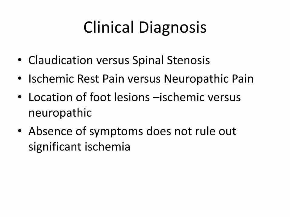
Clinical Diagnosis
• Claudication versus Spinal Stenosis
• Ischemic Rest Pain versus Neuropathic Pain
• Location of foot lesions –ischemic versus neuropathic
• Absence of symptoms does not rule out significant ischemia

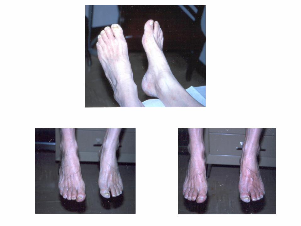
Signs of PVD
• Pulse examination. Frequently inaccurate due to calcified vessels.
• Inflow versus outflow disease
• Autonomic neuropathy
• Dependent Rubor
Non Invasive Studies in PVD
• Many sophisticated tests available eg Ankle Brachial Indices, Segmental pulse volume recordings, Duplex ultrasound, Transcutaneous oxygen, Xenon flow studies.
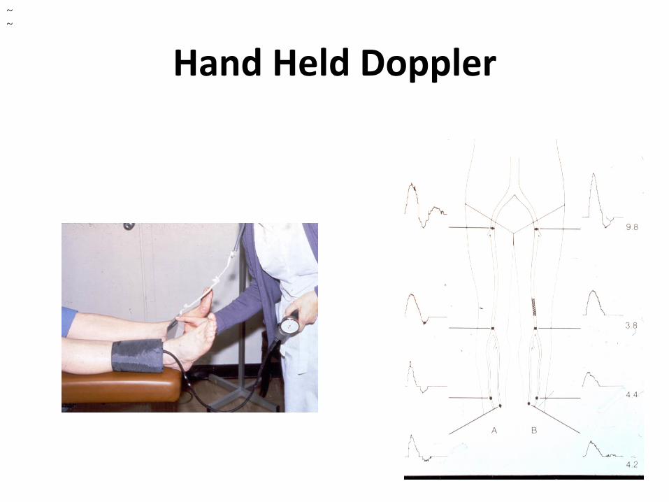
• Most useful and cost effective is a hand held Doppler to assess wave form

Hand Held Doppler
~
~

Interpreting the Ankle–Brachial Index
Adapted from Hirsch AT. Family Practice Recertification. 2000;22:6-12.
ABI Interpretation
0.90–1.30 Normal
0.70–0.89 Mild
0.40–0.69 Moderate
0.40 Severe
>1.30 Noncompressible
vessels
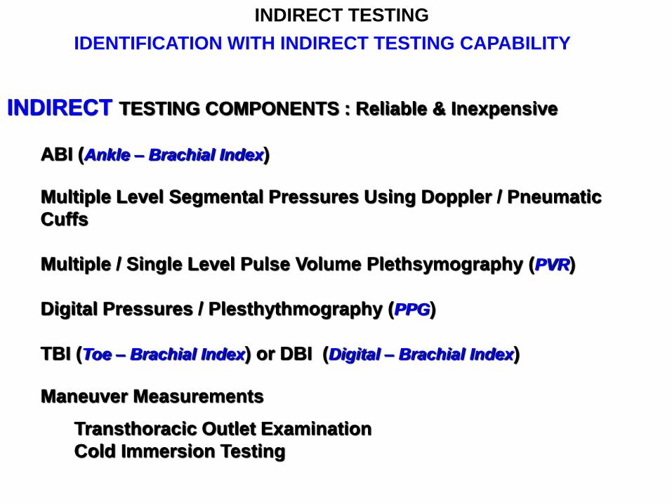
IDENTIFICATION WITH INDIRECT TESTING CAPABILITY
INDIRECT TESTING COMPONENTS : Reliable & Inexpensive
ABI (Ankle – Brachial Index)
Multiple Level Segmental Pressures Using Doppler / Pneumatic
Cuffs
Multiple / Single Level Pulse Volume Plethsymography (PVR)
Digital Pressures / Plesthythmography (PPG)
TBI (Toe – Brachial Index) or DBI (Digital – Brachial Index)
Maneuver Measurements
Transthoracic Outlet Examination
Cold Immersion Testing
INDIRECT TESTING
IDENTIFICATION WITH INDIRECT TESTING CAPABILITY
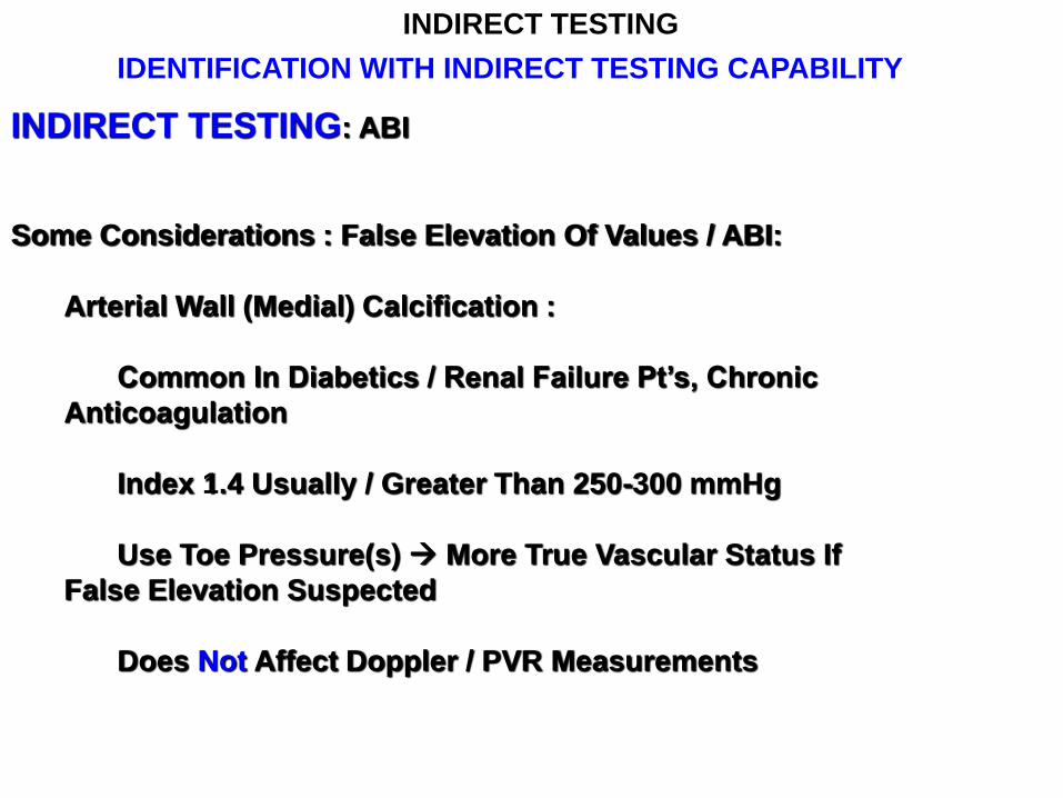
INDIRECT TESTING: ABI
Some Considerations : False Elevation Of Values / ABI:
Arterial Wall (Medial) Calcification :
Common In Diabetics / Renal Failure Pt’s, Chronic
Anticoagulation
Index 1.4 Usually / Greater Than 250-300 mmHg
Use Toe Pressure(s) More True Vascular Status If
False Elevation Suspected
Does Not Affect Doppler / PVR Measurements
INDIRECT TESTING
IDENTIFICATION WITH INDIRECT TESTING CAPABILITY
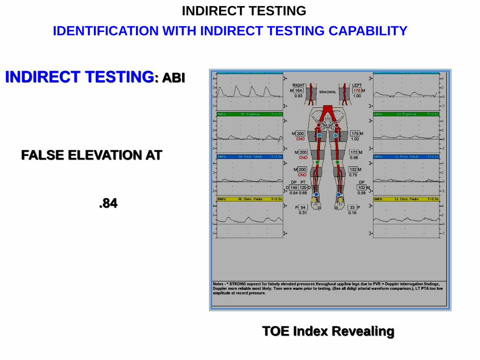
INDIRECT TESTING: ABI
FALSE ELEVATION AT
.84
TOE Index Revealing
INDIRECT TESTING
IDENTIFICATION WITH INDIRECT TESTING CAPABILITY
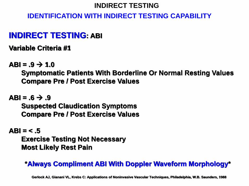
INDIRECT TESTING: ABI
Variable Criteria #1
ABI = .9 1.0
Symptomatic Patients With Borderline Or Normal Resting Values
Compare Pre / Post Exercise Values
ABI = .6 .9
Suspected Claudication Symptoms
Compare Pre / Post Exercise Values
ABI = < .5
Exercise Testing Not Necessary
Most Likely Rest Pain
*Always Compliment ABI With Doppler Waveform Morphology*
Gerlock AJ, Gianani VL, Krebs C: Applications of Noninvasive Vascular Techniques, Philadelphia, W.B. Saunders, 1988
INDIRECT TESTING

INDIRECT TESTING
IDENTIFICATION WITH INDIRECT TESTING CAPABILITY
INDIRECT TESTING: SEGMENTAL PRESSURES
•Can Localize Segment / Location Of Disease
•Vertical Pressure Comparisons
•Horizontal Pressure Comparisons
•Artifacts To Consider
•4 Cuff Or 2 Cuff Method
INDIRECT TESTING IDENTIFICATION WITH INDIRECT TESTING CAPABILITY
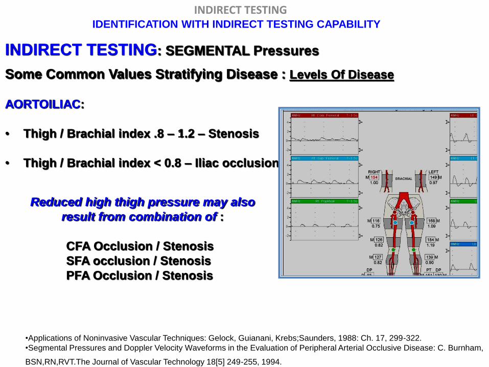
INDIRECT TESTING: SEGMENTAL Pressures
Some Common Values Stratifying Disease : Levels Of Disease
AORTOILIAC:
• Thigh / Brachial index .8 – 1.2 – Stenosis
• Thigh / Brachial index < 0.8 – Iliac occlusion
Reduced high thigh pressure may also
result from combination of :
CFA Occlusion / Stenosis
SFA occlusion / Stenosis
PFA Occlusion / Stenosis
•Applications of Noninvasive Vascular Techniques: Gelock, Guianani, Krebs;Saunders, 1988: Ch. 17, 299-322.
•Segmental Pressures and Doppler Velocity Waveforms in the Evaluation of Peripheral Arterial Occlusive Disease: C. Burnham,
BSN,RN,RVT.The Journal of Vascular Technology 18[5] 249-255, 1994.

INDIRECT TESTING IDENTIFICATION WITH INDIRECT TESTING CAPABILITY
INDIRECT TESTING: SEGMENTAL Pressures
Some Common Values Stratifying Disease : Levels Of Disease
SFA DISEASE:
> 30 mmHg gradient between high thigh pressure and above
knee pressure.
> 25 mmHg gradient between above knee pressure and
contra lateral above knee pressure.
•Applications of Noninvasive Vascular Techniques: Gelock, Guianani, Krebs;Saunders, 1988: Ch. 17, 299-322.
•Segmental Pressures and Doppler Velocity Waveforms in the Evaluation of Peripheral Arterial Occlusive Disease: C. Burnham,
BSN,RN,RVT.The Journal of Vascular Technology 18[5] 249-255, 1994.
INDIRECT TESTING IDENTIFICATION WITH INDIRECT TESTING CAPABILITY
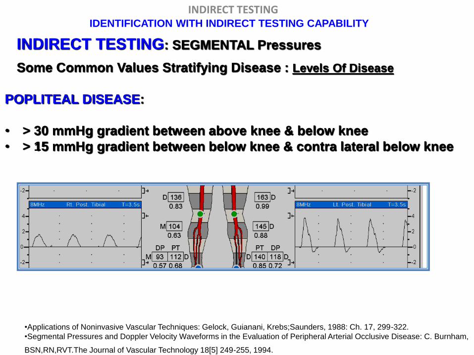
INDIRECT TESTING: SEGMENTAL Pressures
Some Common Values Stratifying Disease : Levels Of Disease
POPLITEAL DISEASE:
• > 30 mmHg gradient between above knee & below knee
• > 15 mmHg gradient between below knee & contra lateral below knee
•Applications of Noninvasive Vascular Techniques: Gelock, Guianani, Krebs;Saunders, 1988: Ch. 17, 299-322.
•Segmental Pressures and Doppler Velocity Waveforms in the Evaluation of Peripheral Arterial Occlusive Disease: C. Burnham,
BSN,RN,RVT.The Journal of Vascular Technology 18[5] 249-255, 1994.
INDIRECT TESTING IDENTIFICATION WITH INDIRECT TESTING CAPABILITY
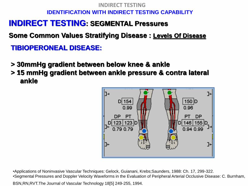
INDIRECT TESTING: SEGMENTAL Pressures
Some Common Values Stratifying Disease : Levels Of Disease
TIBIOPERONEAL DISEASE:
> 30mmHg gradient between below knee & ankle
> 15 mmHg gradient between ankle pressure & contra lateral
ankle
•Applications of Noninvasive Vascular Techniques: Gelock, Guianani, Krebs;Saunders, 1988: Ch. 17, 299-322.
•Segmental Pressures and Doppler Velocity Waveforms in the Evaluation of Peripheral Arterial Occlusive Disease: C. Burnham,
BSN,RN,RVT.The Journal of Vascular Technology 18[5] 249-255, 1994.

INDIRECT TESTING IDENTIFICATION WITH INDIRECT TESTING CAPABILITY
INDIRECT TESTING: SEGMENTAL Pressures
Some Common Values Stratifying Disease : Levels Of Disease
DIGITAL ARTERY DISEASE:
• Digital pressure < 60% of ankle pressure
• Toe / Brachial index < 0.7
• Toe systolic pressure < 30 mmHg Indicates a probable non-healing
lesion
• Digit pressures < 80% of the brachial pressure indicate proximal
disease
•Applications of Noninvasive Vascular Techniques: Gelock, Guianani, Krebs;Saunders, 1988: Ch. 17, 299-322.
•Segmental Pressures and Doppler Velocity Waveforms in the Evaluation of Peripheral Arterial Occlusive Disease: C. Burnham,
BSN,RN,RVT.The Journal of Vascular Technology 18[5] 249-255, 1994.
INDIRECT TESTING
IDENTIFICATION WITH INDIRECT TESTING CAPABILITY
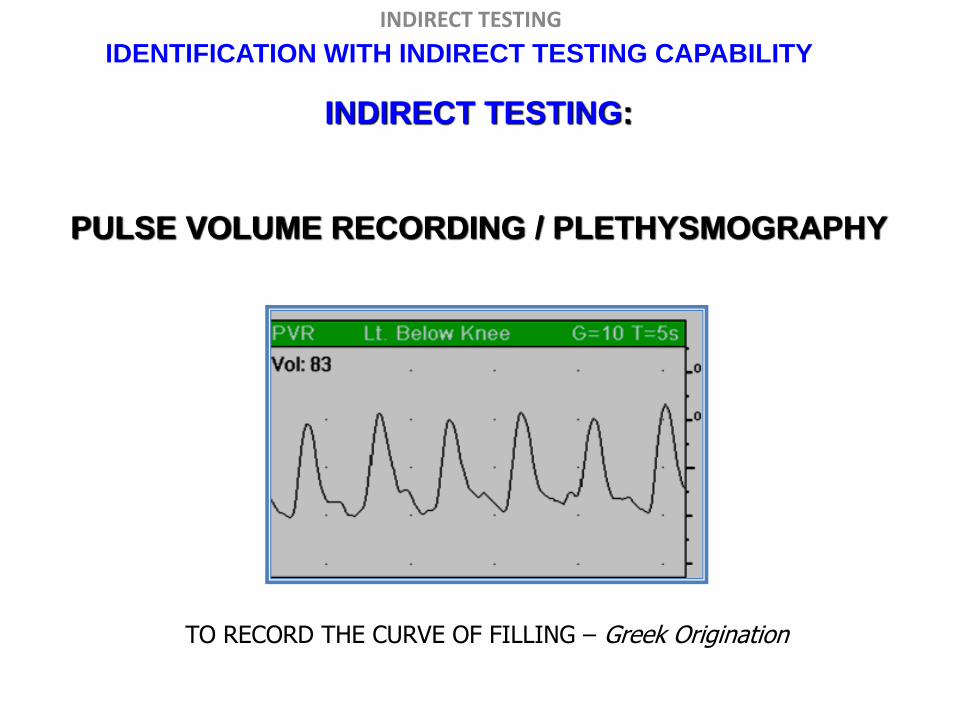
INDIRECT TESTING:
PULSE VOLUME RECORDING / PLETHYSMOGRAPHY
TO RECORD THE CURVE OF FILLING – Greek Origination

INDIRECT TESTING
IDENTIFICATION WITH INDIRECT TESTING CAPABILITY
INDIRECT TESTING: PULSE VOLUME RECORDING / PLETHYSMOGRAPHY
What Does It Do ?
• Measures Changes In Pressure Within The Cuff
Pressure Changes In The Volume Of The Cuff Or Bladder
Relates To
↓
Pressure Changes Within Limb Volume Detected
• Cuffs At Various Levels Compare Volume Changes Between Horizontal +
Vertical Levels
• Typically Inflated To 65 mmHg (Protocols Vary)
Enough To Provide Contact To Skin And To Reflect Pulsatility
• Amplitude Changes On The Graph
INDIRECT TESTING
IDENTIFICATION WITH INDIRECT TESTING CAPABILITY
INDIRECT TESTING: PULSE VOLUME RECORDING / PLETHYSMOGRAPHY
PVR Influenced By :
Blood Pressure
Volume Of Blood (Infection ? Cellulitis ?)
Position Of Extremity
Overall Size Of Extremity
Cardiac Stroke Volume
May Even Be Different On Same Patient B/W Visits
Large Habitus + Edema Will Attenuate PVR Presentation / Wave
Excessive or Not Enough Cuff Inflation
INDIRECT TESTING
IDENTIFICATION WITH INDIRECT TESTING CAPABILITY
INDIRECT TESTING: PULSE VOLUME RECORDING / PLETHYSMOGRAPHY
USEFUL FOR:
Determining Level Of Disease :
Aorto-Iliac + Outflow
Proximal SFA / DFA Involvement
Mid SFA / Abductor Canal
Popliteal / Tibial
Other Uses :
Pre + Post Exercise Measurements
Intra-Op Monitoring
Post-Op Evaluations
Healing Potential
Confirmation Of Rest Pain Symptoms

INDIRECT TESTING
IDENTIFICATION WITH INDIRECT TESTING CAPABILITY
INDIRECT TESTING: PULSE VOLUME RECORDING / PLETHYSMOGRAPHY
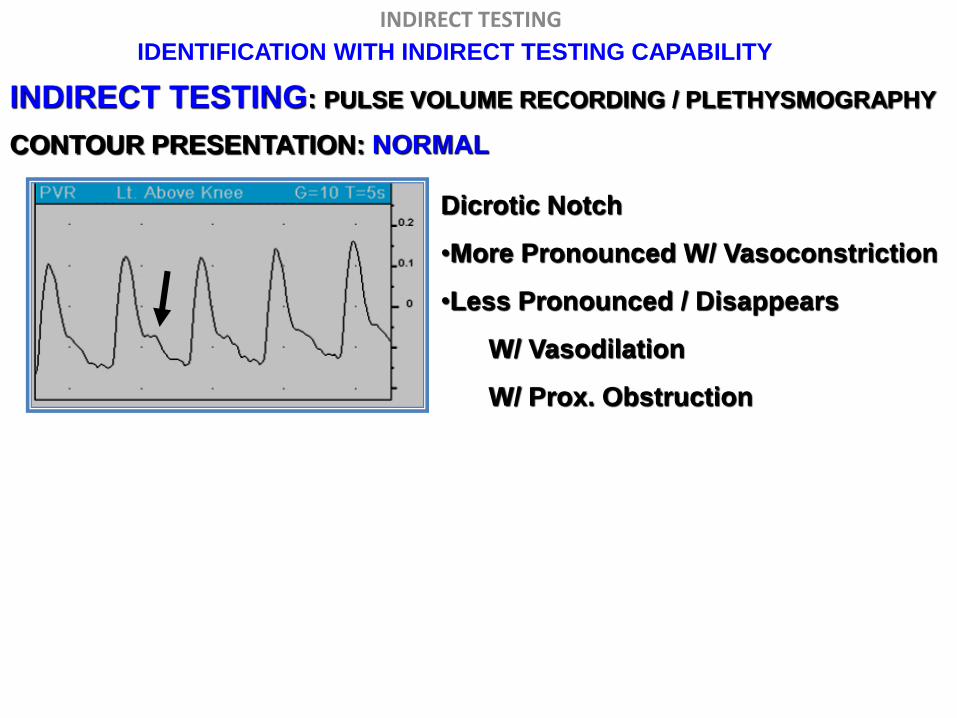
CONTOUR PRESENTATION: NORMAL
Higher Amplitude BK
Dicrotic Notch Present
(Arterial Pulse Reverse Component)
INDIRECT TESTING
IDENTIFICATION WITH INDIRECT TESTING CAPABILITY
INDIRECT TESTING: PULSE VOLUME RECORDING / PLETHYSMOGRAPHY
CONTOUR PRESENTATION: NORMAL
Dicrotic Notch
•More Pronounced W/ Vasoconstriction
•Less Pronounced / Disappears
W/ Vasodilation
W/ Prox. Obstruction
INDIRECT TESTING
IDENTIFICATION WITH INDIRECT TESTING CAPABILITY
INDIRECT TESTING: PULSE VOLUME RECORDING / PLETHYSMOGRAPHY
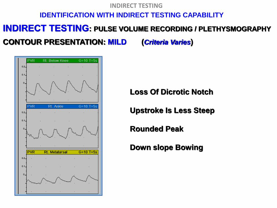
CONTOUR PRESENTATION: MILD (Criteria Varies)
Loss Of Dicrotic Notch
Upstroke Is Less Steep
Rounded Peak
Down slope Bowing

INDIRECT TESTING
IDENTIFICATION WITH INDIRECT TESTING CAPABILITY
INDIRECT TESTING: PULSE VOLUME RECORDING / PLETHYSMOGRAPHY
CONTOUR PRESENTATION: (Criteria Varies)
MODERATE SEVERE
Less Amplitude With Severity

INDIRECT TESTING
IDENTIFICATION WITH INDIRECT TESTING CAPABILITY
INDIRECT TESTING: PULSE VOLUME RECORDING / PLETHYSMOGRAPHY
CONTOUR PRESENTATION: (Criteria Varies)
Tachycardia – Camouflaged Notch
Obviously Normal
INDIRECT TESTING
IDENTIFICATION WITH INDIRECT TESTING CAPABILITY
INDIRECT TESTING: PULSE VOLUME RECORDING / PLETHYSMOGRAPHY
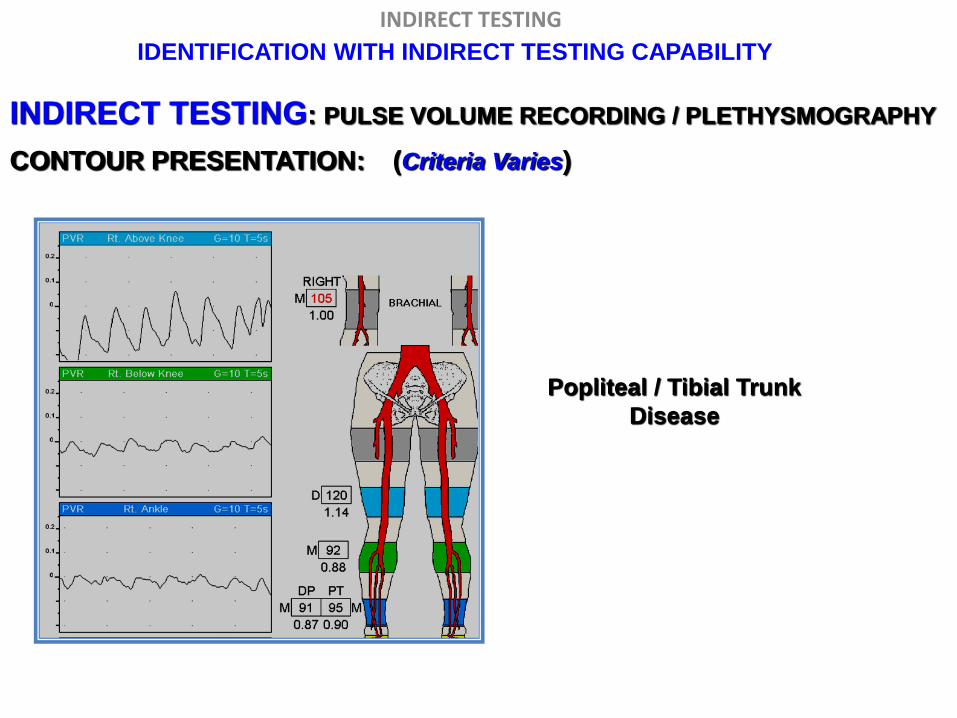
CONTOUR PRESENTATION: (Criteria Varies)
Popliteal / Tibial Trunk
Disease

INDIRECT TESTING
IDENTIFICATION WITH INDIRECT TESTING CAPABILITY
INDIRECT TESTING: PULSE VOLUME RECORDING / PLETHYSMOGRAPHY
CONTOUR PRESENTATION: (Criteria Varies)
Well Developed Collaterization ?
INDIRECT TESTING
IDENTIFICATION WITH INDIRECT TESTING CAPABILITY
INDIRECT TESTING:
CW DOPPLER
IDENTIFICATION WITH INDIRECT TESTING CAPABILITY
INDIRECT TESTING: CW DOPPLER
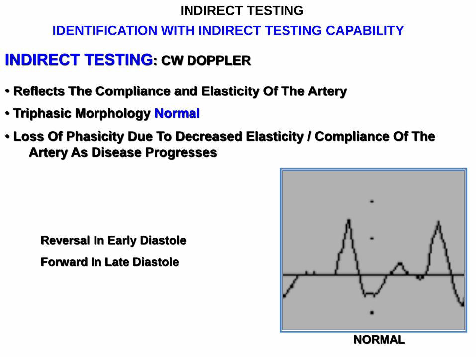
• Reflects The Compliance and Elasticity Of The Artery
• Triphasic Morphology Normal
• Loss Of Phasicity Due To Decreased Elasticity / Compliance Of The
Artery As Disease Progresses
Reversal In Early Diastole
Forward In Late Diastole
NORMAL
INDIRECT TESTING

IDENTIFICATION WITH INDIRECT TESTING CAPABILITY
INDIRECT TESTING: CW DOPPLER
• Reflects The Compliance and Elasticity Of The Artery
• Loss Of Phasicity Due To Decreased Elasticity / Compliance Of The
Artery As Disease Progresses
Forward In Late Diastole Loss
BIPHASIC
MONOPHASIC
INDIRECT TESTING
What's The Difference?

IDENTIFICATION WITH INDIRECT TESTING CAPABILITY
INDIRECT TESTING: CW DOPPLER
NOTE :
• CW Is Qualitative
• Between Region Changes Indicate Disease
• Sensitivity Reduced :
Obesity
Wrong Freq. Selection
Scarring Of Skin
Calcifications W/In Artery Insonated
• Artifacts:
Venous Interference
Movement
• Correct Filter / Scale Adjustment
• Don’t Make A Triphasic Signal Look Biphasic
Requires Expertise In Obtaining True Doppler Insonation Angle &
Clean Signal For True Morphology
INDIRECT TESTING
IDENTIFICATION WITH INDIRECT TESTING CAPABILITY
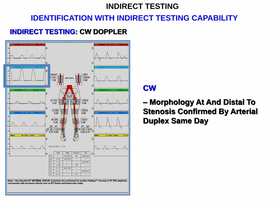
INDIRECT TESTING: CW DOPPLER
CW
– Morphology At And Distal To
Stenosis Confirmed By Arterial
Duplex Same Day
INDIRECT TESTING

IDENTIFICATION WITH INDIRECT TESTING CAPABILITY
INDIRECT TESTING:
TREADMILL EXERCISE TESTING
INDIRECT TESTING

IDENTIFICATION WITH INDIRECT TESTING CAPABILITY
INDIRECT TESTING: TREADMILL EXERCISE TESTING
MAIN INDICATIONS:
• Important for differentiating true vascular claudication from pseudo-
claudication
• Performed on all patients that complain of pain while walking
• Evaluate S/P Revascularization (Iliac Stents, etc..)
ABI’s MAY BE NORMAL AT REST :
• Collateral Development Adequate For Resting Vascular State
• Not Adequate With Increased Demand For Blood Supply
WITH EXERCISE :
• Obstruction Present Will Not Be Able To Meet Perfusion Needs
• Need Will Exceed Collateral Capability
• Significant Pressure Drop As Result
INDIRECT TESTING

IDENTIFICATION WITH INDIRECT TESTING CAPABILITY
INDIRECT TESTING: TREADMILL EXERCISE TESTING
MAIN CONTRA-INDICATIONS:
ABI less than .5 (Varies)
Recent onset of chest pain
Severe Pulmonary Disease
? Cardiac status, known cardio-vasc. dis., prev. MI or CABG
Severe pulmonary disease / Shortness of Breath
Inability to ambulate at treadmill speed
Ischemic rest pain
Ischemic limb ulceration
*If the Patient’s symptoms occur at rest (non-claudication symptoms) and the
resting examination is negative, there is no need to exercise the patient (?)
INDIRECT TESTING

IDENTIFICATION WITH INDIRECT TESTING CAPABILITY
INDIRECT TESTING: TREADMILL EXERCISE TESTING
OPTIONS OTHER THAT TREADMILL :
• Toe Ups / Toe Raises
Simple & Effective
• Reactive Hyperemia
Can Be Painful
Occlusion Of Cuff / Post Release Measurements
Lab Dependant, Personal Physician Preference, Supporting Data
Exists For All Methods Of Post Maneuver Measurements
INDIRECT TESTING
IDENTIFICATION WITH INDIRECT TESTING CAPABILITY
INDIRECT TESTING: TREADMILL EXERCISE TESTING
Discussion: Method
Patient Walks For Specified Time At Specified Grade Or Until Symptoms
Halt Exercise
Protocols Vary :
5 Minutes, 10% (7 º) Grade At 1.5 MPH
5 Minutes, 12% Grade At 2 MPH
More..
INDIRECT TESTING

IDENTIFICATION WITH INDIRECT TESTING CAPABILITY
INDIRECT TESTING: TREADMILL EXERCISE TESTING
METHOD Discussion: Post Exercise Measurements
Protocols Vary:
• Immediate Ankle + Brachial Pressure
30 Second Intervals – First 4 Minutes
Immediate Post Ex PVR
Every Minute Until Pressure Returns To Resting State (< 10 min.)
• Immediate Ankle + Brachial Pressure
2 Minute Intervals Until Pressure Returns To Resting State
(<10 min)
• All Protocols : Record PT Symptoms While Exercising
Post Exercise PVR For Non-Occlusive ABI
INDIRECT TESTING
IDENTIFICATION WITH INDIRECT TESTING CAPABILITY
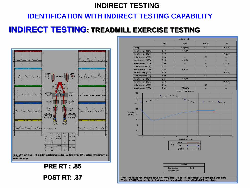
INDIRECT TESTING: TREADMILL EXERCISE TESTING
PRE RT : .85
POST RT: .37
INDIRECT TESTING

IDENTIFICATION WITH INDIRECT TESTING CAPABILITY
INDIRECT TESTING: EXERCISE TESTING
METHOD Discussion: Toe Raises
PT Standing – Raises On Toes – Returns To Flat
Performed Until PT Cannot Continue Or Set Rate (50)
Symptom Onset / Toe Raising #’s Recorded
Has Been Considered As Criteria For Positive (Varies)
> 20 mmHg Drop In Pressure
↓ of 20% Of Resting ABI
Some Considerations :
Can Be Alternative To Treadmill Exercise
Cardiac Risk Factors / Exertional Limitations
Calf Pain May Be Due To General Fatigue
Treadmill Exercise More Accurate For Claudicate
Patients
INDIRECT TESTING
IDENTIFICATION WITH INDIRECT TESTING CAPABILITY
INDIRECT TESTING: EXERCISE TESTING
METHOD Discussion: Reactive Hyperemia
• Inflate Thigh Cuff > 20 mmHg Beyond Thigh Pressure
• Maintain Inflation B/W 3-5 Minutes
• Release And Obtain Ankle Pressures
General Criteria :
↓ In 20 mmHg (+)
Limitations:
• Difficult Differentiating True vs. Pseudoclaudication
• Extremely Painful For Most Patients
Some Considerations:
• Apply Calf Cuff Instead Of Thigh In Suspected Below CFA
Disease
INDIRECT TESTING

IDENTIFICATION WITH INDIRECT TESTING CAPABILITY
COLOR DUPLEX
VS.
SEGMENTAL / INDIRECT PHYIOLOGIC ASSESSEMENT
INDIRECT TESTING

INDIRECT TESTING
SEGMENTAL BP / PVR Suggested For Primary Diagnosis :
• Reimbursement Conditions & Requirements
• Used For 1st Time Diagnosis/ Initial Screen
* * Localize + Characterize Arterial Disease
• Follow Up Exams
Revascularization
Functional Status Of Stents/ Grafts
Treadmill Exercise
• General Limitations :
Cannot Differentiate From Tight Stenosis Vs. Collaterization
False Elevation Of Pressures
Exact Segment Difficult To Quantify

INDIRECT TESTING
COLOR DUPLEX - Suggested In Known Disease States:
• Localizes Stenosis + Severity Of Stenosis
• Collateral Development Visualization
• F/U Revascularization Patentcy
Stent + Graft + Angioplasty
• General Limiations :
Regions Difficult To Asses :
Tibial Vessels + Tibio – Peroneal Trunck
Calcification / Dense Plaque
Iliac Involvement

INDIRECT TESTING
SEG BP / PVR VS DUPLEX SUMMATION
Best Used In Conjunction
Each Have Specific Indications
Follow Reccomendations By ICAVL / Other Associatations
GENRAL ACCEPTED PRACTICES :
General Concept Is To Use Physiological Assessment For PT Management /
Decision Making Initially
Color Duplex Utilization For Further Quantification

INDIRECT TESTING
TAKE NOTE - EXAMPLE : Warm the Digits
Post 5 minutes Toe Warming

INDIRECT TESTING
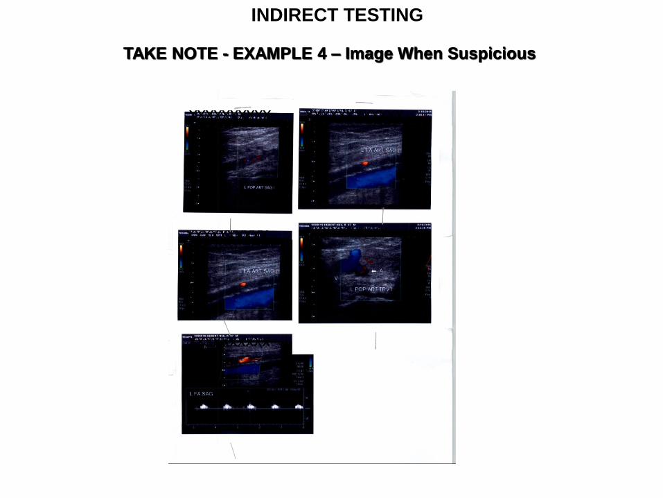
TAKE NOTE - EXAMPLE 4 – Image When Suspicious
INDIRECT TESTING
TAKE NOTE - EXAMPLE 4 – Image When Suspicious
xxxxxxxxxx xxxxxxxxxxxx
xxxxxxxxxx xxxxxxxxxx
xxxxxxxxxx
Hand Held Doppler
~
~

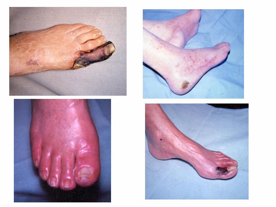
When to Operate on Foot
• In patient with abscess systemic sepsis and an ischemic foot - I+D of the foot as an emergent procedure. Limit procedure to drainage of all pus and dead tissue
• Over extensive debridement may convert ischemic tissue to frank gangrene and thereby reduce options for closure of the foot

Severe infection secondary to MRSA

Chronic infection
• Generally can perform podiatric procedure
48 hours after revascularisation.
• Inflow procedures and revascularisation of
peroneal artery may take 48 hours to obtain
maximal perfusion of foot

Osteomyelitis
Beware!
• Revascularisation may convert dry
gangrene to wet gangrene
• Need to closely monitor and be prepared to
perform urgent debridement

Diabetic Foot

Thank you
Top Related