


![Untitled-7 [] · 2019. 10. 20. · reported in patients receving androgenic anabolic steroid therapy. these cysts are sometimes present w1th minimal hepatic dysfunction, but at other](https://static.fdocuments.us/doc/165x107/5ff78036caac426b10038257/untitled-7-2019-10-20-reported-in-patients-receving-androgenic-anabolic.jpg)


Languages
Pages
Legal

HEPATIC CYSTS Occurrence and effect of single-session alcohol sclerotherapy
Trond Bjerke Larssen
UNIVERSITY OF BERGEN 2006

To Inger Johanne
HEPATIC CYSTS Occurrence and effect of single-session alcohol sclerotherapy
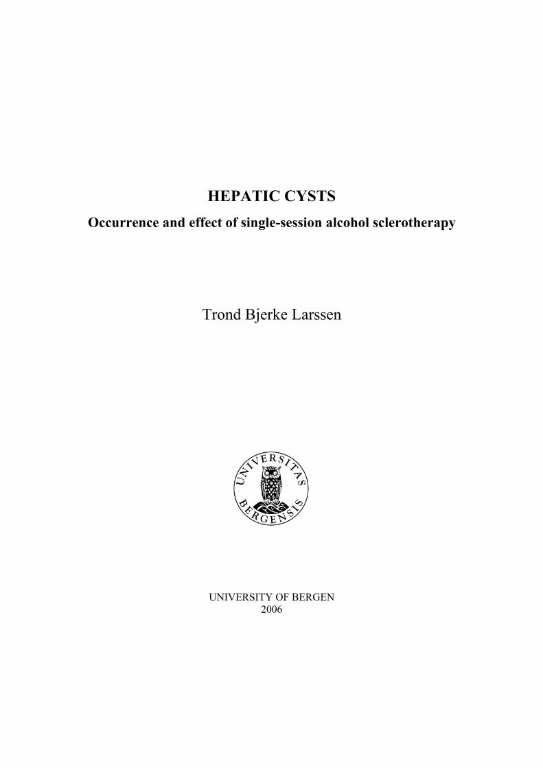
0 mnth 1 mnth 7 mnth 17 mnth
Trond Bjerke Larssen
SECTION FOR RADIOLOGY, DEPARTMENT OF SURGICAL SCIENCES UNIVERSITY OF BERGEN
2006

ISBN 82-308-0244-0
3
CONTENTS 1. Acknowledgements ……………………………………………….………...…….. 5 2. List of abbreviations, errata.…………………….…….……………………….. 7 3. List of Original Papers …………………….……………………………………. 9 4. Background …………………………………………..……..…………………….. 11 4.1. Introduction ……...……..…………….……………………………………………… 11 4.2. Embryology………...………….…………………………………………………...… 12 4.3. Genetic aspects……………………..………..…………………………………...…… 14 4.4. Pathology……...………………...….………………………………….…………….. 14 4.5. Epidemiology……………………….…………………………………...………..….. 15 4.6. Symptoms …………………………………………………………………………… 15 4.7. Diagnosis……………………………….………………………………………...…… 16 4.8. Treatment of liver cysts……………………..…………………………………..…..... 18 4.8.1. Surgery………………………………….………………………………………...… 18 4.8.2. Percutaneous intervention. Sclerotherapy …….……………………………..…….. 19 5. Aims of the study……………………………………………………………...….. 22 6. Materials and methods………………………..………………………..……….. 22 6.1.Patients………………………………………………………………………….…….. 22 6.1.1.Patients, Part I …..…………….…………………………………………………..… 22 6.1.2.Patients, Part II……………………………………………………..............……...... 24 6.2. Methods, part I……………………………………………………………………….. 24 Methods, part II ……………………………………………………………………… 29 6.3. Statistics ………………………………………………………………….………….. 30 7. Summary of results……………………………………………….………………. 30 8. General discussion………………………………………………………..…...….. 33 8.1 Part I. Sclerotherapy of symptomatic liver cysts…………………………………...…. 33 8.1.1. Study design………………………………………………………………………… 33 8.1.2. Evaluation of patient response……………………………………………………… 34 Pre and post-procedure measurements of cyst volume…………………………….. 34 Clinical evaluation of symptoms………………………………………………..…. 36 Evaluation of side effects……………………………………………………….….. 36 8.1.3.Mechanism of sclerotherapy………………………………………………………... 38 8.2. Part II. Occurrence of liver cysts………………………………………………...…… 39 8.2.1. Study design………………………………………………………………...……… 39 8.2.2. Accuracy of our results…………………………………………………………….. 40 9. Conclusions……………………………………………………………………...…. 42 10. References…………………………………………………………………………. 45 11. Appendix: Papers I –V ...................................................................................... 61

4

5
1. Acknowledgements The present investigations were conducted during the years 1993-2001 as a collaboration
between the Departments of Radiology, Surgery, Pathology, and Clinical Biochemistry,
Haukeland University Hospital, Bergen, Norway and Section for Radiology, Department of
Surgical Sciences, University of Bergen.
I am especially grateful to my scientific supervisors, associate Professor Jarle Rørvik MD,
PhD, Professor Karen Rosendahl MD, PhD, Section for Radiology, and Arild Horn MD, PhD,
Section for Surgery, Department of Surgical Sciences, University of Bergen for their patience
and endurance in helping me to complete this thesis.
I am grateful to the scientific support of Ole Martin Pedersen MD, PhD, Department of Heart
Diseases, Haukeland University Hospital. The collaboration with Barbara Karwinski, MD,
PhD, Department of Pathology, Haukeland University Hospital, and with Øyvind Skadberg,
Laboratory of Clinical Biochemistry, Haukeland University Hospital, has also been of great
value.
I am particularly grateful to the Haakon and Sigrun Ødegaards Foundation and to the
Norwegian Cancer Association for their financial support, making this thesis possible. My
thanks are extended to statisticians Geir Egil Eide, MSc, Rolv Schiærven, MSc, and Grethe
Albrektsen, MSc, Department of Public Health and Primary Health Care, University of
Bergen , for their valuable statistical consultation. I am very grateful my fellow radiologist
Ansgar Espeland MD, PhD, Department of Radiology, Haukeland University Hospital, for
helping me during web-publishing and for teaching me the use of the reference manager. I am
also grateful to Judith Mercer Cabot for her help during the preparation of this manuscript.
I wish to thank Aslak Aslaksen, MD, PhD, head of the Department of Radiology, as well as
my fellow radiologists and close colleagues at the Section of Oncologic and Gastrointestinal
Radiology, Anne Taule MD, consultant Gastrointestinal Radiologist, Head of the Section, and
Dag Jensen MD, Arna Mulahasanovic MD, and Marit Bolstad MD. They have had to take on
an extra workload during my periods of scientific work. Their positive attitude has
contributed to the completion of this thesis. Thanks also to Britt Tennebekk, Merethe Lohne
and other radiographers for their interest and support during the sclerotherapy procedures. I
also wish to thank Inger Johanne, my daughters Kathrine and Elisabeth, and the rest of my
family for their support and encouragement during the busy years of research.

6

7
2. List of abbreviations, errata ADPKD Autosomal dominant polycystic kidney disease
ADPLD Autosomal dominant polycystic liver disease without renal involvement
ALP Alkaline phosphatase
ALT Alanine aminotransferase
AST Aspartate aminotransferase
CT Computed tomography
GT Glutamyl transpeptidase
HCC Hepatocellular carcinoma
HU Hounsfield Units
HUS Haukeland University Hospital
LDH Lactic dehydrogenase
mHz Megahertz
MRI Magnetic resonance imaging
US Ultrasound
Errata:
Paper III. Patients and methods: The sentence “From December 1995 to June 1999 all
sclerotherapy procedures in symptomatic, non-parasitic, non-neoplastic liver cysts were
performed with a time of ethanol exposure of 10 minutes” should be corrected to “From
December 1996 to June 1999 all sclerotherapy procedures in symptomatic, non-parasitic, non-
neoplastic liver cysts were performed with an ethanol exposure of 10 minutes”.
Paper V. Results, para.3: The sentence “Of the 174 affected individuals, 14 had polycystic
livers (3.5%)” should be “Polycystic liver disease was found in 14 cases (0.9% of all livers
and 8.1% of 174 livers containing cysts)”.

8
9
3. List of original papers This thesis is based on the following papers, referred to in the text by their Roman numerals:
Paper I Larssen TB, Viste A, Jensen DK, Sondenaa K, Rokke O, Horn A. Single-session alcohol sclerotherapy in benign symptomatic hepatic cysts. Acta Radiol 1997;38(6):993-7. Paper II Larssen TB, Jensen DK, Viste A, Horn A. Single-session alcohol sclerotherapy in symptomatic benign hepatic cysts. Long-term results. Acta Radiol 1999;40(6):636-8. Paper III Larssen TB, Rosendahl K, Horn A, Jensen DK, Rorvik J. Single-session alcohol sclerotherapy in symptomatic benign hepatic cysts performed with a time of exposure to alcohol of 10 min: initial results. Eur Radiol 2003;13(12):2627-32. Paper IV Larssen TB, Rorvik J, Horn A, Karwinski B, Skadberg O, Pedersen OM, Rosendahl K. Biochemical and cytologic analysis of cystic contents in benign non-parasitic symptomatic hepatic cysts before and after ethanol sclerotherapy. Acta Radiol 2004;45(5):504-9. Paper V Larssen TB, Rorvik J, Hoff SR, Horn A, Rosendahl K.. The occurrence of asymptomatic and symptomatic simple hepatic cysts. A prospective, hospital-based study. Clinical Radiology 2005:60;1026-1029.

10
11
4. Background 4.1. Introduction
The term fibropolycystic disease of the liver encompasses a spectrum of disorders, from
simple hepatic cysts to more complex entities such as hepatic fibrosis (Table 1). In this thesis
I have addressed simple hepatic cysts, with or without symptoms. Various terms have been
used for simple hepatic cysts, such as biliary cyst, non-parasitic cyst of the liver, benign
hepatic cyst, congenital hepatic cyst, unilocular cyst of the liver, solitary cyst of the liver 1, or
dysontogenetic liver cyst 2. The term solitary cyst is often inappropriate, since simple (i.e.
thin-walled, fluid-filled) cysts of the liver commonly present as two or more 1. In the
following I have used the terms simple liver (or hepatic) cyst(s) for ten cysts or less 3, and
polycystic liver disease for more than ten cysts.
Traditionally, symptomatic cysts have been treated by open surgery. During the past two
decades the less invasive method of laparoscopic fenestration and the even less traumatic
method of sclerotherapy have been introduced. Because symptomatic liver cysts are rare, little
has been published about either method and the number of patients is low. To our knowledge,
random trials comparing sclerotherapy and laparoscopic surgery have not been published.
However, sclerotherapy has been practised with considerable variation as regards the
selection of sclerosing agent, the volume of sclerosant applied, the time of exposure of the
cyst to the sclerosant as well as the use of one single session or multiple sclerotherapy
sessions. This may explain why the use of sclerotherapy has not gained a wider acceptance.
Leading liver surgeons have expressed an interest in further research being carried out
concerning standardisation and simplification of the method of sclerotherapy4;5.
In this study we have aimed at assessing the short- and long-term effects of procedural
adjustment, so that sclerotherapy would be more acceptable to the patient and more efficient.
For example, we have studied the effect of decreasing the number of sessions of ethanol
injection and the time of ethanol exposure. We also wanted to study the mechanism of
reaccumulation of cyst fluid. Finally, we wanted to assess the number of patients with
asymptomatic and symptomastic liver cysts in patients referred to the Radiology Department
of a Teaching Hospital.

12
Table 1. Fibropolycystic disease of the liver (1)
________________________________________________________________ Heredity Patients age Hepatic pathology at clinical presentation ___________________________________________________________________________ Adults Simple hepatic cysts Not known 40-80 years Cysts, few Autosomal dominant Dominant 30-50 years Cysts, multiple polycystic disease of the kidneys with hepatic involvement (ADPKD) (2) Autosomal dominant Dominant 30-50 years Cysts, multiple polycystic hepatic disease without renal involvement (ADPLD) Carolis disease (3) Not known Adolescence Dilated biliary ducts and extrahepatic bile or young adult duct cysts (choledochal cyst; choledochocele) Congenital Recessive Childhood Fibrosis hepatic fibrosis or adult Dilated biliary ducts ___________________________________________________________________________ Children Infantile Recessive 3-6 months Fibrosis Dilated biliary ducts Neonatal Recessive 1 month Fibrosis Dilated biliary ducts Perinatal Recessive Birth Fibrosis
Dilated biliary ducts __________________________________________________________________________ (1) Modified after Sheila Sherlock6. (2) Between 30%7 and 88%8 of patients suffering from ADPKD also have polycystic disease of the liver. (3) Carolis disease: Cyst-like dilatation of intrahepatic biliary ducts. Choledochal cyst: Dilatation of extrahepatic biliary ducts. Choledochocele:Dilatation of the distal common bile duct localised within the duodenal wall 6.
4.2. Embryology
In 1906, Moschcowitz suggested that non-parasitic hepatic cysts were caused by a
maldevelopment of biliary ducts during intrauterine life9. At three weeks gestational age, a
13
diverticulum appears at the anterior wall of the foregut. This diverticulum differentiates into
two parts, one forming the fetal liver and the other forming the extrahepatic biliary tract,
including the gallbladder 10. The primitive liver cells grow to create sheets of cells. At six
weeks the first bile ductuli appear within these sheets of cells. At 9-10 weeks intrahepatic
biliary ductuli appear as a single layer of epithelial cells surrounding the primitive portal
tracts. At thirteen weeks a second layer or sheet of cells appears. This results in a double
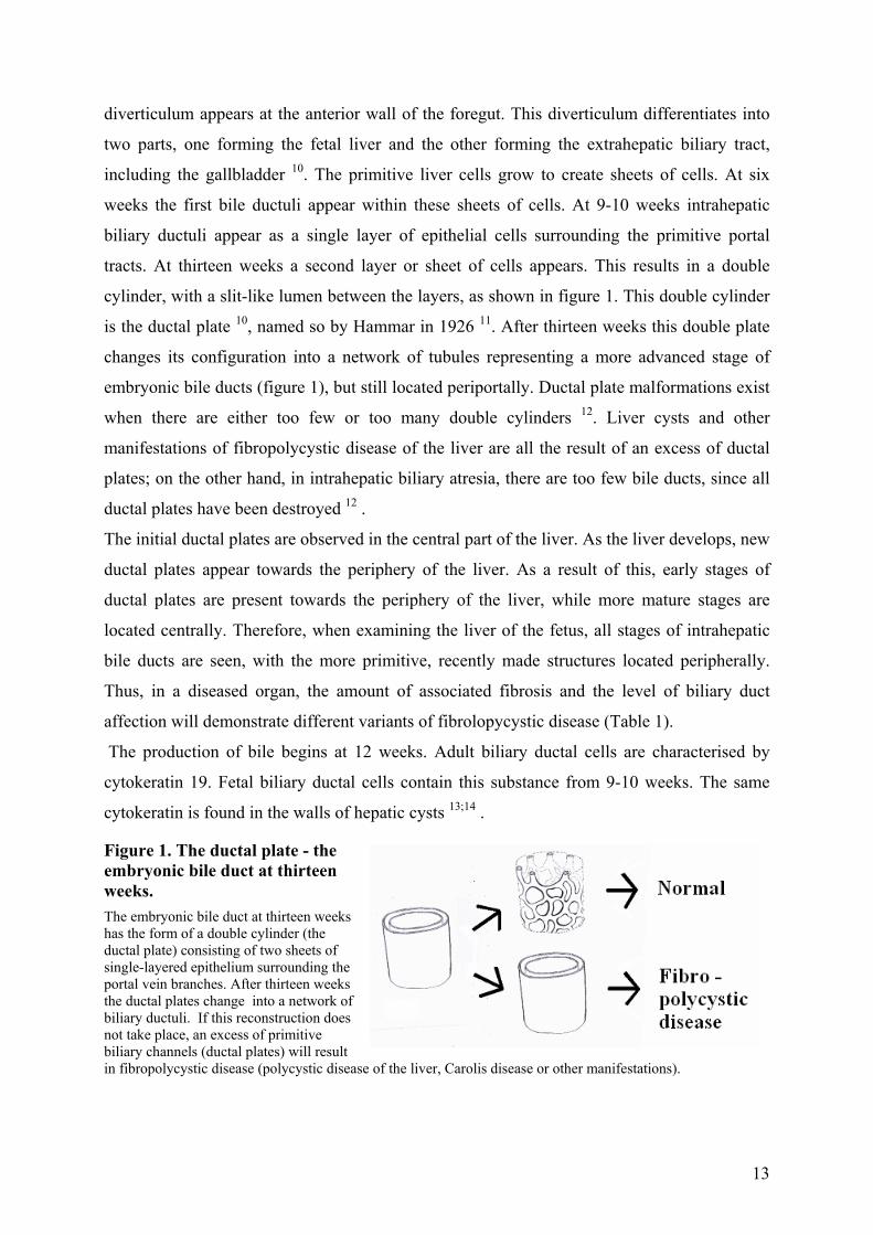
cylinder, with a slit-like lumen between the layers, as shown in figure 1. This double cylinder
is the ductal plate 10, named so by Hammar in 1926 11. After thirteen weeks this double plate
changes its configuration into a network of tubules representing a more advanced stage of
embryonic bile ducts (figure 1), but still located periportally. Ductal plate malformations exist
when there are either too few or too many double cylinders 12. Liver cysts and other
manifestations of fibropolycystic disease of the liver are all the result of an excess of ductal
plates; on the other hand, in intrahepatic biliary atresia, there are too few bile ducts, since all
ductal plates have been destroyed 12 .
The initial ductal plates are observed in the central part of the liver. As the liver develops, new
ductal plates appear towards the periphery of the liver. As a result of this, early stages of
ductal plates are present towards the periphery of the liver, while more mature stages are
located centrally. Therefore, when examining the liver of the fetus, all stages of intrahepatic
bile ducts are seen, with the more primitive, recently made structures located peripherally.
Thus, in a diseased organ, the amount of associated fibrosis and the level of biliary duct
affection will demonstrate different variants of fibrolopycystic disease (Table 1).
The production of bile begins at 12 weeks. Adult biliary ductal cells are characterised by
cytokeratin 19. Fetal biliary ductal cells contain this substance from 9-10 weeks. The same
cytokeratin is found in the walls of hepatic cysts 13;14 .
Figure 1. The ductal plate - the embryonic bile duct at thirteen weeks. The embryonic bile duct at thirteen weeks has the form of a double cylinder (the ductal plate) consisting of two sheets of single-layered epithelium surrounding the portal vein branches. After thirteen weeks the ductal plates change into a network of biliary ductuli. If this reconstruction does not take place, an excess of primitive biliary channels (ductal plates) will result in fibropolycystic disease (polycystic disease of the liver, Carolis disease or other manifestations).
14
4.3. Genetic aspects.
While the infantile forms of hepatic fibropolycystic disease are autosomal recessive (Table 1),
the mode of heritage for the juvenile forms (choledochal cyst and Carolis disease) is
unknown. The adult form of hepatic fibropolycystic disease includes two variants of
polycystic liver disease, both following an autosomal dominant heritage. First, polycystic
liver disease is associated with autosomal dominant polycystic kidney disease (ADPKD). The
autosomal dominant heritage of ADPKD was demonstrated by Dalgaard in 195715. In 1985
the first gene for this disease was localised to the short arm of chromosome 1616. Second,
another variant, autosomal dominant polycystic liver disease (ADPLD) was reported by
Pirson in 199617. ADPLD is not associated with renal involvement. The prevalence of
ADPLD in the general population is not known18. In 2000 the gene for ADPLD was localised
to chromosome 19 by Reynolds 19. The mode of heritage for simple hepatic cysts is not
known 6.
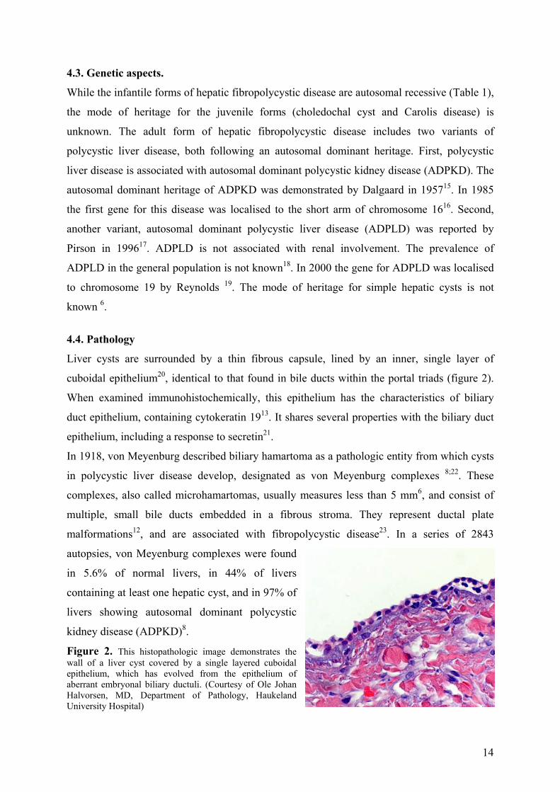
4.4. Pathology
Liver cysts are surrounded by a thin fibrous capsule, lined by an inner, single layer of
cuboidal epithelium20, identical to that found in bile ducts within the portal triads (figure 2).
When examined immunohistochemically, this epithelium has the characteristics of biliary
duct epithelium, containing cytokeratin 1913. It shares several properties with the biliary duct
epithelium, including a response to secretin21.
In 1918, von Meyenburg described biliary hamartoma as a pathologic entity from which cysts
in polycystic liver disease develop, designated as von Meyenburg complexes 8;22. These
complexes, also called microhamartomas, usually measures less than 5 mm6, and consist of
multiple, small bile ducts embedded in a fibrous stroma. They represent ductal plate
malformations12, and are associated with fibropolycystic disease23. In a series of 2843
autopsies, von Meyenburg complexes were found
in 5.6% of normal livers, in 44% of livers
containing at least one hepatic cyst, and in 97% of
livers showing autosomal dominant polycystic
kidney disease (ADPKD)8.
Figure 2. This histopathologic image demonstrates the wall of a liver cyst covered by a single layered cuboidal epithelium, which has evolved from the epithelium of aberrant embryonal biliary ductuli. (Courtesy of Ole Johan Halvorsen, MD, Department of Pathology, Haukeland University Hospital)
15
4.5. Epidemiology
Simple, asymptomatic cysts are the most frequently occurring focal liver lesion, with a
reported prevalence varying between 1% in autopsies24 to 2.5-18% when based on imaging25-
27. These are seldom diagnosed before the age of 40 years, after which the prevalence
increases gradually with age27, without significant differences between the sexes27. Polycystic
liver disease is less frequent than simple hepatic cysts, but the prevalence of polycystic liver
disease as well as of simple hepatic cysts in the general population is not known. That there
are at least two different definitions of polycystic liver does not make the investigation of this
topic easier18. The majority of patients presenting with polycystic liver disease suffer from
autosomal dominant polycystic kidney disease (ADPKD) with a reported prevalence of
0.1%28. The occurrence of liver cysts in ADPKD vary from 30 to 88% in different
studies7;8;29.
Since the occurrence of symptomatic liver cysts is low, true population-based data are sparse.
A report from Sanfelippo in 1974, based on 88 000 explorative laparotomies, revealed
symptomatic liver cysts in 0.2% of the cases30. Between 80 and 90%31;32 of symptomatic liver
cysts, both polycystic and simple, occur in women31;32. The reason for this is unclear, but
gestational hormones33 and estrogen may play a role34. Women who have never been pregnant
and who have not used estrogen medication have a lower statistical risk of symptomatic
hepatic cysts in autosomal dominant polycystic renal disease35.
4.6. Symptoms
Simple liver cysts only rarely cause acute abdominal symptoms. When this occurs, it is due to
spontaneous or traumatic rupture, to hemorrhage into hepatic cysts36, or to torsion of an
exophytic cyst or secondary infection. The most common symptoms are chronic pain,
abdominal mass and early satiety due to compression of the stomach, although jaundice and
respiratory problems have also been reported 37-44. When chronic, even moderate symptoms
may result in a permanent reduction in the patients’ quality of life.
In polycystic liver disease, symptoms may be severe when the degree of hepatomegaly is
extreme45;46. Such symptoms are: lower extremity edema due to inferior vena cava
compression; ascites secondary to hepatic venous outflow obstruction; portal hypertension
with esophageal varices; severe nutritional problems due to compression of the stomach and
gastrointestinal tract; and severely reduced quality of life47;48.
16
Several case reports have been published on cystic disease of the liver complicated by
malignancy such as adenocarcinoma 49, squamous cell carcinoma 50 and cholangiocarcinoma 51;52. It has been claimed that there is an association between polycystic liver disease and
cholangiocarcinoma 51. According to one author, the most frequent complication in autosomal
dominant polycystic liver disease with renal involvement is the infection of liver cysts, with
cholangiocarcinoma as the second most frequent complication 28.
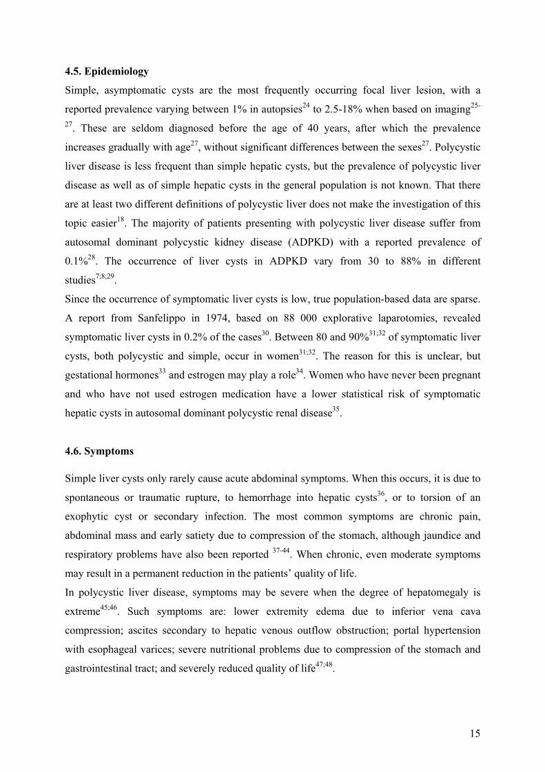
4.7. Diagnosis
The diagnosis of liver cysts is based on cross-sectional imaging, including ultrasound (US),
computed tomography (CT) and magnetic resonance imaging (MRI). The main differential
diagnoses are pyogenic abscess, amoebic abscess, hydatid cysts and necrotic neoplasm 53.
Figure 3. Ultrasound of the liver showing a simple, thin-walled cyst with anechoic contents and posterior echo enhancement.
Ultrasound (US) has been known for its
great potential in differentiating cystic
abdominal lesions from solid ones since the early period of US technology53-60. US has been
the method of choice for investigating the liver for about three decades55;55;59;61-67;67A high
spatial resolution enables a detailed characterization of liver cysts68. The sonographic criteria
of a cyst are a thin, not discernable wall, anechoic cystic contents and posterior echo
enhancement (figure 3). Based on these critera, high diagnostic accuracy has been reported 66;69-71. The major limitations of US occur in obese patients and in patients with severe
deformities, such as kyphoscoliosis.
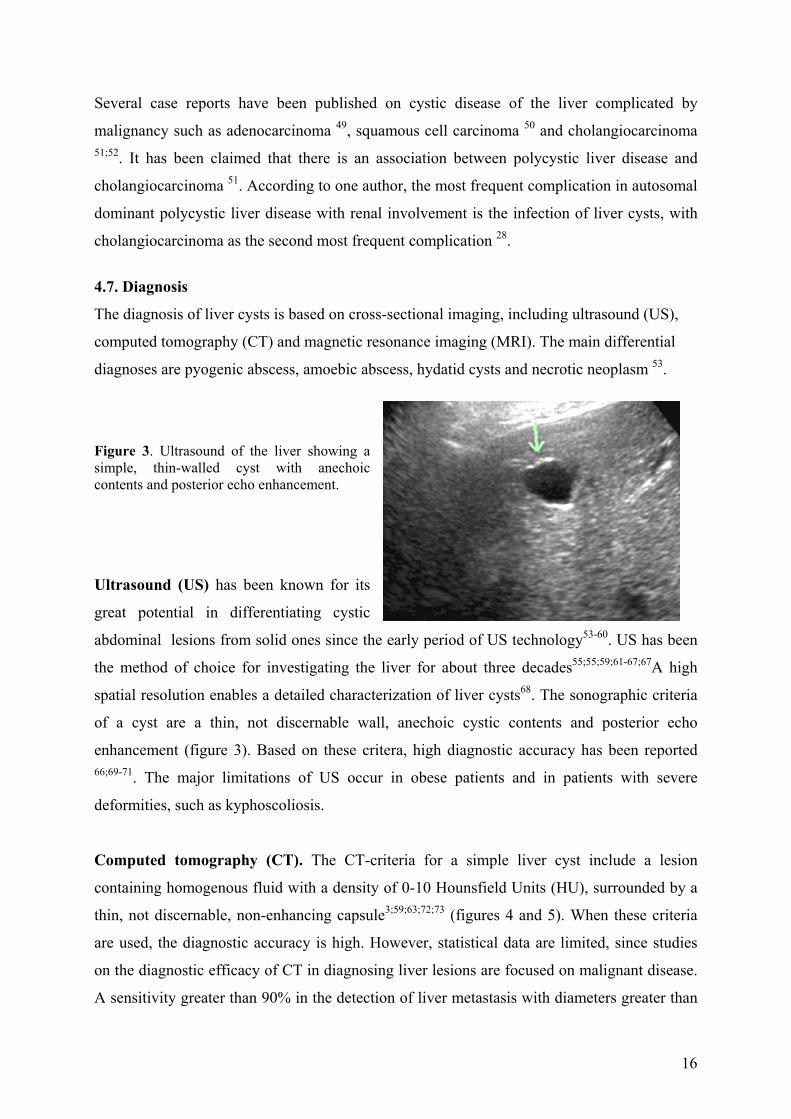
Computed tomography (CT). The CT-criteria for a simple liver cyst include a lesion
containing homogenous fluid with a density of 0-10 Hounsfield Units (HU), surrounded by a
thin, not discernable, non-enhancing capsule3;59;63;72;73 (figures 4 and 5). When these criteria
are used, the diagnostic accuracy is high. However, statistical data are limited, since studies
on the diagnostic efficacy of CT in diagnosing liver lesions are focused on malignant disease.
A sensitivity greater than 90% in the detection of liver metastasis with diameters greater than
17
10 mm has been reported74-79. When CT findings are equivocal, US63or MRI79 are utilized to
distinguish cystic lesions from solid ones. In cases of patchy capsular enhancement or a
thickened capsule, neoplasm or abscess is the more likely diagnosis. After a haemorrhage into
a cystic cavity, CT may show high attenuation consistent with blood clots for 1-3 days
following the event. In these cases, CT is more accurate than US.
Because CT, US and MRI alone are unable to differentiate between a simple liver cyst and a
hydatid cyst, additional tests have to be performed if hydatid disease is suspected 80.
Figure 4. CT scan demonstrating a large liver cyst with a thin wall and homogenous contents.
Figure 5 CT scan showing multiple liver cysts in polycystic liver disease.
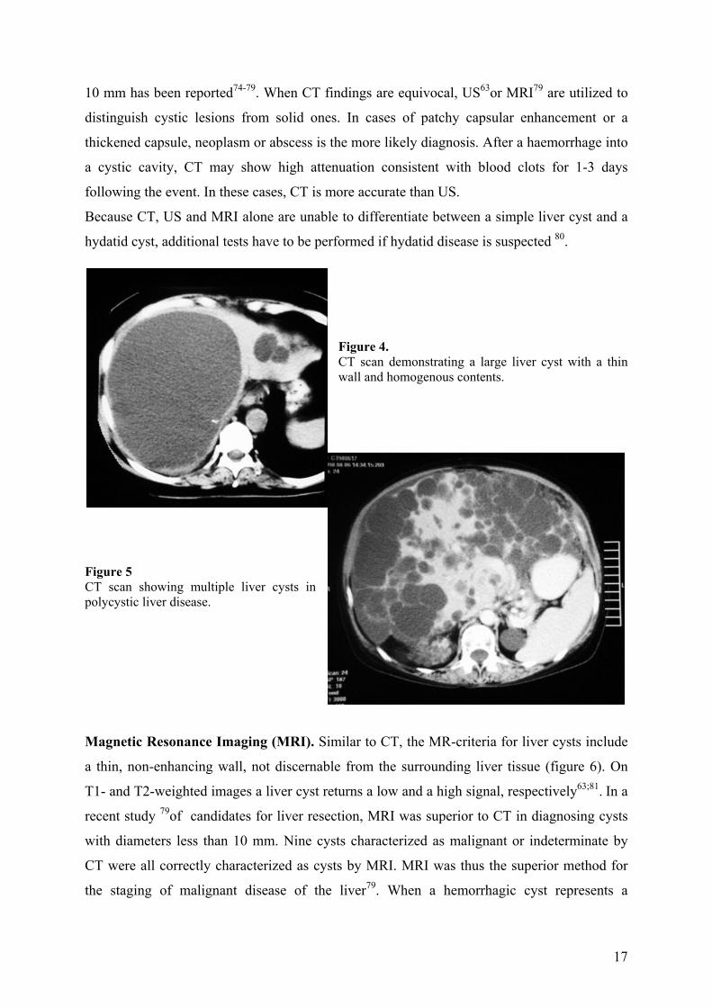
Magnetic Resonance Imaging (MRI). Similar to CT, the MR-criteria for liver cysts include
a thin, non-enhancing wall, not discernable from the surrounding liver tissue (figure 6). On
T1- and T2-weighted images a liver cyst returns a low and a high signal, respectively63;81. In a
recent study 79of candidates for liver resection, MRI was superior to CT in diagnosing cysts
with diameters less than 10 mm. Nine cysts characterized as malignant or indeterminate by
CT were all correctly characterized as cysts by MRI. MRI was thus the superior method for
the staging of malignant disease of the liver79. When a hemorrhagic cyst represents a
18
diagnostic problem on US and CT, MRI may provide characteristic diagnostic
information3;63;82.
Figure 6. Axial T1- weighted MRI in a 56-year-old female with policystic liver disease. High intensity liver- lesions are consistent with cysts containing haemoglobin or haemoglobin degradation products. Low-intensity lesions represent cysts containing ordinary cystic fluid.
4.8.Treatment of liver cysts
Indications for treatment are chronic symptoms which lead to a general reduction of the
quality of life, such as hepatomegaly, pain, local bulging, early satiety due to compression of
the stomach, biliary duct compression, as well as acute abdominal symptoms caused by cystic
rupture or hemorrhage into the cyst. Traditionally, surgery has been the method of choice for
symptomatic liver cysts45;46. During the past two decades, two different percutaneous methods
have been described: laparoscopic fenestration5;80 and percutaneous sclerotherapy31;32;83;84.
4.8.1. Surgery
The alternative surgical methods for treating symptomatic liver cyst are fenestration, liver
resection and liver transplantation.
Fenestration (also termed de-roofing) is the surgical method most commonly used today, and
consists of making an opening - a fenester or window - in the cyst. Thereafter the cystic fluid
is drained into the peritoneal cavity from which it is resorbed and subsequently eliminated via
the urine. This method can be performed laparoscopically. Some publications report
favourable results of laparoscopic cyst fenestration both of simple cysts 5;80;85 and polycystic
liver disease 86.However, this number of studies is few. In addition, the quantity of patients in
each study is low, and the times of observation are short5. Nor have random studies
comparing different surgical methods with sclerotherapy been carried out5. In order to prevent
recurrence, laparoscopic fenestration has been combined with the use of an omental
transposition flap 87. Others have used a combination of laparoscopic fenestration and ethanol
sclerotherapy 88;89. Only after the failure of laparoscopic fenestration should open surgical
fenestration or other surgical methods be used. Fenestration performed in open surgery
combined with liver resection is usually only appropriate in severe cases of polycystic liver
disease45;46.
Until recently, Liver resection has been the traditional surgical method for symptomatic liver
cysts. In solitary or few cysts, partial or total cystectomy and more infrequently formal
19
hepatic resection was performed. The recurrence rate following partial cystectomy has been
reported as from 0 to 37 % 4. In polycystic liver disease, surgical treatment may be indicated
when severe hepatomegaly reduces the quality of life severely or when complications such as
jaundice occur, due to compression of biliary ducts 90. In the largest series published on liver
resection combined with fenestration the mortality and morbidity rates rate were 3% and 58%,
respectively. This report involved severe cases of polycystic liver disease46.
Liver transplantation is a high risk and high cost procedure with a mortality ranging from 0-
20%4;48;91-94. For patients who survive and avoid severe postoperative morbidity, great
improvement of the quality of life may be achieved 29;48;93;95. Lifelong immuno-suppressive
treatment may be necessary 4.
From a surgical point of view, polycystic liver disease may be subdivided into three
categories: Type I, Type II and Type III 4. In Type I, there are a few large cysts, well suited
for laparoscopic fenestration as well as for sclerotherapy. Although the results of liver
resection or total cystectomy for Type I polycystic disease are good, with postoperative
morbidity of only 10%, few recurrences and only one postoperative death reported4, the
procedure of choice should be sclerotherapy5.
In Type II polycystic liver disease, some segments of the liver are relatively free of cysts
while others are diffusely involved, with large numbers of small cysts. If at least two adjacent
liver segments are relatively free of cysts, surgery is worth considering. Liver resection
combined with fenestration of multiple cysts is the method of choice. The reported
peroperative mortality has been 0-20%4, whereas postoperative morbidity may be as high as
50% 4.
In Type III polycystic liver disease, hepatomegaly is due to innumerable small cysts involving
all of the liver segments, and liver tranplantation may be the only therapeutic alternative 29;48;93;95.
4.8.2. Percutaneous interventional procedures.
The alternative methods are aspiration without or with sclerotherapy, using ethanol or other
sclerosing agents. In 1983, Saini reported on 15 sole aspiration procedures on liver cysts in 13
patients. Within two years, all cysts had regained their original size 96. Some cysts recurred
within two weeks. The exact interval before recurrence was not noted, but it became clear that
cysts may recur rapidly following aspiration alone. If the symptoms disappeared after
20
aspiration and reappeared when the cyst recurred, surgery was considered necessary. This
diagnostic procedure was initially used therapeutically, but still has considerable value
in deciding whether to perform sclerotherapy.
Sclerotherapy.
In 1976, Goldstein97, having been inspired by the work of Vestby98;99 on sclerotherapy of
renal cysts by means of panthopaque, injected panthopaque in a large, symptomatic liver cyst.
Although Goldstein’s results were promising with no recurrence after eight or 16 months, no
further work on sclerotherapy using panthopaque has been published. The first paper on liver
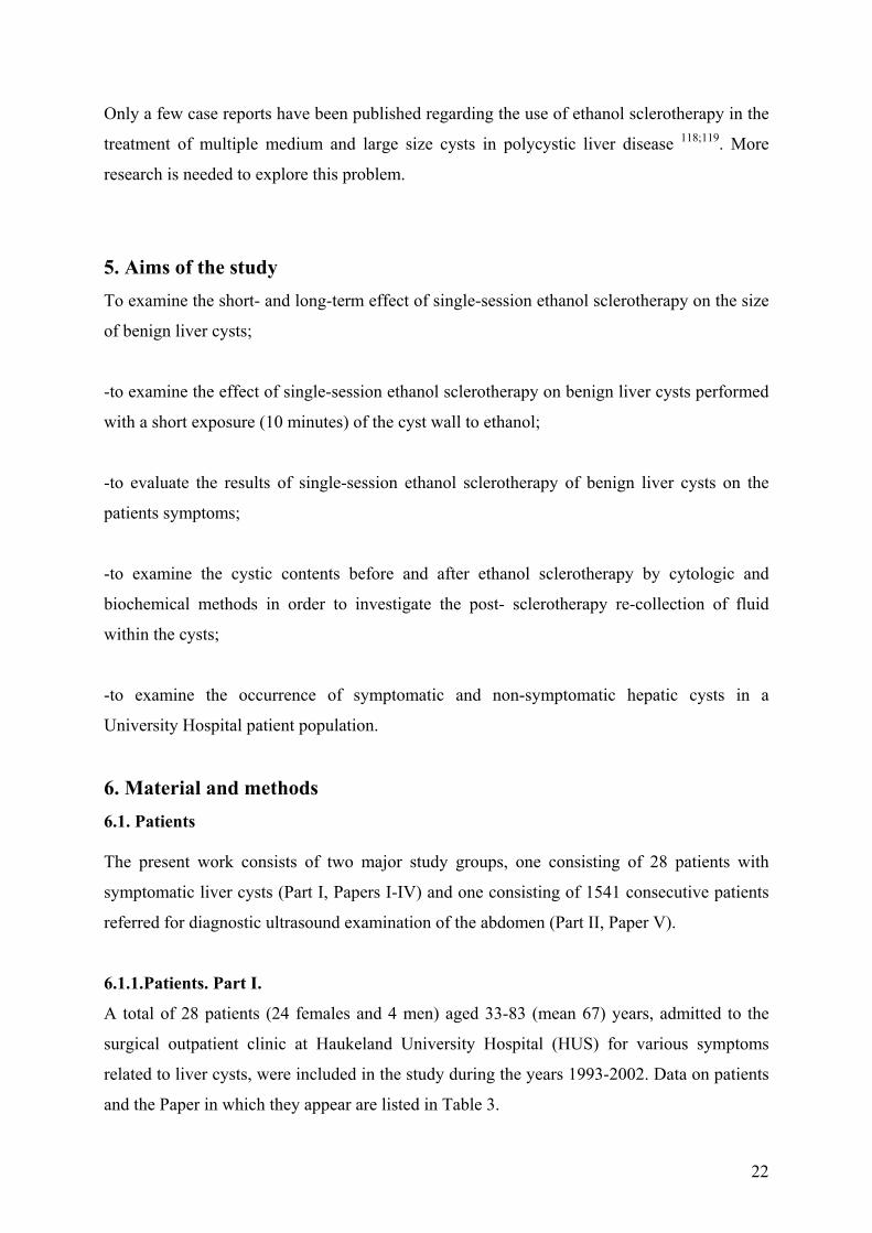
cyst sclerotherapy using ethanol was published by Bean in 1985 84. He reported a good
response to sclerotherapy of six cysts in six patients. His work was followed by others, who
reported high success rates ranging from 70 to100% using differing procedures (Table 2).
Anderson 83, vanSonnenberg 32 and Furuta100 used multiple-session procedures, while other
authors used a single-session procedure 101. Moreover, the time of exposure to ethanol varied
considerably among different authors. Although the results were promising, the method did
not gain general acceptance among liver surgeons. In a review of the literature5 concerning
nearly all studies on ethanol sclerotherapy31;32;83;84;101-105 and laparoscopic surgery85;87;106-114
published in English until 2001, the authors concluded that ethanol sclerotherapy probably
was as effective as laparoscopic fenestration, having the advantage of fewer complications.
They also concluded that a random controlled trial comparing ethanol sclerotherapy with
laparoscopic surgery would be ideal, but that such a study would be difficult to conduct since
symptomatic simple cysts are rare and long-term follow-up would be necessary5.
The use of minocycline hydrochloride as a sclerosing agent has been reported in three studies 115-117. A total of 15 patients were treated for simple hepatic cysts. The results were promising,
but the number of patients is limited, and further clinical research is necessary.
21
Table 2. Ethanol sclerotherapy of benign liver cysts: data from several different authors. ___________________________________________________________________________________ Author Cysts / Sessions Time of Maximum Success patients per exposure alcohol rate (%) Year cyst (minutes) volume(ml) ______________________________________________________________________ Bean 6/6 1 20 200 100 1985 Kairaluoma 15/8 1 60 100 100 1989 (1) Andersson 9/9 1-8 10-20 100 89 1989 Simonetti 30 1? 20-30 20-30% 70 1993 of cyst vol. Montorsi 21/21 ? 20-30 25% 72 1994 of cyst vol. vanSonnen- 14/ (2) 1 –11 (3) 20 -–0 33-50% 88 berg 1994 of cyst vol. Tikkakoski 59/25 1 60 100 97 1996 (1) (1) The study published by Tikkakoski was carried out at the same institution (Oulu University
Hospital, Finland) and includes the same patients as the study published by Kairaluoma. Tikkakoski added patients treated during the period 1987- 1992. Both of these authors applied 100ml alcohol for 20 minutes 3 times (3 times 100ml for a total of 60 minutes) in cyst with volumes over 1000 ml.
(2) In vanSonnenbergs’ study, 24 cysts in 20 patients were treated: 14 cyst with alcohol alone, 10
cysts with tetracycline, doxycycline or a combination of alcohol and tetracycline or doxycycline. (3) Using the multiple session procedure, one single cyst was treated by 11 different sessions
corresponding to 11 doses of ethanol (33-50% of cyst volume x 11), and a cumulated time of exposure to ethanol of 330 minutes during 44 days of catheter drainage.
In the treatment of polycystic liver disease, the comparative roles of ethanol sclerotherapy and
laparoscopic surgery have not been clarified in the review study5. The use of laparascopic
fenestration in these patients was limited. Another study86 has concluded that although the
exact role of laparascopic cyst fenestration in the treatment of polycystic liver disease remains
unclear, the method appears to be of benefit to a limited, selected number of patients. In this
study, this therapy is recommended in massive hepatic cystic disease when the cysts are not
well suited for percutaneous sclerotherapy.
22
Only a few case reports have been published regarding the use of ethanol sclerotherapy in the
treatment of multiple medium and large size cysts in polycystic liver disease 118;119. More
research is needed to explore this problem.
5. Aims of the study To examine the short- and long-term effect of single-session ethanol sclerotherapy on the size
of benign liver cysts;
-to examine the effect of single-session ethanol sclerotherapy on benign liver cysts performed
with a short exposure (10 minutes) of the cyst wall to ethanol;
-to evaluate the results of single-session ethanol sclerotherapy of benign liver cysts on the
patients symptoms;
-to examine the cystic contents before and after ethanol sclerotherapy by cytologic and
biochemical methods in order to investigate the post- sclerotherapy re-collection of fluid
within the cysts;
-to examine the occurrence of symptomatic and non-symptomatic hepatic cysts in a
University Hospital patient population.
6. Material and methods 6.1. Patients The present work consists of two major study groups, one consisting of 28 patients with
symptomatic liver cysts (Part I, Papers I-IV) and one consisting of 1541 consecutive patients
referred for diagnostic ultrasound examination of the abdomen (Part II, Paper V).
6.1.1.Patients. Part I.
A total of 28 patients (24 females and 4 men) aged 33-83 (mean 67) years, admitted to the
surgical outpatient clinic at Haukeland University Hospital (HUS) for various symptoms
related to liver cysts, were included in the study during the years 1993-2002. Data on patients
and the Paper in which they appear are listed in Table 3.
23
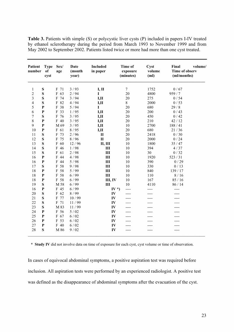
Table 3. Patients with simple (S) or polycystic liver cysts (P) included in papers I-IV treated by ethanol sclerotherapy during the period from March 1993 to November 1999 and from May 2002 to September 2002. Patients listed twice or more had more than one cyst treated. Patient Type Sex/ Date Included Time of Cyst Final volume/ number of age (month in paper exposure volume Time of observ cyst year) (minutes) (ml) (ml/months) __________________________________________________________________________________________ 1 S F 71 3 / 93 I, II 7 1752 0 / 67 2 S F 63 2 / 94 I 20 4800 959 / 7 3 S F 74 3 / 94 I,II 20 275 0 / 54 4 S F 82 4 / 94 I,II 8 2000 0 / 53 5 P F 38 5 / 94 I 20 680 29 / 8 6 P F 33 1 / 95 I,II 20 200 0 / 43 7 S F 76 3 / 95 I,II 20 450 0 / 42 8 P F 40 3 / 95 I,II 20 210 42 / 12 9 P M 69 3 / 95 I,II 10 2700 188 / 41 10 P F 61 8 / 95 I,II 20 680 21 / 36 11 S F 73 2 / 96 II 20 2418 0 / 30 12 S F 75 8 / 96 II 20 2000 0 / 24 13 S F 60 12 / 96 II, III 10 1800 35 / 47 14 S F 46 1 / 98 III 10 394 4 / 37 15 S F 61 2 / 98 III 10 30 0 / 32 16 P F 44 4 / 98 III 10 1920 523 / 31 16 P F 44 5 / 98 III 10 390 0 / 29 17 S F 58 9 / 98 III 10 330 0 / 13 18 P F 58 5 / 99 III 10 840 139 / 17 18 P F 58 6 / 99 III 10 110 8 / 16 18 P F 58 6 / 99 III, IV 10 167 85 / 16 19 S M 58 6 / 99 III 10 4110 86 / 14 16 P F 45 6 / 99 IV *) ---- ---- ---- 20 S F 62 8 / 99 IV ---- ---- ---- 21 S F 77 10 / 99 IV ---- ---- ---- 22 S F 71 11 / 99 IV ---- ---- ---- 23 S M 83 11 / 99 IV ---- ---- ---- 24 P F 56 5 / 02 IV ---- ---- ---- 25 P F 67 6 / 02 IV ---- ---- ---- 26 P F 53 6 / 02 IV ---- ---- ---- 27 P F 40 6 / 02 IV ---- ---- ---- 28 S M 86 9 / 02 IV ---- ---- ---- __________________________________________________________________________________________ * Study IV did not involve data on time of exposure for each cyst, cyst volume or time of observation.
In cases of equivocal abdominal symptoms, a positive aspiration test was required before
inclusion. All aspiration tests were performed by an experienced radiologist. A positive test
was defined as the disappearance of abdominal symptoms after the evacuation of the cyst.
24
The criteria for inclusion in the study were cyst-related symptoms as judged by an
experienced gastrointestinal surgeon and one or more symptomatic liver cysts diagnosed by
imaging.
The criteria for exclusion from the study were: hydatid cyst; neoplastic cyst; cyst-like ectasies
of the intrahepatic biliary ducts (Carolis disease); coagulopathy; or polycystic liver disease
with innumerable small cysts but without large or medium-size cysts suitable for
sclerotherapy.
After contrast injection and before the instillation of ethanol, the following three criteria had
to be met: 1) no communication between the cyst and the biliary tree; 2) no intraperitoneal
leakage of contrast; 3) the ability to aspirate the contrast from the cystic cavity.
Informed consent concerning ethanol sclerotherapy was given by all patients.
6.1.2. Patients, Part II
A total of 1541 patients (869 females and 672 males), aged 0-99 (mean 57.7) years, referred
for abdominal ultrasound to the Department of Radiology, Haukeland University Hospital
were included during the period 21 January 2000 to 11 November 2000. A referring
Department was registered for 1240 of the patients: 404 patients from the Department of
Oncology (all patients had either active or healed malignant disease); 290 patients from the
Department of Internal Medicine; 243 patients from the Department of Surgery; 100 patients
from the Department of Paediatrics; and 243 patients from other departments (Departments of
Neurology, Dermatology, Rheumatology, Psychiatry and Emergency).
6.2. Methods - Part I.
6.2.1. Study design.
Part I was a prospectively conducted, observational and experimental study. The clinical
examinations prior to inclusion were done by an experienced gastrointestinal surgeon, who
classified symptoms as mild, moderate or severe. Pre-procedure liver function tests were
taken. If there was no suspicion of liver tissue damage at that time, these tests were not
repeated at clinical follow-up. To investigate the short- and long-term effect of 20 minutes
exposure to ethanol, 13 patients were studied (Papers I and II), and to investigate the effect of
10 minutes exposure to ethanol, 7 patients were studied (Paper III). To investigate the
cytologic and biochemical aspects of ethanol sclerotherapy, 11 patients were studied (Paper
IV).
25
6.2.2. The effect of sclerotherapy.
The effect of sclerotherapy was evaluated by the measurement of the pre-and post-procedure
cyst volume, by the registration of symptoms and through laboratory tests.
Measurements of cyst volume.
The cyst volume on the day of sclerotherapy was measured by direct measurement of the
aspirated cystic contents. Measurements of cyst volume was performed using CT and the
formula: V = d1 x d2 x d3 x 0.523 before sclerotherapy and at follow-ups scheduled at 3, 6,
12 and 24 months. This method has been applied to uterine and ovary volume measurement
using ultrasound120.
Clinical evaluation of symptoms, and laboratory tests.
The patients were seen at an outpatient clinical consultation scheduled at 3 and 6 and 12
months after sclerotherapy. The symptoms were classified as improved, unchanged or
deteriorated at each follow-up. The following liver function tests were taken before
sclerotherapy: Alanine aminotransferase (ALT), aspartate aminotransferase (AST), glutamyl
transpeptidase (GT), bilirubin, and alkaline phosphatase. Also hemoglobin, thrombocytes,
cephotest and INR were taken. A test for hydatid disease was taken before sclerotherapy when
this diagnosis was suspected.
Evaluation of side effects. Procedural pain was evaluated by the radiologist during the procedure, and classified as
absent, mild, moderate or severe. During our early work with ethanol sclerotherapy we tested
for increased blood ethanol concentration one hour after the sclerotherapy procedure. The
routine use of this test was discontinued due to low values of ethanol and absence of clinical
signs of ethanol intoxication. We later determined ethanol intoxication by constantly
evaluating the patients’ apprehension and level of conciousness. Therefore, a specific ethanol
measurement was only made if there was suspicion of ethanol intoxication based upon clinical
evaluation, or if large cysts requiring the largest volume of ethanol were treated, particularly
if the cyst wall was in close contact with large vessels.
26
Etiology of cystic fluid reproduction after sclerotherapy To investigate the etiology of the re-filling of the cystic cavity, the cystic fluid of 11 cysts in
11 patients was examined on the day of sclerotherapy and 2-8 days (mean 4.5) later and
analysed for cytologic and biochemical parameters. These eleven patients represented all
patients referred for sclerotherapy during two periods, the first period from September 1998 to
November 1999 and the second from May 2002 to September 2002. Two cysts were
examined only by biochemical methods, since too many erythrocytes disturbed the cytologic
examination. As a result, nine cysts were examined by cytologic methods and eleven cysts by
biochemical methods.
The cytologic examination was made by qualified technologists using a manual semi-
quantitative method by which the percentages of the different cellular components could be
calculated. The biochemical analysis included CRP, orosomucoid and haptoglobin indicating
acute inflammatory reaction, protein and albumin indicating capillary permeability, and
bilirubin and alkaline phosphatase as indicators of cystic epithelial function.
In the last five patients, examined in 2002, blood was analysed for the same biochemical
parameters on the day of sclerotherapy. The second blood sample was taken 2-3 days (mean
2.2) after sclerotherapy.
6.3. Sclerotherapy procedure
The patients were admitted to the hospital either the day prior to or early in the morning of the
day the procedure was scheduled. For the first 22 patients, a premedication consisting of 50-
100 mg pethidine and 0.6 mg atropine i.m.was administered 30-45 minutes before the start of
the procedure and supplementary i.v.pethidine injections were given during the procedure if
required. For the last 5 patients a different method of analgesia was used. This method
differed in that an anesthetic nurse was present throughout the procedure, and no
premedication was given. Sedation was achieved by midazolam (Dormicum; Roche) and pain
control by means of short-term analgesics, usually alfentanil (Rapifen “Janssen-Cilag”), and
more infrequently ketamin (Ketalar “Pfizer”), given intravenously during the procedure
according to the individual patient’s needs.
The patient was placed on an angiographic table and the local anesthetic lidocaine 10mg/ml
(Xylocain “Astra Zeneca”) was administered. Figures 7, 8A and 8B show the equipment used
for the sclerotherapy procedure. Under ultrasound guidance, the needle tip was inserted into
the cyst. An 18G, 20 cm needle (COOK , catalogue number SDN- 18- 20 -T ) was preferred.
27
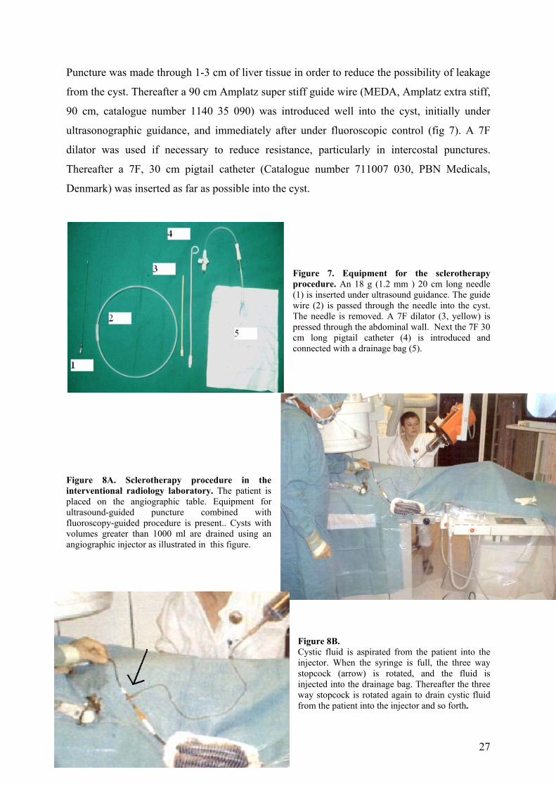
Puncture was made through 1-3 cm of liver tissue in order to reduce the possibility of leakage
from the cyst. Thereafter a 90 cm Amplatz super stiff guide wire (MEDA, Amplatz extra stiff,
90 cm, catalogue number 1140 35 090) was introduced well into the cyst, initially under
ultrasonographic guidance, and immediately after under fluoroscopic control (fig 7). A 7F
dilator was used if necessary to reduce resistance, particularly in intercostal punctures.
Thereafter a 7F, 30 cm pigtail catheter (Catalogue number 711007 030, PBN Medicals,
Denmark) was inserted as far as possible into the cyst.
Figure 7. Equipment for the sclerotherapy procedure. An 18 g (1.2 mm ) 20 cm long needle (1) is inserted under ultrasound guidance. The guide wire (2) is passed through the needle into the cyst. The needle is removed. A 7F dilator (3, yellow) is pressed through the abdominal wall. Next the 7F 30 cm long pigtail catheter (4) is introduced and connected with a drainage bag (5).
Figure 8A. Sclerotherapy procedure in the interventional radiology laboratory. The patient is placed on the angiographic table. Equipment for ultrasound-guided puncture combined with fluoroscopy-guided procedure is present.. Cysts with volumes greater than 1000 ml are drained using an angiographic injector as illustrated in this figure.
Figure 8B. Cystic fluid is aspirated from the patient into the injector. When the syringe is full, the three way stopcock (arrow) is rotated, and the fluid is injected into the drainage bag. Thereafter the three way stopcock is rotated again to drain cystic fluid from the patient into the injector and so forth.
28
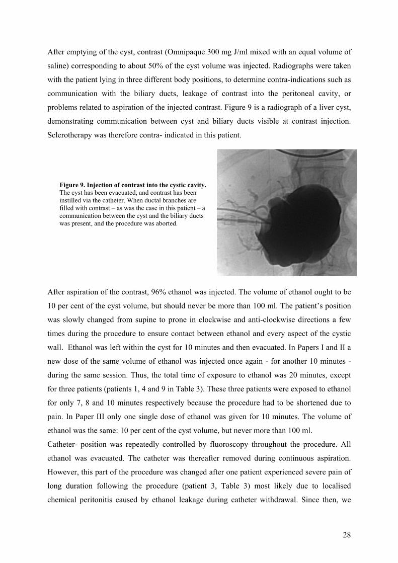
After emptying of the cyst, contrast (Omnipaque 300 mg J/ml mixed with an equal volume of
saline) corresponding to about 50% of the cyst volume was injected. Radiographs were taken
with the patient lying in three different body positions, to determine contra-indications such as
communication with the biliary ducts, leakage of contrast into the peritoneal cavity, or
problems related to aspiration of the injected contrast. Figure 9 is a radiograph of a liver cyst,
demonstrating communication between cyst and biliary ducts visible at contrast injection.
Sclerotherapy was therefore contra- indicated in this patient.
Figure 9. Injection of contrast into the cystic cavity. The cyst has been evacuated, and contrast has been instilled via the catheter. When ductal branches are filled with contrast – as was the case in this patient – a communication between the cyst and the biliary ducts was present, and the procedure was aborted.
After aspiration of the contrast, 96% ethanol was injected. The volume of ethanol ought to be
10 per cent of the cyst volume, but should never be more than 100 ml. The patient’s position
was slowly changed from supine to prone in clockwise and anti-clockwise directions a few
times during the procedure to ensure contact between ethanol and every aspect of the cystic
wall. Ethanol was left within the cyst for 10 minutes and then evacuated. In Papers I and II a
new dose of the same volume of ethanol was injected once again - for another 10 minutes -
during the same session. Thus, the total time of exposure to ethanol was 20 minutes, except
for three patients (patients 1, 4 and 9 in Table 3). These three patients were exposed to ethanol
for only 7, 8 and 10 minutes respectively because the procedure had to be shortened due to
pain. In Paper III only one single dose of ethanol was given for 10 minutes. The volume of
ethanol was the same: 10 per cent of the cyst volume, but never more than 100 ml.
Catheter- position was repeatedly controlled by fluoroscopy throughout the procedure. All
ethanol was evacuated. The catheter was thereafter removed during continuous aspiration.
However, this part of the procedure was changed after one patient experienced severe pain of
long duration following the procedure (patient 3, Table 3) most likely due to localised
chemical peritonitis caused by ethanol leakage during catheter withdrawal. Since then, we

29
irrigated the cyst with saline after aspiration of all ethanol. Saline was thereafter aspirated and
the catheter removed, thus avoiding the leakage of ethanol into the peritoneum.
After the procedure the patient rested in bed for 4 hours. During our initial years of
sclerotherapy all patients were hospitalised for at least 24 hours. Later, patients were usually
able to leave the hospital the same evening, if symptoms were minimal.
Methods - Part II. Study design – method:
Part II was a prospective, cross-sectional study. The ultrasound examinations were performed
by 24 physicians (eight consultants, 16 residents) with experience in US varying from one to
20 years. A Toshiba Power Vision 7000 or an ATL HDI 5000 was used for adults, while
children were examined using an ATL HDI 5000 machine. All the units were equipped with
curved array and phased array sector multifrequency transducers. The ATL HDI 5000
ultrasound machine was equipped with three transducers used for examination of the liver.
The transducer most commonly used had a curved array design and a frequency range of 2.0–
5.0 MHz. The second transducers also had a curved array design, but with a frequency range
from 4.0–7.0 MHz, suitable for small individuals and children, and one minicurved transducer
with a frequency range from 2.0-4.0 MHz. The Toshiba PowerVision 7000 ultrasound
machine was equipped with one curved array and one minicurved transducer, both with a
frequency range of 3.0-6.0 MHz. All transducers of both machines were equipped with 128
piezo-electric crystals.
Criteria for a cyst were: focal liver lesion with anechoic contents; thin wall not distinguishable
from the adjacent liver tissue; and posterior acoustic enhancement. The presence or absence
of liver cysts was continuously recorded in a protocol at the ultrasound laboratory, and further
details with regard to diameter and number of cysts were recorded in the radiology report.
When the measurement of cyst diameter did not appear in the report, it was measured on the
ultrasound images by the investigator.
30
6.3. Statistics
Differences in cystic volumes before and after ethanol liver cyst sclerotherapy were examined
using the Wilcoxon Signed Ranks Test for non- parametric data (papers II-IV). Differences in
cystic volumes before and after sclerotherapy according to time of exposure to alcohol (10 vs.
20 minutes) were examined using a non-parametric test for comparison of two groups (Mann-
Whitney test). In Paper V the association between gender and the occurrence of cysts was
examined using Fisher's exact test and logistic regression analysis. The ability of senior
examiners and residents to diagnose liver cysts was tested using Fisher’s exact test. The
association between the occurrence of cysts, age of the patients and referring department was
examined using logistic regression analysis.
7. Summary of results Paper I.
During the period from March 1993 to August 1995, ten cysts in ten patients were treated
using single-session ethanol sclerotherapy. After a mean observation time of 17.3 (7.8 - 42.0)
months, the volumes of the cysts had decreased from a mean volume of 1375 (200 - 4800) ml
to a mean volume of 126 (0 - 966) ml. This represents a mean volume reduction of 90.8 (77 -
100) %. In eight cysts, post-procedural re-accumulation of cystic contents was observed, but
here too there was a satisfactory volume reduction some time later.
By protocol the cysts were supposed to be exposed to ethanol for 20 minutes, but due to pain
the time of exposure was reduced to 7, 8 and 10 minutes, respectively in three patients
(patients 1,3 and 9 in Table 3). Even though these large cysts, initial volumes 2700, 2000, and
1752 ml, were exposed for only 10 minutes or less, the volumes were reduced to 212 ml, 0
ml and 1 ml, a reduction in volumes of 92%, 100% and 100% after 18, 17 and 42 months
respectively. Pain was severe in one patient and moderate in four others during or
immediately following ethanol instillation. There were no other complications. There were no
abnormalities in liver function tests following sclerotherapy.
Paper II
From March 1993 to November 1998, 23 cysts in 19 patients (18 female and one male) were
treated by single-session ethanol sclerotherapy. Only patients with an observation period of a
minimum of 12 months after single-session ethanol sclerotherapy were deemed eligible for
the final analysis. Eleven patients with eleven treated cysts fulfilled this criteria. After an
31
observation period of mean 38.3 (12 - 67) months, cyst volumes were reduced from mean
1317 (200 - 2700) ml to mean 26 (0 - 188) ml, a volume reduction of mean 98 (93 -100)%.
Thus, the post-sclerotherapy reaccumulation of fluid was followed by a significant volume
reduction in 9 out of 10 patients. There were no complications.
Paper III
During the period from December 1996 to June 1999, 15 symptomatic liver cysts in nine
patients (eight women and one man) were treated with a 10 minute exposure to ethanol. One
70-year-old woman did not turn up for follow-up control examination, and the 4 cysts in her
polycystic liver were therefore excluded. One cyst was excluded because the 83-year-old
female patient died from cardiac disease 7 months after the procedure. Only patients with an
observation period of minimum 12 months after single-session ethanol sclerotherapy were
found eligible for the final analysis. In 10 cysts in 7 patients with observation periods of
median 23 (12-47) months, volumes were reduced from median 392 (30-4110) ml to median
21.5 (0-523) ml, a reduction of the median cyst volume by 95% (p < 0.005).
The only complication was pain during the procedure. During cyst aspiration, pain was severe
in two procedures and moderate in two. During ethanol instillation pain was severe in one
procedure and moderate in two. After evacuation of all ethanol, irrigation with saline and
removal of the catheter, one patient experienced moderate pain, while no patients experienced
severe pain. Liver function tests and clinical follow-up did not reveal any sign of damage to
biliary ducts or liver parenchyma. All patients experienced relief of their clinical symptoms.
Paper IV
During the period from September 1998 to November 1999 and during an additional period
from May 2002 to September 2002, cystic fluid from 11 cysts in 11 patients was examined on
the day of sclerotherapy, and again 2-8 days later(mean 4.5). The fluid was analyzed for
cytologic and biochemical parameters.
Biochemical parameters reflecting acute inflammatory reaction (C-reactive protein (CRP),
haptoglobin and orosomucoid), cyst epithelial function (bilirubin and alkaline phosphatase),
macromolecular leakage (protein and albumin), as well as parameters of hepatocyte function
(alanine aminotransferase (ALT) and aspartate aminotransferase (AST)) were significantly
elevated following sclerotherapy.
Cytologic signs of acute or subacute inflammatory reaction were absent before sclerotherapy,
but present in all cysts after sclerotherapy. The number of cellular elements were increased in
32
all cysts and the relative percentages of different kinds of leucocytes could be calculated. In
four cysts with an interval of two days between the first and the second sample 98-100%
neutrophile granulocytes were present. In one cyst with an interval of two days and in all four
cysts with an interval of six, seven or eight days the neutrophile granulocytes were partially
replaced by lymphocytes and macrophages. In one cyst containing 100% lymphocytes before
sclerotherapy, this was replaced by 98% neutrophile granulocytes and 2 % macrophages two
days after sclerotherapy.
In the last five patients an analysis of biochemical parameters in the blood was performed
both before and following sclerotherapy. Before and after sclerotherapy the median CRP
value in blood was 7 and 71 respectively. In the same patients the corresponding CRP values
in cystic fluid before and after sclerotherapy was 9 (range 0-10) and 10 (range 5-24),
respectively. The second blood sample was taken 2-3 days (mean 2.2) after sclerotherapy. The
other parameters only demonstrated minor differences in the values before and following
sclerotherapy. There were no complications.
Paper V From 21 January 2000 to 11 November 2000, 1541 patients referred for an abdominal
ultrasound examination were included. Liver cysts were diagnosed in 174 (11.3%). No cysts
were found in patients younger than 40 years of age. The occurrence increased with age (p <
0.0005). The occurrence in females was 12.5% (109/869) and in males 9.7% (65/672). This
difference was not statistically significant (p = 0.088). The occurrence of liver cysts did not
differ according to referring department (p = 0,559). Excluding 14 patients with polycystic
liver disease (8.1% of 174 livers containing cysts and 0.9% of all livers), 322 cysts were
recorded according to size in the remaining 160 patients: 91.9% with a diameter of 3 cm or
less; 6.2% with a diameter between 3.1 and 6 cm; and 1.9% with a diameter greater than 6
cm. Symptomatic cysts occurred in six patients, representing 0.4 % of all 1541 livers and 3.5
% of all 174 livers containing cysts. These were all large cysts with a mean diameter of 115
(60 - 180) mm. In examinations performed by senior radiologists cysts were diagnosed in
16.1%, and in examinations performed by residents in 15.2% (p=0.785).
33
8. General discussion Part I. Sclerotherapy of symptomatic liver cysts
Study design.
In principle, evaluation of new procedures and treatments ought to be performed using
random controlled trials. However, in the present study the alternative standard treatment was
surgery, a treatment associated with a higher morbidity and complication rate5;45. Based on
ethical considerations and on earlier reported encouraging results on sclerotherapy, we felt
that an observational trial offering sclerotherapy to patients in need of surgery was justified121.
Moreover, the low prevalence of the disease would have made it difficult to accumulate
enough patients for a random controlled trial5.
Initially (Paper I and II), we examined the effect of single-session ethanol sclerotherapy using
a time of exposure to ethanol of 20 minutes. Ethanol was left within the cyst for 10 minutes
and then evacuated. A new dose of ethanol - the same volume – followed immediately, lasting
for another 10 minutes, resulting in a total exposure to ethanol of 20 minutes. In three of the
initial patients, ethanol was only injected once - for 7, 8 and 10 minutes, respectively, -
because the patients experienced pain during the procedure. Although these cysts were large
(2700ml, 2000ml and 1752 ml respectively) and the time of exposure was reduced, the
sclerotherapy effect was still good, resulting in reductions of volume of 93%, 100% and 100
% at follow-up observation after 41months, 53 months and 67 months, respectively (Table 1,
Paper II). Due to this favourable result, we decided to conduct another study using a 10
minute exposure to ethanol consecutively (Paper III). Ideally, the evaluation of 10 versus 20
minutes exposure could have been performed using a random design. To the present, we have
found no reports on the effect of different sclerotherapy methods using a random design. This
in part reflects the difficulties in obtaining enough cases. Two reports on ethanol
sclerotherapy of simple renal cysts compare single-session with multiple-session
procedure122;123. In one paper, 82 cysts in 82 patients were reported. Forty-two cysts were
treated by single-session and 40 by three different instillations of ethanol at 12 hour intervals.
The mean follow-up periods were 12.9 and 15.4 months, respectively122. In another study, 19
renal cysts in 15 patients were treated by single instillations of ethanol and 13 cysts in 11
patients by repeated instillations of ethanol123. Both these studies concluded that the results
were better when more than one session was used. Another author recommended the use of
multiple sessions for renal cyst ethanol sclerotherapy123;124. In yet another study the long-term
outcome of single-session sclerotherapy of 32 cysts in 32 patients was reported125. The mean
34
follow-up observation period was 55 months. The mean cyst diameter was 7.8cm before and
1.7cm after sclerotherapy (p>0001). They concluded that single-session procedure of
symptomatic renal cysts was efficient.
In the present study, we only included patients with significant symptoms who were judged
by the gastrointestinal surgeon to be in need of surgery. During the study period, all patients
suffering from symptomatic liver cysts were included. No patient also surgically treated was
included except for patient 2, Table III (see below). No patients withdrew from the study after
having given their informed consent.
Another limitation of the study was the deviation from the follow-up protocol. The frequency
of follow-ups had been established at 3, 6 and 12 and 24 months. The intervals did vary, as
illustrated in figures 1 in Papers I, II and III. In spite of these variations, the scleroterapy
effect could still be evaluated. In one patient in Paper II the first follow-up was made 24
months after sclerotherapy. The volume reduction was 100 %, but since no examination was
made at 3 or 6 months it was impossible to determine any presence or absence of a pattern of
temporary post-procedural reaccumulation of fluid. Another patient included in Paper I was
treated for a 4800ml cyst. She was seen at follow-up only once, at 7 months. By that time the
volume of the cyst was reduced by 80%. Nevertheless, the surgeon operated on this patient
one month later because her symptoms had not improved. After surgery her symptoms were
unchanged.
Pre and post-procedure measurements of cyst volume
Accuracy of the method
In the present study the cyst volume was estimated on the basis of the three maximal
diameters (90° angles) measured on the CT images, according to the formula of an ellipsoid:
Volume (V) = d1 x d2 x d3 x 0.523 120. This method has been used for the measurement of
ovarian volume120, and the reported accuracy is good126. The method has also been used for
the measurement of fetal urinary bladder volume for three decades127;128. Still others have
estimated liver cyst volume 101;104and renal cyst volume125 by measuring one diameter .
The effect on cyst volume
The present studies showed that single-session ethanol sclerotherapy reduced cystic volume
significantly over time. Our results support the initial observations of Kairaluoma, who in
35
1989 described that a single-session procedure was sufficient in spite of the refilling of the
cyst after sclerotherapy, since the re-collected fluid was later resorbed. He claimed that this
resorbtion and volume reduction lasted for more than one year102. Kairaluoma treated 15 cysts
in eight patients. His first two patients were treated by a repeat sclerotheraphy procedure, the
initial procedure being repeated after one or two months. He did observe that there was a
temporary re-collection of fluid during the first two months after the initial sclerotheraphy
procedure, but that the cyst thereafter decreased in size for at least two years. Due to this
observation, he performed sclerotheraphy only once, with satisfactory results, on the
following six patients. In our study, all patients of Papers I, II and III who had a follow-up
examination during the first two months after sclerotherapy (16 out of 19 patients) had a re-
collection of fluid within the cyst (fig 1, Paper I, fig 1, Paper II and fig 1, Paper III). In all
these patients this fluid was reduced in volume by 49-100% (median 95%) at later follow-ups.
The post-sclerotherapy production of fluid within the cyst had also been observed by
vanSonnenberg 32and Anderson83 in series of 14 and 9 patients, respectively. However, we
believe that their observations led to an erroneous conclusion. They recommended that a
percutaneous catheter be left in place until the next day, and that a repeated sclerotherapy
procedure be performed if drainage was more than 10 – 15 ml per 24 hours. This resulted in a
maximum time of drainage of 44 days (Table 2), as one single cyst was treated by 11 different
sessions during 44 days of catheter drainage32. In our series, however, we showed that the
long-term effect was excellent after one single sclerotherapy procedure when the catheter was
removed immediately. There was a 99% reduction of cyst volume after seven to 67 months
follow-up. Thus, our studies have shown that the post-sclerotherapy re-collection of fluid is
temporary.
Supplementary analysis:
To examine the effect of sclerotherapy in regard to the duration of ethanol exposure, we
carried out a retrospective analysis on the total study population included in Papers I-III
(Table 3). A total of 19 patients (22 cysts) were studied, 17 women (89.4%) and 2 men (11.6
%) with a median age of 61.2 (32.5-81.7) years. The median volume of cystic fluid was 680
(30-4800) ml before and 6 (0-959) ml after sclerotherapy, a median volume reduction of 99.1
% (p < 0,005). The median follow-up period was 30.8 (7.1 – 67.0) months.
We divided the study population into those who had received an exposure of 10 minutes or
less to ethanol (10 patients, 13 cysts) and those who had received an exposure of 20 minutes
(9 patients, 9 cysts). For the group who had received 10 minutes or less of exposure, the
36
median pre-procedure cyst volumes was 840 ml (30-4110 ml) and the median post-procedure
volume was 8 ml (0-470ml), a median volume reduction of 732 ml (99.1%) (p < 0.005). The
median time of observation was 30.5 (13.1 – 67.0) months. For the 9 patients (9 cysts) treated
with 20 minutes exposure, the median volume was 680 ml (200-4800 ml) before and 0 ml (0-
959ml) after sclerotherapy, a median volume reduction of 680 ml (100%) (p < 0.005). The
median time of observation was 30.0 (7.1 – 54) months. The degree of cyst reduction did not
differ between the two groups (Mann- Whitney test for non-parametric data, p=0.896),
confirming that 10 minutes of exposure is sufficient for the procedure to be successful.
Clinical evaluation of symptoms
In the present study, 26 out of a total of 28 patients reported that their symptoms had
improved following sclerotherapy. Evaluation of symptoms was assessed by a gastrointestinal
surgeon, who obtained a clinical statement on the condition of each patient (patient opinion)
prior to the procedure. This was repeated at all the follow-up examinations at an outpatient
clinical consultation 3, 6 and 12 months after treatment. Although specific methods of
assessing pain and nausea have been developed to impose some structure on the information
we were collecting, none of these were found to embrace the subjective symptom complexity
of liver cysts, which includes different types of pain, early satiety, shortness of breath,
reduced physical mobility and fitness, and psychological problems. Thus, we chose to
evaluate the symptoms in their entirety, classifying them as the patients’ determination of
improved, not changed or worsened. This may be seen to represent a weakness in the present
study, but the examiner’s extensive experience in evaluating clinical symptoms related to liver
disease should in part outweigh these shortcomings. Our results are supported by those of
others 31;32;83;84;101;102;104.
Evaluation of side effects.
Procedural pain.
Other than pain, there were no complications, a finding also reported by others 31;32;83;84;101;102;104. In March 1994, during the second year of this study, 27 ml of ethanol was
used to treat a 275 ml cyst (patient number 3, Table 3). When the catheter was removed after
aspiration of ethanol, the patient experienced severe pain which lasted several days. We
concluded that this may have been due to the leakage of ethanol into the peritoneal cavity.
Even though it was considered that all ethanol was evacuated from the cyst before the catheter
was removed, some ethanol may well have remained within the catheter. This would be
37
sufficient to create a localised chemical peritonitis. Due to this experience we changed our
procedure. At the end of each procedure, when all ethanol had been evacuated, we irrigated
the cyst with saline before removing the catheter. After this change of procedure, we have
never had a similar experience as that described above.
Also at the beginning of this study we observed that pain could be severe during cyst
evacuation, before the application of ethanol, particularly in polycystic liver disease. We also
observed that this pain during cyst evacuation was particularly severe in patients previously
treated by liver surgery, and was probably the result of adhesions. This may well be similar to
the experience of liver surgeons that laparoscopic fenestration of liver cysts is technically
difficult if liver surgery has been done earlier, so that previous surgery is considered as a
contra-indication for laparoscopic surgery120;129.
As mentioned above, to improve pain control during the procedures, we reinforced the team
with an anesthetic nurse who could provide the sedation and analgesics needed (Paper III).
This improved pain control substantially, while still allowing the patient to change body
position during the procedure.
Ethanol intoxication
Since ethanol intoxication was considered a potential side-effect, we examined our first
patients (1993-1995, Paper I) for serum-ethanol one hour post-procedure. Because the values
of ethanol observed were so low, the routine use of this test was discontinued (Paper I). Other
authors have investigated the relationship between blood ethanol concentration, the volume of
ethanol applied and the time of ethanol exposure. Kairaluoma et al found that the total volume
of ethanol applied correlated significantly with the increase in blood ethanol content per
kilogram body weight (p< 0.025, linear regression). Increased blood ethanol levels (maximum
value 1.02 g/L) were measured by them in all eight patients treated for a total of 14
cysts102.As compared to our procedure, Kairaluoma et al and Tikkakoski et al used
significantly longer exposure time (60 minutes), as well as higher volumes of ethanol31;102.
Based on preliminary reports indicating a low risk for serious complications, and based on
previous reports on the effect of ethanol on living cells, we chose ethanol as the sclerosing
agent in the present study 130-134. Several series - involving large series of patients - have been
published on the potential hepatotoxic effect of ethanol tumor ablation therapy 135;136. In a
multicenter study the rate of serious complications associated with this therapy was reported
to be 0.1%132. In another study there were no fatalities in 2485 procedures in 207
38
patients137.Some fatalities due to ethanol tumor ablation therapy have been noted 132;138;139.
One 76-year-old man died from liver necrosis following percutaneous injection of 5 ml
ethanol138, while doses per session as high as 200ml have been given with no reported
fatalities140. In 333 high risk patients (large hepatocellular carcinoma, (HCC), liver cirrhosis)
treated for hepatocellular carcinoma, there were 6 fatalities (1.8%): liver failure in one;
rupture of HCC in one and rupture of esophageal varices in the remaining four141;142.
However, so far no serious complications have been reported as a result of liver cyst ethanol
sclerotherapy 31;32;83;84;101;102;104. While other sclerosing agents, such as minocycline, have
been used in small series, the results are difficult to compare to ours115-117.
Mechanism of sclerotherapy
In in-vitro experiments the toxicity of ethanol upon living cells is dependent upon the duration
of exposure to ethanol and upon the concentration of ethanol130. In animal experiments the
effect of ethanol upon liver tissue has been investigated 131;143. Okano evaluated the
sclerotherapeutic effect of different concentrations of ethanol in patients, concluding that a
concentration below 40% is inefficient 144.
In Paper IV we studied the mechanism of ethanol liver cyst sclerotherapy, evaluated by
cytologic and biochemical parameters before and after sclerotherapy. Cytologic signs of acute
or subacute inflammatory reaction were absent in the fluid from all cysts before sclerotherapy
and present in the fluid from all cysts following sclerotherapy. The biochemical parameters of
acute inflammatory reaction CRP, orosomucoid and haptoglobine were significantly elevated
in cystic fluid after sclerotherapy.
The same parameters were examined both in blood and in cystic fluid in only the last five
patients included in Paper IV. In these patients there was a discrepancy between blood and
cystic fluid as regards the most important biochemical parameter, CRP, the main indicator of
acute inflammation. In blood, the median value of CRP was normal before sclerotherapy and
significantly elevated after the procedure. In cystic fluid, CRP was only slightly elevated
either before or after the procedure. A possible explanation for this could be that cystic fluid is
avascular, whereas the CRP elevation in blood reflects the inflammatory reaction in the
vascularised cystic wall. Of importance was also the observation that there were no signs of
liver tissue damage found in blood when tested for ALT and AST values, nor were these
values high in postsclerotherapy cystic fluid. This accords well with our experience that
ethanol liver cyst sclerotherapy is a method of low risk, causing little or no damage to liver
tissue. The re-collection of cystic fluid after sclerotherapy is probably due to macromolecular
39
leakage through increased gaps that occur between the endothelial cells in capillaries and
venules. This agrees with the increase of protein and albumin concentration found in cystic
contents following sclerotherapy. The significant differences in cystic fluid between pre- and
post-sclerotherapy values of alkaline phosphatase and bilirubin might support the theory that
there is a devitalisation of cystic epithelium when exposed to ethanol. Bilirubin and ALP may
well leak from the adjacent liver tissue into the cystic cavity, due to ethanol damage of the
cystic wall. We have not been able to find similar studies from other authors for the
comparison of results.
The fact that fluid reaccumulates within the cyst after sclerotherapy and then not only is
gradually resorbed but never reappears strongly indicates that this fluid is the product of a
temporary inflammatory reaction in the cystic wall which has lost remaining vital epithelium.
ALT is the main biochemical indicator of liver cell function. The slight elevation of ALT
indicates that the ethanol-induced damage has not only affected the cyst epithelium but also
the adjacent liver tissue. This minor liver tissue damage is of no clinical significance. Severe
liver tissue damage as a complication of ethanol liver cyst sclerotherapy has not been reported
in the literature.
A control group, where cytologic and biochemical tests were taken both before and after
catheterization of the liver cysts - without the use of ethanol - would have been desirable but
was not attempted for ethical as well as practical reasons.
Part II. Occurrence of liver cysts.
Study design
The aim of this prospective, cross-sectional study was to evaluate the occurrence of liver cysts
in our population, using high-resolution ultrasonography. To do so, we examined 1541
patients referred for an ultrasound examination at our hospital during the year 2000. In order
to reduce the possibility of sampling bias, we used a large sample size over a study period of
one year. We closely examined the data for inconsistentcies due to sex, age and referring
department. The data was also evaluated to determine varience due to different examiners.
All examinations were performed by one of 24 radiologists or radiology residents in a daily
practice setting, and the patients were consecutively registered except for those days or
periods when none of the 24 participating radiologists were present at the ultrasound
40
laborotory. Such periods occurred randomly throughout the study period. The data was well
arranged with regard to age, sex and referring department, and the diagnosis of liver cysts did
not differ significantly between radiologists and radiology residents. We therefore believe that
we can make valid generalizations based upon our hospital-based sample. In accordance with
guidelines from the Regional Ethics Committee, no informed consent was judged necessary
for this clinical evaluation, since all data were made anonymous prior to analysis.
Accuracy of the results
The accuracy of ultrasonography in the detection of liver cysts is influenced by several factors
related to the patient, to the ultrasound machine and technique used, and to the examiner. A
restricted ultrasound view related to obesity, small-sized liver positioned far cranially in
combination with bowel tympanism, an uncooperative patient and/or recent abdominal
operation all tend to decrease the sensitivity. However, these factors may in part be
outweighted by the correct positioning of the patients.
Image resolution depends on technical factors such as focusing145, advanced computerised
organisation of emission and receiver functions, emission frequency and post-processing
capabilities. Even though we used high-resolution machines, they were probably insufficient
in diagnosing cysts of 5 - 10 mm diameter at 4-10 cm depth except in lean, small-size patients
optimally suited for US examination. Thus, our results are likely to underestimate the
occurence of very small liver cysts.
We found no statistically significant differences between radiology consultants and residents
in the number of liver cysts detected, suggesting that reporting done by these two groups is
consistent.
The occurrence of liver cysts.
In our hospital-based population we found that 11.3% of the patients had at least one liver
cyst. In previous ultrasound studies of a similar design, the occurrence of hepatic cysts has
varied between 2.5% during the period 1855-87 27, to 4.7% during the period 1990-92 25.
These differences could be due to differences in the ultrasound machines used, to the
experience of the examiners, or to true differences among populations.
In accordance with other authors27 we did not find any significant differences in the
occurrence of liver cysts according to sex. Some authors have reported on a female
41
dominance as regards symptomatic liver cysts31;32; however, we did not see this. In 1974
Sanfelippo 30 reported on an occurrence of symptomatic liver cysts of 0.02% (15 in 88 000
explorative laparatomies). At that time, imaging technology was far less developed than
presently , so Sanfelippo may well have underestimated the occurrence of non-symptomatic
hepatic cysts; however, the rate of symptomatic liver cysts seems quite realistic.
The increasing occurrence of liver cysts with age was highly significant. The absence of cysts
in individuals below the age of 40 is likewise highly significant. This observation is of great
importance in tumor staging in young patients suffering from malignant disease. Regardless
of the patients’ age and regardless of the imaging modality applied, the diagnosis of cysts is
uncertain when the diameter of the lesion is less than 10 mm. because of the averaging effect.
If, for instance, a 5 mm lesion is found in the liver of a 30-year-old cancer patient, and it is
impossible to decide by imaging methods whether this is a cyst or a solid lesion, statistically
this is more likely to be a metastasis, whereas in an 80-year-old patient this lesion is
statistically more likely to represent a cyst.
It is important, when staging cancer patients, to know if there is a correlation between the
occurrence of liver cysts and the presence of malignant disease. No statistically significant
correlation was found between the occurrence of liver cysts and the presence or absence of
malignant disease. This finding is based upon the occurence of liver cysts in Departments
with both a high and a low occurrence of malignant disease. Only with regard to the Pediatric
Department there was a difference in the occurrence of liver cysts compared to other
Departments; cysts were completely abscent in all children examined. This statistically
significant difference is correlated to age and not to the presence or absence of malignant
disease.
We found no differences between residents and senior radiologists in the detection rate of
cysts. The high number of examiners with different levels of experience did not appear to
have biased our results.
In his study, Gaines27 excluded patients suffering from polycystic liver disease, whereas
Carimani25 included them. However, the conclusion for either of these studies as regards the
occurrence of liver cysts would have been unchanged, since polycystic livers represent such a
small proportion of the patients. All polycystic livers included in the present study fulfilled
the criteria of more than 10 cysts as given by Ros3, who in turn refers to Bosniak146.
42
Symptomatic cysts occurred in 3.5% of livers containing cysts. This is a high figure compared
to the results of Sanfelippo (2 symptomatic cyst per 10 000 laparatomies, an occurrence of
0.02%). The number of patients treated for symptomatic liver cysts per year during the period
1993 – 2005 residing in the County of Hordaland was 0-5 patients (mean 3.1), i.e. 1 per
145 566 inhabitants per year (0.0007%). This corresponds better with the results of
Sanfelippo.
9. Conclusions 1. Single-session ethanol sclerotherapy results in a significant reduction of cyst volume in
benign, symptomatic liver cysts.
2. Following ethanol sclerotherapy a re-accumulation of fluid within the cyst is observed.
This re-accumulation of fluid is temporary.
3. Neither repeated sclerotherapy procedures nor prolonged periods of catheter drainage are
necessary.
4. Long term follow-up demonstrated that ethanol sclerotherapy not only has a temporarily
palliative effect, but also results in long-lasting significant cyst volume reduction.
5. Our initial results indicate that it is possible to simplify the method of ethanol liver cyst
sclerotherapy by reducing the time of exposure to ethanol from 20 to 10 minutes, thus
reducing the volume of ethanol used.
6. Our results support the hypothesis that the post-sclerotherapy temporary re-accumulation
of fluid within the cyst is the result of an inflammatory reaction.
7. The reduction in cyst volume achieved by sclerotherapy leads to an improvement in the
patients’ symptoms.
8. The occurrence of liver cysts in a hospital-based population is higher than reported in
previous studies.
9. Symptomatic cysts occurred in 3.5% of livers containing cysts in our hospital patient
population.
10. Future aspects At our institution we now have treated 132 cyst in 60 patients. Twenty-five of these patients
were suffering from polycystic liver disease. In some of these patients more than 10 cysts
have been treated by ethanol sclerotherapy. Among these patients were candidates for liver
transplantation. Preliminary results might indicate that in selected patients ethanol

43
sclerotherapy of multiple cysts might not only replace fenestration and liver resection, but
even liver transplantation as the method of choice. We want to participate in further research
in order to clarify this topic.
Paper III deals with the initial results of sclerotherapy performed with a short time of
exposure to ethanol. Since 1999 we have continued this investigation by consecutively
performing sclerotherapy with a reduced time of exposure to ethanol. We want to continue
our research aiming at further simplification and standardisation of the method of ethanol
sclerotherapy.

44
45
Reference List
1. Farges O, Menu Y, Benhamou J-P. Non-parasitic cystic diseases of the liver and
intrahepatic biliary tree. In Blumgart L, Fong Y, eds. Surgery of the Liver and
Biliary Tract, pp 1245-60. London: W. B. Saunders Company LTD, 2000.
2. Kornprat P, Cerwenka H, Bacher H, El Shabrawi A, Tillich M, Langner C et al.
Minimally invasive management of dysontogenetic hepatic cysts. Langenbecks
Arch.Surg. 2004;389:289-92.
3. Ros PR. Benign liver tumors. In Gore RM, Levine MS, Laufer I, eds. Textbook of
Gastrointestinal Radiology, pp 1861-96. London: WB Saunders company, 1994.
4. Nagorney DM. Surgical management of cystic disease of the liver. In Blumgart
LH, Fong Y, eds. Surgery of the Liver and Biliary Tract, pp 1261-76. new york:
WB Saunders, 2000.
5. Moorthy K, Mihssin N, Houghton PW. The management of simple hepatic cysts:
sclerotherapy or laparoscopic fenestration. Ann.R.Coll.Surg.Engl. 2001;83:409-
14.
6. Sherlock S. Cysts and Congenital biliary Abnormalities. In Sherlock S, Dooley J,
eds. Diseases of the Liver and Biliary System, pp 583-96. New York: Blackwell
Science, 2002.
7. Milutinovic J, Fialkow PJ, Rudd TG, Agodoa LY, Phillips LA, Bryant JI. Liver
cysts in patients with autosomal dominant polycystic kidney disease. Am.J.Med.
1980;68:741-4.
8. Redston MS,.Wanless IR. The hepatic von Meyenburg complex: prevalence and
association with hepatic and renal cysts among 2843 autopsies. Mod.Pathol.
1996;9:233-7.
46
9. Moschcowitz E. Non-parasitic cysts( congenital) of the liver, with a study of
aberrant bile ducts. The American Journal of the medical sciences 1906;674-99.
10. Larsen WJ. Development of the Gastrointestinal Tract. In Larsen WJ, ed.
Human Embryology, pp 235-47. New York: Churchill Livingstone, 2001.
11. Hammar J. Ueber die erste Enstehung der nicht kapillaren intrahepatischen
Gallengange beim Menschen. Zeitschrift.Mikros-Anat.Forsch. 2005;5:59-89.
12. Desmet VJ. Intrahepatic bile ducts under the lens. J.Hepatol. 1985;1:545-59.
13. Perrone RD, Grubman SA, Rogers LC, Lee DW, Moy E, Murray SL et al.
Continuous epithelial cell lines from ADPKD liver cysts exhibit characteristics of
intrahepatic biliary epithelium. Am.J.Physiol 1995;269:G335-G345.
14. Awasthi A, Das A, Srinivasan R, Joshi K. Morphological and
immunohistochemical analysis of ductal plate malformation: correlation with
fetal liver. Histopathology 2004;45:260-7.
15. Dalgaard OZ. Bilateral polycystic disease of the kidneys; a follow-up of 284
patients and their families. Dan.Med.Bull. 1957;4:128-33.
16. Reeders ST, Breuning MH, Davies KE, Nicholls RD, Jarman AP, Higgs DR et al.
A highly polymorphic DNA marker linked to adult polycystic kidney disease on
chromosome 16. Nature 1985;317:542-4.
17. Pirson Y, Lannoy N, Peters D, Geubel A, Gigot JF, Breuning M et al. Isolated
polycystic liver disease as a distinct genetic disease, unlinked to polycystic kidney
disease 1 and polycystic kidney disease 2. Hepatology 1996;23:249-52.
18. Tahvanainen P, Tahvanainen E, Reijonen H, Halme L, Kaariainen H,
Hockerstedt K. Polycystic liver disease is genetically heterogeneous: clinical and
linkage studies in eight Finnish families. J.Hepatol. 2003;38:39-43.
47
19. Reynolds DM, Falk CT, Li A, King BF, Kamath PS, Huston J, III et al.
Identification of a locus for autosomal dominant polycystic liver disease, on
chromosome 19p13.2-13.1. Am.J.Hum.Genet. 2000;67:1598-604.
20. Karani J. Benign Tumors and Cystic Disease of the Liver. In O`Grady JG, Lake
JR, Howdle PD, eds. Comprehensive Clinical Hepatology, pp 1-14. New York:
Mosby, 2000.
21. Everson GT, Emmett M, Brown WR, Redmond P, Thickman D. Functional
similarities of hepatic cystic and biliary epithelium: studies of fluid constituents
and in vivo secretion in response to secretin. Hepatology 1990;11:557-65.
22. von Meyenburg H. Uber die Cystenleber. Beitrage zur pathologischen anatomie,
pp 477-532. Jena: 1918.
23. Anthony PP. Tumors and tumor like lesions of the liver and biliary
tract:etiology,epidemiology and pathology. In MacSween RNM, Burt AD,
Portman BC, Ishak KG, Scheuer PJ, Anthony PP, eds. Pathology of the Liver, pp
711-76. New York: Churchill Livingstone, 2002.
24. Larsen KA. Benign lesions affecting the bile ducts in the post-mortem
cholangiogram. Acta Pathol.Microbiol.Scand. 1961;51:47-62.
25. Caremani M, Vincenti A, Benci A, Sassoli S, Tacconi D. Ecographic epidemiology
of non-parasitic hepatic cysts. J.Clin.Ultrasound 1993;21:115-8.
26. Carrim ZI,.Murchison JT. The prevalence of simple renal and hepatic cysts
detected by spiral computed tomography. Clin.Radiol. 2003;58:626-9.
27. Gaines PA,.Sampson MA. The prevalence and characterization of simple hepatic
cysts by ultrasound examination. Br.J.Radiol. 1989;62:335-7.
28. Ishak KG, Sharp HL. Developmental abnormalities and liver disease in
childhood. In MacSween RNM, Burt AD, Portman BC, Ishak KG, Scheuer PJ,
48
Anthony PP, eds. Pathology of the Liver, pp 107-54. New York: Churchill
Livingstone, 2002.
29. Starzl TE, Reyes J, Tzakis A, Mieles L, Todo S, Gordon R. Liver transplantation
for polycystic liver disease. Arch.Surg. 1990;125:575-7.
30. Sanfelippo PM, Beahrs OH, Weiland LH. Cystic disease of the liver. Ann.Surg.
1974;179:922-5.
31. Tikkakoski T, Makela JT, Leinonen S, Paivansalo M, Merikanto J, Karttunen A
et al. Treatment of symptomatic congenital hepatic cysts with single-session
percutaneous drainage and ethanol sclerosis: technique and outcome.
J.Vasc.Interv.Radiol. 1996;7:235-9.
32. vanSonnenberg E, Wroblicka JT, D'Agostino HB, Mathieson JR, Casola G,
O'Laoide R et al. Symptomatic hepatic cysts: percutaneous drainage and
sclerosis. Radiology 1994;190:387-92.
33. Fick GM, Gabow PA. Natural history of autosomal dominant polycystic kidney
disease. Annu.Rev.Med. 1994;45:23-9.
34. Sherstha R, McKinley C, Russ P, Scherzinger A, Bronner T, Showalter R et al.
Postmenopausal estrogen therapy selectively stimulates hepatic enlargement in
women with autosomal dominant polycystic kidney disease. Hepatology
1997;26:1282-6.
35. Everson GT, Taylor MR, Doctor RB. Polycystic disease of the liver. Hepatology
2004;40:774-82.
36. Zanen AL,.van Tilburg AJ. Bleeding into a liver cyst can be treated
conservatively. Eur.J.Gastroenterol.Hepatol. 1995;7:91-3.
37. Clinkscales NB, Trigg LP, Poklepovic J. Obstructive jaundice secondary to
benign hepatic cyst. Radiology 1985;154:643-4.
49
38. Ergun H, Wolf BH, Hissong SL. Obstructive jaundice caused by polycystic liver
disease. Radiology 1980;136:435-6.
39. Machell RJ,.Calne RY. Solitary non-parasitic hepatic cyst presenting with
jaundice. Br.J.Radiol. 1978;51:631-2.
40. Santman FW, Thijs LG, Van Der Veen EA, Den Otter G, Blok P. Intermittent
jaundice: a rare complication of a solitary nonparasitic liver cyst.
Gastroenterology 1977;72:325-8.
41. Spivey JR, Garrido JA, Reddy KR, Jeffers LJ, Schiff ER. ERCP documentation
of obstructive jaundice caused by a solitary, centrally located, benign hepatic
cyst. Gastrointest.Endosc. 1990;36:521-3.
42. Terada N, Shimizu T, Imai Y, Kobayashi T, Terashima M, Furukawa K et al.
Benign, non-parasitic hepatic cyst causing obstructive jaundice. Intern.Med.
1993;32:857-60.
43. Williams AJ, Wild SR, Palmer KR. Adult hepatic fibropolycystic disease
presenting as obstructive jaundice. Gut 1990;31:1082-3.
44. Wittig JH, Burns R, Longmire WP, Jr. Jaundice associated with polycystic liver
disease. Am.J.Surg. 1978;136:383-6.
45. Newman KD, Torres VE, Rakela J, Nagorney DM. Treatment of highly
symptomatic polycystic liver disease. Preliminary experience with a combined
hepatic resection-fenestration procedure. Ann.Surg. 1990;212:30-7.
46. Que F, Nagorney DM, Gross JB, Jr., Torres VE. Liver resection and cyst
fenestration in the treatment of severe polycystic liver disease. Gastroenterology
1995;108:487-94.
47. Jones WL, Mountain JC, Warren KW. Symptomatic non-parasitic cysts of the
liver. Br.J.Surg. 1974;61:118-23.
50
48. Pirenne J, Aerts R, Yoong K, Gunson B, Koshiba T, Fourneau I et al. Surgical
strategy in liver transplantation for polycystic liver disease. Transplant.Proc.
2001;33:1364-5.
49. Kasai Y, Sasaki E, Tamaki A, Koshino I, Kawanishi N. Carcinoma arising in the
cyst of the liver--report of three cases--. Jpn.J.Surg. 1977;7:65-72.
50. Weimann A, Klempnauer J, Gebel M, Maschek H, Bartels M, Ringe B et al.
Squamous cell carcinoma of the liver originating from a solitary non-parasitic
cyst case report and review of the literature. HPB Surg. 1996;10:45-9.
51. Homer LW, White HJ, Read RC. Neoplastic transformation of v. Meyenburg
complexes of the liver. J.Pathol.Bacteriol. 1968;96:499-502.
52. Honda N, Cobb C, Lechago J. Bile duct carcinoma associated with multiple von
Meyenburg complexes in the liver. Hum.Pathol. 1986;17:1287-90.
53. Brant WE. Liver Biliary Tree, and Gallbladder. In Brant WE, Helms CA, eds.
Fundamentals of Diagnosic Radiology, pp 669-90. New York: Lippincott Williams
&Wilkins, 1999.
54. Birnholz JC. Sonic differentiation of cysts and homogeneous solid masses.
Radiology 1973;108:699-702.
55. Brown RE. Diagnostic ultrasound and liver disease. Semin.Roentgenol.
1975;10:223-8.
56. Carlsen EN. Liver, gallbladder, and spleen. Radiol.Clin.North Am. 1975;13:543-
56.
57. Holm HH, Rasmussen SN, Kristensen JK. Errors and pitfalls in ultrasonic
scanning of the abdomen. Br.J.Radiol. 1972;45:835-40.
58. King DL. Renal ultrasonography: an aid in the clinical evaluation of renal
masses. Radiology 1972;105:633-40.
51
59. Petasnick JP, Ram P, Turner DA, Fordham EW. The relationship of computed
tomography, gray-scale ultrasonography and radionuclide imaging in the
evaluation of hepatic masses. Semin.Nucl.Med. 1979;9:8-21.
60. Schreck WR,.Holmes JH. Ultrasound as a diagnostic aid for renal neoplasms and
cysts. J.Urol. 1970;103:281-5.
61. Melki G. Ultrasonic patterns of tumors of the liver. J.Clin.Ultrasound 1973;1:306-
14.
62. McCarthy CF, Wells PN, Ross FG, Read AE. The use of ultrasound in the
diagnosis of cystic lesions of the liver and upper abdomen and in the detection of
ascites. Gut 1969;10:904-12.
63. Mergo PJ,.Ros PR. Benign lesions of the liver. Radiol.Clin.North Am.
1998;36:319-31.
64. Taylor KJ, Carpenter DA, McCready VR. Grey scale echography in the
diagnosis of intrahepatic disease. J.Clin.Ultrasound 1973;1:284-7.
65. McCarthy CF, Wells PN, Ross FG, Read AE. The use of ultrasound in the
diagnosis of cystic lesions of the liver and upper abdomen and in the detection of
ascites. Gut 1969;10:904-12.
66. Pedersen OM, Odegaard S. Ultrasonography of The Liver ,Biliary System and
Pancreas. In Ødegaard.S, Gilja OH, Gregersen H, eds. Basic and New Aspects of
Gastrointestinal Ultrasonography, pp 75-140. San Diego, California: World
Scientific Publishing Co. Pte. Ltd., 2005.
67. Federle MP, Filly RA, Moss AA. Cystic hepatic neoplasms: complementary roles
of CT and sonography. AJR Am.J.Roentgenol. 1981;136:345-8.
68. Brick SH, Hill MC, Lande IM. The mistaken or indeterminate CT diagnosis of
hepatic metastases: the value of sonography. AJR Am.J.Roentgenol.
1987;148:723-6.

52
69. Dewbury K. Benign focal liver lesions. In Meire HB, Cosgrove DO, Dewbury KC,
Farrant P, eds. Clinical Ultrasound, a comprehensive text. Abdominal and General
Ultrasound., pp 183-208. New York: Churchill Livingstone, 2001.
70. Spiegel RM, King DL, Green WM. Ultrasonography of primary cysts of the liver.
AJR Am.J.Roentgenol. 1978;131:235-8.
71. Taylor KJ,.Viscomi GN. Ultrasound diagnosis of cystic disease of the liver.
J.Clin.Gastroenterol. 1980;2:197-204.
72. Barnes PA, Thomas JL, Bernardino ME. Pitfalls in the diagnosis of hepatic cysts
by computed tomography. Radiology 1981;141:129-33.
73. Levine E, Cook LT, Grantham JJ. Liver cysts in autosomal-dominant polycystic
kidney disease: clinical and computed tomographic study. AJR Am.J.Roentgenol.
1985;145:229-33.
74. Heiken JP, Weyman PJ, Lee JK, Balfe DM, Picus D, Brunt EM et al. Detection of
focal hepatic masses: prospective evaluation with CT, delayed CT, CT during
arterial portography, and MR imaging. Radiology 1989;171:47-51.
75. Haider MA, Amitai MM, Rappaport DC, O'Malley ME, Hanbidge AE, Redston
M et al. Multi-detector row helical CT in preoperative assessment of small (< or =
1.5 cm) liver metastases: is thinner collimation better? Radiology 2002;225:137-
42.
76. Kuszyk BS, Bluemke DA, Urban BA, Choti MA, Hruban RH, Sitzmann JV et al.
Portal-phase contrast-enhanced helical CT for the detection of malignant hepatic
tumors: sensitivity based on comparison with intraoperative and pathologic
findings. AJR Am.J.Roentgenol. 1996;166:91-5.
77. Nelson RC, Chezmar JL, Sugarbaker PH, Bernardino ME. Hepatic tumors:
comparison of CT during arterial portography, delayed CT, and MR imaging for
preoperative evaluation. Radiology 1989;172:27-34.
53
78. Scott DJ, Guthrie JA, Arnold P, Ward J, Atchley J, Wilson D et al. Dual phase
helical CT versus portal venous phase CT for the detection of colorectal liver
metastases: correlation with intra-operative sonography, surgical and
pathological findings. Clin.Radiol. 2001;56:235-42.
79. Ward J, Robinson PJ, Guthrie JA, Downing S, Wilson D, Lodge JP et al. Liver
metastases in candidates for hepatic resection: comparison of helical CT and
gadolinium- and SPIO-enhanced MR imaging. Radiology 2005;237:170-80.
80. Giuliante F, D'Acapito F, Vellone M, Giovannini I, Nuzzo G. Risk for
laparoscopic fenestration of liver cysts. Surg.Endosc. 2003;17:1735-8.
81. Wittenberg J, Stark DD, Forman BH, Hahn PF, Saini S, Weissleder R et al.
Differentiation of hepatic metastases from hepatic hemangiomas and cysts by
using MR imaging. AJR Am.J.Roentgenol. 1988;151:79-84.
82. Vilgrain V, Silbermann O, Benhamou JP, Nahum H. MR imaging in intracystic
hemorrhage of simple hepatic cysts. Abdom.Imaging 1993;18:164-7.
83. Andersson R, Jeppsson B, Lunderquist A, Bengmark S. Alcohol sclerotherapy of
non-parasitic cysts of the liver. Br.J.Surg. 1989;76:254-5.
84. Bean WJ,.Rodan BA. Hepatic cysts: treatment with alcohol. AJR
Am.J.Roentgenol. 1985;144:237-41.
85. Gigot JF, Jadoul P, Que F, Van Beers BE, Etienne J, Horsmans Y et al. Adult
polycystic liver disease: is fenestration the most adequate operation for long-term
management? Ann.Surg. 1997;225:286-94.
86. Robinson TN, Stiegmann GV, Everson GT. Laparoscopic palliation of polycystic
liver disease. Surg.Endosc. 2005;19:130-2.
87. Emmermann A, Zornig C, Lloyd DM, Peiper M, Bloechle C, Broelsch CE.
Laparoscopic treatment of nonparasitic cysts of the liver with omental
transposition flap. Surg.Endosc. 1997;11:734-6.
54
88. Jeng KS, Yang FS, Kao CR, Huang SH. Management of symptomatic polycystic
liver disease: laparoscopy adjuvant with alcohol sclerotherapy.
J.Gastroenterol.Hepatol. 1995;10:359-62.
89. Tanaka S, Watanabe M, Akagi S, Sato S, Niigaki M, Nogami C et al.
Laparoscopic fenestration in combination with ethanol sclerotherapy prevents a
recurrence of symptomatic giant liver cyst. Surg.Laparosc.Endosc. 1998;8:453-6.
90. Howard RJ, Hanson RF, Delaney JP. Jaundice associated with polycystic liver
disease. Relief by surgical decompression of the cysts. Arch.Surg. 1976;111:816-7.
91. Gustafsson BI, Friman S, Mjornstedt L, Olausson M, Backman L. Liver
transplantation for polycystic liver disease--indications and outcome.
Transplant.Proc. 2003;35:813-4.
92. Klupp J, Bechstein WO, Lobeck H, Neuhaus P. [Orthotopic liver transplantation
in therapy of advanced polycystic liver disease]. Chirurg 1996;67:515-21.
93. Swenson K, Seu P, Kinkhabwala M, Maggard M, Martin P, Goss J et al. Liver
transplantation for adult polycystic liver disease. Hepatology 1998;28:412-5.
94. Washburn WK, Johnson LB, Lewis WD, Jenkins RL. Liver transplantation for
adult polycystic liver disease. Liver Transpl.Surg. 1996;2:17-22.
95. Lang H, von Woellwarth J, Oldhafer KJ, Behrend M, Schlitt HJ, Nashan B et al.
Liver transplantation in patients with polycystic liver disease. Transplant.Proc.
1997;29:2832-3.
96. Saini S, Mueller PR, Ferrucci JT, Jr., Simeone JF, Wittenberg J, Butch RJ.
Percutaneous aspiration of hepatic cysts does not provide definitive therapy. AJR
Am.J.Roentgenol. 1983;141:559-60.
97. Goldstein HM, Carlyle DR, Nelson RS. Treatment of symptomatic hepatic cyst
by percutaneous instillation of Pantopaque. AJR Am.J.Roentgenol. 1976;127:850-
3.
55
98. Vestby GW. Percutaneous needle-puncture of renal cysts. New method in
therapeutic management. Invest Radiol. 1967;2:449-62.
99. Vestby GW. [Percutaneous treatment of renal cysts. The triple contrast or
pantopaque method]. Acta Radiol.Diagn.(Stockh) 1971;11:529-44.
100. Furuta T, Yoshida Y, Saku M, Honda H, Muranaka T, Oshiumi Y et al.
Treatment of symptomatic non-parasitic liver cysts--surgical treatment versus
alcohol injection therapy. HPB Surg. 1990;2:269-77.
101. Simonetti G, Profili S, Sergiacomi GL, Meloni GB, Orlacchio A. Percutaneous
treatment of hepatic cysts by aspiration and sclerotherapy.
Cardiovasc.Intervent.Radiol. 1993;16:81-4.
102. Kairaluoma MI, Leinonen A, Stahlberg M, Paivansalo M, Kiviniemi H, Siniluoto
T. Percutaneous aspiration and alcohol sclerotherapy for symptomatic hepatic
cysts. An alternative to surgical intervention. Ann.Surg. 1989;210:208-15.
103. Larssen TB, Viste A, Jensen DK, Sondenaa K, Rokke O, Horn A. Single-session
alcohol sclerotherapy in benign symptomatic hepatic cysts. Acta Radiol.
1997;38:993-7.
104. Montorsi M, Torzilli G, Fumagalli U, Bona S, Rostai R, De Simone M et al.
Percutaneous alcohol sclerotherapy of simple hepatic cysts. Results from a
multicentre survey in Italy. HPB Surg. 1994;8:89-94.
105. McCullough KM. Alcohol sclerotherapy of simple parenchymal liver cysts.
Australas.Radiol. 1993;37:177-81.
106. Paolucci V, Gutt CN, Schaeff B, Encke A. Gasless laparoscopy in abdominal
surgery. Surg.Endosc. 1995;9:497-500.
107. Fabiani P, Mazza D, Toouli J, Bartels AM, Gugenheim J, Mouiel J. Laparoscopic
fenestration of symptomatic non-parasitic cysts of the liver. Br.J.Surg.
1997;84:321-2.
56
108. Krahenbuhl L, Baer HU, Renzulli P, Z'graggen K, Frei E, Buchler MW.
Laparoscopic management of nonparasitic symptom-producing solitary hepatic
cysts. J.Am.Coll.Surg. 1996;183:493-8.
109. Marvik R, Myrvold HE, Johnsen G, Roysland P. Laparoscopic ultrasonography
and treatment of hepatic cysts. Surg.Laparosc.Endosc. 1993;3:172-4.
110. Morino M, De Giuli M, Festa V, Garrone C. Laparoscopic management of
symptomatic nonparasitic cysts of the liver. Indications and results. Ann.Surg.
1994;219:157-64.
111. Nelson J, Davidson D, McKittrick JE. Simple surgical treatment of nonparasitic
hepatic cysts. Am.Surg. 1992;58:755-7.
112. Ooi LL, Cheong LH, Mack PO. Laparoscopic marsupialization of liver cysts.
Aust.N.Z.J.Surg. 1994;64:262-3.
113. Zacherl J, Imhof M, Fugger R, Fritsch A. Laparoscopic unroofing of
symptomatic congenital liver cysts. Surg.Endosc. 1996;10:813-5.
114. Diez J, Decoud J, Gutierrez L, Suhl A, Merello J. Laparoscopic treatment of
symptomatic cysts of the liver. Br.J.Surg. 1998;85:25-7.
115. Cellier C, Cuenod CA, Deslandes P, Auroux J, Landi B, Siauve N et al.
Symptomatic hepatic cysts: treatment with single-shot injection of minocycline
hydrochloride. Radiology 1998;206:205-9.
116. Hagiwara H, Kasahara A, Hayashi N, Kono M, Suzuki K, Kashio S et al.
Successful treatment of a hepatic cyst by one-shot instillation of minocycline
chloride. Gastroenterology 1992;103:675-7.
117. Yamada N, Shinzawa H, Ukai K, Makino N, Matsuhashi T, Wakabayashi H et al.
Treatment of symptomatic hepatic cysts by percutaneous instillation of
minocycline hydrochloride. Dig.Dis.Sci. 1994;39:2503-9.
57
118. Valette PJ, Chataing L, Partensky C, Paliard P. [Treatment of hepatic
polycystosis by intracystic injection of alcohol]. Gastroenterol.Clin.Biol.
1987;11:898-900.
119. Ferris JV. Serial ethanol ablation of multiple hepatic cysts as an alternative to
liver transplantation. AJR Am.J.Roentgenol. 2003;180:472-4.
120. Baltarowich OH. Female Pelvic Organ Measurement. In Goldberg BB., ed.
Atlas of Ultrasound Measurements., pp 190-242. Year book medical publishers
inc, 1990.
121. Pocock, SJ. Clinical Trial - a Practical Approach. Pocock, SJ. 1. 1-1-1983. Avon,
Great Britain, John Wiley & Sons.
122. Chung BH, Kim JH, Hong CH, Yang SC, Lee MS. Comparison of single and
multiple sessions of percutaneous sclerotherapy for simple renal cyst. BJU.Int.
2000;85:626-7.
123. Hanna RM,.Dahniya MH. Aspiration and sclerotherapy of symptomatic simple
renal cysts: value of two injections of a sclerosing agent. AJR Am.J.Roentgenol.
1996;167:781-3.
124. Porpiglia F, Tarabuzzi R, Morra I, Fontana D. [Treatment of voluminous renal
cysts with drainage and repeated percutaneous administration of alcohol. Our
experience]. Arch.Ital.Urol.Androl 1994;66:215-7.
125. Paananen I, Hellstrom P, Leinonen S, Merikanto J, Perala J, Paivansalo M et al.
Treatment of renal cysts with single-session percutaneous drainage and ethanol
sclerotherapy: long-term outcome. Urology 2001;57:30-3.
126. Campbell S, Goessens L, Goswamy R, Whitehead M. Real-time ultrasonography
for determination of ovarian morphology and volume. A possible early screening
test for ovarian cancer? Lancet 1982;1:425-6.
58
127. Campbell S, Wladimiroff JW, Dewhurst CJ. The antenatal measurement of fetal
urine production. J.Obstet.Gynaecol.Br.Commonw. 1973;80:680-6.
128. Fagerquist M, Fagerquist U, Oden A, Blomberg SG. Estimation of fetal urinary
bladder volume using the sum-of-cylinders method vs. the ellipsoid formula.
Ultrasound Obstet.Gynecol. 2003;22:67-73.
129. Gigot JF, Legrand M, Hubens G, de Canniere L, Wibin E, Deweer F et al.
Laparoscopic treatment of nonparasitic liver cysts: adequate selection of patients
and surgical technique. World J.Surg. 1996;20:556-61.
130. Tapani E, Taavitsainen M, Lindros K, Vehmas T, Lehtonen E. Toxicity of
ethanol in low concentrations. Experimental evaluation in cell culture. Acta
Radiol. 1996;37:923-6.
131. Tapani E, Vehmas T, Taavitsainen M. Ultrasound monitoring of experimental
ethanol injection into pig liver. Acad.Radiol. 1994;1:21-4.
132. Di Stasi M, Buscarini L, Livraghi T, Giorgio A, Salmi A, De S, I et al.
Percutaneous ethanol injection in the treatment of hepatocellular carcinoma. A
multicenter survey of evaluation practices and complication rates.
Scand.J.Gastroenterol. 1997;32:1168-73.
133. Vehmas T. Reflux of ethanol during experimental liver ethanol injections. Invest
Radiol. 1992;27:918-21.
134. Vehmas T, Taari K, Kivisaari L, Laurila P, Taavitsainen M. Intra- and peri-
hepatic effects of ethanol injections in the pig liver. Scand.J.Gastroenterol.
1993;28:791-4.
135. Jansen MC, van Hillegersberg R, Chamuleau RA, van Delden OM, Gouma DJ,
van Gulik TM. Outcome of regional and local ablative therapies for
hepatocellular carcinoma: a collective review. Eur.J.Surg.Oncol. 2005;31:331-47.
59
136. Shiina S, Tagawa K, Niwa Y, Unuma T, Komatsu Y, Yoshiura K et al.
Percutaneous ethanol injection therapy for hepatocellular carcinoma: results in
146 patients. AJR Am.J.Roentgenol. 1993;160:1023-8.
137. Livraghi T, Bolondi L, Lazzaroni S, Marin G, Morabito A, Rapaccini GL et al.
Percutaneous ethanol injection in the treatment of hepatocellular carcinoma in
cirrhosis. A study on 207 patients. Cancer 1992;69:925-9.
138. Taavitsainen M, Vehmas T, Kauppila R. Fatal liver necrosis following
percutaneous ethanol injection for hepatocellular carcinoma. Abdom.Imaging
1993;18:357-9.
139. Ebara M, Ohto M, Sugiura N, Kita K, Yoshikawa M, Okuda K et al.
Percutaneous ethanol injection for the treatment of small hepatocellular
carcinoma. Study of 95 patients. J.Gastroenterol.Hepatol. 1990;5:616-26.
140. Tapani E, Soiva M, Lavonen J, Ristkari S, Vehmas T. Complications following
high-dose percutaneous ethanol injection into hepatic tumors. Acta Radiol.
1996;37:655-9.
141. Giorgio A, Tarantino L, de Stefano G, Perrotta A, Aloisio V, Del Viscovo L et al.
Ultrasound-guided percutaneous ethanol injection under general anesthesia for
the treatment of hepatocellular carcinoma on cirrhosis: long-term results in 268
patients. Eur.J.Ultrasound 2000;12:145-54.
142. Livraghi T, Benedini V, Lazzaroni S, Meloni F, Torzilli G, Vettori C. Long term
results of single session percutaneous ethanol injection in patients with large
hepatocellular carcinoma. Cancer 1998;83:48-57.
143. Tapani E, Vehmas T, Voutilainen P. Effect of injection speed on the spread of
ethanol during experimental liver ethanol injections. Acad.Radiol. 1996;3:1025-9.
144. Okano A, Hajiro K, Takakuwa H, Nishio A. Alcohol sclerotherapy of hepatic
cysts: its effect in relation to ethanol concentration. Hepatol.Res. 2000;17:179-84.
60
145. Jaffe CC, Rosenfield AT, Sommer G, Taylor KJ. Technical factors influencing
the imaging of small anechoic cysts by B-scan ultrasound. Radiology
1980;135:429-33.
146. Bosniak MA,.Ambos MA. Polycystic kidney disease. Semin.Roentgenol.
1975;10:133-43.
Top Related