


Languages
Pages
Legal

EDWARD FISHER, MD PGY-3JUNE 11 , 2014
Morning Report

5 yo boy with cough and fever
Presents with cough and fever x5 days, up to 103.5 F
Dx with viral illness at PCP 2 days prior to admission
Intermittent emesis, poor PO intake
Brought to PCH ED for worsening sx

5 yo boy with cough and fever
PMH: Healthy, no surgeriesNo meds, NKDAImms: Up to dateFH: Older brother died at home at 15
months of pneumonia/strep pneumo bacteremia (was fully immunized, prior to PCV 13). Lots of pneumonia on paternal side?
SH: Lives with parents, older brother, no tobacco exposure

5 yo boy with cough and fever
T 37.3, HR 163, RR 36, 94% on RAGen: pale but well-appearing, tachypneic,
NADHEENT: normalCV: Tachycardic, no murmurs, good pulsesResp: Diminished breath sounds on L with
crackles. No retractionsAbd: soft, NT/ND, normal BSSkin: normalNeuro: normal
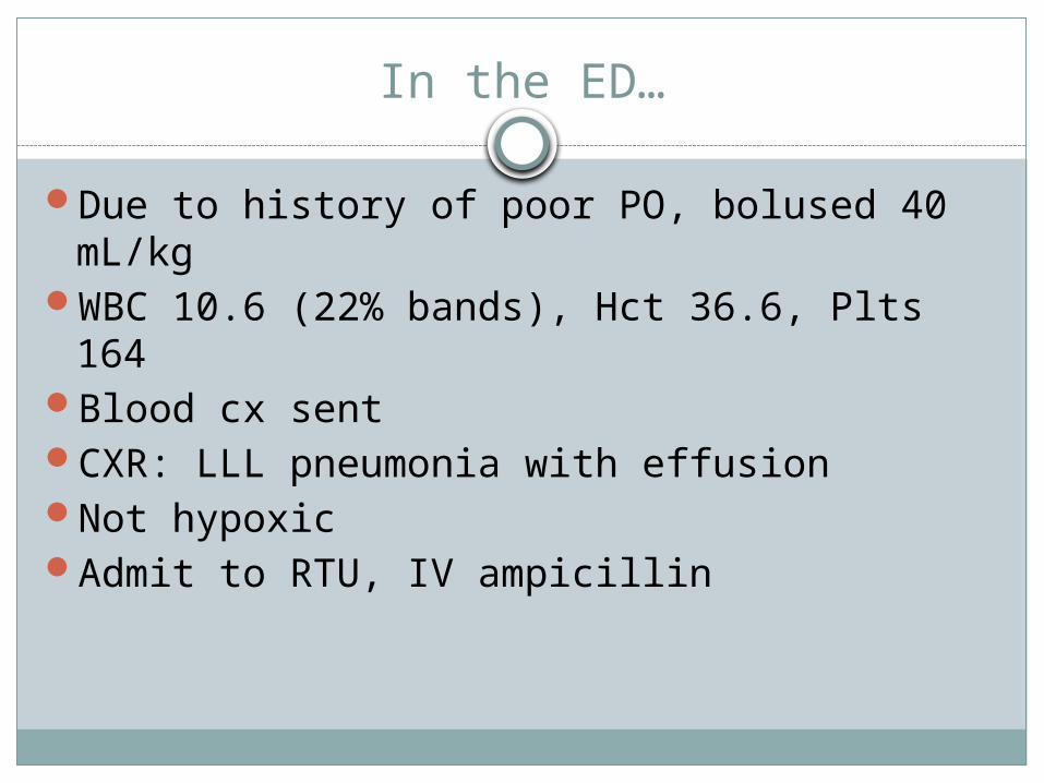
In the ED…
Due to history of poor PO, bolused 40 mL/kgWBC 10.6 (22% bands), Hct 36.6, Plts 164Blood cx sentCXR: LLL pneumonia with effusionNot hypoxicAdmit to RTU, IV ampicillin
In the RTU…
Worsening work of breathing, tachypnea, increasing effusion on CXR
As he is being transferred, blood culture turns positive @16 hrs, DNA for S pneumo detected
Ampicillin --> ceftriaxone

On the floor…
Worsening effusion --> chest tube placement
Mild hyponatremia (SIADH?)
Not hypoxic
Subsequent blood cultures negative

Overnight…
Called to evaluate pt ~2300
Worsened tachycardia (130s --> 170s)
Looks more pale/yellow, more swollen
Nurse is concerned he looks bad, and wants to call a rapid response

Overnight…
T 37.6, HR 167, RR 40, BP 107/62, 88% on RA
Gen: pale/yellow/greenish, not wanting to interact
HEENT: very pale conjunctivaCV: Tachycardic, no murmurs, good pulses,
cap refill hard to elicitResp: Diminished breath sounds on L with
crackles. No retractions. Chest tube in place
Abd: soft, NT/ND, normal BS, no HSMSkin: normalNeuro: normal

Differential diagnosis
Heme Blood loss (Chest tube,
hemothorax, abdomen, GI bleed)
Hemolysis Anemia of
inflammatory state DIC TTP
Rheum Vasculitis
Renal HUS

Labs
WBC 17.6 (23% bands), Hgb 3.9, Hct 11.1, Plts 46
Smear 2+ schistocytes
Na 136, K 4.7, Cl 113, bicarb 17, BUN 33, creat 0.71, gluc 121, alb 2.1, bili 2.1, ALT 26, AST 213
PT/INR 15.4/1.2, PTT 48, fibrinogen 633
D-dimer 4467

Hemolytic-Uremic Syndrome
Microangiopathic hemolytic anemia
Thrombocytopenia
Acute kidney injury

HUS Classifications
Diarrhea positive/negative
Primary (complement gene mutations, antibodies to complement factor B)
Secondary (usually infectious)

Epidemiology
STEC: 90% of HUS cases
Pneumococcus: ~5% (40% of non-STEC cases)
HUS in ~0.5% of pneumococcal infections

Pathogenesis (?)
Certain serotypes of strep pneumo expose Thomsen-Friedenreich (T) antigen (serotype 19A)
Preformed host IgM bind T antigen
Cascade of events lead to HUS
Alternative complement-mediated pathway?
Management
Treat underlying pneumococcal infection - empirically if necessary Consider rates of resistance
HUS therapy largely supportive RBCs and platelets STOP nephrotoxic drugs Nephrology involvement (dialysis?)
Avoid FFP, plasmapheresis (theoretical)

Risk Factors and Outcome
Generally more severe than STEC HUS Mortality higher (5-10% vs 12%) More likely to require dialysis (67-80%) Need more transfusions (PRBs and plts)
Death usually due to infection Biggest risk factor: meningitis (88% of deaths in the
largest review)~10% evolve to ESRD requiring dialysis

References
Banerjee R et al. Streptococcus pneumoniae-associated hemolytic uremic syndrome among children in North America. Pediatr Infect Dis J 30:736, 2011
Copelovitch L, Kaplan BS. Streptococcus pneumoniae-associated hemolytic uremic syndrome. Pediatr Nephrol 23:1951-56, 2008
Copelovitch L, Kaplan BS. Streptococcus pneumoniae-associated hemolytic uremic syndrome: classification and the emergence of serotype 19A. Pediatrics 125:174-182, 2010
Geary DF. Hemolytic uremic syndrome and Streptococcus pneumoniae: improving our understanding. J Pediatr 151:113, 2007
Spinale JM et al. Update on Streptococcus pneumoniae-associated hemolytic uremic syndrome. Curr Opin Pediatr 25:203-8, 2013
Top Related