


Languages
Pages
Legal


Fundamental Biotechnology
Lecture# 6
Haji Akbar
M.Phil

Introduction Bacteriophage
Virus infecting Bacteria. (eater of bacteria)
Short Phage
Groups on structure base
i. taillessii. Head with tailiii. filamentous

Continue!!
Genome: (50% of total mass)
DNA (ssDNA or dsDNA) or
RNA (ssRNA or ds RNA)
in tailless and tailed DNA is encapsulated in capsid.

Historical:
Frederick Twort (1915) and Felix d'Herelle (1917) were the first to recognize viruses which infect bacteria, which d'Herelle called bacteriophages (eaters of bacteria).

Diversity
There are at least 12 distinct groups of bacteriophages, which are very diverse structurally and genetically; the best known ones are the common phages of E.coli

Virulent vs. Temperate Phages
Virulent phages: do not integrate their genetic material into the host cell chromosome and usually kill the host cells (lytic infection) (e.g. T-phages of E.coli).
Temperate phages: may integrate into the host DNA, causing LYSOGENY. (best example λ phage)
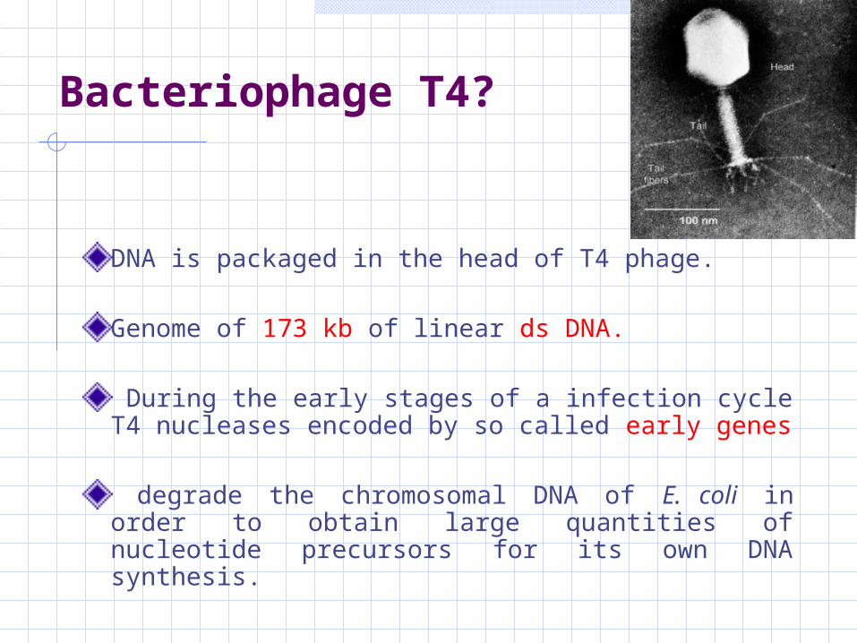
Bacteriophage T4?
DNA is packaged in the head of T4 phage.
Genome of 173 kb of linear ds DNA.
During the early stages of a infection cycle T4 nucleases encoded by so called early genes
degrade the chromosomal DNA of E. coli in order to obtain large quantities of nucleotide precursors for its own DNA synthesis.

λ page
Genome 48.5 kb (46 genes)
Entire Genome has been sequenced and regulatory site are known.
At the ends short (12bp) ss- complementary region “cohesive or sticky” ends--- circulation after infection.
Region is known as cos site.
Phage infection begins!!Adsorption
Lytic or lysogenic cycle depends on # of factors:
Nutrional & metabolic state of host cellMultiplicity of infection (m.o.i- the ratio of page to bacteria during adsorption)
In lysogenic phage genome integrates into host chromroms =prophage
Packaged into mature phage

Bacteriophage infection can be easily monitored by plaque formation. (in the lytic cycle, anyway)
Plaque forming Unit (p.f.u): is a measure of the number of particles capable of forming plaques per unit volume (1,000 PFU/µl indicates that there are 1,000 infectious virus particles in 1 µl of solution)

Page M13
Filamentous
ss-circular DNA (size 6407 bp)DNA enter in to cell converted to double stranded molecule known as replicative form or RF.
Replicates until there are about 100 copies in the cell.

Vectors based on Bacteriophage λ
The λ genome is 48.5 kb, in which 15 kb or so is ‘optional’
it contains genes that are only needed for integration into the E. coli chromosome (controlling lysogenic properties)
These segments can therefore be deleted without impairing the ability of the phage to infect bacteria and direct synthesis of new λ particles by the lytic cycle.

Types of vector
Insertion vectors: (single recognition sit for one or more restriction enzyme)
e.g. λgt10, Charon 16A. (both having EcoRI)
Replacement vectors:/ Substitution vector:Having two restriction sits (RS) which flank a
region known as stuffer fragment (hatched)e.g. EMBL4 and Chapron 40


λgt10: 7.5 kb (insert size), EcoRI lies in cI gene which is basis of selection/ screening.
Charon 16A: 9kb, EcoRI lies in β-galactosidase gene (lac Z).
EMBL4: 13.2 kb (stuffer fragment) and can clone 9 & 22 kb.
Chapron 40: (polystuffer) similar size fragment as EMBL4

Vectors based on M13Two aspect valuble for G.E.1. RF similar to plasmid2. ssDNA useful for sequencing by Dideoxy method.
507 bp intergenic region is the only part available
for manipulation.
Vectors constrict by introducing polylinker /lacZ α-peptide sequence into this region.
Mark reduction in cloning efficiency when DNA fragment longer than 1.5 kb inserted.

Other vectors

CosmidHybrid of plasmid/Phage vectors contain E coli ori that allow it to maintained as a plasmid in the cell and carries a λ cos site.
a plasmid that carries a λ cos site.
cos sites, act as substrates for in vitro packaging
because the cos site is the only sequence that a DNA molecule needs in order to be recognized as a ‘λ genome’ by the proteins that package DNA into λ phage particles.

A cosmid can be 4-8kb in size, so up to 45-47 kb of new DNA can be inserted.
inside the cell the cosmid cannot direct synthesis of new phage particles and instead replicates as a plasmid.
Recombinant DNA is therefore obtained from colonies rather than plaques.

e.g:pLFR-5 has two cos sites from page λ, separated by ScaI RE sit, a multiple cloning site with six unique restriction site origin of replication and Tetr gene

Cosmid Cloning System
cos sites insertedinto a small plasmid
Target DNA ligatedbetween two cosmidDNA molecules
Recombinant DNApackaged and E. coliInfected as before Can clone DNAs upto 45 kb

Cosmid vectors
Advantages: Useful for cloning very large DNA fragments (32 - 47 kbp) Inherent size selection for large inserts Handle like plasmids
Disadvantages: Not easy to handle very large plasmids (~ 50
kbp)

Phagemid vectors
plasmids which contain a small segment of the genome of a filamentous phage, such as M13, fd or f1.
contain all the cis-acting elements required for DNA replication and assembly into phage particles.
They permit successful cloning of inserts several kilobases long.

Continue!!
Following transformation of a suitable E. coli strain with a recombinant phagemid,
the bacterial cells are superinfected with a filamentous helper phage, such as f1, which is required to provide the coat protein.
Phage particles secreted from the superinfected cells will be a mixture of helper phage and recombinant phagemids.

The mixed single-stranded DNA population can be used directly for DNA sequencing.
because the primer for initiating DNA strand synthesis is designed to bind specifically to a sequence of the phagemid vector adjacent to the cloning site.
Commonly used phagemid vectors include the pEMBL series of plasmids and the pBluescript family.


THE END