


Languages
Pages
Legal

Fractures in Abused ChildrenMechanisms of Injury & Estimate of Age
VFPMS Seminar 2016
Anne Smith, Director VFPMS
BackgroundWhat do we want to know?
Does bone injury exist?
What pattern/type of injury is this?
Are there other injuries? (Bone / otherwise)
What is the mechanism of injury?What forces caused it/ contributed?Timing? How long ago did it happen?
Does the ‘explanation offered’ account for the injury?If not, why not? What might better explain it?
How do we know what we know?
• Forensic pathology – child homicides• Anatomical pathology and Histology
• Radiology• Clinical forensic medicine (cause of injury)• Population Health (epidemiology)• Orthopaedic surgery• Research – biomechanics / forces and physics• Accidental bone injury – patterns of injury and
healing• Metabolic & genetic disease states (abnormal
bone)• The courts – criminal justice system• The media
There is MUCH we still do NOT know
6 favourite referencesBilo RAC, Robben SGF, vanRijn RR Forensic Aspects of Paediatric Fractures Differentiating Accidental Trauma from Child Abuse 2010 (Springer)
Kleinman P., Diagnostic Imaging of Child Abuse 3rd ED (Mosby)
Offiah and Hall, Radiological Atlas of Child Abuse 2009 (Radcliffe)
WCPSRG Core-Info @ http://www.core-info.cardiff.ac.uk/fractures/index.htm
Carole Jenny, Child Abuse and Neglect; Diagnosis Treatment and Evidence
Giardino and Alexander, Child Maltreatment: A Clinical Guide and Reference 3rd ED 2005 (GW Publishing)

Anatomy of Long Bone
Basic Medical Anatomy, by Alexander Spence (Benjamin/Cummings 1990).
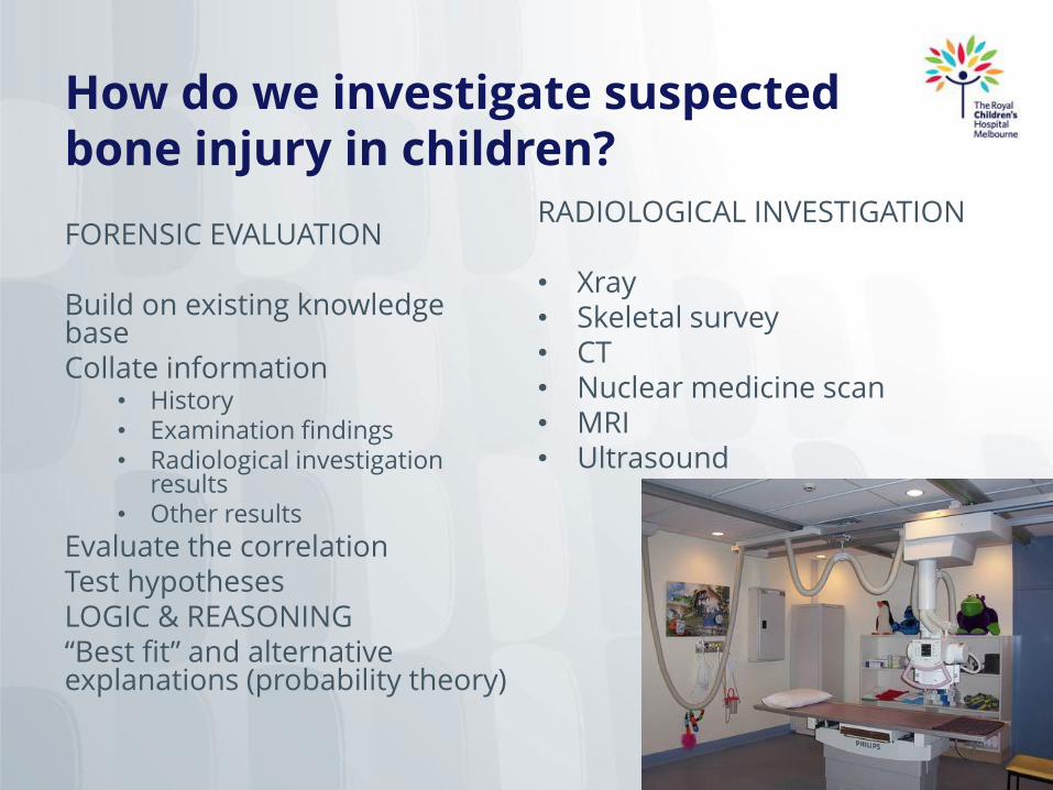
How do we investigate suspected bone injury in children?
FORENSIC EVALUATION
Build on existing knowledge baseCollate information
• History• Examination findings• Radiological investigation
results• Other results
Evaluate the correlationTest hypothesesLOGIC & REASONING“Best fit” and alternative explanations (probability theory)
RADIOLOGICAL INVESTIGATION
• Xray• Skeletal survey• CT • Nuclear medicine scan• MRI• Ultrasound
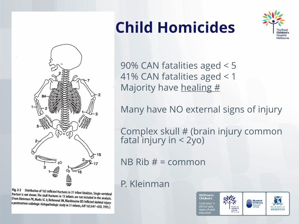
Child Homicides
90% CAN fatalities aged < 541% CAN fatalities aged < 1 Majority have healing #
Many have NO external signs of injury
Complex skull # (brain injury common fatal injury in < 2yo)
NB Rib # = common
P. Kleinman
Serious Assault (physical abuse)
Some studies of Physically Abused children - 11% - 53% have #
Diaphyseal # > metaphyseal # (4:1)
Metaphyseal # around knee and ankles > other limb joints
Bruising > isolated diaphyseal # tranverse > spiral
Of Shaft # , middle 1/3 (50%) distal 1/3 (41%)
Most common long bone (tibia, femur, humerus)
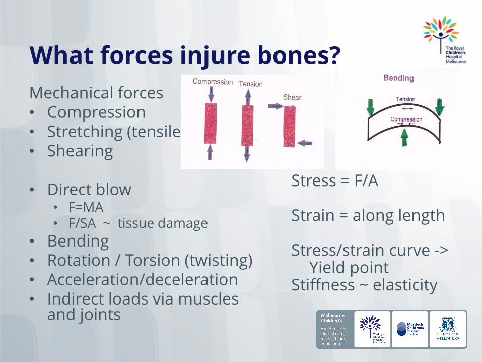
What forces injure bones?
Mechanical forces• Compression • Stretching (tensile)• Shearing
• Direct blow • F=MA• F/SA ~ tissue damage
• Bending• Rotation / Torsion (twisting)• Acceleration/deceleration• Indirect loads via muscles
and joints
Stress = F/A
Strain = along length
Stress/strain curve ->Yield point
Stiffness ~ elasticity
DIRECT INJURY MECHANISMS
Eg. long bones (diaphysis)
• Tapping (blow on small Surface Area)
• Crushing (high force on large area)
• Penetrating (high force on small area)
• Penetrating explosive (high force – lots of tissue damage)
INDIRECT INJURY MECHANISMS
Eg. long bones (diaphysis)• Transverse fracture - tensile force (eg patella #)
• Oblique - axial compressive force (distal femur #)
• Spiral - torsional force (tibia)
• Spiral with small butterfly - bending force (humerus)
• Transverse oblique with large butterfly - axial compression and bending (tibia)
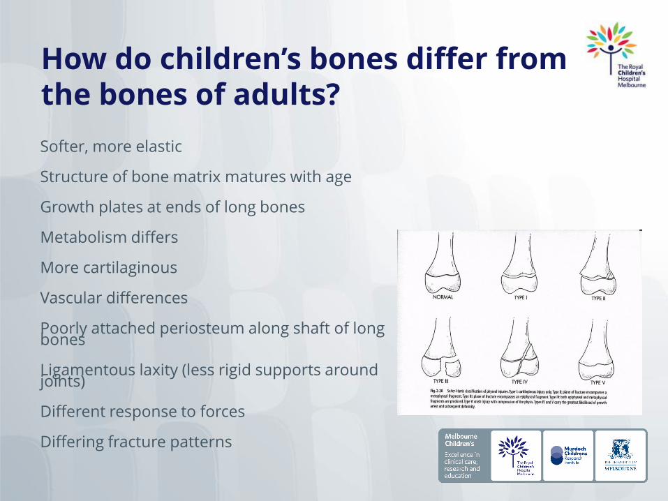
How do children’s bones differ from the bones of adults?
Softer, more elastic
Structure of bone matrix matures with age
Growth plates at ends of long bones
Metabolism differs
More cartilaginous
Vascular differences
Poorly attached periosteum along shaft of long bones
Ligamentous laxity (less rigid supports around joints)
Different response to forces
Differing fracture patterns
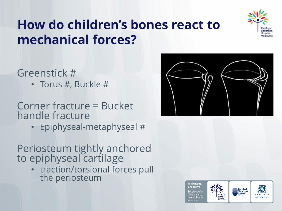
How do children’s bones react to mechanical forces?
Greenstick #• Torus #, Buckle #
Corner fracture = Bucket handle fracture
• Epiphyseal-metaphyseal #
Periosteum tightly anchored to epiphyseal cartilage
• traction/torsional forces pull the periosteum
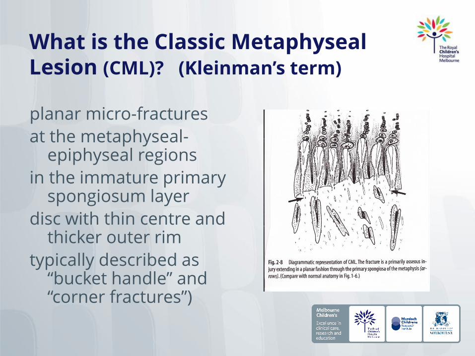
What is the Classic MetaphysealLesion (CML)? (Kleinman’s term)
planar micro-fractures
at the metaphyseal-epiphyseal regions
in the immature primary spongiosum layer
disc with thin centre and thicker outer rim
typically described as “bucket handle” and “corner fractures”)
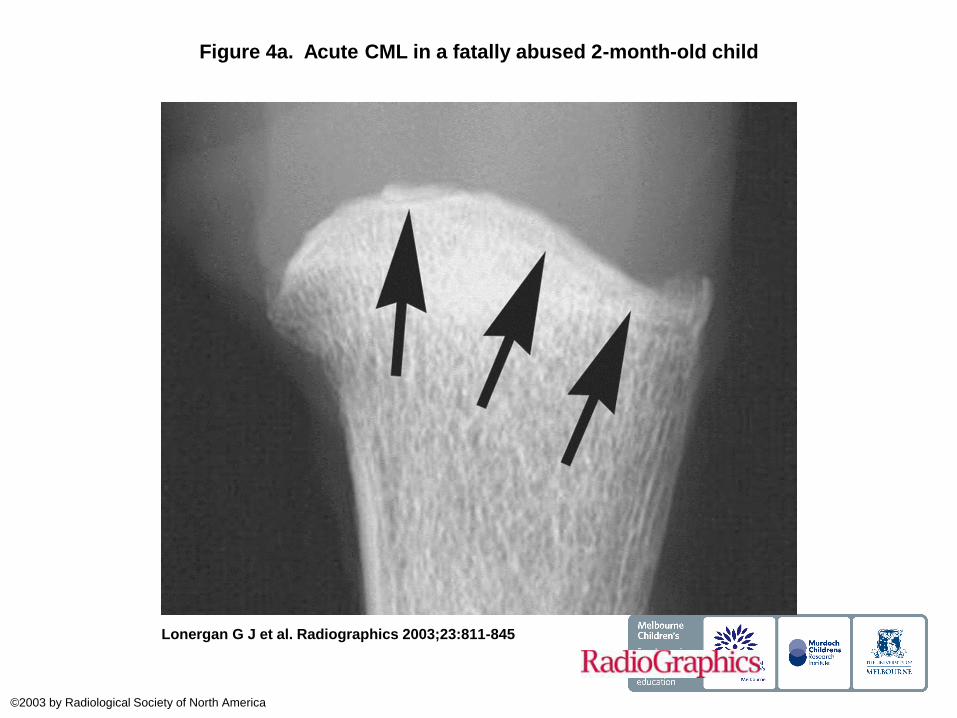
Figure 4a. Acute CML in a fatally abused 2-month-old child
Lonergan G J et al. Radiographics 2003;23:811-845
©2003 by Radiological Society of North America
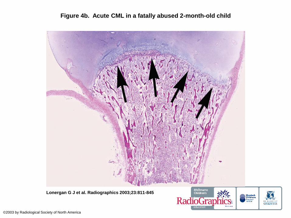
Figure 4b. Acute CML in a fatally abused 2-month-old child
Lonergan G J et al. Radiographics 2003;23:811-845
©2003 by Radiological Society of North America
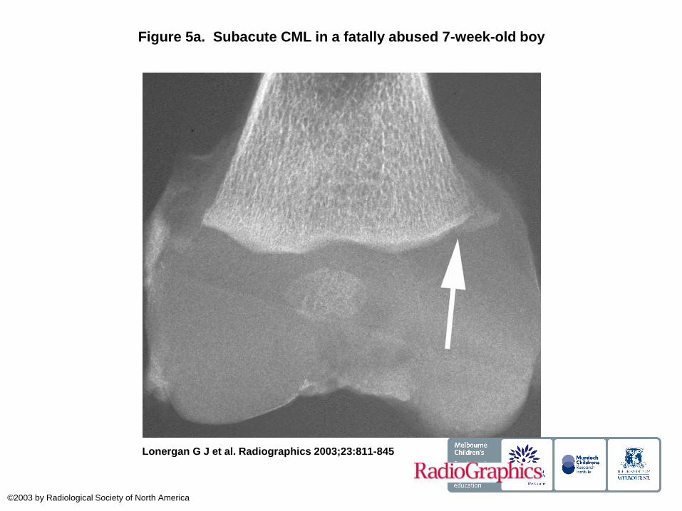
Figure 5a. Subacute CML in a fatally abused 7-week-old boy
Lonergan G J et al. Radiographics 2003;23:811-845
©2003 by Radiological Society of North America
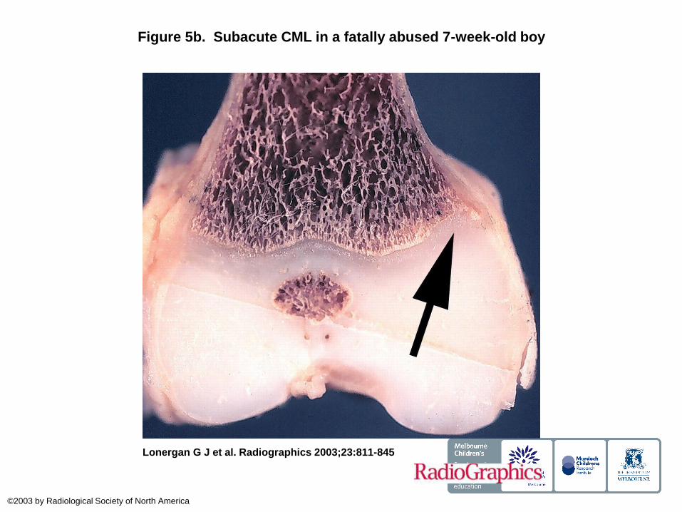
Figure 5b. Subacute CML in a fatally abused 7-week-old boy
Lonergan G J et al. Radiographics 2003;23:811-845
©2003 by Radiological Society of North America

Figure 6a. CML in an abused 2-month-old girl
Lonergan G J et al. Radiographics 2003;23:811-845
©2003 by Radiological Society of North America
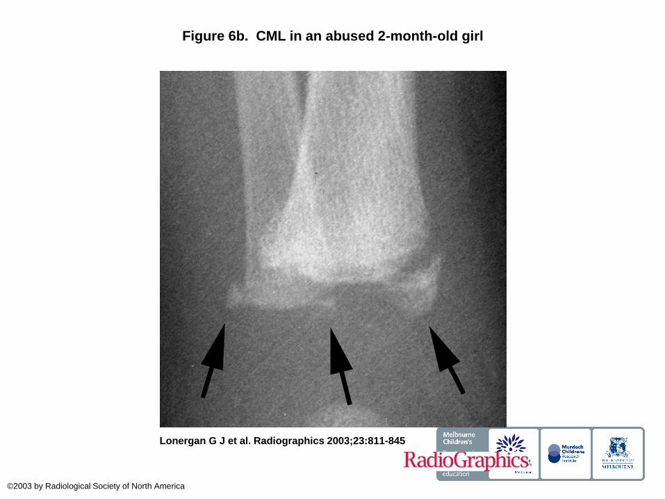
Figure 6b. CML in an abused 2-month-old girl
Lonergan G J et al. Radiographics 2003;23:811-845
©2003 by Radiological Society of North America
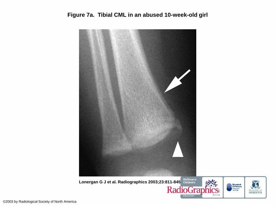
Figure 7a. Tibial CML in an abused 10-week-old girl
Lonergan G J et al. Radiographics 2003;23:811-845
©2003 by Radiological Society of North America
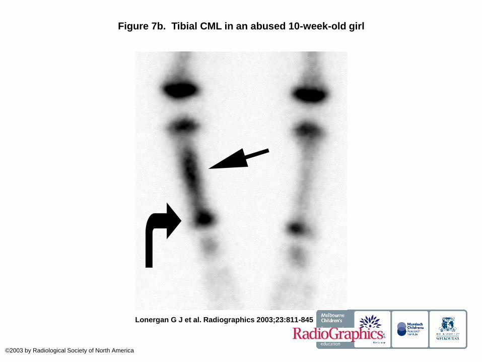
Figure 7b. Tibial CML in an abused 10-week-old girl
Lonergan G J et al. Radiographics 2003;23:811-845
©2003 by Radiological Society of North America
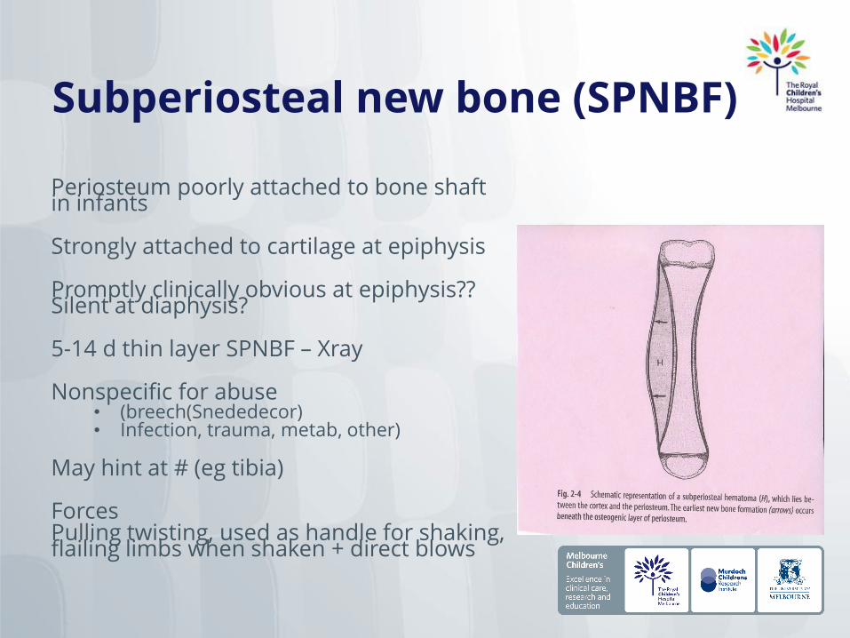
Subperiosteal new bone (SPNBF)
Periosteum poorly attached to bone shaft in infants
Strongly attached to cartilage at epiphysis
Promptly clinically obvious at epiphysis?? Silent at diaphysis?
5-14 d thin layer SPNBF – Xray
Nonspecific for abuse• (breech(Snededecor) • Infection, trauma, metab, other)
May hint at # (eg tibia)
Forces Pulling twisting, used as handle for shaking, flailing limbs when shaken + direct blows
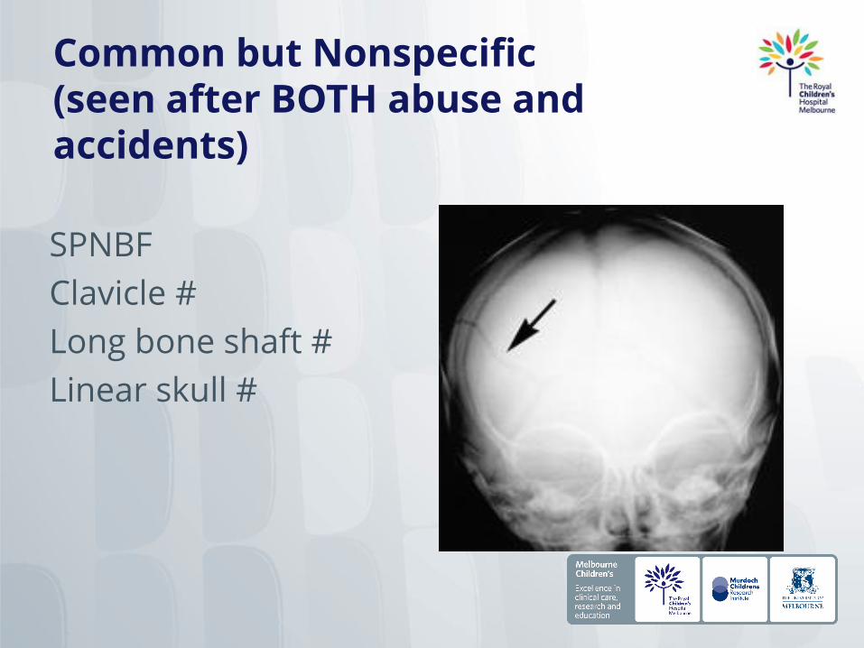
Common but Nonspecific (seen after BOTH abuse and accidents)
SPNBF
Clavicle #
Long bone shaft #
Linear skull #
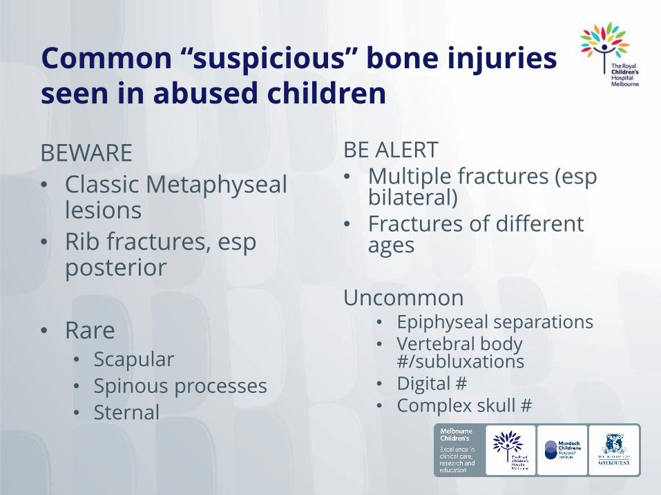
Common “suspicious” bone injuries seen in abused children
BEWARE
• Classic Metaphyseal lesions
• Rib fractures, esp posterior
• Rare• Scapular
• Spinous processes
• Sternal
BE ALERT• Multiple fractures (esp
bilateral)• Fractures of different
ages
Uncommon• Epiphyseal separations• Vertebral body
#/subluxations• Digital #• Complex skull #

How do children’s bones heal?
Osteonal bone healing• Primary bone healing or primary
gap healing (no callus)• Secondary bone healing with callus
formation
Non-Osteonal bone healing• Callus or gap heals with fibrous
tissue that differentiates into lamellar or woven bone (UNCOMMON in children)
Dead bone serves as a mechanical stabiliser until it is remodelled
Remodelling requires weight bearing (Wolff’s law)
Spiral and oblique fractures heal more rapidly than transverse fractures (greater surface area of fracture ends / less surrounding soft tissue damage)
Structure and mechanical properties are restored (unlike so skin and tendon)
Local factors influence rate of healing ( blood flow coexisting tissue damage)
4 phases of bone healing (Radiol)
1. Induction phase
Time of injury to the appearance of new bone at the fracture site.
Inflammatory response may last a few days and reveal itself on x-ray in the form of soft tissue swelling with displacement and obliteration of normal fat and facial planes.
A fracture line that might initially appear sharp can gradually become less well defined + blurs the fracture margins.
A nuclear medicine scan and MRI scan may detect subperiosteal changes that are not yet evident on x-ray.
4 phases of bone healing (Radiol)
2. soft callus (subperiosteal new bone)
In infants this can occur within approximately 7 to 10 days, later (10 to 14 days) in older children.
By approximately 10 days, a cellular collar surrounds the fracture site.
Woven bone calcifies approximately 10 to 15 days after injury.
Exuberant callus formation can be a sign of fracture instability, and/or repetitive injury.
4 phases of bone healing (Radiol)
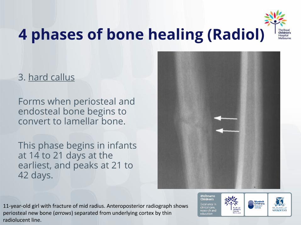
3. hard callus
Forms when periosteal and endosteal bone begins to convert to lamellar bone.
This phase begins in infants at 14 to 21 days at the earliest, and peaks at 21 to 42 days.
11-year-old girl with fracture of mid radius. Anteroposterior radiograph shows periosteal new bone (arrows) separated from underlying cortex by thin radiolucent line.
4 phases of bone healing (Radiol)
4. Remodelling occurs with gradual correction of deformity.
Begins at approximately 3 months and peaks at 1 to 2 years
Heals completely and appears indistinguishable on x-ray from a bone that has not been injured.
Healing generally occurs more rapidly in younger infants
The rate at which bones heal, and remodelling occurs, varies according to the child's age, the anatomy of the injured bone, the site and nature of fracture (including the degree of angulation and separation of bone segments), and metabolic processes that unable healing of bone injury.
Histological evidence of healing
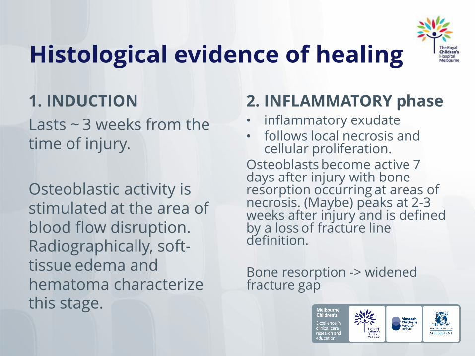
1. INDUCTION
Lasts ~ 3 weeks from the time of injury.
Osteoblastic activity is stimulated at the area of blood flow disruption. Radiographically, soft-tissue edema and hematoma characterize this stage.
2. INFLAMMATORY phase• inflammatory exudate• follows local necrosis and
cellular proliferation. Osteoblasts become active 7 days after injury with bone resorption occurring at areas of necrosis. (Maybe) peaks at 2-3 weeks after injury and is defined by a loss of fracture line definition.
Bone resorption -> widened fracture gap
Histological evidence of healing
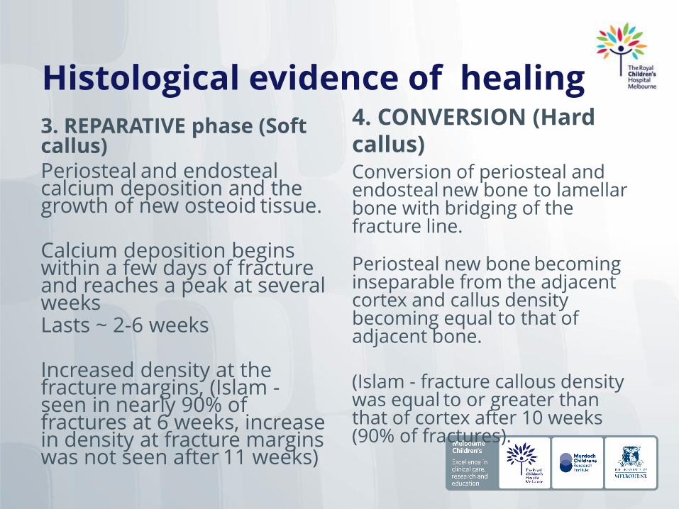
3. REPARATIVE phase (Soft callus)Periosteal and endostealcalcium deposition and the growth of new osteoid tissue.
Calcium deposition begins within a few days of fractureand reaches a peak at several weeks Lasts ~ 2-6 weeks
Increased density at the fracture margins, (Islam -seen in nearly 90% of fractures at 6 weeks, increase in density at fracture margins was not seen after 11 weeks)
4. CONVERSION (Hard callus)Conversion of periosteal and endosteal new bone to lamellar bone with bridging of the fracture line.
Periosteal new bone becoming inseparable from the adjacent cortex and callus densitybecoming equal to that of adjacent bone.
(Islam - fracture callous density was equal to or greater than that of cortex after 10 weeks (90% of fractures).
Histological evidence of healing
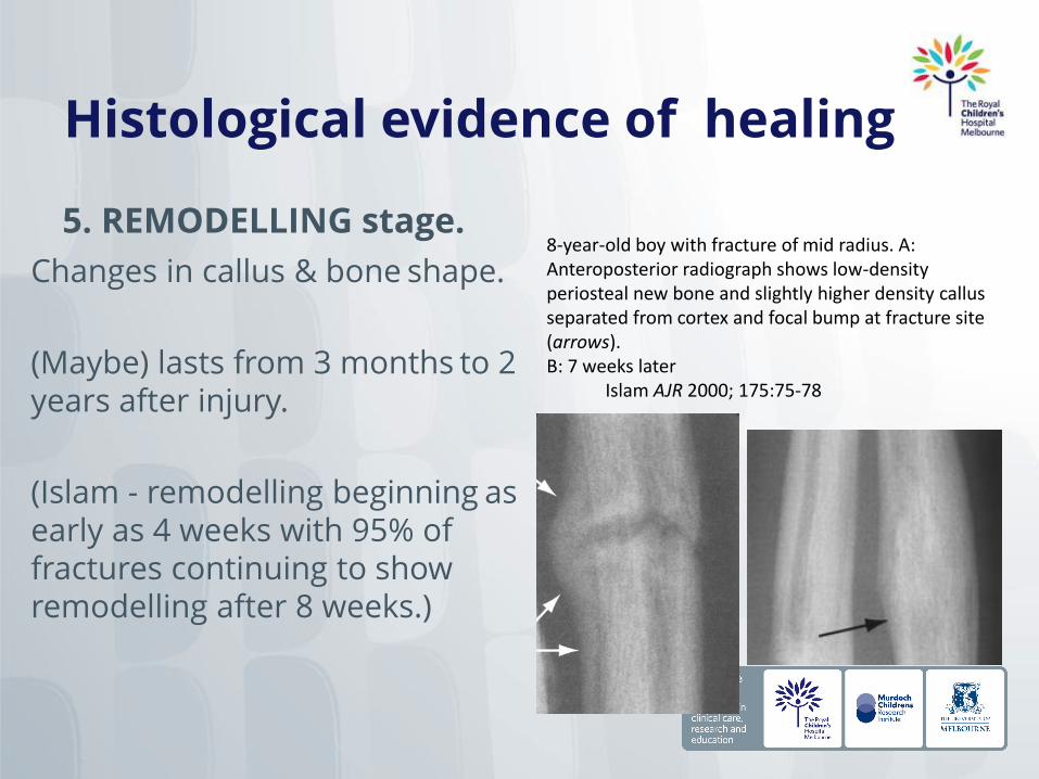
5. REMODELLING stage.
Changes in callus & bone shape.
(Maybe) lasts from 3 months to 2 years after injury.
(Islam - remodelling beginning as early as 4 weeks with 95% of fractures continuing to showremodelling after 8 weeks.)
8-year-old boy with fracture of mid radius. A: Anteroposterior radiograph shows low-density periosteal new bone and slightly higher density callus separated from cortex and focal bump at fracture site (arrows).B: 7 weeks later
Islam AJR 2000; 175:75-78
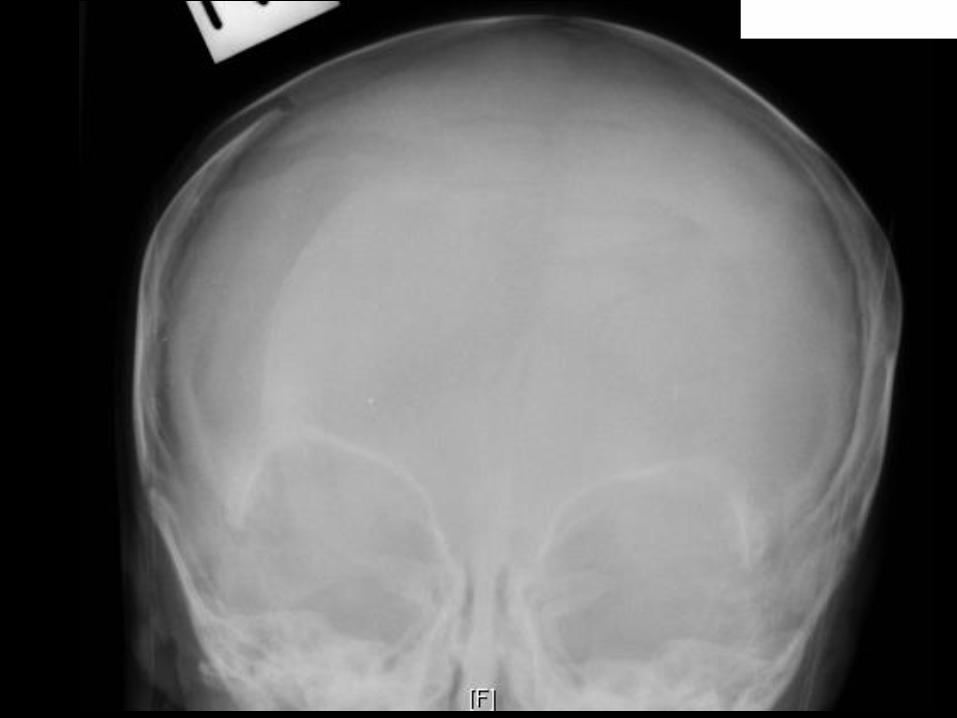
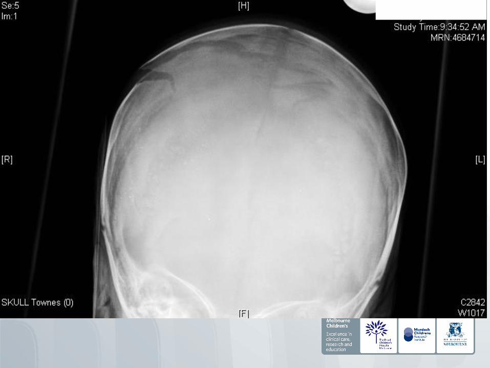
Skeletal survey (NO babygram!)Skull (SXR)
AP and lateral, plus Towne's view for occipital injury. SXRs should be taken with a skeletal survey even if a CT scan has been performed.
Body:AP/frontal chest (including clavicles)Oblique views of the ribs (left and right)AP Abdomen with pelvis and hips
Spine:Lateral spine - cervical and thoraco-lumbar
Limbs:AP humeri, AP forearmsAP femurs, AP Tib/fibPA hands and AP feetSupplemented by:- Lateral views of any suspected shaft fracture.- Lateral coned views of the elbows/wrists/knees/ankles may demonstrate metaphyseal injuries in greater detail than AP views of the limbs alone. The consultant radiologist should decide this, at the time of checking the films with the radiographers.
Brain imaging:CT (brain and bone windows) is the method of choice in the acute phase. A linear skull fracture may not be identified on CT (on bone windows)
PEDIATRICS Vol. 123 No. 5 May 2009, pp. 1430-1435
Bone Scan
“HOT SPOTS”
Subtle bone injury,Infection,Inflammation,
Soft tissue trauma, Growing epiphysesand other pathological causes of increased uptake of radio nucleotide
Bone scans have little place in fracture dating as they become positive within 7 hours and can remain positive for up to one year
Mandelstam SA, Cook D, Fitzgerald M, Ditchfield MR. Complementary use of radiological skeletal survey and bone scintigraphy in detection of bony injuries in suspected child abuse. Arch Dis Child. 2003;88 (5):387 –390
Additional radiology?Coned views of suspicious sites
Re-examination of hot spots
Repeat Xrays in 2+ weeks (eg to see whether callus has developed at site of suspected fracture)
Occasionally = MRI (spinal injury?) CT?
Very Occasionally = Ultrasound (soft tissue injury DDx #?)
NB CONSIDER AND INVESTIGATE POSSIBLE HEAD TRAUMA
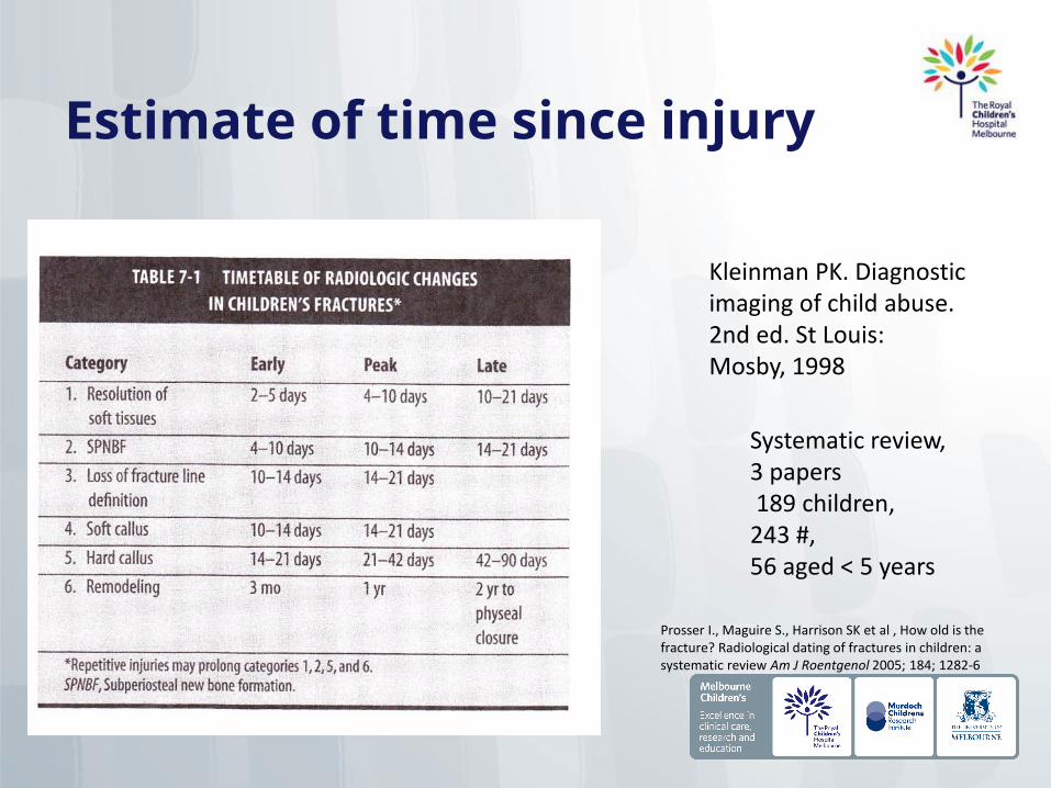
Estimate of time since injury
Kleinman PK. Diagnostic imaging of child abuse. 2nd ed. St Louis: Mosby, 1998
Systematic review,3 papers189 children,
243 #, 56 aged < 5 years
Prosser I., Maguire S., Harrison SK et al , How old is the fracture? Radiological dating of fractures in children: a systematic review Am J Roentgenol 2005; 184; 1282-6
Papers on bone healing in childrenINEXACT SCIENCE
Islam O, Sobeleski D, Symons S, Davidson LK, Ashworth MA, Babyn P. Development and duration of radiographic signs of bone healing in children. Am J Roentgenol 2000;175:75-78
Yeo LI, Reed MH. Staging of healing of femoral fractures in children. Can Assoc Radiol J 1994;45:16-19
Cumming WA. Neonatal skeletal fractures. Birth trauma or child abuse? J Can Assoc Radiol 1979;30:30-33
Hard callus and early remodelling is seen at 8 weeks in majority of cases
Early callus (calcified SPNBF) noted as early as 7 days (neonates)
Dating of fractures is an inexact science
The radiological features of bone healing are a continuum, with considerable overlap
Radiological estimates of time of injury are in terms of weeks rather than days. It is vital that all investigating agencies are aware of these broad time frames
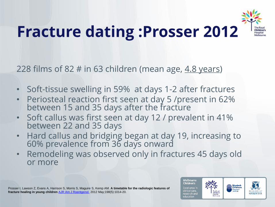
Fracture dating :Prosser 2012
228 films of 82 # in 63 children (mean age, 4.8 years)
• Soft-tissue swelling in 59% at days 1-2 after fractures • Periosteal reaction first seen at day 5 /present in 62%
between 15 and 35 days after the fracture • Soft callus was first seen at day 12 / prevalent in 41%
between 22 and 35 days• Hard callus and bridging began at day 19, increasing to
60% prevalence from 36 days onward • Remodeling was observed only in fractures 45 days old
or more
Prosser I, Lawson Z, Evans A, Harrison S, Morris S, Maguire S, Kemp AM. A timetable for the radiologic features of
fracture healing in young children AJR Am J Roentgenol. 2012 May;198(5):1014-20.
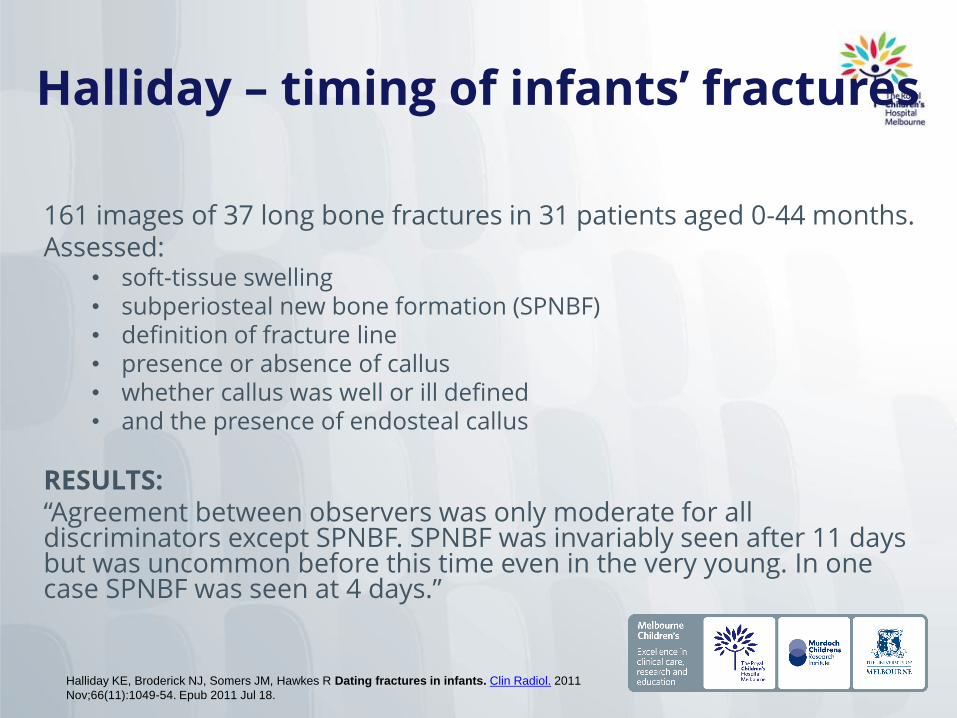
Halliday – timing of infants’ fractures
161 images of 37 long bone fractures in 31 patients aged 0-44 months. Assessed:
• soft-tissue swelling • subperiosteal new bone formation (SPNBF) • definition of fracture line • presence or absence of callus • whether callus was well or ill defined • and the presence of endosteal callus
RESULTS: “Agreement between observers was only moderate for all discriminators except SPNBF. SPNBF was invariably seen after 11 days but was uncommon before this time even in the very young. In one case SPNBF was seen at 4 days.”
Halliday KE, Broderick NJ, Somers JM, Hawkes R Dating fractures in infants. Clin Radiol. 2011
Nov;66(11):1049-54. Epub 2011 Jul 18.
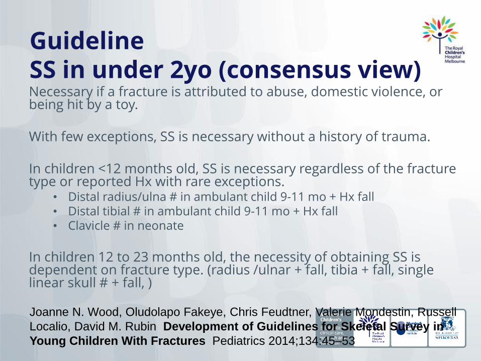
GuidelineSS in under 2yo (consensus view)Necessary if a fracture is attributed to abuse, domestic violence, or being hit by a toy.
With few exceptions, SS is necessary without a history of trauma.
In children <12 months old, SS is necessary regardless of the fracture type or reported Hx with rare exceptions.
• Distal radius/ulna # in ambulant child 9-11 mo + Hx fall• Distal tibial # in ambulant child 9-11 mo + Hx fall• Clavicle # in neonate
In children 12 to 23 months old, the necessity of obtaining SS is dependent on fracture type. (radius /ulnar + fall, tibia + fall, single linear skull # + fall, )
Joanne N. Wood, Oludolapo Fakeye, Chris Feudtner, Valerie Mondestin, Russell
Localio, David M. Rubin Development of Guidelines for Skeletal Survey in
Young Children With Fractures Pediatrics 2014;134:45–53
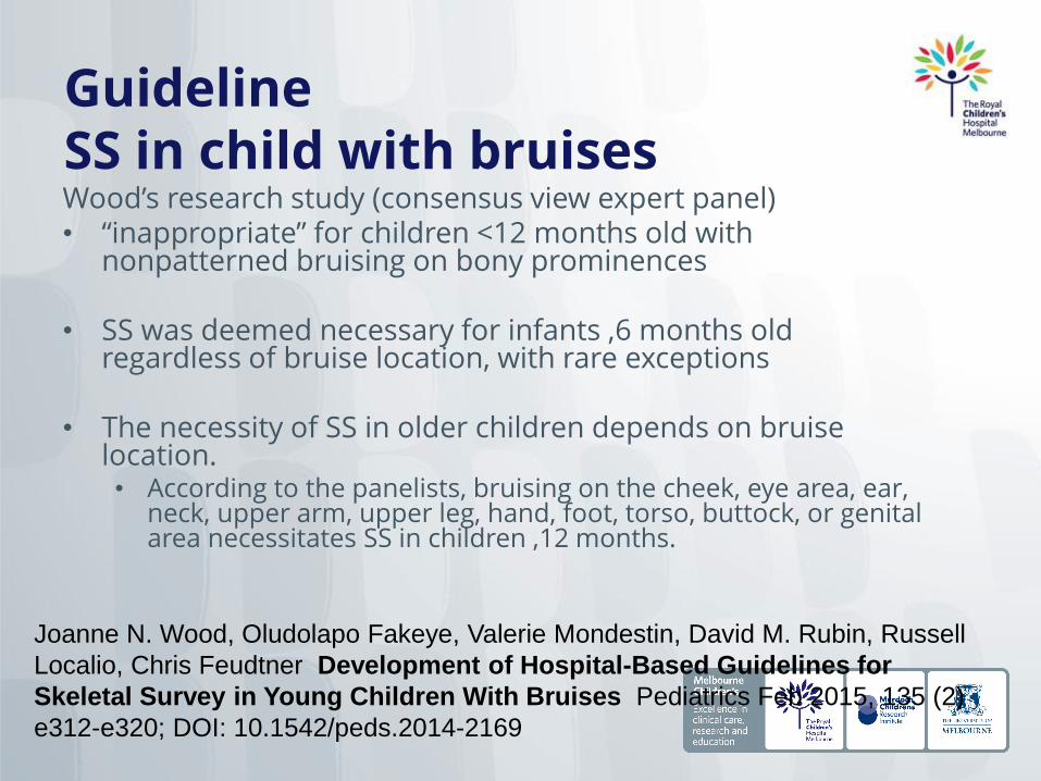
GuidelineSS in child with bruisesWood’s research study (consensus view expert panel)• “inappropriate” for children <12 months old with
nonpatterned bruising on bony prominences
• SS was deemed necessary for infants ,6 months old regardless of bruise location, with rare exceptions
• The necessity of SS in older children depends on bruise location. • According to the panelists, bruising on the cheek, eye area, ear,
neck, upper arm, upper leg, hand, foot, torso, buttock, or genital area necessitates SS in children ,12 months.
Joanne N. Wood, Oludolapo Fakeye, Valerie Mondestin, David M. Rubin, Russell
Localio, Chris Feudtner Development of Hospital-Based Guidelines for
Skeletal Survey in Young Children With Bruises Pediatrics Feb 2015, 135 (2)
e312-e320; DOI: 10.1542/peds.2014-2169

VFPMS guideline
SEEK ADVICE!< 2 years - Consider SS + BS > 2 years X ray suspicious areaBone metabolism – blood tests
• Calcium Phosphate • Alkaline phosphatase (LFT)• U&E, Creat• FBE• Vit D• 2nds line tests – Mg, PTH, syphilis Ab, UMS,,
organic acids, genetic tests…..
Let’s look at some fractures…
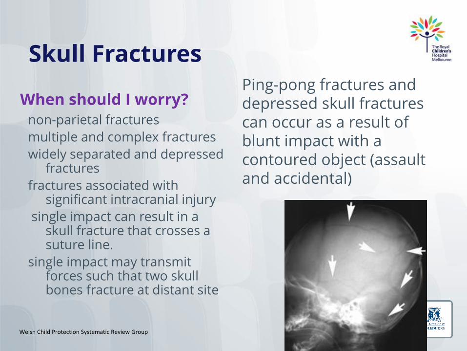
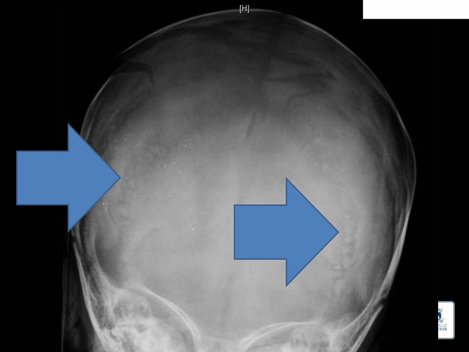
Skull Fractures
When should I worry?non-parietal fractures
multiple and complex fractures
widely separated and depressed fractures
fractures associated with significant intracranial injury
single impact can result in a skull fracture that crosses a suture line.
single impact may transmit forces such that two skull bones fracture at distant site
Ping-pong fractures and depressed skull fractures can occur as a result of blunt impact with a contoured object (assault and accidental)
Welsh Child Protection Systematic Review Group
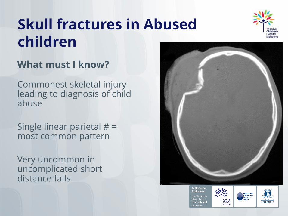
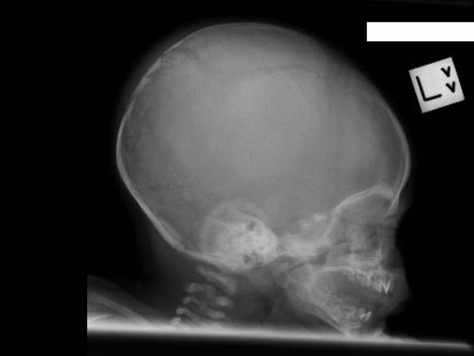
Skull fractures in Abused children
What must I know?
Commonest skeletal injury leading to diagnosis of child abuse
Single linear parietal # = most common pattern
Very uncommon in uncomplicated short distance falls

Rib fractures
What must I know
90% in abused children < 2 years
Can be missed on routine Xray
Posterior rib fractures = highly suggestive of abuse
Rib fractures are a rare complication of CPR in children (only 3 out of 923 children) all multiple and anterior, no posterior (WCPSRG)
Welsh Child Protection Systematic Review Group

Figure 8. Rib fracture mechanism in tight squeezing
Lonergan G J et al. Radiographics 2003;23:811-845
©2003 by Radiological Society of North America
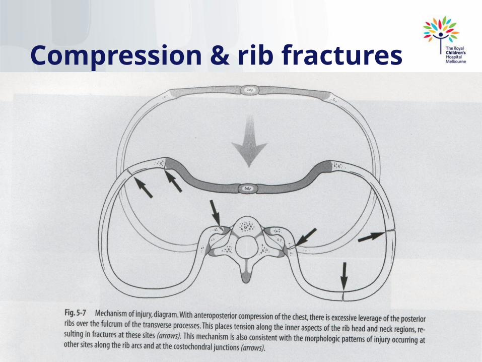
Compression & rib fractures
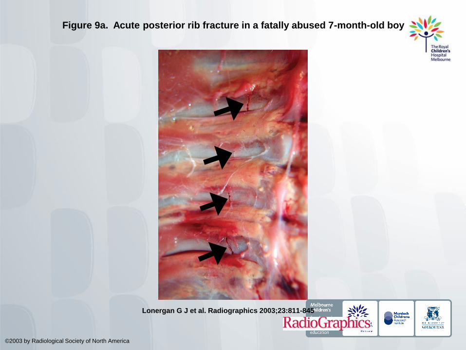
Figure 9a. Acute posterior rib fracture in a fatally abused 7-month-old boy
Lonergan G J et al. Radiographics 2003;23:811-845
©2003 by Radiological Society of North America
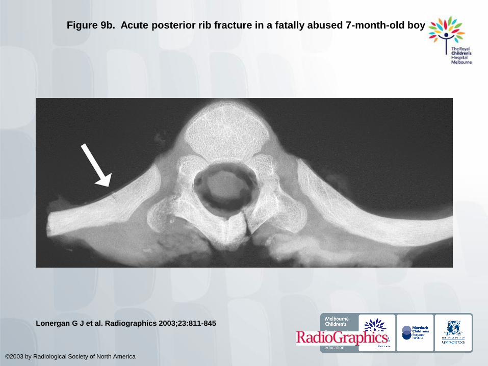
Figure 9b. Acute posterior rib fracture in a fatally abused 7-month-old boy
Lonergan G J et al. Radiographics 2003;23:811-845
©2003 by Radiological Society of North America
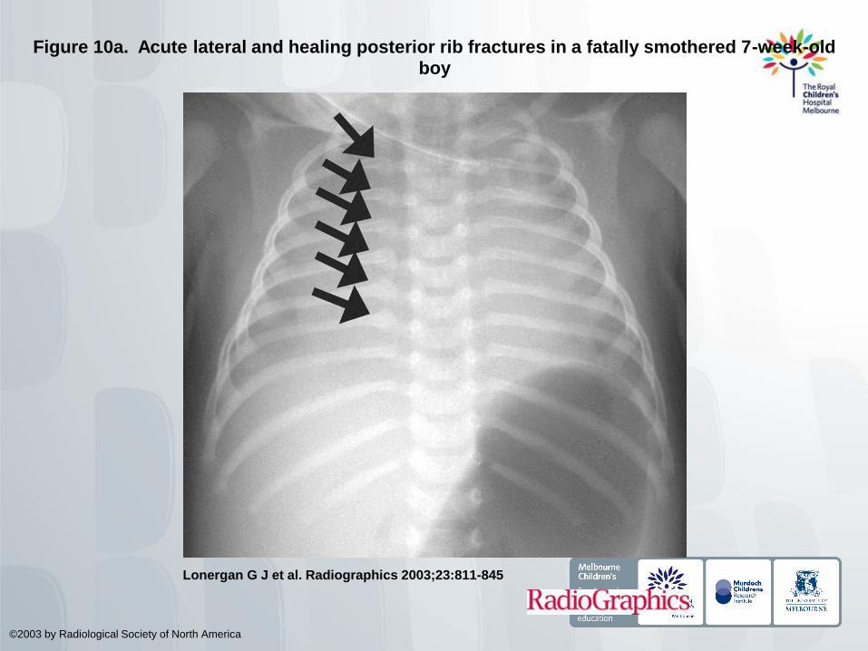
Figure 10a. Acute lateral and healing posterior rib fractures in a fatally smothered 7-week-old boy
Lonergan G J et al. Radiographics 2003;23:811-845
©2003 by Radiological Society of North America
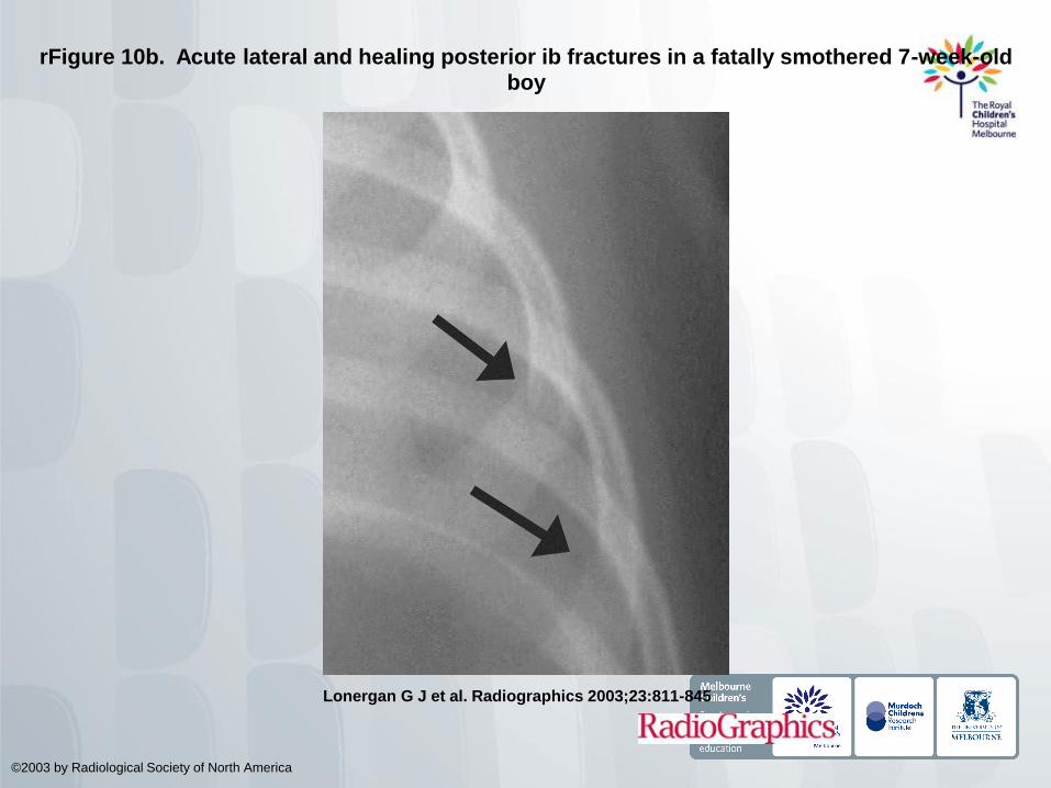
rFigure 10b. Acute lateral and healing posterior ib fractures in a fatally smothered 7-week-old boy
Lonergan G J et al. Radiographics 2003;23:811-845
©2003 by Radiological Society of North America
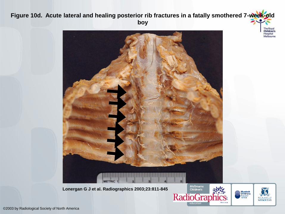
Figure 10d. Acute lateral and healing posterior rib fractures in a fatally smothered 7-week-old boy
Lonergan G J et al. Radiographics 2003;23:811-845
©2003 by Radiological Society of North America

Figure 14b. Healing posterior rib fracture in an abused 2-month-old girl
Lonergan G J et al. Radiographics 2003;23:811-845
©2003 by Radiological Society of North America
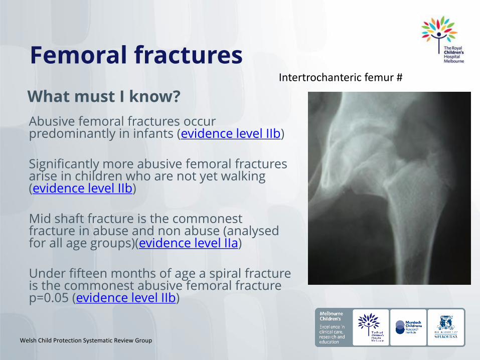
Intertrochanteric femur #
Femoral fractures
What must I know?
Abusive femoral fractures occur predominantly in infants (evidence level IIb)
Significantly more abusive femoral fractures arise in children who are not yet walking (evidence level IIb)
Mid shaft fracture is the commonest fracture in abuse and non abuse (analysed for all age groups)(evidence level IIa)
Under fifteen months of age a spiral fracture is the commonest abusive femoral fracture p=0.05 (evidence level IIb)
Welsh Child Protection Systematic Review Group
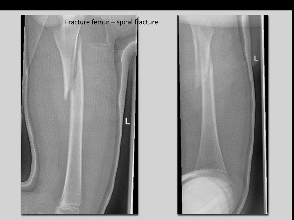
Fracture femur – spiral fracture
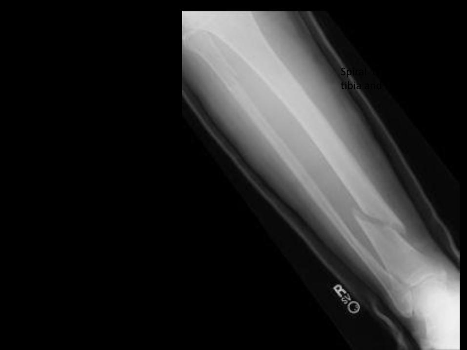
Spiral fracture tibia and fibula
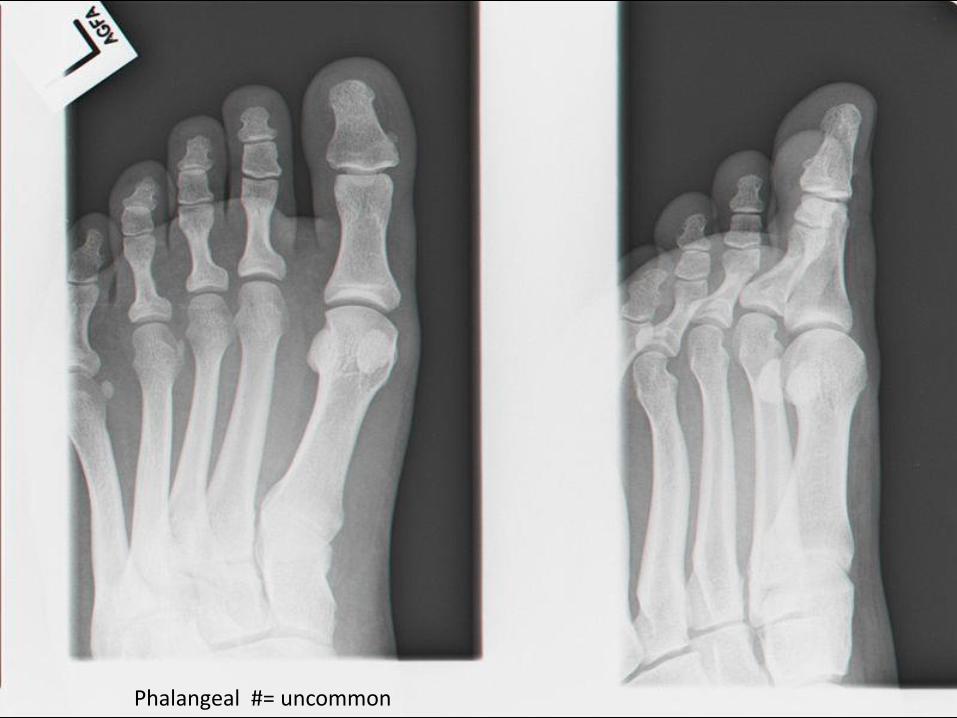
Phalangeal #= uncommon
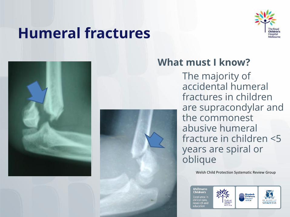
Humeral fractures
What must I know?
The majority of accidental humeral fractures in children are supracondylar and the commonest abusive humeral fracture in children <5 years are spiral or oblique
Welsh Child Protection Systematic Review Group

Lateral condyle fracture elbowFall on outstretched hand – opens jointNOTE direction of force
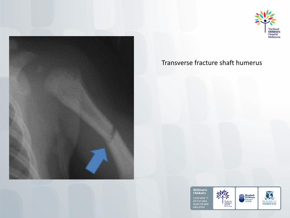
Transverse fracture shaft humerus
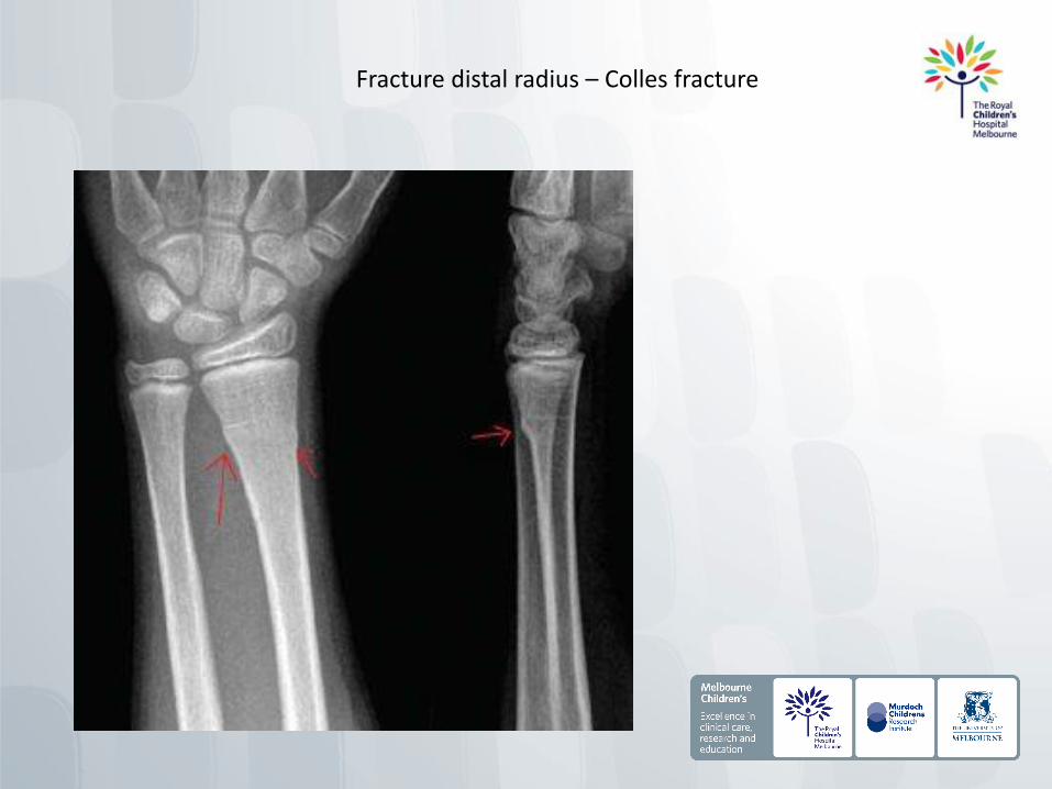
Fracture distal radius – Colles fracture
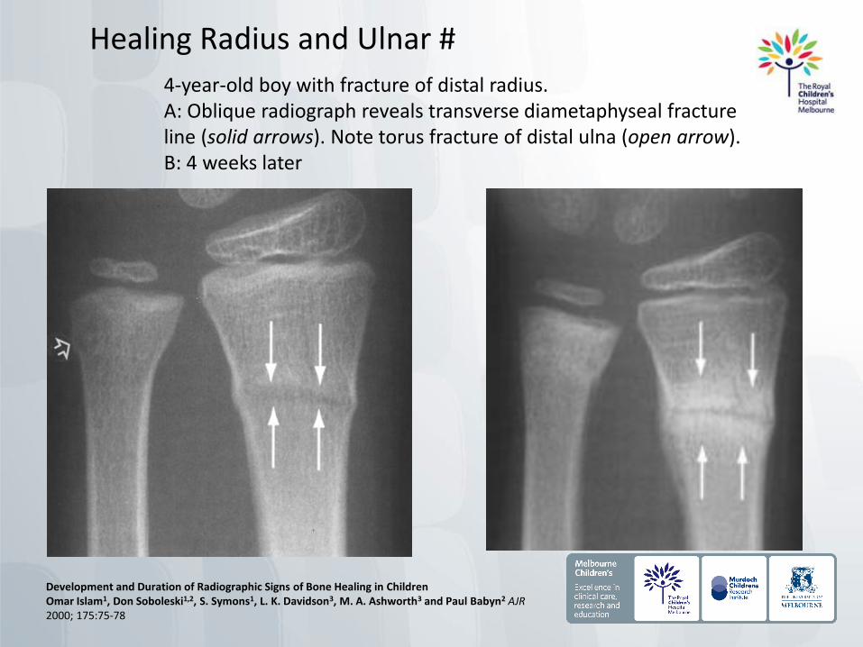
4-year-old boy with fracture of distal radius. A: Oblique radiograph reveals transverse diametaphyseal fracture line (solid arrows). Note torus fracture of distal ulna (open arrow).B: 4 weeks later
Development and Duration of Radiographic Signs of Bone Healing in Children Omar Islam1, Don Soboleski1,2, S. Symons1, L. K. Davidson3, M. A. Ashworth3 and Paul Babyn2 AJR2000; 175:75-78
Healing Radius and Ulnar #
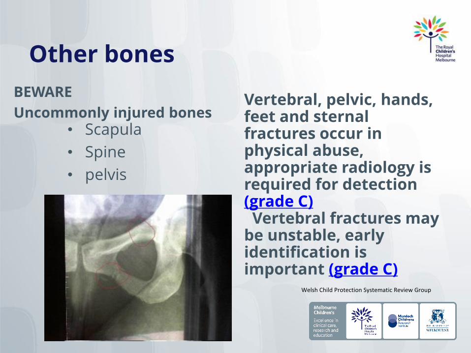
Other bones
Vertebral, pelvic, hands, feet and sternal fractures occur in physical abuse, appropriate radiology is required for detection (grade C)Vertebral fractures may
be unstable, early identification is important (grade C)
• Scapula
• Spine
• pelvis
BEWARE
Uncommonly injured bones
Welsh Child Protection Systematic Review Group

Fracture clavicle midshaft
BE Alert
Uncommon presentations of commonly injured bones
Unusual skull #
Ends of clavicle #
Unusual fracture patterns when long bones injured
Brittle bones
Transient - TBBD
A unique hypothesis (still has proponents) Transient copper defect? No scientific validity?
Deregistration of Dr. Colin Paterson from the Medical Practitioners Board of Scotland
Carol Jenny “ Although frequently offered in court cases as a cause of multiple infant fractures, there is no evidence that this condition actually exists.”Jenny C; Committee on Child Abuse and Neglect.
Evaluating infants and young children with multiple fractures Pediatrics. 2006 Sep;118(3):1299-303
Medical causes• Vit D deficiency• Rickets (renal / other)• OI• Menkes / Copper abn• Scurvey, Vit A toxicosis• Hyper PTH (osteoclasts++)• Hypocalcemia (exprem /
metabolic)• Osteopenia (disuse / other )• Malignancy (leukemia,
N’blastoma, histiocytosis)• Infection – osteomyelitis• Infantile cortical hyperostosis• OTHERS
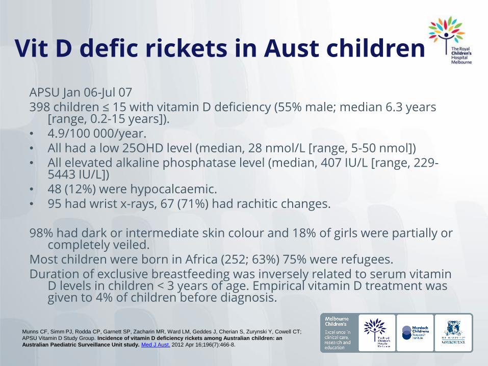
Vit D defic rickets in Aust children
APSU Jan 06-Jul 07398 children ≤ 15 with vitamin D deficiency (55% male; median 6.3 years
[range, 0.2-15 years]). • 4.9/100 000/year. • All had a low 25OHD level (median, 28 nmol/L [range, 5-50 nmol]) • All elevated alkaline phosphatase level (median, 407 IU/L [range, 229-
5443 IU/L])• 48 (12%) were hypocalcaemic. • 95 had wrist x-rays, 67 (71%) had rachitic changes.
98% had dark or intermediate skin colour and 18% of girls were partially or completely veiled.
Most children were born in Africa (252; 63%) 75% were refugees. Duration of exclusive breastfeeding was inversely related to serum vitamin
D levels in children < 3 years of age. Empirical vitamin D treatment was given to 4% of children before diagnosis.
Munns CF, Simm PJ, Rodda CP, Garnett SP, Zacharin MR, Ward LM, Geddes J, Cherian S, Zurynski Y, Cowell CT;
APSU Vitamin D Study Group. Incidence of vitamin D deficiency rickets among Australian children: an
Australian Paediatric Surveillance Unit study. Med J Aust. 2012 Apr 16;196(7):466-8.
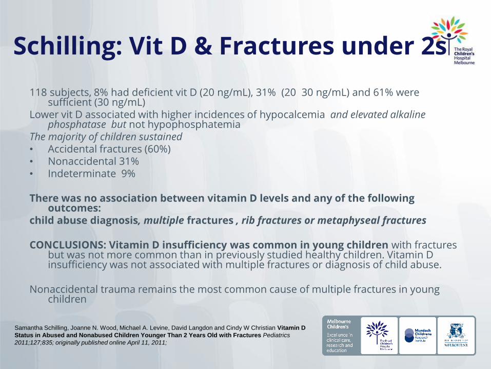
Schilling: Vit D & Fractures under 2s
118 subjects, 8% had deficient vit D (20 ng/mL), 31% (20 30 ng/mL) and 61% were sufficient (30 ng/mL)
Lower vit D associated with higher incidences of hypocalcemia and elevated alkaline phosphatase but not hypophosphatemia
The majority of children sustained• Accidental fractures (60%) • Nonaccidental 31% • Indeterminate 9%
There was no association between vitamin D levels and any of the following outcomes:
child abuse diagnosis, multiple fractures , rib fractures or metaphyseal fractures
CONCLUSIONS: Vitamin D insufficiency was common in young children with fractures but was not more common than in previously studied healthy children. Vitamin D insufficiency was not associated with multiple fractures or diagnosis of child abuse.
Nonaccidental trauma remains the most common cause of multiple fractures in young children
Samantha Schilling, Joanne N. Wood, Michael A. Levine, David Langdon and Cindy W Christian Vitamin D
Status in Abused and Nonabused Children Younger Than 2 Years Old with Fractures Pediatrics
2011;127;835; originally published online April 11, 2011;

How do I assess bone strength?
VERY TRICKY+++
Metabolic tests• Calcium
• Phosphate
• LFT (proteins and AlkPhos)
• Vit D
• U&E, Creat
• FBE
• Other (Mg, PTH, UMS…
• Tests for OI – collagen and genetic
Radiological testsXray osteopenia = very late sign?
Xray signs of other disease (rickets, scurvy, bone dysplasia, OI)
Bone densitometry not clinically useful (forensically) but increasing use in AN, other medical diseases in children
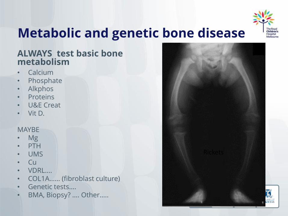
Rickets
Metabolic and genetic bone disease
ALWAYS test basic bone metabolism• Calcium• Phosphate• Alkphos• Proteins• U&E Creat• Vit D.
MAYBE• Mg• PTH• UMS• Cu• VDRL….• COL1A…… (fibroblast culture)• Genetic tests….• BMA, Biopsy? …. Other…..
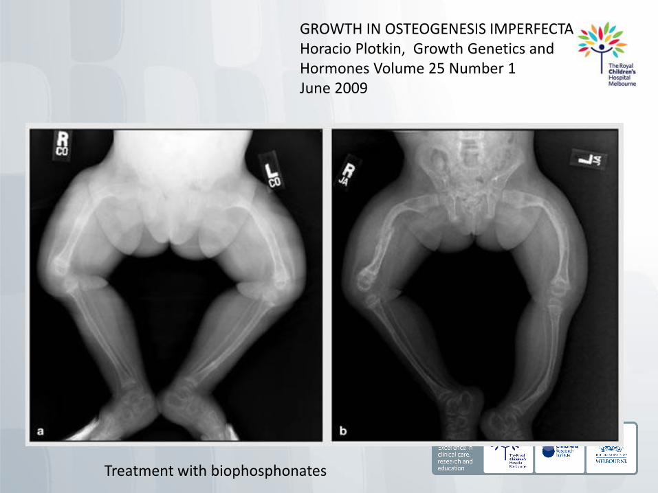
GROWTH IN OSTEOGENESIS IMPERFECTAHoracio Plotkin, Growth Genetics and Hormones Volume 25 Number 1June 2009
Treatment with biophosphonates
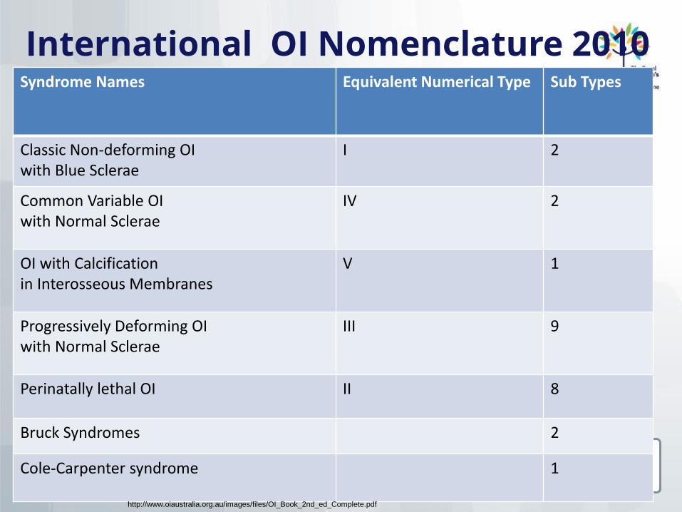
International OI Nomenclature 2010Syndrome Names Equivalent Numerical Type Sub Types
Classic Non-deforming OI with Blue Sclerae
I 2
Common Variable OI with Normal Sclerae
IV 2
OI with Calcification in Interosseous Membranes
V 1
Progressively Deforming OI with Normal Sclerae
III 9
Perinatally lethal OI II 8
Bruck Syndromes 2
Cole-Carpenter syndrome 1
http://www.oiaustralia.org.au/images/files/OI_Book_2nd_ed_Complete.pdf

Don’t forget to examine teeth!
Top Related