


Languages
Pages
Legal

Fast Hybrid Freehand Ultrasound Volume Reconstruction
Athanasios Karamalisa,b, Wolfgang Weinb, Oliver Kuttera and Nassir Navaba
aChair for Computer Aided Medical Procedures (CAMP) Technische Universitat Munchen,Germany
bImaging and Visualization Dept., Siemens Corporate Research, Princeton, NJ, USA
ABSTRACT
The volumetric reconstruction of a freehand ultrasound sweep, also called compounding, introduces additionaldiagnostic value to the ultrasound acquisition by allowing 3D visualization and fast generation of arbitraryMPR(Multi-Planar-Reformatting) slices. Furthermore reconstructing a sweep adds to the general availabilityof the ultrasound data since volumes are more common to a variety of clinical applications/systems like PACS.Generally there are two reconstruction approaches, namely forward and backward with their respective advan-tages and disadvantages. In this paper we present a hybrid reconstruction method partially implemented onthe GPU that combines the forward and backward approaches to efficiently reconstruct a continuous freehandultrasound sweep, while ensuring at the same time a high reconstruction quality. The main goal of this workwas to significantly decrease the waiting time from sweep acquisition to volume reconstruction in order to makean ultrasound examination more comfortable for both the patient and the sonographer. Testing our algorithmdemonstrated a significant performance gain by an average factor of 197 for simple interpolation and 84 foradvanced interpolation schemes, reconstructing a 2563 volume in 0.35 seconds and 0.82 seconds respectively.
Keywords: Visualization, Ultrasound, Volume Reconstruction, GPU
1. DESCRIPTION OF PURPOSE
Today’s most widely used ultrasound imaging technique is freehand ultrasound, that when tracked (optic ormagnetic) allows volume reconstruction from freehand sweeps. A number of reconstruction methods have beenintroduced [4] which can generally be categorized into forward and backward compounding methods.
Forward compounding is qualitatively the inferior approach, projecting the acquired ultrasound slices directlyinto a volume. The lack of any interpolation results in gaps between the slices in the volume. This can be avoidedby increasing the slice thickness for the projection, however the reconstruction quality remains poor. On theother hand, backward compounding is qualitatively the superior approach for which each voxel of the volumeis calculated by taking into account adjacent slices. Different interpolation schemes have been introduced forthe weighting of the adjacent slices [8], reconstruction based on probe trajectory delivering the best qualitativeresults so far [1].
As stated, the main concern of our work is to reduce the waiting time from acquisition to volume recon-struction, which especially becomes irritating when the acquisition has to be repeated multiple times, e.g. inthe context of whole organ imaging for guiding interventional oncology procedures [7], or registration to a pre-operative plan for prostate radiotherapeutic delivery.
In the next sections we will present a high-performance reconstruction method that combines the forwardand the backward approach, using graphics hardware to accelerate some of the steps required.
Further author information:(Send correspondence to Athanasios Karamalis)Athanasios Karamalis: E-mail: [email protected] Wein: E-mail: [email protected] Kutter: E-mail: [email protected] Navab: E-mail: [email protected]
Submitted to SPIE Medical Imaging 2009
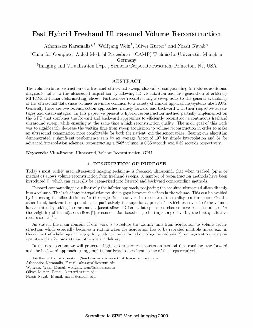
(a)Image edge lines (b)Sweep edge lines (c)Intersection points
Figure 1. Visualizing algorithm steps
2. METHODS
In this section we will briefly present our algorithm starting with the required modeling of the input. Ouralgorithm gets a geometrical representation for each ultrasound image (slice) by defining a line equation foreach edge as demonstrated in figure(1 a). Figure (1 b) shows a visualization of the image edge lines for a givenultrasound sweep.
Algorithm 1 Fast Hybrid Freehand Ultrasound Volume ReconstructionRequire: Sweep image edge lines1: Find optimal sampling direction2: Define sampling layers3: for all sampling layers do4: for all slices do5: if edge line not parallel to sampling layer then6: find valid line-plane intersections7: calculate texture coordinates8: end if9: end for
10: render interpolated quadrilaterals11: end for
As for the algorithm, the sampling layers compose the layers of a compounded volume and are defined using~n · (x− x0) = 0 where the plane normal ~n becomes the optimal sampling direction. Furthermore valid line-planeintersections occur when an intersection between an image edge line and a sampling plane result in a point thatlies on the line segment defining the image edge. Figure (1 c) visualizes the intersection points for a given sweep.Only step 10 (render interpolated quadrilaterals) of the algorithm is performed on the GPU. More on calculatingtexture coordinates and rendering quadrilaterals follows.
2.1 MPR generation
For each sampling layer we render quadrilaterals (quads) utilizing the intersection points and texture coordinatesof adjacent ultrasound slices by using common graphic APIs like OpenGL (in our case) or DirectX. Texturecoordinate are simply calculated for each slice by dividing the Euclidean distance of an intersection point to animage edge point, by the length of the edge it lies on. Forming n− 1 quadrilaterals for n slices produces a MPRof the sweep for a given sampling layer. Furthermore by using quadrilaterals to connect adjacent ultrasoundslices we avoid gaps that might arise, as in the case of forward compounding.
If the acquisition images the same anatomy twice, the most recent sweep imaging data will be reconstructeddue to the successive drawing of slices. Repeatedly imaging the same anatomy with freehand ultrasound usuallydoes not yield the exact same data, due to the highly dynamic imaging process (speckle noise, tissue deformation
Submitted to SPIE Medical Imaging 2009
etc.); hence this behavior is desirable, unless explicit averaging is needed (similar to aperture compounding onultrasound transducers).
In case of discontinues sweeps we cannot use quadrilaterals and have to fall back to rendering lines for eachintersection between a slice and a sampling plane. This would result in a forward reconstruction with decreasedquality.
2.2 Quadrilateral interpolation
Each pixel on the quadrilaterals is the result of a linear interpolation between the bilinear interpolated valuesof the adjacent ultrasound slices. However, even modern GPU hardware does not support direct renderingof quadrilaterals and decomposes them into two triangles which are processed separately. This results in adiscontinuous attribute interpolation inside the quadrilateral [2], which is highly visible since we use irregularconvex quadrilaterals for rendering. In order to deal with this problem we used the following two approaches.The first approach approximates an improved interpolation by splitting the quadrilateral into four trianglesusing the center point inside the quadrilateral. The final interpolation is performend in a simple fragment shaderprogram that runs on the GPU [5]. The second approach is more sophisicated and defines a continuous attributeinterpolation inside the quadrilateral by implementing general barycentric coordinates for irregular polygons [3]in a fragment shader program, making this approach the computationally more demanding one.
2.3 Volume generation
Nowadays most graphic cards provide the ability to ’render-to-volume’ which allows redirecting the renderingfrom the display to an internal volume on the graphics card, without much effort. This volume can directly beused to generate arbitrary MPRs or for 3D rendering of the entire volume, whereas trilinear interpolation insidethe volume is supported by graphic cards without any performance penalty. Moreover the volume can be readback into the RAM for further processing.
3. RESULTS
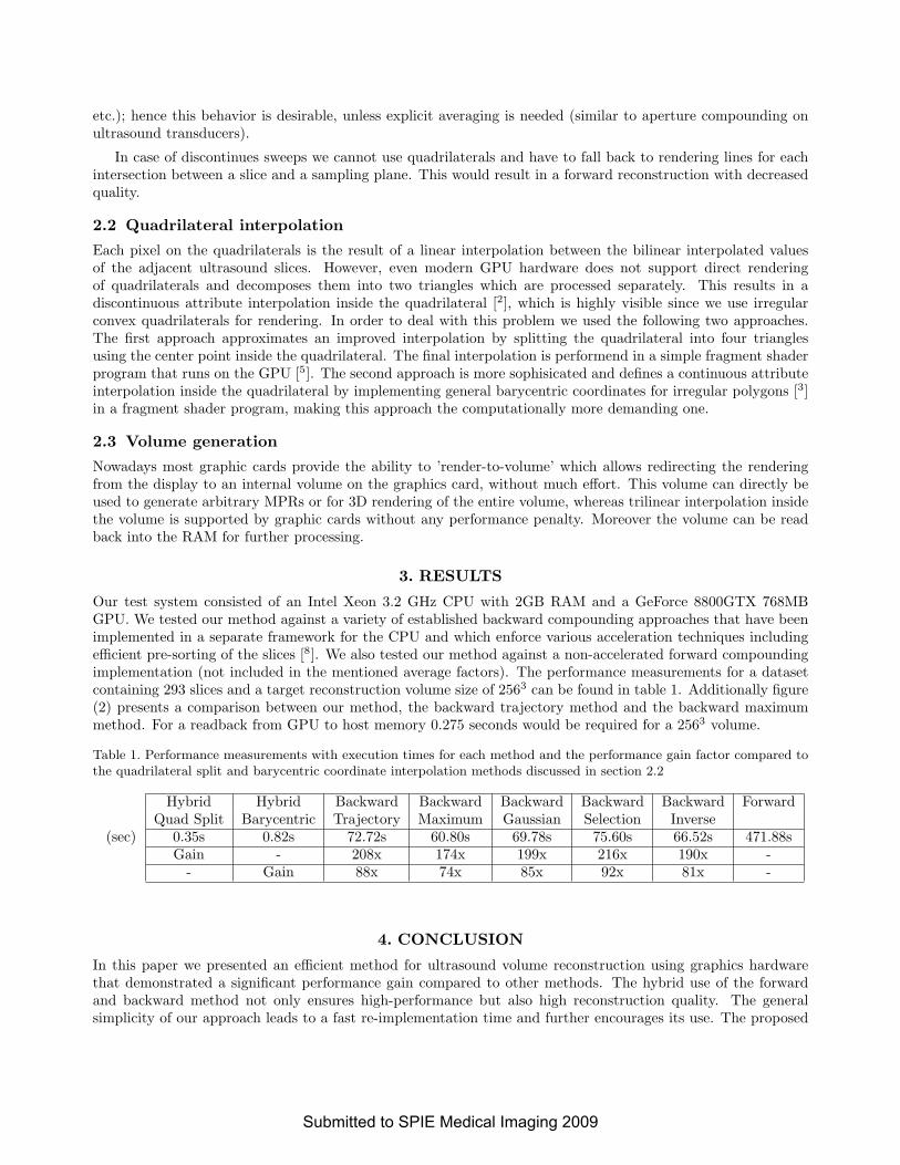
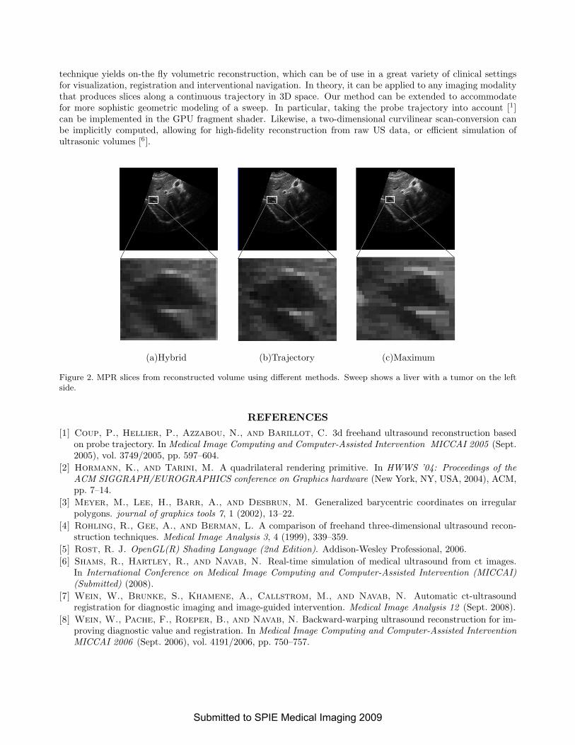
Our test system consisted of an Intel Xeon 3.2 GHz CPU with 2GB RAM and a GeForce 8800GTX 768MBGPU. We tested our method against a variety of established backward compounding approaches that have beenimplemented in a separate framework for the CPU and which enforce various acceleration techniques includingefficient pre-sorting of the slices [8]. We also tested our method against a non-accelerated forward compoundingimplementation (not included in the mentioned average factors). The performance measurements for a datasetcontaining 293 slices and a target reconstruction volume size of 2563 can be found in table 1. Additionally figure(2) presents a comparison between our method, the backward trajectory method and the backward maximummethod. For a readback from GPU to host memory 0.275 seconds would be required for a 2563 volume.
Table 1. Performance measurements with execution times for each method and the performance gain factor compared tothe quadrilateral split and barycentric coordinate interpolation methods discussed in section 2.2
Hybrid Hybrid Backward Backward Backward Backward Backward ForwardQuad Split Barycentric Trajectory Maximum Gaussian Selection Inverse
(sec) 0.35s 0.82s 72.72s 60.80s 69.78s 75.60s 66.52s 471.88sGain - 208x 174x 199x 216x 190x -
- Gain 88x 74x 85x 92x 81x -
4. CONCLUSION
In this paper we presented an efficient method for ultrasound volume reconstruction using graphics hardwarethat demonstrated a significant performance gain compared to other methods. The hybrid use of the forwardand backward method not only ensures high-performance but also high reconstruction quality. The generalsimplicity of our approach leads to a fast re-implementation time and further encourages its use. The proposed
Submitted to SPIE Medical Imaging 2009
technique yields on-the fly volumetric reconstruction, which can be of use in a great variety of clinical settingsfor visualization, registration and interventional navigation. In theory, it can be applied to any imaging modalitythat produces slices along a continuous trajectory in 3D space. Our method can be extended to accommodatefor more sophistic geometric modeling of a sweep. In particular, taking the probe trajectory into account [1]can be implemented in the GPU fragment shader. Likewise, a two-dimensional curvilinear scan-conversion canbe implicitly computed, allowing for high-fidelity reconstruction from raw US data, or efficient simulation ofultrasonic volumes [6].
(a)Hybrid (b)Trajectory (c)Maximum
Figure 2. MPR slices from reconstructed volume using different methods. Sweep shows a liver with a tumor on the leftside.
REFERENCES[1] Coup, P., Hellier, P., Azzabou, N., and Barillot, C. 3d freehand ultrasound reconstruction based
on probe trajectory. In Medical Image Computing and Computer-Assisted Intervention MICCAI 2005 (Sept.2005), vol. 3749/2005, pp. 597–604.
[2] Hormann, K., and Tarini, M. A quadrilateral rendering primitive. In HWWS ’04: Proceedings of theACM SIGGRAPH/EUROGRAPHICS conference on Graphics hardware (New York, NY, USA, 2004), ACM,pp. 7–14.
[3] Meyer, M., Lee, H., Barr, A., and Desbrun, M. Generalized barycentric coordinates on irregularpolygons. journal of graphics tools 7, 1 (2002), 13–22.
[4] Rohling, R., Gee, A., and Berman, L. A comparison of freehand three-dimensional ultrasound recon-struction techniques. Medical Image Analysis 3, 4 (1999), 339–359.
[5] Rost, R. J. OpenGL(R) Shading Language (2nd Edition). Addison-Wesley Professional, 2006.[6] Shams, R., Hartley, R., and Navab, N. Real-time simulation of medical ultrasound from ct images.
In International Conference on Medical Image Computing and Computer-Assisted Intervention (MICCAI)(Submitted) (2008).
[7] Wein, W., Brunke, S., Khamene, A., Callstrom, M., and Navab, N. Automatic ct-ultrasoundregistration for diagnostic imaging and image-guided intervention. Medical Image Analysis 12 (Sept. 2008).
[8] Wein, W., Pache, F., Roeper, B., and Navab, N. Backward-warping ultrasound reconstruction for im-proving diagnostic value and registration. In Medical Image Computing and Computer-Assisted InterventionMICCAI 2006 (Sept. 2006), vol. 4191/2006, pp. 750–757.
Submitted to SPIE Medical Imaging 2009
Top Related
