


Languages
Pages
Legal


J. Algal Biomass Utln. 2011, 2 (3): 82– 93 Antioxidant activities and antiproliferative activity of green microalgae
© PHYCO SPECTRUM INC
Evaluation of in vitro antioxidant activities and antiproliferative activity
of green microalgae, Desmococcus olivaceous and Chlorococcum humicola
R.Uma, V. Sivasubramanian and S.Niranjali Devaraj*
Vivekananda Institute of Algal Technology (VIAT), RKM Vivekananda College, Chennai, INDIA
*Dept of Biochemistry, University of Madras, Guindy, Chennai, INDIA
Abstract
The potential antioxidant and antiproliferative activities of acetone and methanolic extracts of green micro algae,
Desmococcus olivaceous and Chlorococcum humicola, were evaluated in vitro. Total phenolic and flavonoid content were
determined in both the extracts. Free radical (DPPH) scavenging potential was higher in Desmococcus olivaceous than
cholorococcum humicola. Reducing power ( H2O2 ) scavenging assay, Ferric reducing antioxidant power assay (FRAP),
lipid peroxidation inhibition efficiency ( TBAR’s assay) of both extracts were also evaluated. Methanolic extract of D
.olivaceous and C.humicola showed higher reducing power and promising activity in preventing lipid peroxidation that
might prevent oxidative damage to biomolecules. DNA fragmentation analysis was done by agarose gel electrophoresis.
The results obtained suggest that methanolic extract of D.olivaceous and acetone extract of C.humicola were able to
protect DNA from oxidative damage. Tested extracts showed strong selective cell proliferation inhibition on Hep-2 cell
line, especially the methanolic extract of D. olivaceous with IC50 value of 1.56µg / ml and the acetone extract of
C.humicola with IC50 value of 0.625µg / ml. Cell growth inhibition was mainly due to apoptosis proved by fragmentation
analysis. The results obtained suggest that the methanolic extracts of D. olivaceous and acetone extracts of C. humicola
may be promising alternative to synthetic substances as natural compound with high anti-proliferative activity.
Keywords: Phenolics,Flavanoids Free radicals, LPO, DPPH, HRSA, TBARS, MDA,FRAP, MTT, IC50
Abbreviations
LPO,Lipid peroxidation; DPPH, 1,1-diphenyl 1-2 picrylhydrazyl; HRSA, Hydrogenperoxide radical scavenging assay;TBARS,
thiobarbituricacid reactive assay;MDA, malon dialdehyde; FRAP, ferric reducing antioxidant power assay; MTT,3-[4,5-
dimethylthiazole-2y] 2,5- diphenyltetrazoliumbromide; IC50, Inhibitory concentration.
Introduction
Reactive Oxygen species (ROS) such as
superoxide anion, hydroxyl radicals and
hydrogen peroxide which are generated
by normal physiological process and
various exogenous factors initiate
peroxidation of membrane lipids as well
as a wide range of other biological
molecule through a process that is
believed to be implicated in the etiology
of several disease conditions including
Coronary heart disease, Stroke,

J. Algal Biomass Utln. 2011, 2 (3): 82– 93 Antioxidant activities and antiproliferative activity of green microalgae
© PHYCO SPECTRUM INC
Rheumatoid arthritis, Diabetes and
Cancer. Antioxidants play an important
role in inhibiting and scavenging radicals
thus providing protection to humans
against infections and degenerative
diseases. The two most commonly used
synthetic antioxidants, butylated hydroxyl
anisole ( BHA) and butylated hydroxyl
toluene ( BHT) have begun to be
restricted because of their toxicity and
DNA damage induction. Therefore,
natural antioxidants from plant and algal
extracts have attracted increasing interest
due to the safety. Recent researches have
been interested in finding novel
antioxidants to combat and / or prevent
ROS mediated diseases.
Green microalgae are widely used in the
life science as the source of compounds
with diverse structural forms and
biological activities. Marine algae have
been historically and exceptionally rich
source of pharmacologically active
metabolites with antineoplastic,,
antimicrobial and antiviral effects.(
Faulkner, 2000 ; Tziveleka et. al., 2003.)
Green micro algae like Scenedesmus and
Chlorella contain rich source of active
metabolites with anticarcinogenic effects.(
Farouk et. al., 2002.) Antitumor promoting
glyceroglycolipids from the green alga
chlorella (Morimoto et. al., 1995 ). More
marine algae have been suspected of
having strong antiproliferative. and
antioxidants properties, including Fucus
vesiculosis and brown alga, Ecklonia cava
(Yasantha et. al., 2000)
The aim of the present study was to
evaluate antioxidant properties and anti
proliferative activity of methanolic and
acetone extracts of green microalgae, D.
olivaceous and C. humicola by measuring
scavenging activity against free radicals,
reducing capacity and protection of
biological molecules from ROS induced
damage. These micro algae are extensively
employed in the phycoremediation of
various types of industrial effluents by
Vivekananda Institute of Algal Technology
(VIAT), Chennai (Sivasubramanian et al.,
2010). The algal extracts were evaluated for
their suppressive effect on tumor cell
growth by using MTT assay and DNA
fragmentation analysis.
Materials and Methods:
Culturing and growth of algal organisms

J. Algal Biomass Utln. 2011, 2 (3): 82– 93 Antioxidant activities and antiproliferative activity of green microalgae
© PHYCO SPECTRUM INC
Fresh water green microalgae
Desmococcus olivaceous and Chlorococcum
humicola were obtained from the culture
collection of Vivekananda Institute of
Algal technology (VIAT) Chennai. Algal
Biomass was obtained by growing algal
cultures in 20L of water and 0.25g / L of
NPK fertilizer was added with a facility
to pump the culture with aeration pump.
The algae was grown for 10 days and
harvested.
Preparation of Algal extract:
0.5g of dried algal material was
extracted in 20ml of acetone and
methanol kept in an orbital shaker for
overnight. The obtained extracts were
filtered with Whatman no.1 filter paper
and the filtrate was collected. The solvents
were removed under reduced pressure at
50°C to yield a concentrated extract
(12% and 11%) respectively.
Quantitative analysis of antioxidative
compounds
Determination of Total phenolic compounds
Total phenolic content was determined
with Folin & Ciocalteau reagent
according to the method described by
Singleton et. al., (1999 ) using gallic acid
as standard.
Determination of Total Flavonoids:
Total Flavonoids content was determined
by the method described by Zhishen et.
al., (1999)
Antioxidant activities assays
DPPH Radical Scavenging Assay
Free radical scavenging ability of the
extracts was tested by DPPH radical
scavenging assay as described by Blois,
(1958). Different concentration of sample
50, 100 &150µl (0.25,0.5 &0.75 mg) of the
extracts were taken in the test tubes.
3.0ml of 0.1mM DPPH in ethanol was
added to each tube and incubated in
dark at room temperature for 30minutes.
The absorbance was read at 517nm using
UV-visible spectrophotometer. BHT
(butylated hydroxy toluene) was used in
standard calibration. The % inhibition
(I%) was calculated using the formula :
I% = [ Abs ( control) - Abs ( sample)] x
100
Abs ( control
Hydrogen peroxide(H2o2) scavenging
assay (HRSA)
H2O2 scavenging activity was determined
according to Ruch etal., (1988). A
solution of H2O2(10mM) was prepared in
J. Algal Biomass Utln. 2011, 2 (3): 82– 93 Antioxidant activities and antiproliferative activity of green microalgae
© PHYCO SPECTRUM INC
phosphate buffer ( pH7.4). Reaction
mixture containing 2.5ml of H2O2
solution and 0.1, 0.3, 0.5ml of varying
concentration of algal extracts (0.5, 1.5
&2.5 mg ) was made up of to 3ml with
phosphate buffer. The absorbance was
measured at 0min and after 60 min at
240nm. Ascorbic acid was used as the
control. Total H2O2 scavenging activity
was expressed in %.
HRSA(%) = [ Abs(control) – Abs (sample ) / Abs
(control) ] x 100
Thiobarbituric Acid Reactive Assay
(TBARS)
The assay was performed as described
by Halliwell Gutteridge (1999 ) ,in which
the extent of LPO was estimated from
the concentration of Malondialdehyde,, a
thiobarbituric acid reacting substance which
is produced due to lipid peroxidation.
50µg, 10 µg, 150 µg, 200 µ g and 250 µg
of the algal extract were taken in a test
tubes and were evaporated to dryness at
80°C .1 ml of 0.15M potassium chloride
was added to the tubes and followed by
0.5ml of goat liver homogenate (10%
W/V in Phosphate buffered saline; calcium
magnesium free). Peroxidation was
initiated by the addition of 100 µl of
2mM ferric chloride. After incubating the
tubes for 30min at 37°C, the
peroxidation reaction was stopped by
adding 2ml of ice-cold HCL ( 0.25N)
containing 15% TCA & 0.38% TBA. The
tubes were kept at 80°C for 1 hr, cooled
and centrifuged at 7500rpm. The
absorbance of the supernatant, containing
TBA-MDA complex was read at 532nm.
The anti-lipid peroxidation activity
(ALP%) was calculated using the
formula :
ALP% = [ Abs ( control) - Abs ( sample)] x 100
Abs (control)
Ferric Reducing antioxidant Power
Assay
Ferric Reducing antioxidant Power Assay
was determined by the method described by
Omidreza et. al ., (2000).
MTT Assay
In this study, cancer cell growth inhibition
activity was measured by using MTT assay
(Mossman,1983; Carmichael et.al., 1987).
Hep-2 cell lines were maintained in minimal
essential media( MEM) containing 10% heat
inactivated fetal calf serum (FCS)
supplemented with penicillin (100 µg/ml)

J. Algal Biomass Utln. 2011, 2 (3): 82– 93 Antioxidant activities and antiproliferative activity of green microalgae
© PHYCO SPECTRUM INC
and streptomycin (100 µg/ml) at 37°c
under 5% co2 in air. 48hrs monolayer
culture of Hep2 cells at a concentration of
1 lakh / ml /well seeded in 24 well titre
plate.
The monolayer of cells was washed twice
with MEM without FCS to remove the
dead cells and excess FCS. To the washed
cell sheet, add 1ml of medium (without
FCS) containing defined concentration of
the drug in respective wells. Each dilution
of the drug ranges from 1:1 to 1:64 and
they were added to the respective wells of
the 24 well titre plates. To the cell control
wells add 1 ml MEM without FCS. Plates
were incubated at 37oc in 5% co2
environment and observed for cytotoxicity
using inverted microscope. In each well
add 200ul of MTT concentration (5
mg/well) cells were then incubated for
cytotoxicity. Add 1ml of DMSO in each
well and mix, leave for 45 seconds. If
any viable cells present formazan crystals
after adding solubilizing reagent (DMSO)
shows the purple colour formation. The
suspension is transferred into the cuvette
of spectro photometer and an OD values
was read at 595mm by taking DMSO as
a blank. Graph is plotted by taking
concentration of the drug on X axis and
relative cell viability on Y axis.
Cell Viability (%) = Mean OD/Control OD x 100
All determinations were carried out in
triplicates. The IC50, the antiproliferative
activity of the tested enzymatic fractions
was determined in terms of the amount
(µg/ml) of the extract necessary for
inhibiting 50% of the cell growth.
DNA fragmentation Analysis
A semiquantitative method for measuring
apoptosis was described by Bortner, et. al (
1995).
Results and Discussion
Total phenolic and flavonoid contents in
the methanolic and acetone extracts were
expressed as mg/g and were presented in
Fig 1. The methanolic extract had higher
phenolic and flavonoid content than
acetone extract of D. olivaceous, and .
C.humicola . This may be due to the
differences in the polarity of the two
solvents used and there by the different
phenolic components differentially eluted.

J. Algal Biomass Utln. 2011, 2 (3): 82– 93 Antioxidant activities and antiproliferative activity of green microalgae
© PHYCO SPECTRUM INC
FIG. 1. TOTAL PHENOLIC AND FLAVONOID CONTENTS OF METHANOLIC AND ACETONE EXTRACTS OF
DESMOCOCCUS OLIVACEOUS AND CHLOROCOCCUM HUMICOLA
DPPH radical scavenging assay
Both methanolic and acetone extracts of
D. olivaceous and C . humicola showed a
significant dose dependent reduction of
DPPH radicals. The scavenging action
was higher in Desmococcus (95.8%) in
comparison with the Chlorococcum
(93%). This assay revealed that the
extracts might prevent reactive radical
species from damaging biomolecules
such as Lipoprotein, DNA, aminoacids,
sugar, proteins and PUFA in biological
and food systems.(Table 1 )
TABLE.1. DPPH RADICAL SCAVENGING ACTIVITY OF METHANOL AND ACETONE EXTRACTS OF DESMOCOCCUS
OLIVACEOUS AND CHLOROCOCCUM HUMICOLA
Concentraction
Of Extracts
(µg)
Inhibition %
Desmococcus olivaceous Chlorococcum humicola
Methanol Acetone Methanol Acetone
250
92.6 + 0.5 92 + 1.6 91 + 1.4 87.3 + 1.5
500
94 + 1.5 94 + 1.6 92 + 1.6 89 + 1
750
95.8 + 0.7 95.8 + 0.5 94 + 1.8 93 + 1.5
The values were expressed as inhibition %; Data’s are mean of triplicate determination mean + S.D (n=3)

J. Algal Biomass Utln. 2011, 2 (3): 82– 93 Antioxidant activities and antiproliferative activity of green microalgae
© PHYCO SPECTRUM INC
Hydrogen peroxide scavenging assay
(HRSA)
In this study, methanolic extract of D.
olivaceous at 2.5mg/0.5ml exhibited 39%
scavenging activity (Fig 2) and the
acetone extracts of C. humicola exhibited,
15% scavenging activity. Acetone extract
of Desmococcus and methanolic extract
of Chlorococcum showed relatively low
H2O2 scavenging activities. However, the
activity increases with the sample
concentration.(Fig 2)
FIG 2 HYDROGEN PEROXIDE SCAVENGING ASSAY OF METHANOLIC AND ACETONE EXTRACTS OF DESMOCCOCUS
OLIVACEOUS AND CHLOROCOCCUM HUMICOLA
TBARS assay
Both extracts were capable of preventing
the formation of MDA in a dose
dependent manner. The methanolic
extracts of D. olivaceous & C. humicola
was observed to be significantly better
inhibitor of Lipid peroxidation compared
to acetone extract. (Fig III) shows the ALP
% potential of the extracts of
Desmococcus and Chlorococcum.
FIG 3 ANTI LIPID PEROXIDATION ACTIVITY OF METHANOLIC AND ACETONE EXTRACTS OF DESMOCOCCUS
OLIVACEOUS & CHLOROCOCCUM HUMICOLA

J. Algal Biomass Utln. 2011, 2 (3): 82– 93 Antioxidant activities and antiproliferative activity of green microalgae
© PHYCO SPECTRUM INC
FRAP ASSAY
Methanolic extracts of Desmococcus and
acetone extracts of Chlorococcum showed
maximum ferric reducing antioxidant
power.(Table 2 &Fig 4)
TABLE 2 & FIG 4. FERRIC REDUCING ANTIOXIDANT POWER ASSAY (FRAP)
MTT assay
Of the extracts tested, the methanolic
extract of D. olivaceous had the highest
cell growth inhibition activity with an
average IC50 value of 0.156mg/ml. In
addition, the acetone extract of
Chlorococcum also showed good cell
growth inhibition activity on HEP-2 cell
line ( 0.625mg/ml IC50). These extracts
induced high antiproliferative activity in
a dose dependent manner.. ( Fig 5 a & b)
Sl.
No. Contents
BHT control
( 0.16mg/ml)
Desmococcus sp Chlorococcum sp
Methanol Acetone Methanol Acetone
1 Absorbance at
593 nm
0.063
0.211 0.101 0.035 0.183

J. Algal Biomass Utln. 2011, 2 (3): 82– 93 Antioxidant activities and antiproliferative activity of green microalgae
© PHYCO SPECTRUM INC
FIG 5 a INVITRO ANTI PROLIFERATIVE ACTIVITY OF METHANOL EXTRACTS OF DESMOCOCCUS
OLIVACEOUS
020406080
100
5 1.25 0.3125 0.078 0.019
% c
ell
viab
ility
Concentration (mg/ml)
FIG 5 B INVITRO ANTI PROLIFERATIVE ACTIVITY OF ACETONE EXTRACTS OF
CHLOROCOCCUM HUMICOLA
0
20
40
60
80
100
120
5 2.5 1.25 0.625 0.3125 0.156 0.078 0.039 0.019 0.097
% c
ell
viab
ility
Concentration (mg/ml)
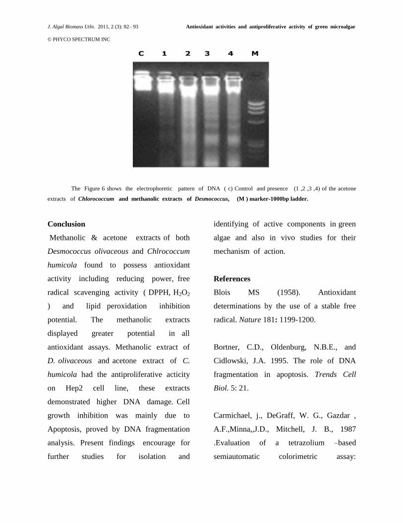
DNA Fragmentation analysis
In control lane, DNA is not fragmented. In
lane 1 ,2 ,3 & 4 acetone extracts of
Chlorococcum and methanolic extracts of
Desmococcus treated with Hep-2 cell line,
DNA was fragmented. Hence, 3 bands
ranging from 180 to 220bp was obtained.
(Fig 6)
FIG 6 DNA FRAGMENTATION BY AGAROSE GEL ELECTRO PHORESIS
J. Algal Biomass Utln. 2011, 2 (3): 82– 93 Antioxidant activities and antiproliferative activity of green microalgae
© PHYCO SPECTRUM INC
The Figure 6 shows the electrophoretic pattern of DNA ( c) Control and presence (1 ,2 ,3 ,4) of the acetone
extracts of Chlorococcum and methanolic extracts of Desmococcus, (M ) marker-1000bp ladder.
Conclusion
Methanolic & acetone extracts of both
Desmococcus olivaceous and Chlrococcum
humicola found to possess antioxidant
activity including reducing power, free
radical scavenging activity ( DPPH, H2O2
) and lipid peroxidation inhibition
potential. The methanolic extracts
displayed greater potential in all
antioxidant assays. Methanolic extract of
D. olivaceous and acetone extract of C.
humicola had the antiproliferative acticity
on Hep2 cell line, these extracts
demonstrated higher DNA damage. Cell
growth inhibition was mainly due to
Apoptosis, proved by DNA fragmentation
analysis. Present findings encourage for
further studies for isolation and
identifying of active components in green
algae and also in vivo studies for their
mechanism of action.
References
Blois MS (1958). Antioxidant
determinations by the use of a stable free
radical. Nature 181: 1199-1200.
Bortner, C.D., Oldenburg, N.B.E., and
Cidlowski, J.A. 1995. The role of DNA
fragmentation in apoptosis. Trends Cell
Biol. 5: 21.
Carmichael, j., DeGraff, W. G., Gazdar ,
A.F.,Minna,,J.D., Mitchell, J. B., 1987
.Evaluation of a tetrazolium –based
semiautomatic colorimetric assay:

J. Algal Biomass Utln. 2011, 2 (3): 82– 93 Antioxidant activities and antiproliferative activity of green microalgae
© PHYCO SPECTRUM INC
assessment of chemosensitvity testing.
Cancer Research 47 , 936 -942.
Farouk k.El-Baz, Ahmed M, Aboul- Eneim
2002 , Anticarcinogenic activity of algal
extracts, Journal of Medical Science, 2(5-6
):243-251.
Faulkner DJ (2000) Marine natural
products.Nat.Prod.Rep.17: 7: 55
Halliwell, B., & Gutteridge, J.M.C(1999)
.Free radicals in Biology and Medicine,. 3rd
edition.Oxford:Oxford, University press.
Morimoto, T. A, Nagatu and N. Murakami ,
1995. Anti tumor promoting glycerol
glycolipids from the green alga Chlorella
vulgaris. Phytochem., 40 : 1433- 1437 .
Mossman, T ., 1983. Rapid colorimetric
assay for cellular growth and survival:
application to proliferation and cytotoxicity
assays. Journal of Immunological Methods ,
65,55-63.
Noda, H.H.A., K. Arashima and K .
Nisizawa , 1990. Antitumor activity of
marine algae. Hydrobiology, 2
Omidreza,F.,Antonio, L,. P., Giancarlo,
M.,& Luciano, s.. (2005) Evaluation of the
antioxidant activity of flavonoids by ferric
reducing amtioxidant power Q assay and
cyclic voltammetry. Biochemica et
Biophtsica Acta, 1721 , 174- 181.
Ruch RJ, Cheng SJ, Klaunig JE (1989).
Prevention of cytotoxicity and inhibition of
intercellular communication by antioxidant
catechins isolated from Chinese green tea.
Carcinogenesis, 10: 1003-1008.
Singleton VL,Orthofer R and Lamuela-
Raventos RM, Analysis of total phenols and
oxidation substrates and antioxidants by
means of Folin –Ciocalteau reagent.
Methods Enzymol , 1999 ,299, 152 – 177.
Sivasubramanian,V, V V Subramanian and
M Muthukumaran.2010.Production of algal
biomass integrated with Phycoremediation –
A sustainable and economically viable
approach. J. Algal Biomass utln. 1(4): 10 –
57
Tziveleka L.A, Vagias C, Roussis V (2003)
Natural products with anti HIV activity
from marine organism Current Trop . Med
.Chem .3(13) :1512 -11535.

J. Algal Biomass Utln. 2011, 2 (3): 82– 93 Antioxidant activities and antiproliferative activity of green microalgae
© PHYCO SPECTRUM INC
Yasantha, A., Kim K, N.,& Jeon, Y . J. (
2006). Antiproliferative and antioxidant
properties of an enzymatic hydrolysate
from brown algae, Ecklonia cava. Food and
chemical Toxicology, 44, 1065- 1074
Zhishen I,Mengcheng T and Jianming W,
The determination of flavanoids contents in
mulberry and their scavenging effects on
Super radicals, Food Chem, 1999 ,64, 555 -
559.
Top Related