


Languages
Pages
Legal

ESM 1. Additional figures of UFRJ-DG 031 Av
Supplementary Figure 1 Counterslab of UFRJ-DG 031 Av. a, main slab. b,
interpretative drawing of the skeleton and feathers Abbreviations: cd, free caudal
vertebrae; dv, dorsal vertebrae; fr, frontals; il, ilium; lc, left coracoid; lf, left femur; lh,
left humerus; lr, left radius; lt, left tibiotarsus; lu, left ulna; rcp, right carpometacarpus;
rh, right humerus; rib, dorsal rib; rr, right radius; ru, right ulna; scl, sclerotic ring. Scale
bar, 1 cm.
Supplementary Figure 2 ESM. Details of cranial bones of UFRJ-DG 031 Av. a,
main slab. b, interpretative drawing. Abbreviations: al, alular feather; cf, cover feathers;
cpm, carpometacarpus; fr, frontals; I, digit I; II, digit II; mx, maxilla; par, parietal; r,
radius; scl, sclerotic ring. Scale bar, 1 cm.
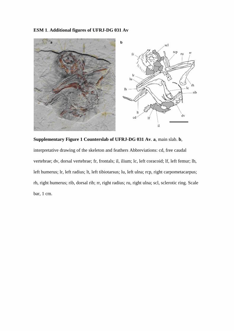
Supplementary Figure 3. Detail of the thorax of UFRJ-DG 031 Av. a, main slab. b,
interpretative drawing. Abbreviations: dv, dorsal vertebrae; lc, left coracoid; lf, left
femur; ls, left scapula; rc, right coracoid; rib, dorsal rib. Scale bar, 0,7 cm.

Supplementary Figure 4. Details of the skeleton of UFRJ-DG 031 Av. a, main
slab. b, interpretative drawing. Abbreviations: cd, free caudal vertebrae; il, caudal end
of left ilium; lc, left coracoid; lf, left femur; ls, left scapula; pub, pubes; py, pygostyle;
rc, right coracoid; rh, right humerus; rr, right radius; ru, right ulna; tb, tibiotarsus; tmt,
metatarsus. Scale bar, 0,2 cm.
Supplementary Figure 5. Reconstruction of selected elements of UFRJ-DG 031
Av. a, anterior free caudal vertebra in dorsal view; b, right manus in lateral view.

Abbreviations: mc, metacarpal; ns, neural spine; ph, phalanx; poz, postzygapophyses;
prz, prezygapophyses; tp, transverse process. Not to scale.
Supplementary Figure 6. Details of the feet of UFRJ-DG 031 Av. a, main slab. b,
interpretative drawing. At left an incomplete left foot, at right a nearly complete right
foot. Abbreviations: d, digit; I, one; II, two; III, three; mt, metatarsal; tb, tibiotarsus.
Scale bar, 0,5 cm.

Supplementary Figure 7. Strict consensus tree showing the phylogenetic position
of UFRJ-DG 031 Av within Enantiornithes.

Supplementary Note 1. Additional description of the skeleton of UFRJ-DG 031 Av.
UFRJ-DG 031 Av consists on a nearly complete skeleton, preserved in two slabs,
including skull, nearly complete fore and hindlimbs, vertebral column and pectoral and
pelvic girdles. However, as is usual in fossils preserved in two slabs, the bones are
cracked. Some crushing and displacement of bones has occurred. Several bones,
however, remain in articulation. The skull and neck are rotated ventrally with respect to
the rest of the skeleton, and are exposed on the left side, whereas most of remaining
skeleton is exposed on the right side. The skull and mandible suffered strong crushing
and deformation, and most bones cannot be individualized. The pelvic girdle and
hindlimb remain in anatomical position, whereas the pectoral girdle has been displaced
ventrally with respect of the dorsal column. The minor slab shows better preserved
bones than the main slab. It contains the impression of the skull, and both forelimbs and
fragments of pectoral girdle. The ischia and sternum are not preserved.
The very small body size, large orbit, elongate caudal series, poorly developed
proximal humerus and distal ends of other long bones (femur, tibia), as well as the lack
of fusion in the metatarsus indicates that the specimen may be a juvenile
The skull is highly distorted and poorly preserved, and only some bones can be
properly recognized. We identify several skull roof elements including parietals,
frontals and nasal, as well as the maxilla and lacrimal bone. Some other elements
located at the level of the snout, nearly perpendicular to the main axis of the skull are
problematic, since they are much more robust compared to other cranial elements, and
their morphology do not match with bones of the rostrum.
The parietals and frontals are dorsally convex, indicating a vaulted braincase. The
snout is highly pointed and subtriangular in contour. The maxilla is subtriangular and

the presence of minute alveoli on its mid portion indicate the existence of small-sized
teeth, which are not preserved in the slab. The lacrimal bone is incompletely preserved
but shows a pillar-shaped ventral process. Due to weathering, remaining skull and
dentary bones do not offer interesting anatomical information.
The vertebral column of UFRJ-DG 031 Av is represented by cervical, dorsal, sacral,
and caudal vertebrae, but each element is very poorly preserved. Five cervical vertebrae
have been preserved in articulation, and are exposed on their right side. The complete
number of cervicals is unknown. The centra are rather elongate and lack any sign of
lateral excavations or pleurocoels. A longitudinal ridge separates the centra from the
neural arch. The neural spines are dorsoventrally tall and show a subrectangular contour
in lateral view. Although incompletely preserved, vertebral centra are stouter and
shorter than those of other enantiornithes, such as Gobipteryx. Six dorsal vertebrae are
preserved. Although they are highly distorted, they show a conformation similar to that
of other enantiornithes. The vertebral centra are relatively short, resembling the
proportions of basal enantiornithes such as Iberomesornis [1], being different from
derived enantiornithines such as Gobipteryx, in which they are longer [2] and resembles
basal taxa, as for example, The centra exhibit a deep lateral longitudinal excavation, as
frequently occurs in Enantiornithes [3]. The neural spine is well developed and
subrectangular in contour, being slightly fan-shaped distally. Parapophyses are located
high on the centrum, a diagnostic character of Enantiornithes [1].
Caudal vertebrae are poorly preserved, the is represented only by its transverse
processes. There are 8 free caudal vertebrae. The second and third caudals are
completely preserved, but exposed in dorsal view. The first half of the fourth caudal has
been preserved as bone, whereas the posterior half has been preserved as a mold.
Caudals fifth through eight are preserved as impressions of the ventral face of their
centra. The vertebral centra are not well-preserved, but appear exhibit biconvex articular
surfaces. On caudals 2-4 the neural spine is transversely thick, and the transverse
processes are very well-developed, robust, and craniocaudally expanded. A composite
reconstruction of the neural arch of free caudals in dorsal view, indicates transversely
compressed zygapophyses, thick neural spine, and a wide fossa lateral to the
postzygapophyses, for the articulation of the prezygapophysis of the posterior vertebrae.
This combination of characters is different from the more reduced caudal neural spine,
transverse processes and zygapophyses seen in most enantiornithine taxa.
The pygostyle of UFRJ-DG 031 Av is a rod-like structure very similar to that
present in other enantiornithes. It is composed by 8 caudal vertebrae, and is cone-shped,
being longer than the combined length of free caudals. The proximal end is forked,
showing two sub-parallel longitudinal processes, as is diagnostic of Enantiornithes [4].
Vertebral bodies are short and sub-cilindrical, and lack any sign of transverse processes.
The pygostyle exhibits a longitudinally extended ventrolateral process, as frequently
occurs among Enantiornithes [4]. The elongate tail feathers insert at the level of the
third pygostyle vertebra.
No cervical ribs have been preserved, or they cannot be identified on the available
cervicals. Most of the right dorsal ribs are preserved, as well as the distal tips of some
left ribs. The rib pattern is similar to that described for the enantiornithine
Iberomesornis [1]. The first dorsal rib is very long. Posterior dorsal ribs decrease in
length. The proximal end of the ribs is bicapitate, and its distal end is slightly expanded
for articulation with sternal ribs. There are some fragmentary preserved sternal ribs.
These show a slightly expanded distal articular surface.
Both coracoids are preserved partially as natural molds, and are exposed in ventral
view. They are elongate and narrow, strut-like bones. The proximal end is typical of
enantiornithine birds, showing a highly reduced procoracoid and a convex boss for
articulation with the scapula. The acrocoracoid is reduced and rounded in contour. The
lateral margin of coracoid is convex, whereas the medial one is slightly concave. As in
most Enantiornithes the lateral process is absent [2]. The distal margin of the bone
appears to be only slightly concave.
The left scapula is preserved in dorsal view. The proximal end is badly damaged,
and most structures are obscure. The acromion is well-developed and subtriangular in
contour, and is laterally oriented, a condition similar to that of other Enantiornithes. The
distal end of the scapular blade is acute and transversely compressed.
Both humeri are exposed in posterior view. The right humerus is nearly complete,
whereas the left one is eroded on its distal half. The humeral head is rounded in contrast
with the majority of Enantiornithes, in which it is apomorphycally saddle-shaped [1].
This head is located central to the main axis of the bone, as commonly occurs in
enantiornithine birds (e.g., Iberomesornis, Gobipteryx; [2,5]). On its centre exhibits a
poorly concave longitudinal groove. The bicipital tubercle is a poorly differentiated and
rounded process, the capital groove and the transverse ligamental groove are absent, a
combination of characters that contrast with more derived Enantiornithes (e.g.,
Martinavis, Enantiornis, Gobipteryx, Halimornis [6]). The bicipital crest is absent, a
plesiomorphic condition present in Enantiornithes and Archaeopteryx [2]. The distal end
of the humerus is transversely expanded, although not at the degree seen in
Euenantiornithes. There is not clear evidence of the presence of a distal caudal fossa.
The distal condyles are not exposed; however, in caudal view, a ventral extension of
the distal margin indicates that the dorsal condyle was nearly sub-parallel to the humeral
diaphysis, a diagnostic condition to Enantiornithes [1].

The left and right ulnae are preserved in posterior aspect. Both are highly
fragmented and show poorly preserved proximal and distal ends, precluding the
description of most features. The ulna is nearly as long as the humerus, a condition that
contrast with that of derived Enantiornithes, including Elbretornis and Enantiornis, in
which the ulna is much shorter than humerus [6]. The proximal end of the bone shows a
well-developed olecranon and the external condyle is prominent and rounded. The distal
end of ulna is nearly flat and subcuadrangular in contour, contrasting with the pulley-
shaped morphology seen in other Enantiornithes [6].
Left and right radii are preserved in posterior view. The proximal end shows a well
developed median concavity. In posterior view its distal end is rounded in contour. It
shows a longitudinal groove, as diagnostic of Enantiornithes [3]. The shaft of the radius
appears to the wider proximally than its distal end, a peculiar condition shared with
Iberomesornis and Enantiornis [1].
UFRJ-DG 031 Av manus is sub-equal to total ulnar length. The left
carpometacarpus has been preserved in lateral view, whereas the right carpometacarpus
is represented by highly crushed scraps of bone. Proximally, the carpometacarpus is
tightly-fused, whereas the distal end is unfused. The proximal end of carpometacarpus
is badly crushed, with por anatomical details. A small fragment of bone may represent
metacarpal I. Phalanx 1-I is elongate, although its incompleteness precludes estimation
of its total length. The distal end of ungual 2-I is preserved; it is acute and slightly
curved. Metacarpal II is shorter and thinner than metacarpal III, a diagnostic condition
of Euenantiornithes [6]. Phalanx 1-II is robust and subrectangular in contour, lacking of
mid-constriction. Phalanx 2-II is preserved mainly as a bone impression. It is thinner
and slightly shorter than 1-II and shows a well-defined mid constriction. Ungual 3-II is
acute and sligtly curved, and appears to be sub-equal in size and morphology to ungual

2-I. Only the base of phalanx 1-III has been preserved, indicating a reduced digit III, as
frequently occurs in birds.
The right ilium is represented by part of the acetabulum. The acetabular portion of
ilium lacks of supracetabular crest, and the presence of a caudal antitrochanter can be
observed on the posterior margin of the acetabulum. The left ilium preserves its
posterior blade. It is subtriangular in contour and dorsoventrally low.
The pubes are observed in caudal view but their proximal and distal ends are
broken. A well-developed pubic symphysis appears to be present.
The right femur is exposed in caudal view; its mid-shaft has been broken away, and
its distal end is badly damaged. The left femur is hidden by a large amount of scraps of
indeterminate bones, the right femur and the pubes. The only preserved portions are its
proximal end in medial view, and the medial condyle in posterior view. The length of
the left femur does not matches with the right one (see measurements), suggesting that
there was some movement of proximal and distal parts. The femoral shaft appears to be
nearly straight. The femoral head is anterodorsally oriented, and a fovea capitis appears
to be absent. It shows a continuous articular surface with the trochanteric crest. The
latter one is well developed, and proximally reached the level of the proximal margin of
the femoral head. The iliotrochanteric drepression is wide and deep, subtriangular in
contour. In caudal view, a bulbous posterior trochanter is present distal to the
trochanteric crest. The distal end is not very well preserved. A patellar groove appears
to be absent, and the medial condyle is bulbous and ellipsoidal in side view.
Both tibiotarsi show a badly crushed surface, and their distal and proximal ends are
poorly preserved. The right tibiotarsus is exposed in lateral view, whereas the left
tibiotarsus is exposed in medial view. The left tibiotarsus shows hidden proximal and

distal ends. The tibiotarsus is much shorter than the femur, and is subequal in length to
the metatarsals. Due to its poor preservation additional data cannot be afforded.
The proximalmost portion of the metatarsals is not fused, and a small metatarsal cap
is absent. In posterior view the hypotarsus is absent. The right metatarsals are exposed
in posterior aspect, most of the shaft of metatarsals III and II are preserved as
impressions. The left metatarsals are exposed in anterior view, but their proximal halves
are hidden by the right metatarsals. The metatarsals are elongate and transversely
narrow. Metatarsals are unfused along most of its length. Metatarsal IV is transversely
compressed, specially at its distal end, and is thinner than other metatarsals, as
diagnostic of Enantiornithes [1]. Its distal end is very transversely compressed and
shows a ginglymoid articulation. Metatarsal I is small and has strongly posteriorly
deflected distal condyles, and distal condyles are proximally joined, both characters
considered as diagnostic of Enantiornithes [1]. Metatarsal II is relatively robust. Its
distal end and the articular surface of phalanx 1-II does not appears to be wider than
metatarsal III, in contrast with remaining Enantiornithes [1]. Metatarsal III is the stouter
bone of the foot, it is transversely wide and homogeneous in width along all its length.
Its distal ginglymoid is poorly preserved.
Pedal phalanges are relatively well preserved, although most unguals are preserved
only as impressions. Non-ungual phalanges 1-IV and 2-IV are relatively elongate and its
combined length clearly surpasses the distal end of phalanx 1-III, whereas in
Iberomesornis they do not surpasses such level [1]. Phalanges 3-IV and 4-IV are not
preserved. Non-ungual phalanges of pedal digit III are very narrow and extremelly
elongate, being much longer than metatarsal III. In other Enantiornithes (e.g.,
Iberomesornis [1]) the combined length of these phalanges is subequal or shorter than
metatarsal III length. Phalanx 1-III is the stouter of the foot and shows a very wide and

transversely expanded proximal end. Phalanx 2-IIII is not well preserved; it was
relatively slender and constitutes the shortest non-ungual phalanx of the foot. Phalanx 3-
III is very elongate and narrow, being the more gracile element of the pes. Its distal
ginglymoid appears to be small and only slightly excavated. Phalanges 1-II and 2-II are
elongate and subequal in length and morphology each other. Phalanx 1-I is very
elongate and robust, being the stouter element of the foot, as commonly occurs in
Enantiornithes [7]. Pedal unguals are elongate, and show a slightly curved ventral
surface. Ungual 5-IV does not preserves its proximal half, but available information
suggests that it was relatively elongate and smaller than other unguals. Ungual 4-III is
relatively robust and is larger than unguals of digit II and III. Ungual 3-II is subequal in
length and morphology to other unguals of the foot. Pedal ungual of digit I with very
curved ventral margin, much more than remaining unguals.
Remarkably, the specimen preserves different kind of feathers around the skeleton.
The skeleton of UFRJ-DG 031 Av is covered by filamentous feathers, including a
crown of feathers located at the top of the head. 10 secondary asymmetrical remiges
anchored on the forearm are preserved. None of the remiges are completely preserved,
so its total length and morphology are nearly unknown. Left and right alulae are
represented by feathers attached to digit I, a condition widespread among Enantiornithes
[8]. At least three alular feathers have been preserved on the left hand. Alular feathers
are asymmetrical, a condition widespread among birds.
A preserved pair os tail feathers is rather elongate, being 30% longer than total body
length. They insert on the third pygostylian vertebra and conform the typical ribbon-like
morphology present in Enantiornithes and Confusiusornithidae [9] these tail feathers
gradually increase in width distally. The calamus of UFRJ-DG 031 Av is extremelly
well-developed, and shows on its midline a narrow longitudinal groove that runs from

the base to the distal end of the feather. A cross-section at the base of the calamus
indicates that the structure was nearly flat, whereas the cross-section at the mid-length
indicates that at this portion the calamus was very convex. A reconstruction indicates
that mid calamus was nearly 8-shaped in cross section. At the distal third, the calamus
gradually flattens towards the rachis. The first sign of barbs is observed at the distal half
of the tail, and barb size increases towards its distal end. The barbs are clearly
distinguishable at the tip of the tail (approximately the distal 2 centimeters of the tail
feather). Each barb was dorsoventrally thick and dorsally convex, and of uniform
thickness and width for most of its length. They are angled at approximate 16º from the
rachis and lack any sign of interlocking barbules. Both vanes are sub-equal, suggesting
a symmetrical feather. At the distal end, and near its contact with the third pygostylian
vertebra, the calamus narrows abruptly. One remarkable aspect of these ribbon-like
feathers is the existence at the base of each feather of a longitudinal row of five brown
spots at the calamus, probably representing the original tail coloration of UFRJ-DG 031
Av. No remains of hindlimb feathers are recognized.

Supplementary Note 2. Measurements of UFRJ-DG 031 Av.
Right humerus, total length: 14 mm.
Coracoid, total length: 7.7 mm
Left ulna, total length: 13.3 mm
Left metacarpal III, total length: 7.2 mm.
Left manual phalanx 1-III, total length: 3.2 mm
Right femur, total length*: 11.7 mm
Left femur, total length: 12.8 mm
Left tibia, total length*: 12 mm
Left metatarsal III, total length: 8.9 mm
Right pedal phalanx 1-I, total length: 2.6 mm
Right pedal ungual of digit I, total length: 3.5 mm
Pygostyle, total length: 9.4 mm
Caudal series, total length: 8.3 mm
Caudal tail feather, total length: 79.9 mm
Preserved portion of right remiges, total length of longest feather: 10.3 mm
Preserved portion of left remiges, total length of longest feather: 8.6 mm
* Indicates estimated size due to incomplete preservation of the element.

Supplementary Note 3. Phylogenetic analysis
Present phylogenetic analysis is based on the version of O´Connor and Zhou [10]
data set, which constitutes the most comprehensive analysis regarding the phylogeny of
Enantiornithes. The matrix was only modified by the inclusion of UFRJ-DG 031 Av.
The data matrix is composed of 245 characters distributed among 51 taxa.
The phylogenetic analysis was performed using TNT 1.1 [11]. All characters were
equally weighted and treated as unordered. Heuristic searches were performed after
1,000 pseudoreplications of WAG+TBR search strategy, with 10 random addition
sequences after each search and 100 trees were saved at each replicate. The
phylogenetic analysis resulted in the recovery of 82 Most Parsimonious Trees (MPTs),
of 71 steps, with a consistency index of 0.40, and a retention index of 0.71. The strict
consensus tree (Supplementary Figure 7) resulted on a large polytomy at the base of
Enantiornithes that comprised most genera, including UFRJ-DG 031 Av.

Supplementary Note 4. Scoring for UFRJ-DG 031 Av in the data matrix of
O´Connor and Zhou [10]
UFRJ-DG 031 Av
??????1??????????????????????????????????????????0??????00????0?????24
010?0110000111110100?1??000?0?????????????????????010?0100002?1???????
???11?????1???1???0?0100001200100?122??????????????????01?0012????????
??????????100?0010??01??20?0?0?1210

Supplementary references
1- Sereno, P.C. Iberomesornis romerali (Ornithothoraces, Aves) re-evaluated as an
enantiornithine bird Neues Jahr. Geol. Paläont. Abhand. 215, 365–395 (2000).
2- Kurochkin, E.N. A new enantiornithid of the Mongolian Late Cretaceous, and a
general appraisal of the Infraclass Enantiornithes (Aves). Russ. Acad. Sci., Special
Issue: 1–50 (1996).
3- Chiappe, L.M. & Calvo, J.O. Neuquenornis volans, a new Late Cretaceous bird
(Enantiornithes: Avisauridae) from Patagonia, Argentina. J. Vert. Paleont. 14, 230–
246 (1994).
4- Chuong, C.M., Wu, P., Zhang, F.C., Xu, X., Yu, M., Widelitz, R.B., Jiang, T.X. &
Hou, L. Adaptation to the sky: Defining the feather with integument fossils from
Mesozoic China and experimental evidence from molecular laboratories. J. Exp. Zool. B
Mol. Dev. Evol. 298, 42–56 (2003).
5- Sanz, J.L. & Bonaparte, J.F. A New Order of Birds (Class Aves) from the Lower
Cretaceous of Spain. Nat. Hist. Mus. of Los Angeles County Contrib. Sci. 36, 38–49
(1992).
6- Chiappe, L.M. & Walker, A. Skeletal morphology and systematics of the Cretaceous
Euenantiornithes (Ornithothoraces: Enantiornithes). In Mesozoic birds: above the heads
of dinosaurs (eds. Chiappe LM, Witmer L), pp. 240–267. Univ. California press (2002).

7- Martin, L.D. The Enantiornithes: terrestrial birds of the Cretaceous. Cour. Forsch.
Senck. 181, 23–36 (1995).
8- Sanz, J.L., Chiappe, L.M., Pérez–Moreno, B.P., Buscalioni, A.D., Moratalla, J.J.,
Ortega, F. & Poyato–Ariza, F.J. An Early Cretaceous bird from Spain and its
implications for the evolution of avian flight. Nature 382, 442–445 (1996).
9- O’Connor, J.K., Chiappe, L.M., Chuong, C., Bottjer, D.J. & You, H. Homology and
Potential Cellular and Molecular Mechanisms for the Development of Unique Feather
Morphologies in Early Birds. Geosciences 2, 157–177 (2012).
10- O’Connor, J.K. & Zhou, Z. A redescription of Chaoyangia beishanensis (Aves)
and a comprehensive phylogeny of Mesozoic birds. J. Syst. Paleont. 11, 889-906
(2012).
11- Goloboff PJ, Farris J, Nixon K. 2008. A free program for phylogenetic analysis.
Cladistics 24: 774-786.
Top Related