


Languages
Pages
Legal


Sleep-disordered breathing-
Impact on the brain and body
function
Ronald M. Harper, Ph.D.
Distinguished Professor of NeurobiologyDavid Geffen School of Medicine, UCLA
Objectives
• What types of sleep disordered breathing
• Effects on cardiovascular control, memory,
cognition, mood, hormonal, metabolic
control (diabetes)
• How does this happen
• What interventions are useful?

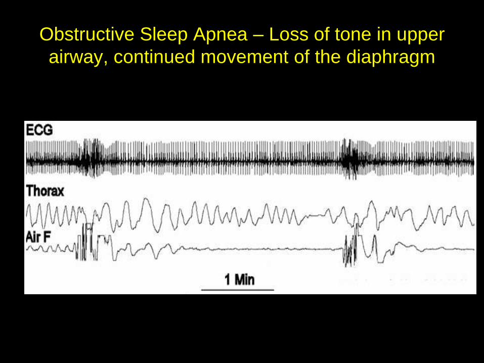
Obstructive Sleep Apnea – Loss of tone in upper
airway, continued movement of the diaphragm

Tongue genioglossal activity during sleep
Harper and Sauerland, 1978

If muscle tone is lost during REM sleep…..
But, not the only mechanism in OSA
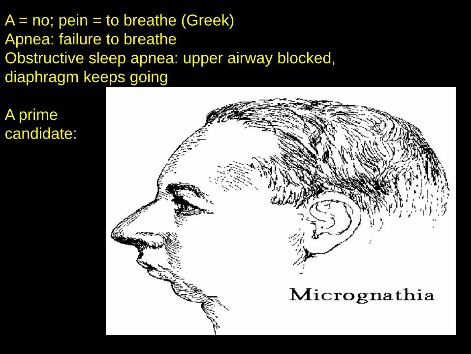
A = no; pein = to breathe (Greek)
Apnea: failure to breathe
Obstructive sleep apnea: upper airway blocked,
diaphragm keeps going
A prime
candidate:
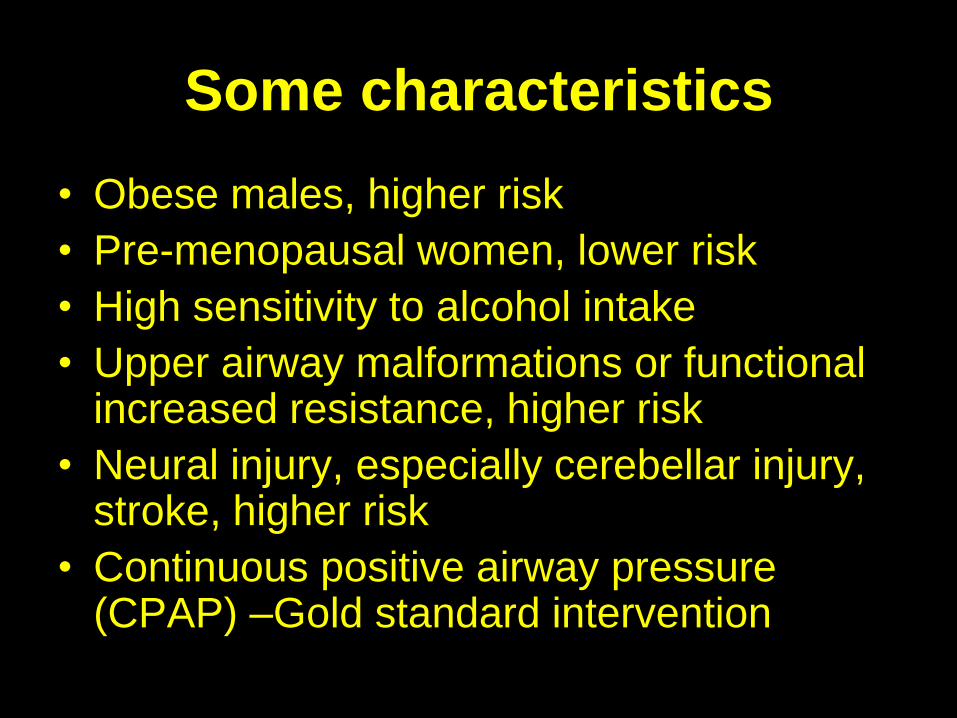
Some characteristics
• Obese males, higher risk
• Pre-menopausal women, lower risk
• High sensitivity to alcohol intake
• Upper airway malformations or functional increased resistance, higher risk
• Neural injury, especially cerebellar injury, stroke, higher risk
• Continuous positive airway pressure (CPAP) –Gold standard intervention
Obstructive sleep apnea
• It’s not just obese, adult males
• Pediatric cases; often hypertrophied
tonsilar tissue
• Maldevelopment of facial structures,
micrognathia, can be a factor
• Childhood obesity an increasingly major
concern
Upper Airway Resistance Syndrome (UARS)
Increased airway resistance (not complete
blockage, as in OSA), similar muscle relaxation
Usually evaluated by esophageal pressure;
increased esophageal pressure, followed by arousals
Arousals- sympathetic activation, hypertension
Abnormal sleep architecture (from arousals), snoring,
daytime sleepiness, hypertension (hypotension also)
Intervention similar; CPAP, mandibular devices

Periodic or Cheyne-Stokes Breathing
-often found in heart failure, sometimes in
OSA
A coordination issue- matching peripheral CO2 sensing with central chemoreception
Similar O2 desaturation and reperfusion concerns!

Why hypertension in OSA?
Resting sympathetic nervous activity is
exaggerated in OSA, even during waking states!
Sympathetic discharge can be recorded from peroneal nerve
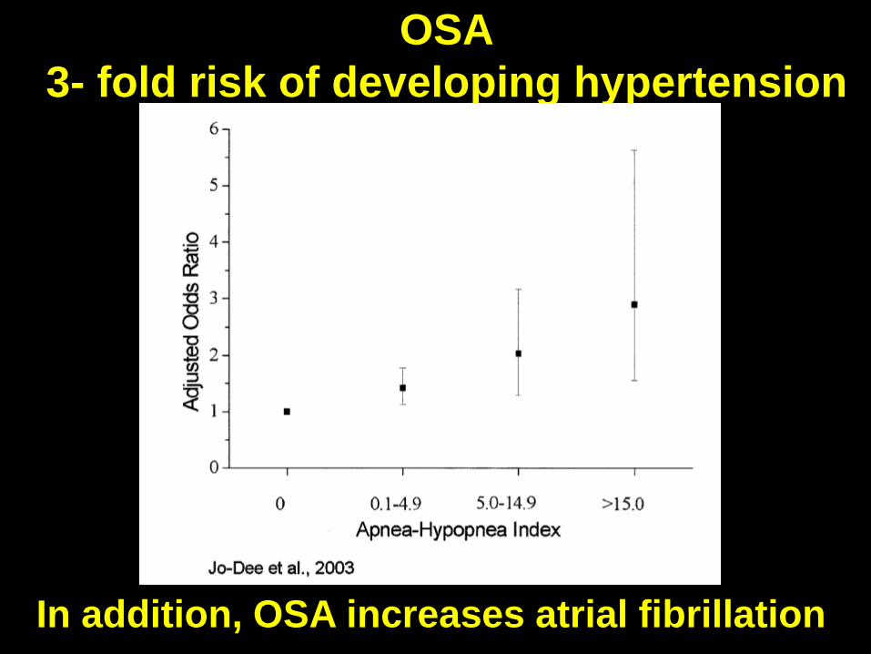
OSA
3- fold risk of developing hypertension
In addition, OSA increases atrial fibrillation

Heart failure patients have OSA and Cheyne-Stokes breathing, show substantial brain injury, especially in right insular cortices, just like OSA patients
Insular
cortex
Insular
cortex
Right insular cortex regulates sympathetic tone)
Right insular damage

The structural damage has consequences; the insular
cortex responds improperly in OSA
3 Valsalva efforts (blood pressure challenge; breathe
against a resistance): OSA patients vs Controls
From: Henderson et al., J. Appl. Physiol., 94:1063-1074, 2003

Obstructive Sleep Apnea: gray matter loss in
cerebellum and hippocampus
Macey et al., 2002

Cerebellar injury will affect blood pressure
regulation (keeps blood pressure from
falling too low or elevating too high); injury
will also affect motor coordination
(including breathing!!) – coordination is
what the cerebellum does!
How can cerebellar injury
affect physiology?
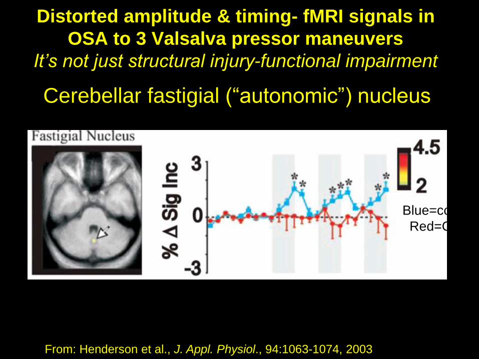
Distorted amplitude & timing- fMRI signals in
OSA to 3 Valsalva pressor maneuvers
It’s not just structural injury-functional impairment
From: Henderson et al., J. Appl. Physiol., 94:1063-1074, 2003
Cerebellar fastigial (“autonomic”) nucleus
Blue=control
Red=OSA

Brain stem areas also affected: ventrolateral
medulla, essential for breathing and blood pressure
regulation, injured in OSA (Mean Diffusivity)
Kumar et
al., 2012
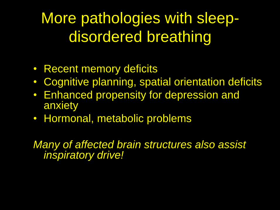
More pathologies with sleep-
disordered breathing
• Recent memory deficits
• Cognitive planning, spatial orientation deficits
• Enhanced propensity for depression and anxiety
• Hormonal, metabolic problems
Many of affected brain structures also assist inspiratory drive!
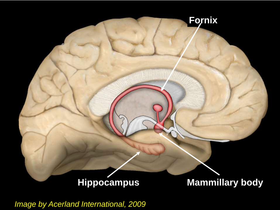
Hippocampus Mammillary body
Fornix
Image by Acerland International, 2009

0.05
0.1
0.15
P value
Regional volume reduction in
hippocampus of OSA patients
10 OSA
vs
10 Controls


Mammillary volume loss- thiamine deficiency
(chronic alcoholics, Beriberi), but how about OSA?
• Thiamine deficiency - common in those with high fluid
loss, malnutrition, diuresis, malabsorption, sulfites,
thiaminase in raw fish
• B12 deficiency- fluid loss, other meds- proton pump
inhibitor antiacids, metformin, antibiotics,
• Diabetics- urination, HF patients often subjected to
dialysis and diuretics; HF patients, frequent
malabsorption,
• OSA patients frequently diabetic, often have fluid
regulation issues- profuse nocturnal sweating

With high fluid release (sweating,
diuresis, urination)……..
• Essential vitamins can be flushed from the body
• Potassium
• Magnesium
• Thiamine (Vitamin B1)
Thiamine is necessary to transport carbohydrates
into cells. If cells become too excited (through
hypoxia), and insufficient thiamine is present,
cells can die.
Damage in brain sites which regulate depression: hippocampus, anterior cingulate
Cingulate cortex plays a role in depression; electrical stimulation can rapidly reverse symptoms.
The cingulate cortex shows structural injury and functional deficits in OSA
Depression and anxiety associated with OSA
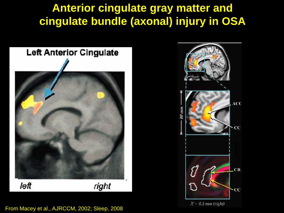
Anterior cingulate gray matter and
cingulate bundle (axonal) injury in OSA
From Macey et al., AJRCCM, 2002; Sleep, 2008
Voxel-based morphometry Fractional anisotropy
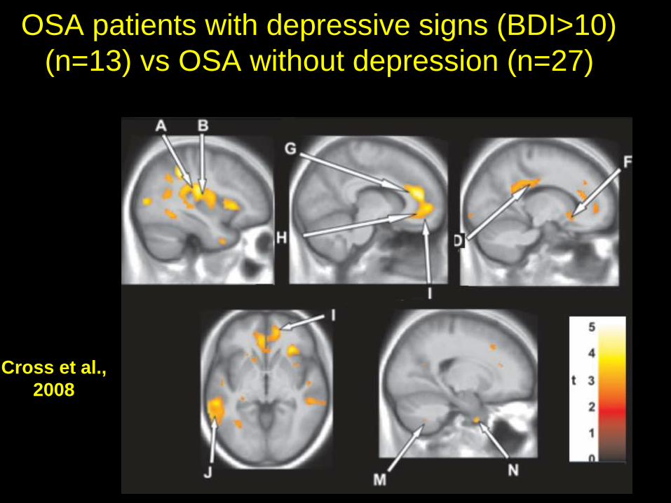
OSA patients with depressive signs (BDI>10)
(n=13) vs OSA without depression (n=27)
Cross et al.,
2008
Obstructive Sleep Apnea, hormones, and
Diabetes75% of obese Type II diabetics- moderate-to-
severe OSA!
Remainder- mild OSA!
Two hrs sleep deprivation- 50 mg/dl rise in glucose
OSA- neural injury in areas influencing
hypothalamus, potential for hormonal
dysruption
OSA: significant decline in testosterone

Enhanced injury
in Type II
diabetics
with OSA over
OSA patients
without
diabetes (T2
relaxation time)
Harper et al., 2009

An OSA treatment intervention
The Australian
didgeridoo- a
native instrument
requiring precise
neural control
over upper airway
muscles.
Relearning of cerebellar and motor circuits!
Not just a small airway!

Summary
• Sleep disordered breathing affects cardiovascular, cognitive, memory, pain, mood, and hormonal regulatory sites; a result of brain injury in condition
• Three types: OSA, UARS, Cheyne Stokes
• Cerebellar injury - loss of breathing and cardiovascular coordination in OSA
• Hormonal dysregulation in sleep-disordered breathing, likely via hypothalamic injury
• Close association, OSA with diabetes
• Interventions are available, and Dentistry can make significant contributions!

Acknowledgments
These studies were supported by the National
Institutes of Health, through multiple institutes,
including the Heart, Lung and Blood Institute, the
Nursing Institute, and the National Institutes of
Child Health and Human Development. Dr. R.
Kumar, Dr. P. Macey, Dr. M. Woo, Dr. J. Ogren,
Dr. R. Cross, and Dr. F. Yan-Go contributed
substantially to the work.