


Languages
Pages
Legal


1
Effects of a PACS-integrated Quality Assurance Reporting Tool
EncouragingEncouragingEncouragingEncouraging Squeakier WheelsSqueakier WheelsSqueakier WheelsSqueakier Wheels
Justin Cramer, MD
Matthew Morgan, MD
Tony Jones
George Milliner
Peter Jenkins, PhD
Ulrich Rassner, MD
Department of RadiologyUniversity of Utah School of MedicineSalt Lake City, UT
PURPOSE
2
Purpose
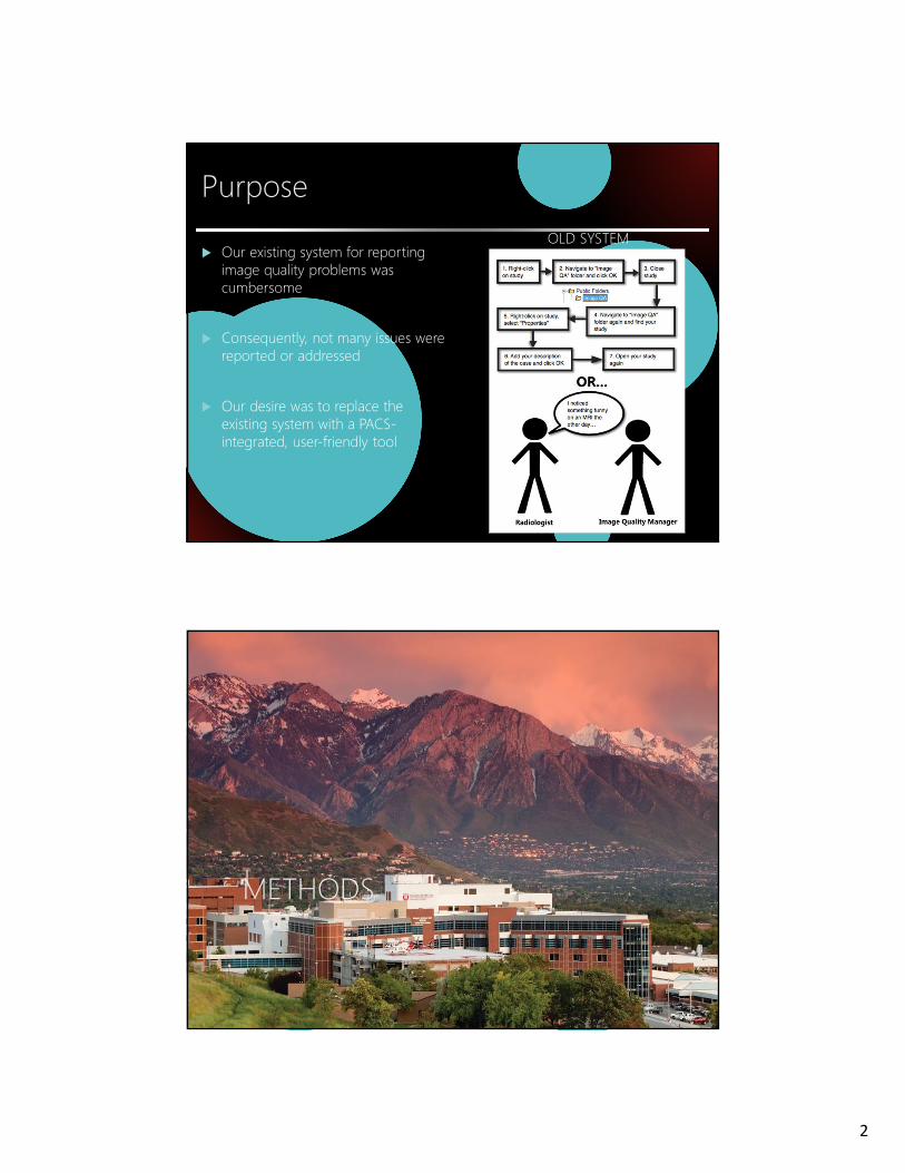
OLD SYSTEM� Our existing system for reporting image quality problems was cumbersome
� Consequently, not many issues were reported or addressed
� Our desire was to replace the existing system with a PACS-integrated, user-friendly tool
METHODS

3
Methods
� Determine functional requirements
� Project Team:
� Chief of Imaging Informatics
� Director of Radiology IT
� Director of Clinical Services
� Medical Director of Modality Safety
� Medical physicist
� Modality managers
� Software engineer
Methods
� PACS integration via a right-click context menu
� Launches image QA reporting tool for the radiologist (AKA the “Image Quality Report Form”)
Functional Requirements – Image Quality Report Form
4
Methods
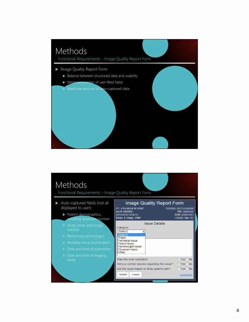
� Image Quality Report Form
� Balance between structured data and usability
� Minimize number of user-filled fields
� Maximize amount of auto-captured data
Functional Requirements – Image Quality Report Form
Methods
� Auto-captured fields (not all displayed to user):
� Patient demographics, including accession number
� Study series and image number
� Performing technologist
� Modality name and location
� Date and time of submission
� Date and time of imaging study
Functional Requirements – Image Quality Report Form

5
Methods
� User-filled fields:
� Issue Category
� Issue description (free text)
� Whether the issue required the scan to be repeated
� Whether anyone was contacted regarding the issue
� Whether the issue impacted patient care
Functional Requirements – Image Quality Report Form
Methods
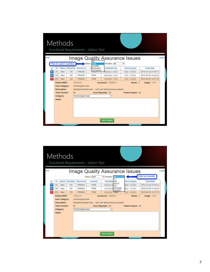
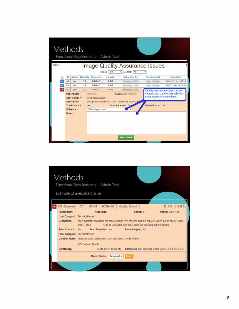
� Administrative interface for tracking submitted issues (aka “Admin Tool”) was also designed
� Required functions:
� List all outstanding issues
� Display issues by modality
� Sub-categorize issues into additional categories not visible to the end-user (see next slides)
� Apply a status to an issue (outstanding, resolved, etc.)
� Allow free-text notes on an issue
Functional Requirements – Admin Tool

6
MethodsFunctional Requirements – Admin Tool – Additional Curation Categories
SubmittorSubmittorSubmittorSubmittor CuratorCuratorCuratorCurator
Praise Praise
Technical issue Technical - artifact: avoidable
Technical - artifact: Improvable
Technical - artifact: Unavoidable
Technical – Contrast issues
Technical – Incorrect or incomplete recons
Technical – Scanner failure
Patient issue Patient – incorrect patient
Patient - Refused
Patient - Uncooperative
Patient issue
MethodsFunctional Requirements – Admin Tool – Additional Curation Categories
SubmittorSubmittorSubmittorSubmittor CuratorCuratorCuratorCurator
Technologist Technologist – Incorrect parameters
Technologist – Incorrect protocol
Technologist – Incorrect technique
Technologist issue
Physician Physician – Incorrect protcol
Physician – Incorrect/poor technique
Physician – Unclear/confusing instructions
Physician issue
Other Other
Other – Physician
Other - Technologist
Technologist issue
7
MethodsFunctional Requirements – Admin Tool
MethodsFunctional Requirements – Admin Tool
8
MethodsFunctional Requirements – Admin Tool
MethodsFunctional Requirements – Admin Tool
Example of a resolved issue
9
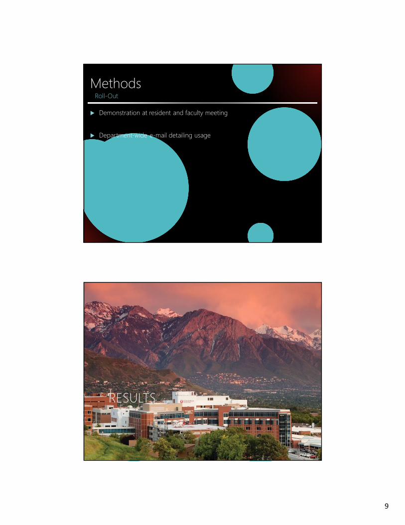
Methods
� Demonstration at resident and faculty meeting
� Department-wide e-mail detailing usage
Roll-Out
RESULTS
10
Results
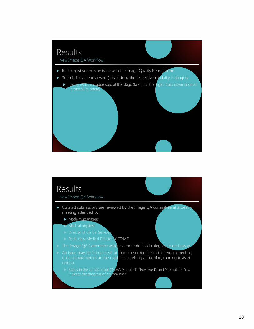
� Radiologist submits an issue with the Image Quality Report Form
� Submissions are reviewed (curated) by the respective modality managers
� Many issues are addressed at this stage (talk to technologist, track down incorrect protocol, et cetera)
New Image QA Workflow
Results
� Curated submissions are reviewed by the Image QA committee at a weekly meeting attended by:
� Modality managers
� Medical physicist
� Director of Clinical Services
� Radiologist Medical Director of CT/MRI
� The Image QA Committee assigns a more detailed category to each issue
� An issue may be “completed” at that time or require further work (checking on scan parameters on the machine, servicing a machine, running tests et cetera).
� Status in the curation tool (“New”, “Curated", “Reviewed", and “Completed”) to indicate the progress of a submission
New Image QA Workflow

11
Results
� Large rise in the number of issues submitted
� Pre-implementation
� 8 issues/month
� Post-implementation
� 75 issues/month over 11 months
Number of Issues Submitted
Results
� Post-implementation
� Totals over 11 months:
� 831 total submissions
� 416 radiography
� 221 CT
� 190 MRI
Number of Issues Submitted

12
Results
� Large rise in the number of participating radiologists
� Pre-implementation
� 4 radiologist participants
� Post-implementation
� 38 radiologist participants
Number of Participating Radiologists
Results
� Differing protocols on different scanners
� Different technical parameters for same protocol on different scanners
� Incorrect servicing of an x-ray machine
� Incorrect calculation of deviation indices
� Incorrect dose modulation adjustment parameters
� Faulty AEC cell in radiography system
� Faulty CR reader creating subtle artifact
� Different radiologists giving technologists different guidelines about studies (e.g. when to use a grid on portable radiographs, when to repeat a study or additional views). We asked the responsible section to discuss what criteria they wanted to use in order to give consistent guidance to technologists
Examples of Newly Discovered Issues

13
CONCLUSIONS
Conclusions
� To increase image QA participation, we needed to make it painless to submit issues
� A PACS-integrated image QA submission tool minimally disrupted radiologist’s workflow
� This resulted in a dramatic increase in the number of submitted issues and number of participating radiologists
� The rise in the rate of QA issues reflects an increase in participation, NOT a reflection of worse system performance
� We now have a much more accurate view into our imaging quality and can take appropriate steps to improve
Top Related