


Languages
Pages
Legal


1
DNA: Molecular basis of Inheritance
Lecture 15: 05/24/16

2
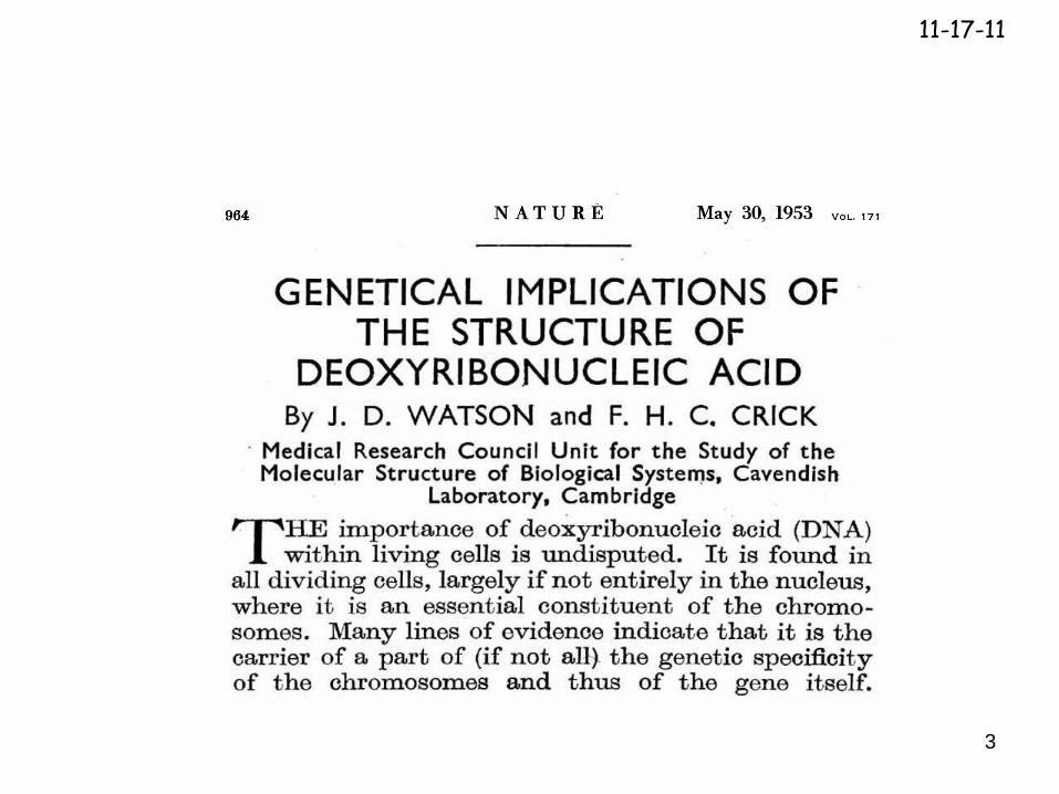
DNA Double Helix
11-17-11
3
11-17-11

4
DNA: Molecular basis of InheritanceHistorical Overview!
First Isolation of DNA
DNA as genetic material
Transformation
DNA vs Protein
Chargaff’s Rules
Structure of DNA
Franklin’s X-ray Diffraction photograph of DNA
Watson and Crick – The Double Helix
---------------------------------------------------------DNA Replication: Meselson and Stahl’s Semi-conservative model.
A change in phenotype by the acquisition of external DNA
A:T and G:C composition in cells
11-17-11
5
DNA: Molecular basis of Inheritance
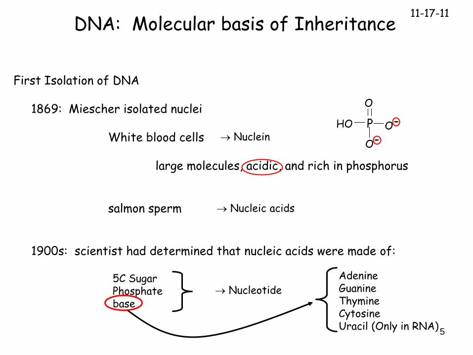
First Isolation of DNA
1869: Miescher isolated nuclei
White blood cells
large molecules, acidic, and rich in phosphorus
salmon sperm
1900s: scientist had determined that nucleic acids were made of:
Nuclein
Nucleic acids
5C SugarPhosphatebase
Nucleotide
AdenineGuanineThymineCytosineUracil (Only in RNA)
HO P
O
O-
O-
11-17-11

6
DNA as genetic material
By The 1930’s universally accepted:
1928: Griffith discovered the Transformation Phenomenon
Studied the pathogenicity of Streptococcus pneumoniae
Can the genetic trait of pathogenicity be transferred between bacteria?
DNA: Molecular basis of Inheritance
All cells contained DNA
11-17-11

7
DNA as genetic material – Griffith (1928)
Can the genetic trait of pathogenicity be transferred between bacteria?
DNA: Molecular basis of Inheritance
2 Strains of S. pneumoniae
Smooth (S) – have a capsule - pathogenic
Rough (R) – no capsule – non pathogenic
Inject
11-17-11

8
DNA: Molecular basis of Inheritance
Extracted all molecular material
DNA as genetic material – Avery (1930s-40s)
Isolated the TRANSFORMATION factor
Systematically remove or destroy various componenents
RNaseProteaseDNase
11-17-11

9
DNA: Molecular basis of Inheritance
DNA as genetic material – Hershey-Chase (~1952)
DNA or Protein as the genetic material of bacteriophages?
bacteriophages are viruses that infect bacteria
DNA encapsulated by proteins
Colorized TEM
11-17-11

10
DNA: Molecular basis of Inheritance
Life Cycle of bacteriophage
Inject DNA into bacteria
Hijacks bacteria replication and translation machinery to replicate its phage DNA and make head and tail proteins
Package replicated DNA
Lyse bacteria and release new bacteriophages
11-17-11

11
DNA: Molecular basis of Inheritance
32P labelled DNA
35S labelled protein
(Cys & Met a.a.)
(phosphate)
Grow phages with radioactive materials
Infect bacteriaSeparate Bacteria
and phage by mixing
Centrifuge mixture
pellet
pellet
supernatent
supernatent
DNA or Protein as the genetic material of bacteriophages to produce more phage?
11-17-11

12
H
DNA: Molecular basis of Inheritance
Structure of DNA
Phosphate
Base
5C sugar (deoxyribose)
C1
C2C3
C4
5
C1 is attached to basePurines A and G (2 rings)Pyrimidines C and T (1 ring)
C2 is missing OH C3 is reactive site (3’end or 3’OH)C5 attached to phosphate: reactive (5’end or 5’ phosphate)
11-17-11

13
DNA: Molecular basis of Inheritance
Structure of DNA
DNA is polymer of nucleotides
Condensation reaction: loss of water
H
H
5’3’
5’
3’
11-17-11

14
DNA: Molecular basis of Inheritance
Structure of DNA
Chargaff’s Rule
Diversity of species
1. All had DNA
2. Total amounts differed – BUT……
Pecentage of A and T the same“ “ G and C “ “
11-17-11
15
DNA: Molecular basis of Inheritance
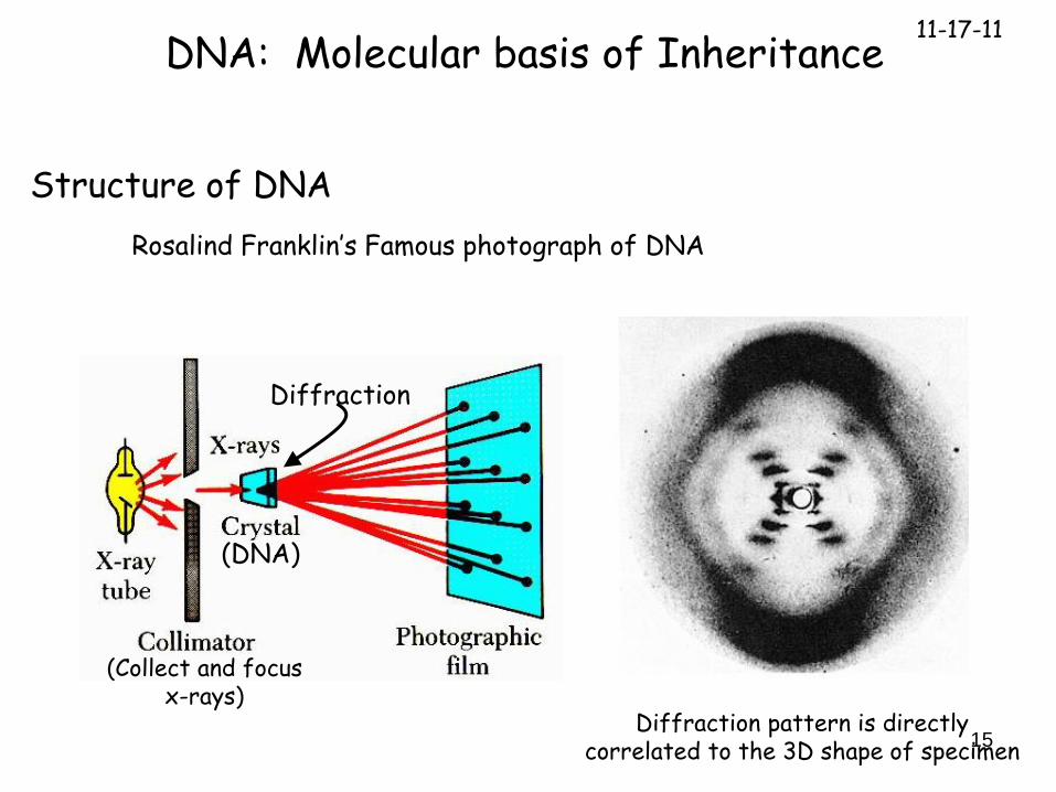
Structure of DNA
Rosalind Franklin’s Famous photograph of DNA
(DNA)
Diffraction
(Collect and focus x-rays)
Diffraction pattern is directly correlated to the 3D shape of specimen
11-17-11
16
DNA: Molecular basis of Inheritance
Structure of DNA
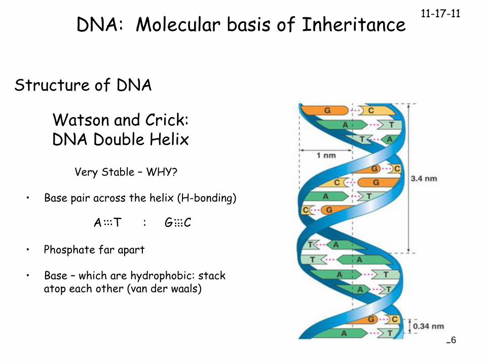
Watson and Crick: DNA Double Helix
Very Stable – WHY?
• Base pair across the helix (H-bonding)
• Phosphate far apart
• Base – which are hydrophobic: stack atop each other (van der waals)
A T : G C
11-17-11

17
DNA: Molecular basis of Inheritance
Structure of DNA Double Helix UniformDiameter
Right-handed helix
Helix has Uniform Diameter
Strands are antiparallel
0.34 nm rise per nucleotide
3.4nm rise per turn (10 nucleotides per turn)
11-17-11

18
DNA: Molecular basis of Inheritance
A, T, G, C
This is basis for amazing genetic diversity among species – HOW?
Central DogmaDNA RNA Protein
11-17-11

19
DNA: Molecular basis of Inheritance
How is DNA replicated?
3 Hypothesis to Test?
1. Conservative Hypothesis
2. Semi Conservative Hypothesis
3. Dispersive Hypothesis
Parent DNA helix remains intact, and a 2nd new copy is made
2 strands of Parent DNA helix separate, each function as a template to make “COMPLEMENTARY” strands.
Daughter DNA helix get a mix of new DNA.
11-17-11

20
How is DNA replicated?
Conservative
Semi-Conservative
Dispersive
DNA Replication11-17-11
21
DNA Replication
A
C
T
A
G
A
C
T
A
G
A
C
T
A
G
A
C
T
A
G
T
G
A
T
C
T
G
A
T
C
A
C
T
A
G
A
C
T
A
G
T
G
A
T
C
T
G
A
T
C
T
G
A
T
C
T
G
A
T
C
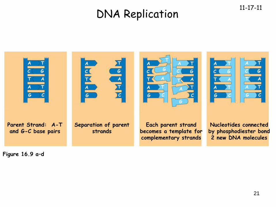
Figure 16.9 a–d
Parent Strand: A-T and G-C base pairs
Separation of parent strands
Each parent strand becomes a template for complementary strands
Nucleotides connected by phosphodiester bond2 new DNA molecules
11-17-11

22Figure 16.11
2
3) DNA sample centrifuged after 20 minutes (first
replication)
1) Bacteria cultures in medium containing 15N
2) Bacteria transferred to medium containing
14N
Meselson and Stahl’s Experiment
4) DNA sample centrifuged after 20 minutes (first
replication)
Less dense
More dense
: mechanism of DNA Replication

23
First replication Second replication
Conservativemodel
Semiconservativemodel
Dispersivemodel
Meselson and Stahl’s Experiment: mechanism of DNA Replication
15N
15N + 14N
14N

24
DNA Replication
Bacteria has 4.6 million bases pairs to replicate
Human diploid cell has 3 billion base pairs to replicate
46 (long) DNA molecules
Has to be fast and accurate
> 2000 bases per second are polymerized!!
only 1 mistake per 10 billion bases added!!
Many proteins involved!
(Polymers)
11-17-11

25
DNA Replication
Origin of Replication - specialized site where replication begins
Easy to unwind because high number of A/T base pairs
Bacteria has 1
Eukaryotic cells - 100s to 1000s per chromosome
Replication fork - Ends of replication bubble (origin)
Helicase – special protein which unwinds DNA (uses ATP)
Single stranded binding proteins (SSBP’s) - Binds single stranded DNA and keeps it from base pairing
Replication Fork
Replication Fork
11-17-11

26
DNA Replication
Initiation
Fusion
Lateral Expansion(Elongation)
11-17-11

27
DNA Replication
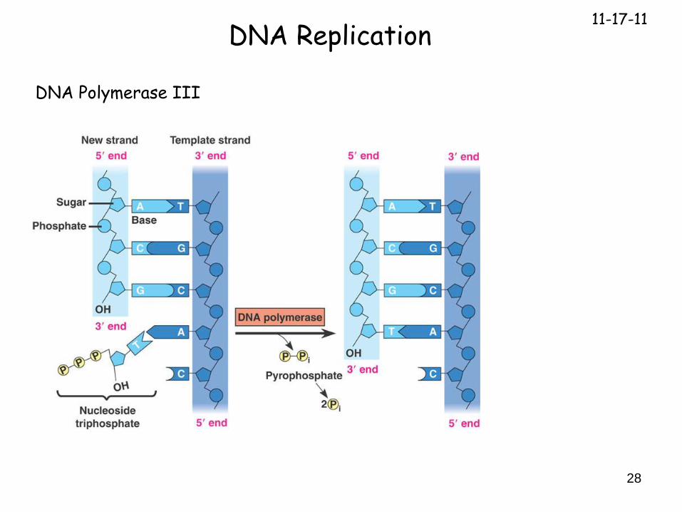
DNA Polymerase III
catalyses the addition of deoxyribose nucleotides to the 3’end (3’OH)
dATP, dTTP, dGTP, dCTP
looks at opposite (template) strand and chooses correct base
catalyses the phosphodiester bond
energy source: energy released from PPi 2 Pi (14 kcal of energy)
high fidelity (very seldom makes a mistake) : 1 in 10 billion base additions
11-17-11
28
DNA Replication
DNA Polymerase III
11-17-11

29
DNA Replication
Primase – synthesizes short (5-10 bases in length) RNA primer
supplies the 3’OH needed by DNA Pol III to add bases
DNA Polymerase I - removes RNA primer and replaces it with DNA
DNA ligase – joins (ligates) DNA together to make one continuous strand
(Note: Make sure you know Table 16.1 (Campbell – 7th edition)!)
11-17-11
30
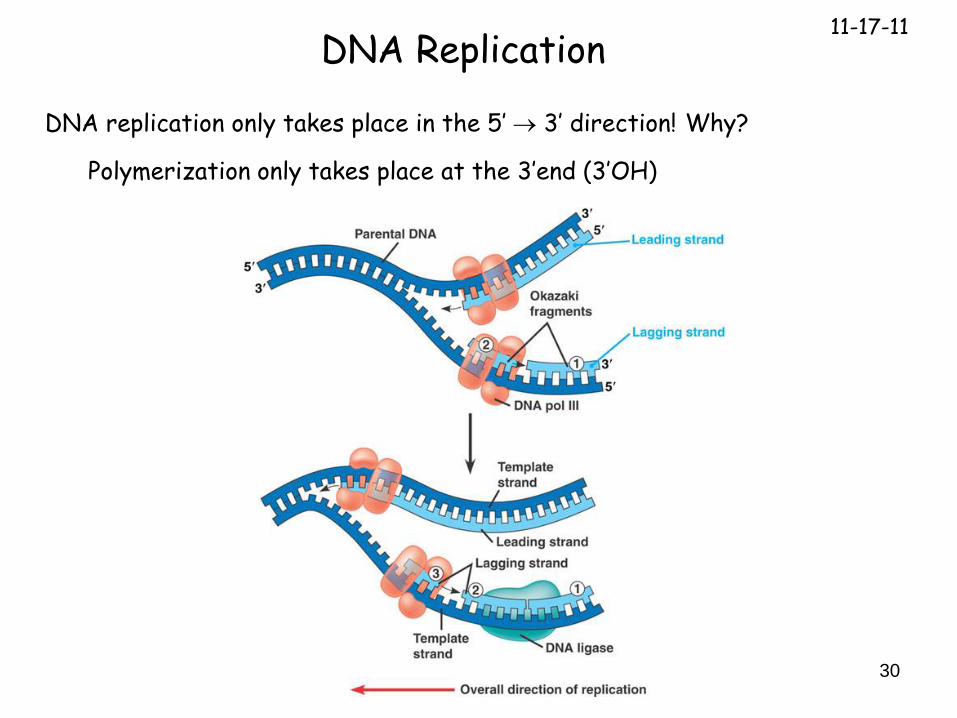
DNA Replication
DNA replication only takes place in the 5’ 3’ direction! Why?
Polymerization only takes place at the 3’end (3’OH)
11-17-11
31
Overall direction of replication
3
3
3
35
35
35
35
3
5
3
5
3
5
3 5
5
1
1
21
12
5
5
12
35
Templatestrand
RNA primer
Okazakifragment
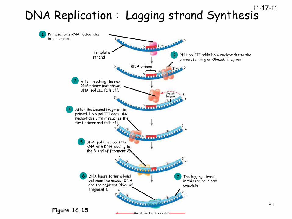
Figure 16.15
Primase joins RNA nucleotides into a primer.
1
DNA pol III adds DNA nucleotides to the primer, forming an Okazaki fragment.
2
After reaching the next RNA primer (not shown), DNA pol III falls off.
3
After the second fragment is primed. DNA pol III adds DNAnucleotides until it reaches the first primer and falls off.
4
DNA pol 1 replaces the RNA with DNA, adding to the 3 end of fragment 2.
5
DNA ligase forms a bond between the newest DNAand the adjacent DNA of fragment 1.
6 The lagging strand in this region is nowcomplete.
7
DNA Replication : Lagging strand Synthesis11-17-11

32
Helicase
SSBP
DNA Replication: Overview11-17-11

33
DNA Replication: Telomeres
Dilemma: What about the ends of linear DNA?
11-17-11

34
DNA Replication: Telomeres
Dilemma: What about the ends of linear DNA?
Telomerase is the answer!
~2500 base pairs at the end of DNA is a repeatTTAGGGAATCCC
Telomerase is a protein with a built-in RNA template
AAUCCCAAU
Adds back Telomere ends
11-17-11

35
DNA Replication: Telomeres
Telomerase adds back nucleotides at the ends of DNA
Repeat again and again
11-17-11
Top Related