


Languages
Pages
Legal


UW/GTC/UO
BAL © 2016
Benjamin A. Lipsky, MD FACP, FIDSA, FRCP (London), FFPM RCPS (Glasg)
Emeritus Professor Medicine, University of Washington Visiting Professor (Infectious Diseases), Associate Fellow,
Green Templeton College, University of Oxford
Diabetic Foot Osteomyelitis: What is New in Diagnosis &Treatment
UNIVERSITY OF OXFORD

UW/GTC/UO
BAL © 2016
Bone of contention
Lipsky BA, “Bone of Contention: Diagnosing DFO”. Clin Infect Dis 2008;47:528

UW/GTC/UO
BAL © 2016
• Epidemiology: common problem with high morbidity
• Pathophysiology: spread from soft tissue infection
• Microbiology: mostly S. aureus; often polymicrobial
• Diagnosis: tests insensitive early; non-specific late –Clinical (especially probe-to-bone); biomarkers
– Imaging: X-ray; MRI; SPECT/CT; PET/CT
–Bone culture/histopathology
• Treatment: clinician/center dependent –Surgical: standard; combined with antibiotics
–Antibiotic: can be used alone; long duration
• Follow-up: difficult; at least 1 year
Overview of Diabetic Foot Osteomyelitis

UW/GTC/UO
BAL © 2016
UW/GTC/UO
BAL © 2016
Clinical Infectious Diseases 2012;54(12):132–173 Published by Oxford University Press on behalf of the Infectious Diseases Society of
America 2012. DOI: 10.1093/cid/cis346

UW/GTC/UO
BAL © 2016
iwgdf.org/guidelines/guidance Diab Metab Res Rev 2016;32 Suppl 1:45

UW/GTC/UO
BAL © 2016
Epidemiology of Diabetic Foot Infections
Population having diabetes: 12+%
:5-25% Moderate: 30-60%
Wound infected at presentation: ~55%
Mild: 35+%
Develop a foot wound: ~25%
Lipsky et al, Clin Infect Dis 2012;54:132
DFO: ≤20% DFO: ~30-40% DFO: ~50-80%

UW/GTC/UO
BAL © 2016
Pathogenesis Diabetic Foot Osteomyelitis
• Intramedullary spread of infection → bone death, persistence of infection (biofilm)
• Infection → soft tissue via sinus tracts; new bone (involucrum) may form
• Infection/inflam’tion kills bone sequestrum; may detach
Lipsky, Berendt. American College Physicians Medicine 2011
• Soft tissue loss & infection leads to contiguous cortical bone infection & necrosis

UW/GTC/UO
BAL © 2016
Implications of Presence of Osteomyelitis in DFI
Osteomyelitis No Osteo p-
(n=37) (n=36) Value
Length hospitlzn (d) 42 (29-51) 20 (13-30) <0.001
Durtn antibiotic (d) 47 ± 20 22 ± 15 <0.001
Durtn IV antibiotic(d) 44 (31-65) 33 (23-46) 0.030
Time wound heal (d) 183 ±95 141 ± 65 0.030
Surgical procedures 24 (65%) 11 (31%) <0.003
Minor amputation 22 (59%) 5 (14%) <0.001
Multluoglu, Lipsky et al, Scand J Infect Dis 2013;45:497
Pts hospitalized for DFI; 1 center (Istanbul) in 2 years

UW/GTC/UO
BAL © 2016
DFI: Worse Outcomes with Bone vs Soft Tissue Infxn
• Retrospective review 200 pts hospitalized with DFI
• DFO dx: + bone culture or histology
• 133 pts (67%) had DFO, 80% forefoot
• Compared to STI, DFO significantly
– Overall amptns: 61% v 22%, OR 5.4
– Minor amptns: 47% v 18%, OR 4.0
– Major amptns: 17% v 5%, OR 3.6
– Mean length of stay: 9.8 v 7.7 d, p=0.06
Hobizal et al, ISDF, The Hague 20 May 2015

UW/GTC/UO
BAL © 2016
Microbiology: Pathogens on Bone Biopsy DFO
Variables Lesens Senneville Aragon-Sanchez
# Samples 80 76 176
Mean isol/sample 1.6 ± 1 1.54 –
Lesens et al, Clin Microbiol Infect 2011;17:285
%, by pathogen
Staph aureus 33% 26% 47%
[MRSA 19% 10% 17%]
Coag-neg staph 14% 26% 11%
Streptococci 9% 12% 3%
Enterococci 12% 8% 1%
Gram − rods 20% 18% 29%
Pseud aerug 8% 2% 9%
Anaerobes 4% 5% –

UW/GTC/UO
BAL © 2016
Conventional Culture (n=26) 16s rRNA Sequencing (n=23)
Gram + cocci 20 (77%) Gram+ cocci 23 (100%)
S. aureus, total 13 (50) Staphylococcus spp. 20 (87)
Coag – staphylococci 11 (42) Coag – staphylococci Not tested
Streptococcus spp. 6 (23) Streptococcus spp. 13 (57)
Enterococcus spp. 2 (8) Enterococcus spp. 0
Gram + bacilli 1 (4) Gram + bacilli* 18 (78)
Corynebacterium spp. 1 (4) Corynebacterium spp.18 (78)
Gram – bacilli 13 (50) Gram – bacilli 10 (44)
P. aeruginosa 4 (15) Pseudomonas spp. 5 (22)
Anaerobes 6 (23) Anaerobes* 20 (87)
- Facultative 3 (12) - Facultative 17 (74)
- Obligate 3 (12) - Obligate 20 (87)
Polymicrobial infxn 16 (64) Polymicrobial infxn 21 (91)
* p<0.001 for conventional vs molecular 16s
Microbiome in DFO: PCR vs Culture of Bone
van Asten et al, Eur J Clin Micro Inf Dis 2016;35:293

UW/GTC/UO
BAL © 2016
Diagnosing DFO: Current Methods
• Clinical –History: long wound duration; recurrent infections
–Exam: deep (>3mm)/large (>2 cm2) ulcer; bony
prominence; visible bone/joint; “sausage” toe
–Probe-to-bone: useful if done/interpreted correctly
Dinh et al Clin Inf Dis 2008;47:519; Butalia et al JAMA 2008;299:806 Berendt et al. Diabetes Metab Res Rev 2008;24 Suppl 1:S145
UW/GTC/UO
BAL © 2016
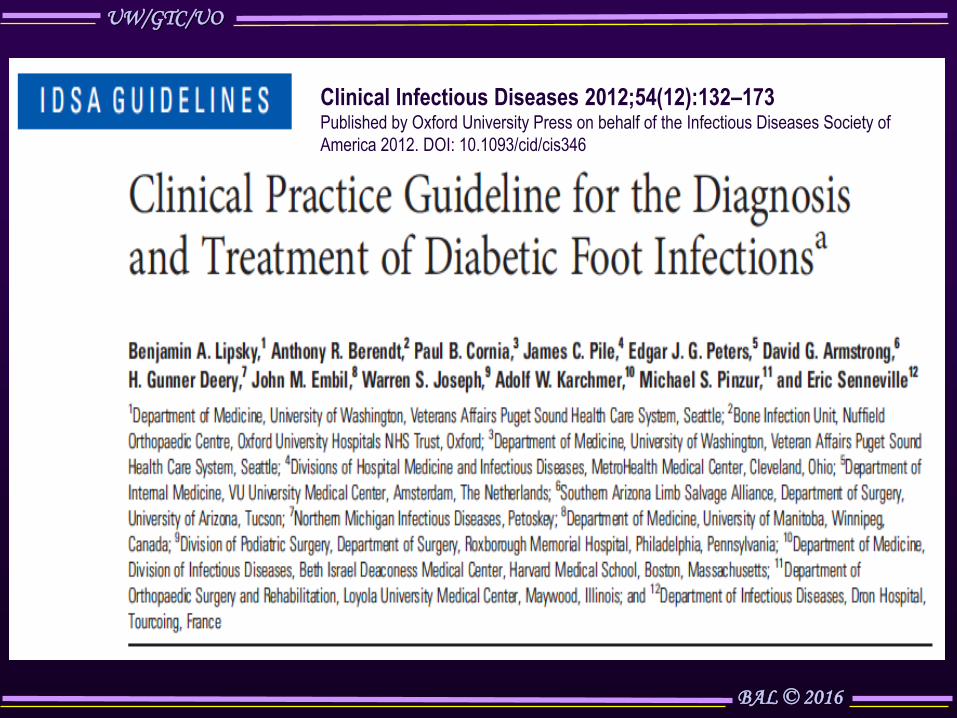
The Probe-to-Bone Test in DF Osteomyelitis
Ertugrul BM, Lipsky BA. Diab Foot Ankle 2013;4:10
Method (Grayson, JAMA 1995):
• 14 F blunt metal probe
• Bone: gritty, hard feel

UW/GTC/UO
BAL © 2016
Reference,yr Pts Sensi Specif PPV NPV DxOR Prev
Grayson, 1995 75 .66 .85 .89 .56 11 66%
Lavery, 2007 247 .87 .91 .57 .98 64 12%
Aragon, 2011 338 .94 .98 .99 .83 630 79%
Lozano, 2010 132 .98 .78 .94 .91 180 80%
Mutluoglu, 2012 65 .67 .85 .87 .63 11 60%
Zaiton, 2014 102 .83 .77 .92 .59 16 75%
----------------------------------------------------------------------------------
Pooled 959 .87 .88 .92 .84 49 58%
Performance Characteristics PTB Test
Lam et al, Clin Infect Dis 2016 (in press)
UW/GTC/UO
BAL © 2016
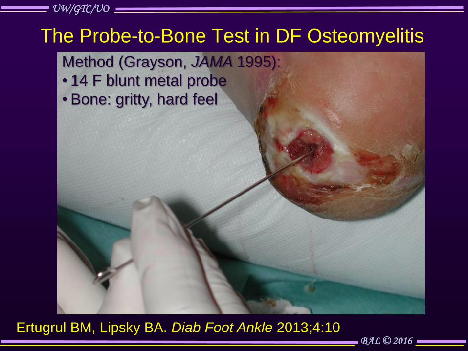
The Importance of Prevalence in PTB Test
Calculated positive & negative predictive values given pooled sensitivity & specificity.
Prevalence: PPV, but NPV
Lam et al, Clin Infect Dis 2016 (in press)

UW/GTC/UO
BAL © 2016
Diagnosing DFO: Current Methods
• Clinical – History: long wound duration, recurrent infection
– Exam: deep(>3mm)/large (>2 cm2) ulcer, bony
prominence, visible bone/joint, “sausage” toe
– Probe-to-bone: useful if done/interpreted correctly
– Blood tests: WBC, ESR, C-RP, PCT, ? biomarkers
Dinh et al Clin Inf Dis 2008;47:519; Butalia et al JAMA 2008;299:806 Berendt et al. Diabetes Metab Res Rev 2008;24 Suppl 1:S145

UW/GTC/UO
BAL © 2016
Meta-analysis*: Biomarkers for Diagnosing DFO
Test Sensitivity Specificity
Single test (pooled studies)
- ESR (n=6) .81 (.71-.88) .90 (.75-.96)
- WBC (n=3) .56 (.36-.74) .84 (.76-.90)
Combinations (1 study each)
- CRP (>3.2) .85
+ depth (>3mm) 1.00 .55
- CRP (>1.4) .85 .83
+ PCT (>0.3) .81 .71
van Asten et al, Curr Diabetes Rev 2015 (epub Jul 12)
*8/195 studies met inclusion criteria

UW/GTC/UO
BAL © 2016
• 35 pts hospitalized with infected DFU; 24 with DFO
• Inflammatory markers measured @ baseline, 3, 6 wks
ESR, CRP, PCT, IL-6, IL-8, tumor necrosis factor alpha (TNFα),
monocyte chemotactic protein-1 (MCP-1), macrophage
inflammatory protein-1 alpha (MIP1α)
• Results: no significant differences between grps except:
– PCT in non-osteo group at baseline (p=0.05)
–CRP, ESR, PCT, IL-6 w/ therapy in DFO (but not
non-osteo) group (p=0.05)
–MCP-1 increased with therapy (p=0.002)
• Conclusion: inflammatory markers have limited use for
differentiating bone vs soft tissue DFI
Value of Inflammatory Markers in DFO: Pilot Study
van Asten et al, Int Wound J 2015 (epub 3 Dec)

UW/GTC/UO
BAL © 2016
Inflammatory Markers Over 1 Year F/U of DFO
ESR CRP
122 pts with bone bx proven DFO; overall ulcer healing rate 38%
Stagnating values associated with poor outcomes
van Asten et al, Int Wound J 2016 (epub March)
Remission Healing Remission Healing
Reinfection Reamputation Reamputation
–– Yes
–– No

UW/GTC/UO
BAL © 2016
Diagnosing DFO: Current Methods
• Clinical – History: long wound duration, recurrent infection
– Exam: deep (>3mm)/large (>2 cm2) ulcer, bony
prominence visible bone/joint, “sausage” toe
– Probe-to-bone: useful if done/interpreted correctly
– Blood tests: WBC, ESR, C-RP, PCT, ? biomarkers
• Imaging – Plain x-ray: limited sensi (early) & specif (late)
– Radionuclide scans: WBC>bone; non-specific
– MRI: best current test; marrow edema, soft tissue
– SPECT/CT, PET/CT, PET/MRI promising; F/U
Dinh et al Clin Inf Dis 2008;47:519; Butalia et al JAMA 2008;299:806 Berendt et al. Diabetes Metab Res Rev 2008;24 Suppl 1:S145

UW/GTC/UO
BAL © 2016
Typical Features DFO on Plain X-rays
• Periosteal reaction or elevation
• Loss of bone cortex with bony erosion
• Focal loss cortical trabecular pattern
• Lucency in bone marrow
• Bone sclerosis, with or without erosion
• Presence of sequestrum: dead bone; radiodense
• Presence of involucrum: layer of new bone growth
• Presence of cloacae: opening in involucrum or cortex
• Evidence of sinus tract from bone to soft tissue
Lipsky et al, Diab Metab Res Rev 2016 Jan;32 Suppl 1:45

UW/GTC/UO
BAL © 2016
Radiographic Features of Osteomyelitis

UW/GTC/UO
BAL © 2016
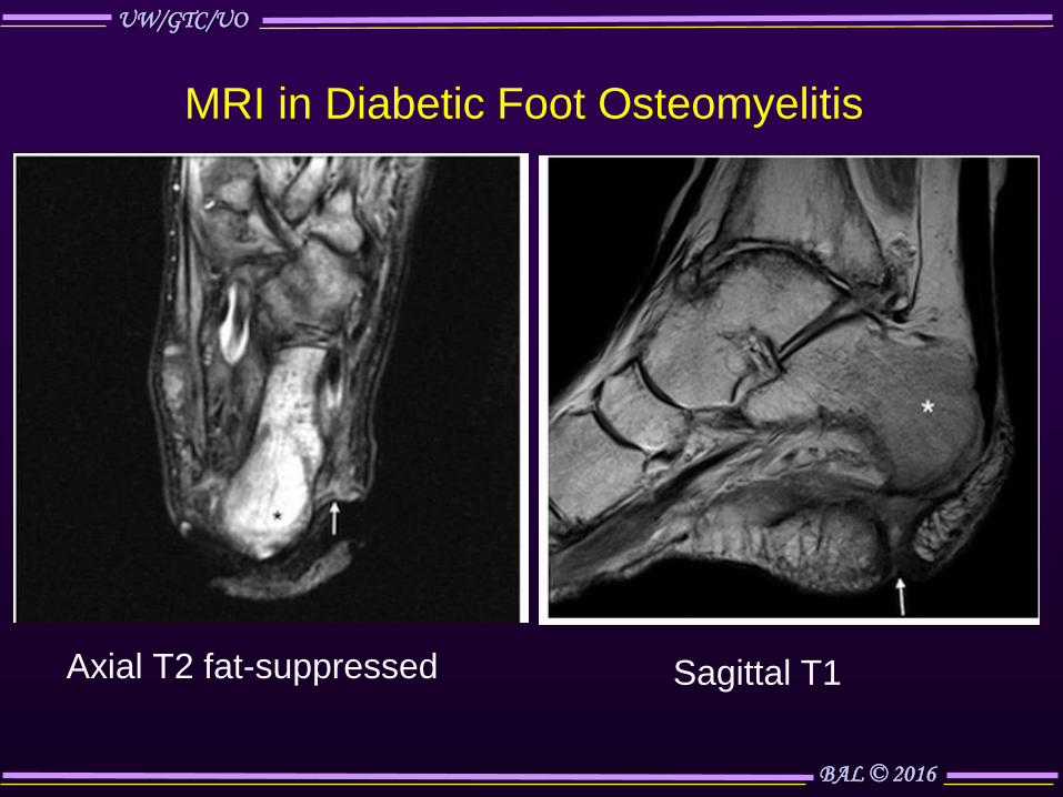
Relative Utility MRI Findings for Dx DFO Finding Relative Utility marrow intensity T1-w; on fluid-sensitive Equivocal sequences; post-contrast enhancement • Joint effusion & enhancement, subluxation Equivocal
& dislocation, bone fragmentation & proliferation, erosion &destruction, intraarticular bodies
• Periosteal reaction * • Subtending skin ulcer * • Ulcer: area >2 mm or depth >3 mm ** • Tenosynovitis * • Focal involvement * • Septic arthritis *** • The “ghost sign” (contrast) *** • Sinus tract, or abscess ***
Leone et al, Skel Radiol 2016; epub 17 Feb
UW/GTC/UO
BAL © 2016
MRI in Diabetic Foot Osteomyelitis
Axial T2 fat-suppressed Sagittal T1

UW/GTC/UO
BAL © 2016
SPECT/CT 3 phase bone scan: can’t differentiate recent post-op change in 1st & 2nd toes from midfoot osteomyelitis
Fridman et al, Clin Pod Med Surg 2014;31:43
In111 WBC differentiates infection from post-op changes but poor anatomical correlation
Fusion WBC SPECT/CT specificity & good anatomical detail

UW/GTC/UO
BAL © 2016
FDG-PET/CT for Diabetic Foot Osteomyelitis
L foot plantar ulcer (A) FDG-PET maximal intensity projection with focus of FDG uptake 1st toe; (B) coronal, (C) sagittal CT PET/CT: focus of uptake in fragmented 1st MT (+ bone culture)
Israel, Sconfienza, Lipsky. QJ Nucl Med Mol Imag 2013;58:33
A
CT PET PET/CT
FDG-PET
CT
PET

UW/GTC/UO
BAL © 2016
Advanced Imaging for Diabetic Foot Osteomyelitis
Lipsky et al, Diab Metab Res Rev 2016 Jan;32 Suppl 1:45
bone
[*Specificity=100%]

UW/GTC/UO
BAL © 2016
Approach to Diagnosing Diabetic Foot Osteomyelitis
Pt with suspected diabetic foot osteomyelitis (OM)
Plain X-rays
Negative
Appropriate infection management
MRI
Soft tissue
infection
Positive Equivocal Positive:
OM
OM
WBC [SPECT/CT] or FDG PET/CT
Negative Negative
Appropriate wound care
Appropriate wound care
Equivocal
Charcot
Bone marrow scintigraphy
Discordant w/ WBC
Concordant w/ WBC
Israel, Sconfienza, Lipsky. Quart J Nucl Med Molec Imag 2013;58:33

UW/GTC/UO
BAL © 2016
Likelihood Ratios of Diagnostic Tests for DFO* Diagnostic Test + LR − LR
Clinical gestalt 5.5 0.54
Ulcer area >2cm2 7.2 0.48
Ulcer inflammation 1.5 0.84
Probe-to-bone test 6.4 0.39
ESR >70 mm/hr 11.0 0.34
Plain X-rays 2.3 0.63 99Tc 3 phase bone scan 1.4 0.40
Tc/In labeled WBC scan 4.7/2.3 0.12/.038
MRI 3.8 0.14
SPECT/CT 3.0 0.18
FDG-PET 5.6 0.40
*Approximations based on variable number of heterogeneous studies Lipsky et al, IWGDF DFI guidance 2015; Markanday OFID 2014; 1(2):ofu060
Imag
ing
B
iom
arke
r
C
linic
al
UW/GTC/UO
BAL © 2016
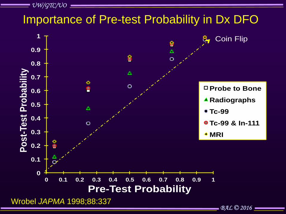
Importance of Pre-test Probability in Dx DFO
0
0.1
0.2
0.3
0.4
0.5
0.6
0.7
0.8
0.9
1
0 0.1 0.2 0.3 0.4 0.5 0.6 0.7 0.8 0.9 1
Po
st-
Te
st
Pro
ba
bil
ity
Pre-Test Probability
Probe to Bone
Radiographs
Tc-99
Tc-99 & In-111
MRI
Coin Flip
Wrobel JAPMA 1998;88:337

UW/GTC/UO
BAL © 2016
Diagnosing DFO: Current Methods
• Clinical – History: long wound duration, recurrent infection
– Exam: deep (>3mm)/large (>2 cm2) ulcer, bony
prominence visible bone/joint, “sausage” toe
– Probe-to-bone: useful if done/interpreted correctly
– Blood tests: WBC, ESR, C-RP, PCT, ? biomarkers
• Imaging – Plain x-ray: limited sensi (early) & specif (late)
– Radionuclide scans: WBC>bone; non-specific
– MRI: best current test; marrow edema, soft tissue
– SPECT/CT, PET/CT, PET/MRI quite promising
• Bone Biopsy: culture & histology- criterion standard
Dinh et al Clin Inf Dis 2008;47:519; Butalia et al JAMA 2008;299:806 Berendt et al. Diabetes Metab Res Rev 2008;24 Suppl 1:S145

UW/GTC/UO
BAL © 2016
Courtesy: Drs. E. Senneville & E. Beltrand
Bone Biopsy for Diabetic Foot Osteomyelitis
Microbiology
Histology
UW/GTC/UO
BAL © 2016
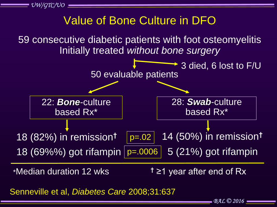
22: Bone-culture based Rx*
28: Swab-culture based Rx*
18 (82%) in remission† 14 (50%) in remission†
Value of Bone Culture in DFO
Senneville et al, Diabetes Care 2008;31:637
† ≥1 year after end of Rx
59 consecutive diabetic patients with foot osteomyelitis Initially treated without bone surgery
*Median duration 12 wks
50 evaluable patients 3 died, 6 lost to F/U
p=.02
18 (69%%) got rifampin 5 (21%) got rifampin p=.0006

UW/GTC/UO
BAL © 2016
Value of a Negative Bone Culture
341 pts with suspected DFO: percutaneous bone bx; ≥2 yr F/U
275 culture + 66 culture −
[25 lost to F/U or excluded]
41 enrolled
16 (39%) healed 25 unhealed
10 clinically/imaging stable 15 repeat bone bx
6 likely not osteo 4 suspected osteo 9 culture − 6 cult +
Senneville et al, Diabet Med 2012;29:56
True negative : 31 (75%) False negative: 10 (25%)

UW/GTC/UO
BAL © 2016
In Which Situations Is Diagnostic Bone Biopsy
Most Recommended?
• Uncertainty regarding the diagnosis of osteomyelitis
despite clinical and imaging evaluations
• Culture data from soft tissue specimens unclear
• Failure to respond to empiric antibiotic therapy
• Plan to insert metalware in bone at affected site
• Desire to use antibiotic agents that may be especially
effective for osteomyelitis but have a high potential
for selecting resistant bacteria (eg, rifampin, FQs)
Lipsky et al, [IDSA DFI guidelines] Clin Infect Dis 2012;54:1679
Lipsky et al, [IWGDF DFI guidance] Diab/Metab Res Rev 2015 (in press)

UW/GTC/UO
BAL © 2016
Treating Diabetic Foot Osteomyelitis

UW/GTC/UO
BAL © 2016
Key Questions About Treatment of Diabetic
Foot Osteomyelitis
• What are the most appropriate antibiotic regimens
–Specific single, or combinations of, agent(s)
–Route of therapy (IV vs oral vs local)
–Duration of therapy
• When is surgical resection of bone required
• How do we know when we have achieved remission
or cure?

UW/GTC/UO
BAL © 2016
Exclusively Surgical Treatment of Osteomyelitis
September 1907 3 months after injury
March 1912 Radiological &
clinical resolution
10 yo with post-traumatic osteomyelitis of L femur; pus drained, sequestrum removed; later sinus curetted
R. Hammond, J Bone Joint Surg Am 1913;s 2-10:569

UW/GTC/UO
BAL © 2016
Exclusively Antibiotic Therapy of DFO
Of 51 cases, 53% resolved with antibiotic w/o bone surgery
DFO of 4th MT head 11 months later
Bamberger et al Am J Med 1987;83:653

UW/GTC/UO
BAL © 2016
Resolution of DFO With Antibiotic Therapy Alone
31 Oct 2013 13 Dec 2013 16 Jan 2014 11 Dec 2015
Mutluoglu, Lipsky (in submission)

UW/GTC/UO
BAL © 2016
Can Osteomyelitis Be Cured Without Surgical Resection of Bone?
• Review of reported patients managed with
antibiotics & little or no surgical debridement1
–546 total patients in 11 studies from 1987-2002
–Mostly given oral fluoroquinolones, for 3 mos
–Satisfactory response seen in ~65% (25-88%)
• In 4 more recent observational studies2-5 (total of
443 patents): 63-79% remission rates
• All retrospective studies, mostly forefoot cases
1Jeffcoate, Lipsky. Clin Infect Dis 2004;39 Suppl 2:S115; 2Acharya et al, Diab Res Clin Pract 2013 Sep;101(3):e18 3Ulcay et al, Pak J Med Sci 2014;30:28; 4Zeun et al, IJLEW 2015 (1 Sept) 5Jordano-Montanez et al, Enferm Infecc Microbiol Clin 2014;32:555

UW/GTC/UO
BAL © 2016
Outcome of S. aureus DFO by Treatment Type
Medical Surgical P
-Favorable outcome 87% 80% NS
-Hospitalized >24 hours 49% 94% <.001
-Mean hospital LOS (days) 17 ± 3 50 ± 12 .004
-Median duration abx (wks) 8 (6-52) 5 (2-44) .001
-Antibiotic d/c 2° side effects 33% 9% .01
Lesens et al, Int J Lower Extrem Wounds 2014; Dec 16 pii
Retrospective cohort study 74 pts; + bone culture (35% MRSA)
Bone surgery in 47% (mostly forefoot); rest antibiotic alone
Mortality on F/U (mean 21 months): 20%
New episode (noncontiguous) DFO: 32% No significant diff
medical v surgical

UW/GTC/UO
BAL © 2016
RCT of Primarily Antibiotic vs Surgery for DFO
52 patients met inclusion criteria
25 randomized to antibiotics (90 days, culture modified)
27 randomized to surgery (conservative, + 10 d abx)
1 dropped out 24 treated 22 operated 5 dropped out
2 died 4 required surgery
18 (75%) cured
19 (86%) cured
3 needed minor amputation
1 required minor amputation
3 healed w/ surgery
No signif. differences: -Percent cured -Time to cure -Complications
Lazaro-Martinez et al, Diabetes Care 2014; 37:789
Reulceration: 9.5% abx; 21% surgery

UW/GTC/UO
BAL © 2016
Primarily Surgical vs Medical Treatment for
DFO: Individualizing the Choice
Surgical
• Substantial bone necrosis
• Fxnly non-salvageable foot
• Pt is non-ambulatory
• risks antibiotic problems
• No available active antibiotic
• Uncorrectable foot ischemia
• Patient preference
Medical
• Pt too unstable for surgery
• Bad post-op mechanics likely
• No other need for surgery
• Small, forefoot lesion
• No skilled surgeon available
• Surgery costs prohibitive
• Patient preference
Lipsky BA. Diabetes Care 2014;37:593

UW/GTC/UO
BAL © 2016
Do We Need IV Antibiotic Therapy for Oseo?
• Pilot study in Oxford; then enrolled 1060 pts in 30 UK centers
• Bacterial infection types: osteomyelitis (DFO); prosthetic joint
infection; orthopedic device/bone-graft; spinal infection
• Open label: ~1 wk IV abx for all, then randomized to ≥5 wks of
IV or oral antibiotic regimen (clinicians’ choice of agent[s])
• Outcomes: assessed at one year follow up visit - 1° is definite failure (+ culture or dx histology from bone or
periprosthetic tissue, draining sinus or pus from bone - 2°: SAE, IV line compltn, Rx failure; early Rx termination;
resource allocation; QoL; hip/knee score; Rx adherence
• Endpoint review committee (blind to Rx): all potential failures
Li et al. Trials 2015;16:583
Oral vs IV Antibiotics for bone & joint infections

UW/GTC/UO
BAL © 2016
Bone Penetration of Antibiotics
Antibiotic Mean [Bone/Serum] Ratios
Levofloxacin 0.36-1.0
Ciprofloxacin 0.27-1.2
Moxifloxacin 0.33-1.05
Vancomycin 0.05-0.67
Linezolid 0.2-0.51
Daptomycin 1.17
Clindamycin 0.21-0.45
Cefazolin 0.179
Ceftriaxone 0.07-0.17
Cefuroxime 0.04-0.55
Rifampin 0.08-0.57
Lalani T. UpToDate 2014

UW/GTC/UO
BAL © 2016
Bone Penetration of Antibiotic Agents in DFO
• Fosfomycin 1
• Daptomycin 2
• Linezolid 3
1Schintler V et al. J Antimicrob Chemother 2009; 2Traunmüller F et al. J
Antimicrob Chemother 2010; 3Traunmüller F Int J Antimicrob Agents 2010
Substantial variability in mean bone penetration among drugs, studies

UW/GTC/UO
BAL © 2016
Duration of Antibiotic Treatment for DFO
• Multicenter RCT: 6 vs 12 weeks of antibiotic therapy
– Pts w/ + bone culture; w/o ischemia; no surgical Rx
– Remission: no evidence infection, stable/improved
x-rays, overlying wound healed X ≥12 months f/u
• Results: 40 pts enrolled 2007-2009
– Characteristics of 2 treatment groups similar
– Remission rates:
6 weeks: 12/20 (60%)
12 weeks: 14/20 (70%) [p=NS]
- Signif. GI adverse events with 6 wks (15 v 45%)
- No differences: relapse; need for resection; spread
Tone et al, Diabetes Care 2015;38:302

UW/GTC/UO
BAL © 2016
Antibiotic Beads or Bone
Grafts for Treating DFO
Panagopoulos Int J Low Extrem
Wounds 2015;14:87
Xanthopoulou et al, ECCMID 2016

UW/GTC/UO
BAL © 2016
Local Antibiotics for Treating Osteomyelitis
Advantages
• Hi local antibiotic level
• Fills dead space
• Low systemic concentrations
• PLA & CaSO4 don’t require bead removal
Barth et al, Int J Antimicrob Ag 2011;38:371
PMMA (beads, cement), CaSO4 (pellets, beads), Polylactic acid
Disadvantages
• Variable formulations, sizes
• Requires pharmaceutical grade antibiotic powders
• Subinhib conc → resistance
• ? Inhibition of bone healing
• Operation to remove PMMA
*Literature search (July 2014) 3770 papers: ? best system,
antibiotic, duration, need for surgery or systemic antibiotics
*Panagopoulos, et al Int J LE Wound 2015;14:87

UW/GTC/UO
BAL © 2016
IDSA DFI Guidelines on Treating Bone Infection
Extent of Surgery Route of Rx Duration of Rx
No residual infected Parenteral 2 - 5 days tissue (eg, post- or oral amputation)
Residual infected Parenteral 2 – 4 weeks
soft tissue only or oral
Residual infected Initial parenteral 4 – 6 weeks
(but viable) bone then oral switch
Dead bone present Initial parenteral ? ≥ 3 months no bone resection then oral switch
Lipsky et al,[IDSA DFI guidelines] Clin Infect Dis 2012;54:132

UW/GTC/UO
BAL © 2016
Proposed Management Scheme DFO: IWGDF Definite • + bone culture & histology • Pus in bone at surgery • Atraumatically detached bone fragment in ulcer • Intraosseous abscess on MRI
Berendt et al, Diab Metab Res Rev 2008;24 Suppl 1:S145
Probable • Visible cancellous bone in wound • MRI: characteristic findings • Bone: + culture, − or no histology • Bone: + histology, − or no culture Possible • X-rays: cortical destruction • MRI: bone edema/cloacae • Probe to bone positive • Visible cortical bone • ESR >70 mm/h • Non-healing wound for >6 wk

UW/GTC/UO
BAL © 2016
Proposed Sequential Approach for Concomitant Soft Tissue Infection & Suspected Osteomyelitis
Berthol, Int Symp Diab Foot, Hague 2015; Markanday OFID 2014; 1(2):ofu060
41% had DFO; 59% in remission @ final f/u (mean 3.9 y)
[If DFO?, repeat dx tests]
Retrospective study, 32 pts, Tourcoing 2006-13

UW/GTC/UO
BAL © 2016
Negative margin
Positive margin
Failure-free survival (years)
Effect of Residual Osteomyelitis After Resection
111 DFO pts: 39 (35%) histology + margins for osteomyelitis1
1Kowalski et al, J Foot Ankle Surg 2011;50:171 2Atway et al, J Foot Ankle Surg 2012;51:749
27 pts with DFO who had a forefoot amptn 2
- Rate of residual osteomyelitis: 41%
- Poor outcome: 82% for + vs 25% for − margin

UW/GTC/UO
BAL © 2016
How Do We Know if a Patient is Responding
To Antibiotic Treatment?
• Decreasing inflammatory markers
• Resolution of local soft tissue infection
• Healing of adjacent/overlying wound
• Evolving radiographic findings c/w healing
• Lack of uptake on radionuclide studies
Lipsky BA, et al. IDSA: Clin Infect Dis 2012;54:132
Lipsky BA, IWGDF: Diab/Metab Res Rev 2016;32 Suppl 1:45
Consider “in remission” until F/U @ least 1 year
UW/GTC/UO
BAL © 2016
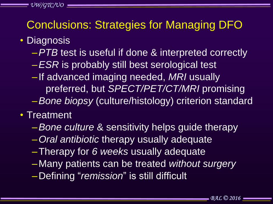
Conclusions: Strategies for Managing DFO
• Diagnosis
–PTB test is useful if done & interpreted correctly
–ESR is probably still best serological test
– If advanced imaging needed, MRI usually
preferred, but SPECT/PET/CT/MRI promising
–Bone biopsy (culture/histology) criterion standard
• Treatment
–Bone culture & sensitivity helps guide therapy
–Oral antibiotic therapy usually adequate
–Therapy for 6 weeks usually adequate
–Many patients can be treated without surgery
–Defining “remission” is still difficult

UW/GTC/UO
BAL © 2016

UW/GTC/UO
BAL © 2016
Teşekkür ederim
Top Related