


Languages
Pages
Legal


Design, Synthesis and Evaluation of Peptide-Based Affinity
Labels for Mu Opioid Receptors
By
C2009
Bhaswati Sinha
Submitted to the Department of Medicinal Chemistry and the Faculty of the Graduate School of the University of Kansas in partial fulfillment of the requirements for the
degree of Doctor of Philosophy.
Dissertation Committee:
____________________________________
Chairperson: Dr. Jane V. Aldrich
____________________________________
____________________________________
____________________________________
____________________________________
Dissertation Defended __________________
Dr. Teruna J. Siahaan
Dr. Michael F. Rafferty
Dr. Emily E. Scott
Dr. David S. Moore

ii
The Dissertation Committee for Bhaswati Sinha certifies that this is the approved version of the following dissertation:
Design, Synthesis and Evaluation of Peptide-Based Affinity Labels for Mu
Opioid Receptors
____________________________________
Chairperson: Dr. Jane V. Aldrich
____________________________________
____________________________________
____________________________________
____________________________________
Date Approved _________________
Dr. Michael F. Rafferty
Dr. Teruna J. Siahaan
Dr. Emily E. Scott
Dr. David S. Moore

iii
Dedicated to:
My parents
Kumkum DattaChowdhury
Prithwish Chandra DattaChowdhury
My brother
Atish DattaChowdhury
My husband
Sandipan Sinha

iv
Acknowledgements
I would like to take this opportunity to thank all those who have helped me earn the
doctorate degree from the University of Kansas.
First and foremost, I would like to thank my advisor Prof. Jane V. Aldrich for her
excellent mentorship, support and guidance through out my graduate career at the
University of Kansas. I was fortunate to have some great colleagues to work with and
I would like to thank all of them for their co-operation, encouragement and helpful
inputs. They are Anand Joshi, Wendy Hartsock, Kendra Dresner, Katherine Smith,
Angela Peck, Dr. Wei-Jie Fang, Dr. Xin Wang, Dr. Santosh Kukarni, Dr. Mark Del
Borgo, Dr. Kshitij Patkar, Dr. Nicolette Ross, Dr. Tatyana Yakovleva and Dr. Sandra
Barlett. Special thanks to Wendy for always being so friendly and helpful.
I am grateful to Prof. Mike Rafferty, Prof. Teruna Siahaan, Dr. Emily Scott and Dr.
David Moore for being in my thesis committee. I would like to specially thank Dr.
Rafferty for his valuable suggestions.
I made some wonderful friends in Lawrence and life here would not have been so
much enjoyable without them. Rashida, Deb, Mrinal, Barnali, Sumit M, Deepti, Sasi,
Aparna, Nadim, Vinya, Naveen, Anurupa, Gagandeep, Diptesh, Ramu and Sanjibani
thank you all for your friendship. I express my deepest gratitude to my old friends:
Soma, Dhriti, Peuli, Prajna, Pranamita, Iman, Abira, Indranil, Arpana, Pinaki, Sumit
G, Paroma and Suchitra for their constant encouragement.
Sandipan, my husband has been a pillar of support for me. His love, motivation and
patience were instrumental behind my success. I am grateful to my parents-in-law
Ashoka Sinha and Salil Kumar Sinha for their affection and encouragement. My
brothers- in-law Suman and Sovan Sinha, my sisters-in-law Paromita and Bhaswati

v
Sinha have always had faith in my ability. I am fortunate to have such a supportive
family including my two cute nephew and niece: Ayush and Ashmita Sinha.
Words can not do justice to express my indebtedness to my father Prithwish Chandra
DattaChowdhury and my mother Kumkum DattaChowdhury. My father has always
been a source of inspiration. Whatever I achieved today is because of their constant
motivation, love and trust. My caring and loving brother Atish DattaChowdhury has
been equally instrumental towards my achievement through his unconditional
support. I am grateful to my sister-in-law Anindita DattaChowdhury for her
friendship and affection and my sweet nephew Arnab DattaChowdhury- a bundle of
joy. Finally I owe my success to my entire family and would like to thank you all
from the bottom of my heart.

vi
Table of Contents
Page
Acknowledgements………………………………………………………………….iv List of Tables………………………………………………………………………..xii List of Figures………………………………………………………………………xiv List of Schemes……………………………………………………………………xviii Abbreviations………………………………………………………………………xix Abstract………………………………………………………………………………..1
Chapter 1
Summary of Thesis Projects…………………………………………………………3
1.1.Background and Significance……………………………………………………..4
1.2. Research Projects…………………………………………………………………8
1.2.1. Project 1: Discovery of Dermorphin-Based Affinity Labels with
Subnanomolar Affinity for Mu Opioid Receptors (Chapter 3)…………………8
1.2.2. Project 2: Synthesis and Evaluation of DAMGO-Based Affinity Labels
for MOR and Discovery of an Unexpected Side Reaction (Chapter 4)……….10
1.2.3. Project 3: Design, Synthesis and Evaluation of a Dermorphin-Based
Multifunctional Affinity Label Probe for Mu Opioid Receptors (Chapter 5)...12
1.3. Conclusions……………………………………………………………………...14
1.4. Bibliography…………………………………………………………………….14

vii
Chapter 2
Literature Review…………………………………………………………………. 22
2.1. Opioid Receptors………………………………………………………………..23
2.1.1. Structure and Function of Opioid Receptors…………………………...24 2.2. Mu Opioid Receptors (MOR)…………………………………………………...25
2.2.1. Mutagenesis Studies of MOR…………………………………………27 2.2.2. Computational Studies on MOR………………………………………30 2.3. Ligands for MOR………………………………………………………………..33
2.3.1. Small Molecule Ligands for MOR……………………………………….33
2.3.2. Opioid Peptides…………………………………………………………...34
2.3.2.1. Endogenous Opioid Peptides Interacting with MOR……………34
2.3.2.2. MOR selective Linear Enkephalin Analogs ……………………36
2.3.2.3. MOR Selective Conformationally Constrained Enkephalin
Analogs…………………………………………………………………..37
2.3.2.4. MOR Selective Peptides from Amphibian Skin ………………..38
2.3.2.4.1. SAR Study of Dermorphins………………………...40
2.4. Affinity Labels…………………………………………………………………..44 2.4.1. Photoaffinity Labels………………………………………………………46 2.4.2. Electrophilic Affinity Labels……………………………………………..49 2.4.2.1. Small Molecule-Based Electrophilic Affinity Labels: β-
Funaltrexamine and Other Analogs………………………………………50

viii
2.4.2.2. Peptide-Based Electrophilic Affinity Labels……………………54
2.5. Significance: Peptide vs. Small Molecule-Based Affinity Labels………………57
2.6. Bibliography……………………………………………………………………..58
Chapter 3
Discovery of Dermorphin-Based Affinity Labels with Subnanomolar Affinity for
Mu Opioid Receptors……………………………………………………………….84
3.1. Introduction……………………………………………………………………...85
3.2. Background……………………………………………………………………...87
3.3. Dermorphin and Previous Dermorphin-Based Analogs………………………...88 3.3.1. Dermorphin-Based Affinity Labels……………………………………....90 3.4. Rationale for the Design of New Affinity Labels……………………………….92 3.5. Results and Discussion………………………………………………………….93 3.6. Conclusions……………………………………………………………………...99 3.7. Experimental…………………………………………………………………...100 3.7.1. Solid Phase peptide Synthesis (SPPS) of Dermophin-Based Affinity Labels ………………………………………………………………………….100 3.7.2. Cleavage from Resin…………………………………………………….102 3.7.3. Purification and Analysis………………………………………………..103 3.7.4. Pharmacological Assays………………………………………………...104 3.7.4.1. Radioligand Binding Assays…………………………………...104 3.7.4.2. Wash-Resistant Inhibition of Binding Assays…………………105

ix
3.8. Bibliography…………………………………………………………………...105
Chapter 4
Synthesis and Evaluation of DAMGO-Based Affinity Labels for MOR and
Discovery of an Unexpected Side Reaction………………………………………113
4.1. Introduction…………………………………………………………………….114
4.2. DAMGO-Based Affinity Labels: Design Strategy………………………….....118
4.3. Results and Discussion………………………………………………………...120
4.3.1. Loading of Fmoc-Gly-ol onto DHP-HM Resin and Synthesis of
DAMGO Analogs……………………………………………………………121
4.3.2. Side Reaction: Formation of Cyclic O-Alkyl Thiocarbamates………..122
4.3.2.1. Proposed Side Reaction………………………………………125 4.3.2.2. Characterization of the Side Products: FTIR of Fr A and Fr B………………………………………………………….126 4.3.2.3. Characterization of the Products by Proton NMR……………..................................................................................127
4.3.3. Radioligand Binding and Wash-resistant Inhibition of Binding
Assays………………………………………………………………………..128
4.3.4. Overcoming the Side Reaction: Design and Synthesis of DAMGA ([D-
Ala2,N-MePhe4,Gly5]enkephalinamide) Analogs…………………………....131
4.4. Conclusions…………………………………………………………………….136 4.5. Experimental…………………………………………………………………...138

x
4.5.1. Loading of Fmoc-Gly-ol onto DHP-HM Resin and SPPS of DAMGO
Analogs……………………………………………………………………….140
4.5.2. Solid Phase Peptide Synthesis of the DAMGA Derivatives…………..140
4.5.3. Cleavage from Resin…………………………………………………...140
4.5.4. Purification and Analysis………………………………………………141
4.5.5. Instrumentation………………………………………………………...141
4.5.6. Pharmacological Assays……………………………………………….141
4.6. Bibliography…………………………………………………………………...142
Chapter 5
Design, Synthesis and Evaluation of a Dermorphin-Based Multifunctional
Affinity Label Probe for Mu Opioid Receptors…………………………………148
5.1. Introduction……………………………………………………………………149
5.2. Design of a Dermorphin-Based Affinity Labels: Design Strategy……………155
5.3. Results and Discussion………………………………………………………...157
5.3.1 Synthesis of the Multifunctional
[D-Lys(=C=S)2]dermorphin Derivative………………………………………157
5.3.2. Results from Preliminary Microscopy Experiments ……………….. …162
5.4. Conclusions……………………………………………………………………164
5.5. Experimental…………………………………………………………………...165
5.5.1 Synthesis of Dermorphin-based Multifunctional
Affinity Label…………………………………………………………………165

xi
5.5.2 Cleavage from Resin……………………………………………………168
5.5.3 Purification and Analysis………………………………………………..168
5.5.4 Separation of Isomers from Rhodamine WT……………………………169
5.5.5 Microscopy Experiments………………………………………………...171
5.6. Bibliography…………………………………………………………………...172
Chapter 6
Conclusions and Future Work…………………………………………………...180
6.1. Introduction……………………………………………………………………181
6.2. Conclusions from Research Projects…………………………………………..181
6.2.1 Project 1: Discovery of Dermorphin-Based Affinity Labels with
Subnanomolar Affinity for Mu Opioid Receptors (Chapter 3).......................182
6.2.2 Project 2: Synthesis and Evaluation of DAMGO-Based Affinity
Labels for MOR and Discovery of an Unexpected Side
Reaction (Chapter 4)…………………………………………………………183
6.2.3 Project 3: Design, Synthesis and Evaluation of a Dermorphin-Based
Multifunctional Affinity Label Probe for Mu Opioid Receptors
(Chapter 5)……………………………………………………………………186
6.3. Future work……………………………………………………………………188
6.4. Bibliography…………………………………………………………………...191

xii
List of Tables PageTable 2.1: Opioid affinities and activity in GPI and MVD of selected
MOR agonists and antagonists
34
Table 2.2: Mammalian opioid peptides with their precursor proteins
35
Table 2.3: Opioid receptor affinities and selectivity in the GPI and MVD of MOR selective enkephalin.
36
Table 2.4: Affinities and MOR selectivity of natural dermorphins (amidated and acid forms) and biological activities on GPI and MVD.
40
Table 2.5: Affinity and selectivity of dermorphin analogs including tetrapeptide analogs of dermorphin modified at the 2nd position.
42
Table 2.6: Enkephalin-based photoaffinity labels for opioid receptors.
48
Table 2.7: Progress towards developing peptide-based affinity labels for MOR: analogs of endomorphin, DAMGO and dermorphin.
56
Table 3.1: Dermorphin-based potential affinity labels reported previously.
91
Table 3.2: Analytical data for dermorphin analogs 1-6
94
Table 3.3: Binding affinities of dermorphin derivatives for MOR and DOR.
95
Table 4.1: Binding affinity of previously prepared DAMGO affinity labels.
119
Table 4.2: Analytical data for DAMGO analogs 2, 3, 5 and 6
123

xiii
Table 4.3: HPLC retention times and molecular weights of two pure fractions (A and B) obtained from attempted synthesis of [D-Lys(=C=S)2]DAMGO
123
Table 4.4: 1H NMR data of fractions A and B obtained from the attempted synthesis of [D-Lys(=C=S)2]DAMGO, 4
128
Table 4.5: Binding affinities of DAMGO derivatives for MOR and DOR under standard assay conditions
129
Table 4.6: Analytical data for DAMGA analogs 7-12
134
Table 4.7: Binding affinities of DAMGA derivatives for MOR and DOR.
134
Table 5.1: Analytical data for multilabeled dermorphin analogs 1-4
162
Table 6.1: Binding affinities of dermorphin derivatives for MOR and DOR
182
Table 6.2: Binding affinities of DAMGO and DAMGA derivatives for MOR and DOR
184

xiv
List of Figures PageFigure 1.1: Design of potential affinity labels for MOR and the
corresponding reversible control peptides based on the parent peptide dermorphin
10
Figure 1.2: Proposed affinity labels for MOR and the corresponding reversible control peptides based on the parent peptide DAMGO
11
Figure 1.3: Proposed reaction for the formation of the cyclic O-alkyl thiocarbamate
11
Figure 1.4: Design of dermorphin-based multifunctional affinity label for MOR
13
Figure 2.1: Serpentine model of MOR
25
Figure 2.2: Morphine and morphine-derived alkaloids for MOR
32
Figure 2.3: MOR selective enkephalin analogs including conformationally constrained derivatives
37
Figure 2.4: Dermorphin peptides
39
Figure 2.5: An illustration of the two steps involved in covalent binding of an affinity label to its target
45
Figure 2.6: Precursors of reactive species in photoaffinity labels
46
Figure 2.7: A: Photoaffinity labels for opioid receptors, B: Amino acid or
47

xv
acid derivative of photoaffinity labels used to characterize
opioid receptors
Figure 2.8: Small molecule electrophilic and reporter affinity labels for MOR
51
Figure 2.9: Location of the attachment point (Lys233) of β-FNA on MOR
52
Figure 3.1: Structure of dermorphin
89
Figure 3.2: Potential affinity label derivatives for MOR and the corresponding reversible control peptides based on the parent peptide dermorphin
93
Figure 3.3: (A) Wash-resistant inhibition of binding of [D- Orn]2dermorphin and (B) [D-Lys]2dermorphin derivatives.
97
Figure 3.4: Concentration-dependent wash-resistant inhibition of binding of dermorphin analogs
98
Figure 4.1: DAMGO (Tyr-D-Ala-Gly-NMePhe glyol)
118
Figure 4.2: Previously designed DAMGO-based affinity labels
119
Figure 4.3: Potential affinity labels for MOR and the corresponding reversible control peptides based on the parent peptide DAMGO
120
Figure 4.4: Loading of Fmoc-Glyol onto DHP-HM resin.
121
Figure 4.5: HPLC spectra of pure fractions A (top) and B (bottom)
obtained during purification of the products obtained from the
attempted synthesis of [D-Lys(=C=S)2]DAMGO, 4
124

xvi
Figure 4.6: Reactions for the formation of two possible cyclic O-alkyl thiocarbamates.
125
Figure 4.7: IR spectra of fractions A (top) and B (bottom) obtained from
the attempted synthesis of [D-Lys(=C=S)2]DAMGO, 4.
127
Figure 4.8: Wash-resistant inhibition of binding by [D-Orn2]DAMGO analogs 2 and 3.
130
Figure 4.9: Wash-resistant inhibition of binding by [D-Lys2]DAMGO analogs 5 and 6
131
Figure 4.10: Modifications incorporated in the DAMGA analogs.
132
Figure 4.11: Wash-resistant inhibition of binding of [D-Lys2]DAMGA analogs: 10 and 11
135
Figure 5.1: The fluorescent and purification tags and the PEG-like linker for incorporation into multifunctional dermorphin derivatives
155
Figure 5.2: Design of dermorphin-based multifunctional affinity labels for MOR
156
Figure 5.3: Structure of the fully protected dermorphin intermediate.
157
Figure 5.4: Separation of 5- and 6-carboxy isomers of rhodamine B
160
Figure 5.5: HPLC spectra of the crude multilabeled peptide.
161
Figure 5.6: Labeling of SH-SY5Y cells by the [D- Lys(N=C=S)2]dermorphin derivative 1 containing Oregon
163

xvii
Green.
Figure 5.7: Isomers of carboxyrhodamine B
170
Figure 6.1: Cyclization reaction leading to the cyclic O-alkyl thiocarbamate side product.
185
Figure 6.2: The design of the dermorphin-based multifunctional affinity
label for MOR
187
xviii
List of Schemes PageScheme 3.1: Fmoc-based solid phase synthesis of the peptide affinity labels
based on the parent peptide dermorphin
94
Scheme 4.1: Synthetic scheme for DAMGO analogs
122
Scheme 4.2: Synthesis of DAMGA analogs
133
Scheme 5.1: Solid phase synthetic strategy for dermorphin-based multilfunctional peptides.
158

xix
Abbreviations
Abbreviations used for amino acids follow the rules of the IUPAC-IUB Joint
Commission of Biochemical Nomenclature in Eur. J. Biochem. 1984, 138, 9-37.
Amino Acids are in the L-configuration except where indicated otherwise. Additional
abbreviations used in this dissertation are as follows:
Aloc: allyloxycarbonyl;
Boc: tert-butyloxycarbonyl;
cAMP: cyclic adenosine monophosphate;
cDNA: complementary DNA;
CHO: Chinese hamster ovary;
CNS: central nervous system;
CSU: confocol-scanning unit;
CTOP: D-Phe-cyclo[Cys-Tyr-D-Trp-Orn-Thr-Pen]-Thr-NH2
Dab: 2,4-diaminobutyric acid;
DADLE: [D-Ala2,D-Leu5]enkephalin;
DALCE: [D-Ala2,Leu5,Cys6]enkephalin chloromethyl ketone;
DALDA: Tyr-D-Arg-Phe-Lys-NH2
DALECK: [D-Ala2,Leu5]enkephalin
DAMGA: [D-Ala2,NMePhe4,Gly5]enkephalinamide;
DAMGO: [D-Ala2,MePhe4,glyol]enkephalin;
DAMK: Tyr-D-Ala-Gly-NMePhe-chloromethyl ketone
Dap: 2,3-diaminopropionic acid;
xx
DBU: 1,8-diazabicyclo[5.4.0]undec-7-ene;
DCM: dichloromethane;
DIEA: N,N-diisopropylethylamine;
DMF: N,N-dimethylformamide;
Dmt: 2,6-dimethyltyrosine
DOR: δ opioid receptor
DPDPE: cyclo[D-Pen2,D-Pen5]enkephalin;
DSB: d-desthiobiotin
DSLET: [D-Ser2,Leu5,Thr6]enkephalin
Dyn A: dynorphin A;
EL: extracellular loop;
EMCCD: electron multiplier charge-coupled device;
ESI-MS: electrospray ionization mass spectrometry;
FBS: fetal bovine serum
Fmoc: 9-fluorenylmethoxycarbonyl;
GPCR: G-protein coupled receptor;
GPI: guinea pig ileum;
HOBt: 1-hydroxybenzotriazole;
HPLC: high-performance liquid chromatography;
Hyp: hydroxyproline
i.t.: intrathecal
IL: intracellular loop;

xxi
ivDde: 1-(4,4-dimethyl-2,6-dioxocyclohex-1-ylidene)-3-methylbutyl
JOM 6: [Tyr-cyclo[D-Cys-Phe-D-Pen]NH2(Et)]
KOR: κ opioid receptor
MOR: µ opioid receptor;
MTSEA: methanethiosulfonate ethylammonium;
Mtt: 4-methyltrityl;
MVD: mouse vas deferens;
NMR: nuclear magnetic resonance;
Npys : 3-nitro-2-pyridinesulphenyl;
ORL-1: opioid-receptor like-1;
PAL: peptide amide linker;
PBS: phosphate buffered saline;
PEG: poly(ethylene glycol);
Pen: penicillamine
PS: polystyrene;
PyBOP: benzotriazole-1-yloxytripyrrolidinophosphonium
hexafluorophosphate;
RPMI: Roswell Park Memorial Institute;
SAR: structure-activity relationship;
s.c.: subcutaneous
SEM: standard error of mean;
SPPS: solid-phase peptide synthesis;

xxii
TAPS: Tyr-D-Arg-Phe-Ser-OH
TFA: trifluoroacetic acid;
Tic: 1,2,3,4-tetrahydroisoquinoline-3-carboxylic acid;
TIPP: Tyr-Tic-Phe-Phe
TIPS: triisopropylsilane;
TM: transmembrane;
Trt: trityl;
WRIB: wash-resistant inhibition of binding

1
Abstract
Narcotic analgesics produce pain relief generally through activation of µ opioid
receptors (MOR), but the use of these analgesics is limited by their side effects,
namely respiratory depression, tolerance, physical dependence and constipation.
Understanding receptor-ligand interactions at the molecular level could facilitate the
design of novel opioid ligands potentially with less deleterious side effects. This task
is challenging since there is no crystal structure available for opioid receptors.
With the aim of understanding MOR-ligand interactions, we designed novel MOR
selective peptide ligands containing a reactive affinity label group. Affinity labels that
interact with the receptor in a non-equilibrium manner can provide information about
specific receptor-ligand interactions. We selected two MOR selective peptides:
dermorphin, an endogenous ligand present in South American frog skin, and the
synthetic enkephalin analog DAMGO ([D-Ala2,NMePhe4,glyol]enkephalin), for
developing electrophilic affinity label derivatives. We substituted D-Orn or D-Lys in
position 2 (in place of D-Ala) in both dermorphin and DAMGO, and attached a
bromoacetamide or an isothiocyanate group as the electrophilic functionality to the
side chain amines of the D-amino acids.
For the dermorphin derivatives, we successfully identified several affinity labels with
high MOR affinity (IC50 = 0.1-5 nM) and high selectivity for MOR that exhibit wash-
resistant inhibition of binding to these receptors. Among these, [D-

2
Lys(=C=S)2]dermorphin was further modified to include a purification tag (d-
desthiobiotin) and a fluorescent tag (Oregon Green or 5-carboxyrhodamine B). This
multifunctional affinity label peptide was synthesized successfully using an Fmoc-
solid phase synthetic strategy. Initial fluorescent microscopy studies suggest
irreversible labeling of MOR expressed on SH-SY5Y cells by this multifunctional
peptide, thus demonstrating the utility of the fluorescent tag.
For the DAMGO series of analogs, the bromoacetamide derivatives exhibited
subnanomolar binding affinity (IC50 = 0.45 nM) to MOR. However, the
isothiocyanate derivatives resulted in the formation of an unexpected cyclic O-alkyl
thiocarbamate side product. This side reaction was successfully overcome by
replacing the glyol in DAMGO by the glycylamide, yielding affinity label derivatives
that exhibited subnanomolar affinity (IC50 = 0.3-0.8 nM) and wash-resistant
inhibition of MOR binding.
These high affinity peptide-based affinity labels will be useful pharmacological tools
to study MOR.

3
Chapter 1.
Summary of Thesis Projects

4
1.1 Background and Significance
Narcotic analgesics such as morphine produce pain relief mainly through
activation of µ opioid receptors (MOR) which belong to the family of G-protein
coupled receptors (GPCR).1 However a plethora of side effects associated with the
clinically used analgesics acting at MOR, such as respiratory depression,
constipation, tolerance and physical dependence limit their therapeutic use.1-3
Therefore, there is an urgent need to develop potent analgesics devoid of these severe
side effects. To achieve this goal, it is of utmost importance to study the interactions
of MOR selective ligands with their receptor at the molecular level.
Since the cloning of the opioid receptors in the 1990s and subsequent
determination of their sequences,4-8 considerable advancements have been made in
understanding receptor-ligand interactions at the molecular level. Information
obtained from chimeric opioid receptors and receptors containing point mutations has
demonstrated the complexities of ligand-receptor interactions, including differences
in interactions of the same ligands with different receptors, and of different ligands
with the same receptor.9 Through such studies, it was also found that the opioid
peptides and alkaloids use common sites for binding, but their modes of interaction
are different.9-13 Information has also been obtained on the roles of individual
residues in opioid receptors from site-directed mutagenesis.9 For example, results
from the mutation of Asp in transmembrane (TM) 2 suggested that agonists and
antagonists may bind differently to this residue.14, 15

5
In the absence of crystal structures of opioid receptors, computational models of
these receptors remain an important tool for understanding structure-function
relationships for these receptors.16 Homology modeling of opioid receptors based on
the existing crystal structures of rhodopsin17, 18 is the most common approach and
several groups have reported computational models of opioid receptors based on
homology modeling.19-23 Several reports have emerged on computational models of
nonpeptide ligands binding to their receptors.24-31 The rigid structures of some of
these ligands make the docking studies less complicated. However, the flexible nature
of opioid peptides makes the computational modeling of such ligands bound to their
receptor quite challenging, and therefore reports for these compounds in the literature
have been limited. There is only one example of a MOR selective peptide agonist (the
tetrapeptide JOM6)32 whose computational model has been developed using
structural constraints.33, 34 Comparisons of models of agonist-bound MOR with MOR
in an inactive state33 suggested that rotation of the side chain of Trp293 in TM6 is a
major change that takes place upon agonist binding to MOR. However, a serious
limitation of such homology modeling is the lack of identity between opioid receptor
sequences and rhodopsin (only ~20% identity for all residues and ~29% identity in
the TM regions). Therefore, homology modeling of rhodopsin and opioid receptors
may generate many errors, mostly from misalignment of sequences.35-37
Although information obtained from both molecular biology techniques and
computational models has provided tremendous insight into the complexities of
opioid receptor-ligand interactions, these techniques suffer from potential drawbacks.

6
Changes in the primary sequence of receptors by site-directed mutagenesis or in
chimeric receptors can affect protein secondary and / or tertiary structure, and in turn
affect the interactions and affinities of various ligands for such receptors.9 The use of
computational models of opioid receptors have inherent drawbacks associated with
the low sequence homology between the template and the protein being modeled, and
additional receptor specific and ligand specific experimental constraints are needed to
improve the accuracy of such models.9 From the above discussion it is evident that
there is a need to develop more direct methods to identify specific receptor-ligand
interactions for opioid receptors.
Affinity labels, which are compounds that interact with their receptors in an
irreversible, two-step recognition process,38 can provide direct information on
receptor-ligand interactions. The first step is the reversible binding of the affinity
label to the receptor, followed by covalent attachment of the affinity label to the
receptor, provided that the affinity label has sufficient reactivity and is properly
oriented to react with an appropriate functionality on the receptor.38 By identifying
the attachment point of an affinity label to its receptor, direct evidence can be
obtained on specific receptor-ligand interactions. Such information can then be used
as an ‘anchor point’ to assess and improve existing computational models. This
concept forms the central hypothesis of this research.
The objective of this research is to develop peptide-based electrophilic affinity
labels selective for MOR. Since MOR is the primary opioid receptor targeted to
modulate pain, it is of utmost importance to understand the molecular interactions of

7
MOR-selective ligands at the molecular level. Since irreversible binding of an
electrophilic affinity label depends on the reactivity of the label as well as the
proximity of a nearby nucleophile in the receptor, an increase in specificity can be
achieved by using such labels.38 We chose to design peptide-based affinity labels for
two reasons. First, there is considerable evidence in the literature, based on site-
directed mutagenesis of opioid receptors, suggesting different modes of binding for
peptide vs nonpeptide ligands to opioid receptors.9-13 Since endogenous ligands for
opioid receptors are peptides, it is important to explore the interactions of such
peptides with their receptors and also understand the differences in their binding to
receptors compared to nonpeptide ligands. Examples of peptide ligands with high
affinity for MOR are the enkephalins, β-endorphin1 and the recently identified
endomorphins39 the mammalian peptides and dermorphin, the only example of an
endogenous MOR selective opioid peptide found in amphibian skin.40 Furthermore,
complimentary information can be obtained from studying interactions of peptides
and nonpeptides with opioid receptors, and this information can be utilized in
developing novel drugs targeting opioid receptors. Secondly, peptide-based affinity
labels offer unique advantages over nonpeptide ligands. The polymeric nature of
peptides permits easy incorporation of additional functionalities (e.g. biotin and / or a
fluorescent group) which can aid in receptor isolation and characterization.
There have been very few reports of electrophilic peptide-based affinity labels
selective for MOR. The only reported examples of such compounds are DAMK ([D-

8
Ala2,NMePhe4]enkephalin-1-4 chloromethyl ketone)41 and [D-
Ala2,Leu(CH2S)Npys5]-enkephalin (where Npys is 3-nitro-2-pyridinesulphenyl).42
Previous attempts in our research group to prepare affinity labels for MOR by
incorporating an electrophilic functionality such as a bromoacetamide or an
isothiocyanate on the para position of either Phe3 or Phe4 of endomorphin-2 (Tyr-Pro-
Phe-PheNH2) were unsuccessful because the modified analogs exhibited large (40- to
80-fold) decreases in MOR binding affinity compared to endomorphin-2.43
The goal of the present research was to design, synthesize and evaluate
electrophilic affinity labels selective for MOR. Two MOR selective ligands were
chosen for further modification: dermorphin, and DAMGO, a synthetic analog of
enkephalin.44
1.2 Research Projects
1.2.1 Project 1
Discovery of Dermorphin-Based Affinity Labels with Subnanomolar Affinity for
Mu Opioid Receptors (Chapter 3)
The objective of this project was to design, synthesize and evaluate the binding
affinity of peptide-based electrophilic affinity labels for MOR based on dermorphin,
an endogenous heptapeptide present in South American frog skin45 exhibiting
exceptionally high affinity (IC50 = 0.72 nM) and selectivity (250-fold) for MOR over
DOR.

9
Previously, the para position of Phe3 or a Phe in position 5 of dermorphin and
[Lys7]dermorphin, was modified by introducing an electrophilic functionality such as
a bromoacetamide or isothiocyanate group.46 Modification of the ‘message’ domain
(Phe3) resulted in >1000-fold decrease in MOR affinity. Introduction of a Phe residue
in position 5 of dermorphin and [Lys7]dermorphin was well tolerated and the peptides
retained nanomolar affinity for MOR, but the analogs containing the affinity label
group did not exhibit wash-resistant inhibition of binding to MOR, as would be
expected for an affinity label.46
In the present study we chose an alternative location in the ‘message’ sequence,
position 2, to incorporate a reactive functionality. Larger D-amino acids are tolerated
at this position in peptides by MOR,45 suggesting that introduction of an affinity label
into the side chain of this residue would not interfere with the binding of these ligands
to the receptor. In the present study, D-Ala at position 2 was replaced by D-Orn or D-
Lys. The free amine on the side chain of these amino acids was used as a suitable
handle to incorporate the electrophilic bromoacetamide or isothiocyanate
functionalities (Figure 1.1). This strategy also permitted varying the length of the
amino acid side chain to optimize binding of the affinity label to its receptor. For
these series of analogs, [D-Orn(COCH3)2]- and [D-Lys(COCH3)2]dermorphin served
as reversible control peptides for the respective series of compounds in the
pharmacological assays.

10
Figure 1.1. Design of potential affinity labels for MOR and the corresponding reversible control peptides based on the parent peptide dermorphin (Tyr-D-Ala-Phe-Gly-Tyr-Pro-SerNH2). The new series of analogs were successfully synthesized following a solid-phase
synthesis procedure. From the pharmacological assays, high affinity ligands were
identified that exhibited wash-resistant inhibition of binding to MOR.47
1.2.2 Project 2
Synthesis and Evaluation of DAMGO-Based Affinity Labels for MOR and
Discovery of an Unexpected Side Reaction (Chapter 4)
Continuing our effort to design new, selective and potent peptide-based affinity
labels for MOR, we chose DAMGO, a highly potent and selective agonist for MOR,44
as a parent ligand for further modification. Based on the successful design of
dermorphin-based affinity label by substituting D-Orn or D-Lys in position 2 and
attaching an electrophilic functionality, i.e. an isothiocyanate or a bromoacetamide,
we decided to apply the same design to develop DAMGO based-affinity labels
(Figure 1.2).

11
Figure 1.2. Potential affinity labels for MOR and the corresponding reversible control peptides based on the parent peptide DAMGO (Tyr-D-Ala-Gly-NMePhe-glyol) During the attempted synthesis and purification of the isothiocyanate containing
analogs of DAMGO an unexpected side reaction occurred resulting in the formation
of cyclic-O-alkyl thiocarbamate derivatives (Figure 1.3). The identities of the
products were determined by various techniques: (HPLC, IR, NMR and MS).48
Figure 1.3. Proposed reaction for the formation of the cyclic O-alkyl thiocarbamate. Here [D-Lys(=C=S)2]DAMGO is shown as the example.
Based on the pharmacological assays analogs with high binding affinities were
identified that also exhibited wash resistant inhibition of binding to MOR.
The isothiocyanate analogs of [D-Orn2] and [D-Lys2]DAMGO were modified to
overcome the side reaction. This was achieved by replacing the glyol functionality by
a glycylamide. Both the bromoacetamide and isothiocyanate affinity labels were

12
synthesized and evaluated in the glycylamide series. The affinity label functionalities
were well tolerated by MOR, but most of the affinity labels in this series lost
selectivity for MOR over DOR compared to the corresponding DAMGO analogs.
1.2.3 Project 3
Design, Synthesis and Evaluation of a Dermorphin-Based Multifunctional
Affinity Label Probe for Mu Opioid Receptors (Chapter 5)
From the series of new dermorphin-based affinity labels, described in Project 1
above, we identified [D-Lys(CS)2]dermorphin as the lead peptide for designing a
multifunctional probe with the long range goal of identifying the attachment point of
this peptide to MOR. This ligand was selected due to its high affinity (IC50 = 0.38
nM), selectivity for MOR over DOR (255-fold) and wash-resistant inhibition of
binding to MOR. Peptides, due to their polymeric nature, provide definite advantages
over nonpeptides in receptor isolation studies. For example additional residues can be
incorporated which can bear a purification tag such as biotin or d-desthiobiotin. Such
a tag enables receptor enrichment via affinity purification with a streptavidin-based
extraction procedure.49-51 Opioid receptors are membrane proteins that are expressed
at very low concentrations in different cell lines.51 Therefore affinity purification via
d-desthiobiotin-streptavidin interaction would enrich the available receptor.
Additionally, a fluorescent label could also be incorporated to facilitate the detection
of labeled receptors. In this project, Oregon Green or 5-carboxyrhodamine was used
as the fluorophore. Figure 1.4 shows the design of the multifunctional affinity label
probe for MOR.

13
Figure 1.4. Design of the dermorphin-based multifunctional affinity labels for MOR
The affinity label derivative [D-Lys(CS)2]dermorphin was extended at the C-
terminus by incorporation of two Lys residues, separated from each other and the
peptide by hydrophilic poly(ethylene glycol) (PEG)-like linkers to decrease
hydrophobicity of the peptides and minimize non-specific binding. The functional
tags, d-desthiobiotin for purification and either Oregon Green or 5-carboxyrhodamine
B as fluorescent labels, were attached to the side chains of the additional Lys
residues. Solid-phase synthetic methodology was developed to selectively incorporate
each functionality (the purification tag, fluorescent label and the affinity label)
sequentially without any interference from the other side chain functionalities in the
peptide. A C-terminal β-alanine was incorporated in order to facilitate introduction of
14
the bulky fluorescent group to the resin-bound peptide during the synthesis. The [D-
Lys(CS)2]dermorphin-based multilabeled peptides containing either Oregon Green or
5-carboxyrhodamine B, along with the reversible controls, were successfully
synthesized following this methodology. Preliminary microscopy experiments
examining the interaction of the fluorescent affinity label peptide containing Oregon
Green with MOR on SH-SY5Y cells suggest wash-resistant binding of the
multifunctional affinity label dermorphin derivative, thus demonstrating the utility of
this approach.
1.3 Conclusions
The peptide-based affinity labels with high MOR affinity described in this thesis
will be useful pharmacological tools to study MOR and will aid in understanding
MOR-ligand interactions at the molecular level.
1.4 Bibliography 1. Aldrich, J. V.; Vigil-Cruz, S. C. Narcotic analgesics. In Burger's Medicinal
Chemistry and Drug Discovery, Abraham, D. J., Ed. John Wiley and Sons: New
York, 2003; Vol. 6, pp 329-481.
2. Gutstein, H. B.; Akil, H. Opioid analgesics. In Goodman and Gilman's The
Pharmacological Basis of Therapeutics, 10th ed.; Hardman, J.; Limbird, L.;
Gilman, A., Eds. McGraw-Hill: New York, 2001; pp 569-619.

15
3. Mather, L. E.; Smith, M. T. Clinical Pharmacology and Adverse Effects. In
Opioids in Pain Control: Basic and Clinical Aspects, Stein, C., Ed. Cambridge
University Press: Cambridge, UK, 1999; pp 188-211.
4. Chen, Y.; Mestek, A.; Liu, J.; Hurley, J. A.; Yu, L. Molecular cloning and
functional expression of a mu-opioid receptor from rat brain. Mol. Pharmacol.
1993, 44, 8-12.
5. Evans, C. J.; Keith, D. E., Jr.; Morrison, H.; Magendzo, K.; Edwards, R. H.
Cloning of a delta opioid receptor by functional expression. Science 1992, 258,
1952-1955.
6. Fukuda, K.; Kato, S.; Mori, K.; Nishi, M.; Takeshima, H. Primary structures and
expression from cDNAs of rat opioid receptor delta- and mu-subtypes. FEBS Lett.
1993, 327, 311-314.
7. Kieffer, B. L.; Befort, K.; Gaveriaux-Ruff, C.; Hirth, C. G. The delta-opioid
receptor: isolation of a cDNA by expression cloning and pharmacological
characterization. Proc. Natl. Acad. Sci. U. S. A. 1992, 89, 12048-12052.
8. Minami, M.; Toya, T.; Katao, Y.; Maekawa, K.; Nakamura, S.; Onogi, T.;
Kaneko, S.; Satoh, M. Cloning and expression of a cDNA for the rat kappa-opioid
receptor. FEBS Lett. 1993, 329, 291-295.
9. Law, P. Y.; Wong, Y. H.; Loh, H. H. Mutational analysis of the structure and
function of opioid receptors. Biopolymers 1999, 51, 440-455.

16
10. Wang, J. B.; Johnson, P. S.; Wu, J. M.; Wang, W. F.; Uhl, G. R. Human kappa
opiate receptor second extracellular loop elevates dynorphin's affinity for human
mu/kappa chimeras. J. Biol. Chem. 1994, 269, 25966-25969.
11. Fukuda, K.; Kato, S.; Mori, K. Location of regions of the opioid receptor involved
in selective agonist binding. J. Biol. Chem. 1995, 270, 6702-6709.
12. Onogi, T.; Minami, M.; Katao, Y.; Nakagawa, T.; Aoki, Y.; Toya, T.; Katsumata,
S.; Satoh, M. DAMGO, a mu-opioid receptor selective agonist, distinguishes
between mu- and delta-opioid receptors around their first extracellular loops.
FEBS Lett. 1995, 357, 93-97.
13. Xue, J. C.; Chen, C.; Zhu, J.; Kunapuli, S.; DeRiel, J. K.; Yu, L.; Liu-Chen, L. Y.
Differential binding domains of peptide and non-peptide ligands in the cloned rat
kappa opioid receptor. J. Biol. Chem. 1994, 269, 30195-30199.
14. Kong, H.; Raynor, K.; Yasuda, K.; Moe, S. T.; Portoghese, P. S.; Bell, G. I.;
Reisine, T. A single residue, aspartic acid 95, in the delta opioid receptor specifies
selective high affinity agonist binding. J. Biol. Chem. 1993, 268, 23055-23058.
15. Surratt, C. K.; Johnson, P. S.; Moriwaki, A.; Seidleck, B. K.; Blaschak, C. J.;
Wang, J. B.; Uhl, G. R. -mu opiate receptor. Charged transmembrane domain
amino acids are critical for agonist recognition and intrinsic activity. J. Biol.
Chem. 1994, 269, 20548-20553.
16. Pogozheva, I. D.; Przydzial, M. J.; Mosberg, H. I. Homology modeling of opioid
receptor-ligand complexes using experimental constraints. AAPS J. 2005, 7,
E434-448.

17
17. Palczewski, K.; Kumasaka, T.; Hori, T.; Behnke, C. A.; Motoshima, H.; Fox, B.
A.; Le Trong, I.; Teller, D. C.; Okada, T.; Stenkamp, R. E.; Yamamoto, M.;
Miyano, M. Crystal structure of rhodopsin: A G protein-coupled receptor. Science
2000, 289, 739-745.
18. Scheerer, P.; Park, J. H.; Hildebrand, P. W.; Kim, Y. J.; Krauss, N.; Choe, H. W.;
Hofmann, K. P.; Ernst, O. P. Crystal structure of opsin in its G-protein-interacting
conformation. Nature 2008, 455, 497-502.
19. Chaturvedi, K.; Christoffers, K. H.; Singh, K.; Howells, R. D. Structure and
regulation of opioid receptors. Biopolymers 2000, 55, 334-346.
20. Kane, B. E.; Svensson, B.; Ferguson, D. M. Molecular recognition of opioid
receptor ligands. AAPS J. 2006, 8, E126-137.
21. Zhang, Y.; Sham, Y. Y.; Rajamani, R.; Gao, J.; Portoghese, P. S. Homology
modeling and molecular dynamics simulations of the mu opioid receptor in a
membrane-aqueous system. ChemBioChem. 2005, 6, 853-859.
22. Strahs, D.; Weinstein, H. Comparative modeling and molecular dynamics studies
of the delta, kappa and mu opioid receptors. Protein Eng. 1997, 10, 1019-1038.
23. Bissantz, C.; Bernard, P.; Hibert, M.; Rognan, D. Protein-based virtual screening
of chemical databases. II. Are homology models of G-Protein coupled receptors
suitable targets? Proteins 2003, 50, 5-25.
24. Cappelli, A.; Anzini, M.; Vomero, S.; Menziani, M. C.; De Benedetti, P. G.;
Sbacchi, M.; Clarke, G. D.; Mennuni, L. Synthesis, biological evaluation, and
quantitative receptor docking simulations of 2-[(acylamino)ethyl]-1,4-

18
benzodiazepines as novel tifluadom-like ligands with high affinity and selectivity
for kappa-opioid receptors. J. Med. Chem. 1996, 39, 860-872.
25. Metzger, T. G.; Paterlini, M. G.; Portoghese, P. S.; Ferguson, D. M. Application
of the message-address concept to the docking of naltrexone and selective
naltrexone-derived opioid antagonists into opioid receptor models. Neurochem.
Res. 1996, 21, 1287-1294.
26. Pogozheva, I. D.; Lomize, A. L.; Mosberg, H. I. Opioid receptor three-
dimensional structures from distance geometry calculations with hydrogen
bonding constraints. Biophys. J. 1998, 75, 612-634.
27. Subramanian, G.; Paterlini, M. G.; Larson, D. L.; Portoghese, P. S.; Ferguson, D.
M. Conformational analysis and automated receptor docking of selective
arylacetamide-based kappa-opioid agonists. J. Med. Chem. 1998, 41, 4777-4789.
28. Topham, C. M.; Mouledous, L.; Poda, G.; Maigret, B.; Meunier, J. C. Molecular
modelling of the ORL1 receptor and its complex with nociceptin. Protein Eng.
1998, 11, 1163-1179.
29. Filizola, M.; Laakkonen, L.; Loew, G. H. 3D modeling, ligand binding and
activation studies of the cloned mouse delta, mu; and kappa opioid receptors.
Protein. Eng. 1999, 12, 927-942.
30. Subramanian, G.; Paterlini, M. G.; Portoghese, P. S.; Ferguson, D. M. Molecular
docking reveals a novel binding site model for fentanyl at the mu-opioid receptor.
J. Med. Chem. 2000, 43, 381-391.

19
31. Lavecchia, A.; Greco, G.; Novellino, E.; Vittorio, F.; Ronsisvalle, G. Modeling of
kappa-opioid receptor/agonists interactions using pharmacophore-based and
docking simulations. J. Med. Chem. 2000, 43, 2124-2134.
32. McFadyen, I. J.; Sobczyk-Kojiro, K.; Schaefer, M. J.; Ho, J. C.; Omnaas, J. R.;
Mosberg, H. I.; Traynor, J. R. Tetrapeptide derivatives of [D-Pen2,D-Pen5]-
enkephalin (DPDPE) lacking an N-terminal tyrosine residue are agonists at the
mu-opioid receptor. J. Pharmacol. Exp. Ther. 2000, 295, 960-966.
33. Fowler, C. B.; Pogozheva, I. D.; Lomize, A. L.; LeVine, H., 3rd; Mosberg, H. I.
Complex of an active mu-opioid receptor with a cyclic peptide agonist modeled
from experimental constraints. Biochemistry 2004, 43, 15796-15810.
34. Fowler, C. B.; Pogozheva, I. D.; LeVine, H., 3rd; Mosberg, H. I. Refinement of a
homology model of the mu-opioid receptor using distance constraints from
intrinsic and engineered zinc-binding sites. Biochemistry 2004, 43, 8700-8710.
35. Eswar, N.; John, B.; Mirkovic, N.; Fiser, A.; Ilyin, V. A.; Pieper, U.; Stuart, A.
C.; Marti-Renom, M. A.; Madhusudhan, M. S.; Yerkovich, B.; Sali, A. Tools for
comparative protein structure modeling and analysis. Nucleic Acids Res. 2003, 31,
3375-3380.
36. John, B.; Sali, A. Comparative protein structure modeling by iterative alignment,
model building and model assessment. Nucleic Acids Res. 2003, 31, 3982-3992.
37. Shacham, S.; Topf, M.; Avisar, N.; Glaser, F.; Marantz, Y.; Bar-Haim, S.;
Noiman, S.; Naor, Z.; Becker, O. M. Modeling the 3D structure of GPCRs from
sequence. Med. Res. Rev. 2001, 21, 472-483.

20
38. Takemori, A. E.; Portoghese, P. S. Affinity labels for opioid receptors. Annu. Rev.
Pharmacol. Toxicol. 1985, 25, 193-223.
39. Zadina, J. E.; Hackler, L.; Ge, L. J.; Kastin, A. J. A potent and selective
endogenous agonist for the mu-opiate receptor. Nature 1997, 386, 499-502.
40. Montecucchi, P. C.; de Castiglione, R.; Piani, S.; Gozzini, L.; Erspamer, V.
Amino acid composition and sequence of dermorphin, a novel opiate-like peptide
from the skin of Phyllomedusa sauvagei. Int. J. Pept. Protein. Res. 1981, 17, 275-
283.
41. Benyhe, S.; Hepp, J.; Simon, J.; Borsodi, A.; Medzihradszky, K.; Wollemann, M.
Tyr-D-Ala-Gly-(Me)Phe-chloromethyl ketone: a mu specific affinity label for the
opioid receptor. Neuropeptides 1987, 9, 225-235.
42. Shimohigashi, Y.; Takada, K.; Sakamoto, H.; Matsumoto, H.; Yasunaga, T.;
Kondo, M.; Ohno, M. Discriminative affinity labelling of opioid receptors by
enkephalin and morphiceptin analogues containing 3-nitro-2-pyridinesulphenyl-
activated thiol residues. J. Chromatogr. 1992, 597, 425-428.
43. Choi, H.; Murray, T. F.; Aldrich, J. V. Synthesis and evaluation of potential
affinity labels derived from endomorphin-2. J. Pept. Res. 2003, 61, 58-62.
44. Handa, B. K.; Land, A. C.; Lord, J. A.; Morgan, B. A.; Rance, M. J.; Smith, C. F.
Analogues of beta-LPH61-64 possessing selective agonist activity at mu-opiate
receptors. Eur. J. Pharmacol. 1981, 70, 531-540.
45. Melchiorri, P.; Negri, L. The dermorphin peptide family. Gen. Pharmacol. 1996,
27, 1099-1107.
21
46. Choi, H.; Murray, T. F.; Aldrich, J. V. Dermorphin-based potential affinity labels
for mu-opioid receptors. J. Pept. Res. 2003, 61, 40-45.
47. Sinha, B.; Cao, Z.; Murray, T. F.; Aldrich, J. V. Discovery of dermorphin-based
affinity labels with subnanomolar affinity for mu opioid receptors. J. Med. Chem.
2009, Accepted.
48. Dattachowdhury, B.; Murray, T. F.; Aldrich, J. V. The synthesis of DAMGO-
based potential affinity labels with high mu opioid receptor affinity and the
formation of cyclic O-alkyl thiocarbamates. Adv. Exp. Med. Biol. 2009, 611, 265-
266.
49. Girault, S.; Sagan, S.; Bolbach, G.; Lavielle, S.; Chassaing, G. The use of
photolabelled peptides to localize the substance-P-binding site in the human
neurokinin-1 tachykinin receptor. Eur. J. Biochem. 1996, 240, 215-222.
50. Eppler, C. M.; Hulmes, J. D.; Wang, J. B.; Johnson, B.; Corbett, M.; Luthin, D.
R.; Uhl, G. R.; Linden, J. Purification and partial amino acid sequence of a mu
opioid receptor from rat brain. J. Biol. Chem. 1993, 268, 26447-26451.
51. Kempf, J.; Snook, L. A.; Vonesch, J. L.; Dahms, T. E.; Pattus, F.; Massotte, D.
Expression of the human mu opioid receptor in a stable Sf9 cell line. J.
Biotechnol. 2002, 95, 181-187.

22
Chapter 2.
Literature Review

23
2.1 Opioid Receptors
The opioid system modulates a variety of complex physiological functions
including analgesia, stress response, immunity, neuroendocrine function and
cardiovascular control. This wide spectrum of neurobiological effects by the opioid
system is mediated by activation of specific membrane bound receptors.1
The term ‘opioid’ is derived from opium from which morphine, the prototypical
opioid analgesic, was isolated. Although the analgesic effects of opium were known
for thousands of years, opioid binding sites were first proposed by Beckett et al. only
in the early 1950s,2 followed by Portoghese et al. in the 1960s3 and Martin et al. in
1970s.4 The stereospecific binding of opiates to specific receptors in mammalian
brain tissue was first reported around the same time in 1973.5-8 But it took almost two
and a half decades of extensive pharmacological research since the first discovery of
opiate binding sites by Beckett et al. to characterize the different types of opioid
receptors. To date, three different types of opioid receptors have been cloned, the µ
opioid receptor (MOR: µ for morphine),9 the κ opioid receptor (KOR: κ for
ketocyclazocine),10 and the δ opioid receptor (DOR: δ for mouse vas deferens)10. A
fourth opioid-like orphan receptor has been identified by homology screening which
is generally referred to as the opioid-receptor-like-1 (ORL-1) receptor, although it
does not bind the classical opioid receptor ligands.11, 12 There is also considerable
pharmacological evidence for the existence of opioid receptor subtypes, particularly
for subtypes of MOR, which may be products of alternative mRNA splicing,13
24
posttranslational modifications of the receptors, or homo- or hetero- receptor
dimerization14 of the existing MOR, KOR, DOR and ORL-1 proteins.
2.1.1 Structure and Function of Opioid Receptors
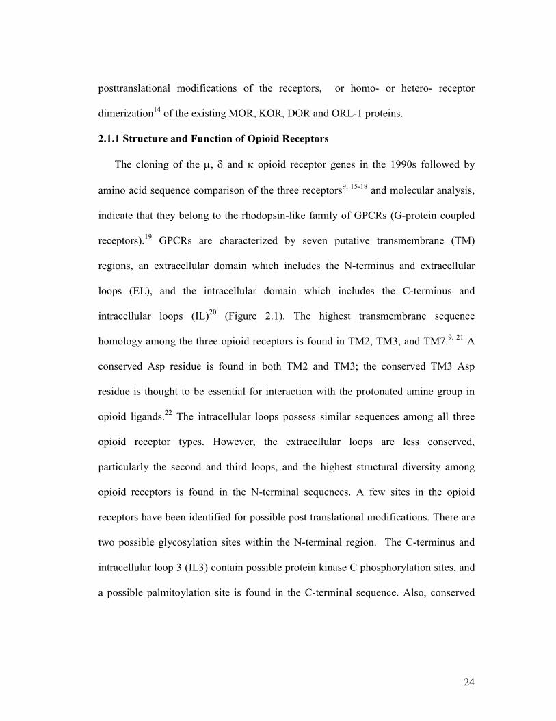
The cloning of the µ, δ and κ opioid receptor genes in the 1990s followed by
amino acid sequence comparison of the three receptors9, 15-18 and molecular analysis,
indicate that they belong to the rhodopsin-like family of GPCRs (G-protein coupled
receptors).19 GPCRs are characterized by seven putative transmembrane (TM)
regions, an extracellular domain which includes the N-terminus and extracellular
loops (EL), and the intracellular domain which includes the C-terminus and
intracellular loops (IL)20 (Figure 2.1). The highest transmembrane sequence
homology among the three opioid receptors is found in TM2, TM3, and TM7.9, 21 A
conserved Asp residue is found in both TM2 and TM3; the conserved TM3 Asp
residue is thought to be essential for interaction with the protonated amine group in
opioid ligands.22 The intracellular loops possess similar sequences among all three
opioid receptor types. However, the extracellular loops are less conserved,
particularly the second and third loops, and the highest structural diversity among
opioid receptors is found in the N-terminal sequences. A few sites in the opioid
receptors have been identified for possible post translational modifications. There are
two possible glycosylation sites within the N-terminal region. The C-terminus and
intracellular loop 3 (IL3) contain possible protein kinase C phosphorylation sites, and
a possible palmitoylation site is found in the C-terminal sequence. Also, conserved

25
Cys residues are found in the first and second extracellular loops which are thought to
be involved in a disulfide linkage.
Figure 2.1.: Serpentine model of MOR23
2.2 Mu Opioid Receptors (MOR)
Opioid analgesics (e.g. morphine), produce pain relief mainly through activation
of MOR and are considered indispensable drugs for the management of pain.24-26
However, there are severe deleterious side effects associated with opioid analgesics,
namely respiratory depression, addiction liability and constipation.27, 28 Martin et al.
in 1976 first differentiated the pharmacological profile of MOR activation vs. KOR in
vivo using morphine as the prototype agonist.4, 8 Administration of morphine to dogs
resulted in a myriad of effects, including miosis, hypothermia, bradycardia, and
analgesia and physical dependence after chronic administration.4 Also,

26
discontinuation of morphine resulted in an abstinence syndrome that could be
suppressed by morphine but not by ketocyclazocaine. The latter drug has its own
spectrum of actions, such as pupillary constriction, sedation and depression of flexor
reflexes, which Martin attributed to activation of a separate type of receptors now
referred to as KOR. These studies established the existence of different opioid
receptor types.4, 8
Based on radioligand binding experiments in brains of several species, the
proportion of MOR is found to be 41% of the opioid receptor population in rat, 25%
in guinea pig and 25% in the mouse.29 A more precise and direct method to
characterize regional differences in receptor distribution is autoradiography.30 The
most common radioligands used for autoradiography studies for MOR are
[3H]DAMGO ([D-Ala2,MePhe4,glyol]enkephalin)31-33 and [3H]CTOP(D-Phe-
cyclo[Cys-Tyr-D-Trp-Orn-Thr-Pen]-Thr-NH2).34 Based on autoradiographic studies
of MOR in rat brain, the highest densities are found in the striatum, the accessory
olfactory bulb, and several areas of thalamic nuclei; in contrast, lower levels of MOR
were found in the cerebral cortex and cerebellum.35
The cloning of the MOR from rat brain was first reported by Chen et al. and
Fukuda et al. in 19939, 18 and showed 64% homology in amino acid sequences to the
DOR cloned earlier by Evans et al. and Keiffer et al.15, 16 The expected high affinity
for selective MOR ligands such as morphine and DAMGO and likewise low affinity
for DOR the KOR selective ligands were observed for cloned MOR expressed on
COS-7 cells. 36

27
2.2.1 Mutagenesis Studies of MOR
Since the cloning of the opioid receptors in 1990s, numerous efforts have been
undertaken to investigate and identify the molecular basis of ligand recognition and
selectivity for particular opioid receptor types. Generally, two approaches have been
utilized to analyze receptor structures. The first approach is to design receptor
chimeras where domains of one opioid receptor type have been replaced by the
corresponding domain from a different opioid receptor.22, 37 The other approach
involves site-directed mutagenesis of critical amino acid residues of a specific
receptor type to investigate the effect of such changes on ligand binding to the
receptor.38, 39 Although both of these approaches have been widely used in examining
the domains of receptors involved in binding, these techniques suffer from the
drawback of potential alteration of the three dimensional structure of the ligand
binding region by the structural changes in primary sequences.22 Nevertheless, these
techniques have been widely used over the past decade and a plethora of information
has been obtained with regard to the specific domains of the opioid receptors possibly
involved in binding and selectivity.
Several groups have reported the involvement of extracellular loops in the binding
through constructing opioid receptor chimeras. Through such studies, it was also
proposed that the binding sites for the opioid peptides and alkaloids are different. 22,
40-43 Results obtained from constructing MOR / DOR chimeric receptors revealed the
importance of extracellular loop 1 (EL1) and TM2 for the binding of MOR selective
ligands.41 It was also reported that a DOR chimeric receptor which included the EL1

28
from MOR bound the MOR selective peptide DAMGO with high affinity, whereas
the MOR receptor chimera bearing the EL1 from DOR was resulted in 100-fold lower
affinity for DAMGO.42 The contribution of Lys108 in EL1 of DOR to ligand
selectivity was demonstrated replacing Lys108 with Asn in the DOR sequence, which
enabled binding of the MOR selective ligand dermorphin. However, the EL1 does not
seem to play a critical role for the binding of MOR selective alkaloids.44 An
investigation of MOR / KOR chimeric receptors suggested that EL3 was critical for
the high-affinity binding of DAMGO to MOR.17, 45 Additional studies identified four
residues (Lys303, Val316, Trp318 and His319) in EL3 of MOR that may be
important for recognition of DAMGO.43 Also, TM6, TM7, and EL3 were found to be
important for the selective binding of sufentanil to MOR over KOR.44 The
importance of EL1 and EL3 was further established with chimeric receptors between
MOR and angiotensin II receptors. When EL1 and 3 from MOR were substituted with
the corresponding regions of angiotensin II receptors, there were reductions in opioid
receptor affinities.46 Separate chimeric receptor studies reported by Varga et al.47 and
Meng et al.48 it was demonstrated that Lys300 in EL3 of MOR represents a critical
site for the selectivity of peptidic ligands. In addition to demonstrating the
involvement of the ELs of MOR in conferring selectivity to peptidic ligands, Seki et
al. also reported ligand-dependent selectivity. While incorporation of EL3 of MOR
into a KOR chimera imparted high affinity binding for DAMGO, this result did not
extend to the MOR-selective agents dermorphin and fentanyl.46 Therefore the

29
molecular basis for ligand affinity and selectivity for different opioid receptors
remains to be fully elucidated.
For high affinity binding to opioid receptors, the presence of protonated nitrogen
in opioid ligands is required. Therefore, an aspartic or a glutamic acid residue in the
binding pocket of the opioid receptors would potentially serve as a counter ion for
ligand binding.22 Conflicting evidence concerning the importance of Asp147 on TM3
of MOR can be found in the literature. Although binding affinities of peptide agonists
to MOR was eliminated through mutation of Asp147 to Ala in MOR, this mutation
did not affect the binding of opioid antagonists diprenorphine and naloxone.22
Furthermore, mutation of Asp147 to Glu resulted in a decrease in binding for
DAMGO, but not for morphine.39 Therefore, it is speculated that other acidic
residues, such as the conserved Asp in TM2, could act as the counter ion in agonist
binding.22 Other charged amino acid residues, such as His297 of MOR, have also
been implicated in the binding of opioid ligands. Thus, mutation of His297 of MOR
to Ala resulted in a several fold loss in [3H]DAMGO binding to MOR.39, 49
Interestingly, this mutation to MOR instilled partial agonistic properties in classical
opioid antagonists (e.g. naloxone).50
One other useful approach to determine the domains of opioid receptors involved
in opioid binding is through the study of irreversible ligands. Chen et al.
demonstrated that β-funaltrexamine (βFNA), a MOR selective antagonist, labeled the
Lys233 in TM5 (see details in section 2.4.2.1).51, 52 This result supports the proposal

30
of ligand binding sequences other than EL1 and EL3 for nonpeptide (opiate)
antagonists. 41-43, 45
2.2.2 Computational Studies on MOR
Currently, there are no high-resolution crystal structures of any of the opioid
receptors. The only transmembrane receptor proteins in GPCR family whose crystal
structure have been solved are rhodopsin in its dark state bound to 11-cis retinal,53, 54
human β2 adrenergic receptor,55, 56 and recently the crystal structure of rhodopsin in
its G-protein interacting conformation.54 In the absence of crystal structures,
computational models of opioid receptors are the other available option for
developing structure-function relationships.57 Homology modeling of opioid receptors
based on the crystal structure of rhodopsin is the most common approach, and several
applications of this approach have been reported in the literature.37, 58-62 Fowler and
coworkers reported a homology model of the agonist bound receptor state of MOR in
complex with the MOR selective cyclic peptide JOM6,63 using structural
constraints.64, 65 Comparison of models of agonist-bound MOR with MOR without a
ligand64 predicted that the rotation of side chain of Trp293 was the major change that
takes place upon agonist binding to MOR, resulting in the relocation of the indole
ring of Trp293 from the interface between TM6 and TM7 to the interface between
transmembrane domains 3, 5, and 6. These movements of TM domains were
proposed to form a π stacking interaction with the aromatic ring of Tyr1 of JOM6.
TM6 movement would then reorient the side chain of Met151, Asp147, Lys233,
Lys303 and Trp318.64, 65 The importance of Trp293 to the activation of MOR had

31
been previously proposed by other groups based on mutagenesis data on rhodopsin
and agonist-activated leukotriene receptors.66, 67
The homology model and molecular dynamics simulation of MOR in
phospholipids bilayer-aqueous environment was subsequently reported by Zhang et
al. where they demonstrated the conformational changes observed in TMs as well as
EL and IL regions.62 They further evaluated the molecular dynamics simulation with
naloxone (Figure 2.2), the universal opioid antagonist, to MOR. At least three main
binding domains of naloxone were observed: a polar and aromatic domain composed
of Asp147, Phe289, Trp293, Cys321 and Tyr326, possibly involved in cation-π
interactions with the protonated nitrogen of naloxone; a hydrophobic domain
consisting of Tyr148 from TM3, Tyr210 and Phe221 from EL2 and another
hydrophobic region involving Trp318, Leu219, Ile322, Ile296 and Ile144.62 Based on
their mutational analysis and computational study, Li and coworkers reported that
mutation of Asp164 in the highly conserved Asp-Arg-Tyr (DRY) motif in TM3 to
either His, Gln, Tyr or Met resulted in constitutive activation of MOR.68
Computational modeling based on the crystal structure of rhodopsin suggests the
differences in conformation resulting from the mutation are probably due to changes
in the interaction between the cytoplasmic ends of TM3 and TM6 involving the
conserved Arg116 in TM3 and Arg280 in TM6.61 Very recently, conformational
changes in the transmembrane domains of the constitutively active Asp164Tyr MOR
were reported based on identification of accessible cysteine residues within the TM
domains labeled by methanethiosulfonate ethylammonium (MTSEA).69

32
As discussed earlier (Chapter 1, section 1.1), the low sequence homology between
MOR and rhodopsin represents a potential for significant error in homology modeling
experiments.70-72 One way to improve the accuracy of such theoretical model is by
providing adequate receptor-specific (in this case opioid receptors) and ligand-
specific experimental constraints. Based on the above findings there is clearly a need
to develop a more direct method to examine receptor-ligand interactions and the
selectivity for different opioid receptor ligands (peptide vs. non-peptide).
Figure 2.2. Morphine and morphine-derived alkaloids for MOR
O
CH3N
HO OH
H1
2
34 5
6
7
8
910
11
12 1314
(-) Morphine
O
CH3N
H3CO OCH3
Thebaine
O
CH3N
H3CO OH
Codeine
N
OOCH3
H3C
Mepiridine
N
H
NCH3
O
Fentanyl
H3C
NH
OCH3
H3C
H3C
Methadone
NOCH3
OOCH3
Cyprodime
NOH
OO
OHNaloxone
NOH
OO
OHNaltrexone
Agonists
Antagonists
H

33
2.3 Ligands for MOR
2.3.1 Small Molecule Ligands for MOR
Morphine, the prototypical MOR agonist, was first isolated from poppy seeds by
Serturner in 1803. He named the compound after Morpheus, the Greek god of sleep
and dreams.28 But it was more than a century later that the complex structure of
morphine was confirmed through total synthesis by Gates and Tschudi.73, 74 Later, the
relative stereochemistry of morphine was established by chemical synthesis and X-
ray crystallography.75 The absolute configuration (Figure 2.2) was later proved with
application of various techniques.76 In addition to morphine, related alkaloids
discovered in opium are codeine and thebaine77 (Figure 2.2). Although thebaine is not
active as an analgesic, it serves as an important synthetic intermediate for the
preparation of several other potent analgesics.78 The elucidation of the structure of
morphine was followed by extensive structure-activity relationship (SAR) studies of
analogs of morphine and related synthetic compounds including morphinans,
benzomorphinans and phenylpiperidines. Some of the MOR selective agonists
discovered from such studies include meperidine, fentenyl and methadone (Figure
2.2) which are also routinely used for the treatment of pain, alone or in combination
therapy, or opiate addiction (methadone, Table 2.1).
The antagonists naltrexone and naloxone (Table 2.1 and Figure 2.2) have been
extensively used to study MOR pharmacology. These compounds also exhibit
significant affinity towards DOR and KOR. Naloxone is primarily used to reverse

34
opiate overdose whereas naltrexone is employed for treatment of narcotic addiction
and alcohol dependence. The antagonist cyprodime exhibits higher selectivity for
MOR over DOR and KOR.
Table 2.1 Opioid affinities and activity in guinea pig ileum (GPI) and mouse vas deference (MVD) of selected MOR agonists and antagonists79
2.3.2 Opioid Peptides
2.3.2.1 Endogenous Opioid Peptides Interacting with MOR
The first endogenous ligands for mammalian opioid receptors discovered back in
1970s were the two pentapeptides: leucine and methionine enkephalin80 followed by
dynorphin A81, 82 and β-endorphin.83 Since these peptides were structurally different
from the alkaloid opiates, they were referred to as opioids to include all nonpeptides
and peptides with opiate-like activity. All of these endogenous peptides share a
common N-terminal tetrapeptide sequence (Tyr-Gly-Gly-Phe, Table 2.2), but they
differ in their C-terminal sequences and also in their preferential interaction with
different opioid receptor types. Based on this observation, Goldstein proposed the
common N- terminal sequence as the ‘message’ sequence required for activation of
opioid receptors and the unique C-terminal sequences as ‘address’ sequences which
Ki (nM) Ki ratio IC50 (nM) a. Agonists MOR DOR KOR MOR/DOR/KOR GPI MVD
Morphine 1.8 90 317 1/50/175 28 478 Meperidine 385 4,350 5,140 1/11/13 1,109 16,000 Fentanyl 7.0 150 470 1/21/67 0.92 26 Methadone 4.5 15 1,630 1/3.3/360 22 523
Ki (nM) Ki ratio Ke (nM) b. Antagonists MOR DOR KOR MOR/DOR/KOR GPI MVD
Cyprodine 9.4 356 176 1/38/19 31 6110 Naloxone 1.8 23 4.8 1/13/2.7 1.9 12 Naltrexone 1.1 6.6 8.5 1/6.0/7.7 0.36 3.6

35
Table 2.2 Mammalian opioid peptides with their precursor proteins
provide the required affinity for a particular ligand for a particular opioid receptor
type.84 Some synthetic enkephalin analogs and the amphibian peptide dermorphin
(see below)85 interact with the MOR preferentially. Endomorphins which were
relatively recently discovered by Zadina et al.86, 87 exhibit high affinity and highest
selectivity for MOR among the endogenous mammalian peptides. Other enkephalin
Precursor Protein Endogenous Peptide Sequence
Leu-Enkephalin Tyr-Gly-Gly-Phe-Leu
Met-Enkephalin Tyr-Gly-Gly-Phe-Met
Met-Enkephalin-Arg6-Phe7 Tyr-Gly-Gly-Phe-Met-Arg-Phe
Proenkephalin
Met-Enkephalin-Arg6-Phe7-Leu8 Tyr-Gly-Gly-Phe-Met-Arg-Gly-Leu
Dynorphin A Tyr-Gly-Gly-Phe-Leu-Arg-Arg-Ile-Arg-Pro-Lys-
Leu-Lys-Trp-Asp-Asn-Gln
Dynorphin B Tyr-Gly-Gly-Phe-Leu-Arg-Arg-Gln-Phe-Lys-Val-
Val-Thr
α-Neoendorphin Tyr-Gly-Gly-Phe-Leu-Arg-Lys-Tyr-Arg-Pro-Lys
Prodynorphin
β-Neoendorphin Tyr-Gly-Gly-Phe-Leu-Arg-Lys-Tyr-Arg-Pro
Proopiomelanocortin β-Endorphin Tyr-Gly-Gly-Phe-Met-Thr-Ser-Glu-Lys-Ser-Gln-
Thr-Pro-Leu-Val-Thr-Leu-Phe-Lys-Asn-Ala-Ile-
Ile-Lys-Asn-Ala-Tyr-Lys-Lys-Gly-Glu
Endomorphin-1 Tyr-Pro-Trp-PheNH2 Unknown
Endomorphin-2 Tyr-Pro-Phe-PheNH2

36
analogs (e.g. DPDPE) as well as the deltorphin family of amphibian peptides88
preferentially bind to DOR 89, 90 where as dynorphin A binds preferentially to KOR.91
2.3.2.2 MOR Selective Linear Enkephalin Analogs
The enkephalin class of opioid peptides has been extensively studied since their
identification.89, 92-95 The endogenous enkephalins show some preference for binding
to DOR but are labile to degradation by a variety of proteases. Therefore, extensive
Table 2.3 Opioid receptor affinities and selectivity in the GPI and MVD of MOR selective enkephalin
aIC50 values
research has been carried out to develop modified analogs with increased metabolic
stability and different selectivities. As a result both MOR and DOR selective
enkephalin analogs have been identified.89, 92, 94, 95 For example, amidation, reduction,
or complete elimination of the C- terminus results in analogs with retention of MOR
affinity and an appreciable increase in MOR selectivity. DAMGO (Figure 2.3), the
most commonly used MOR selective peptide ligand, is an example of a reduced C-
terminus with high affinity and selectivity for MOR (Table 2.3).99 Other related MOR
Ki (nM)
Ki Ratio
IC50 (nM)
Peptide MOR DOR MOR/DOR GPI MVD
References
DAMGO 1.9 345 180 4.5 33 79
Syndyphalin-25
(Tyr-D-Met-Gly-NMePheol)
0.29a 1,250a 4300 0.0025 - 96
LY 164929 (Figure 2.3) 0.6a 900a 1500 - - 97
Tyr-cyclo[D-Dab-Gly-Phe-Leu] 13.8 1158 83 14.1 81.4 98

37
selective C-terminally modified tetrapeptide analogs include syndyphalin-25 (Table
2.3),100 and LY164929 (Figure 2.3)97 that exhibit significantly higher MOR
selectivity than DAMGO (Table 2.3). Also, the characteristic Tyr-Gly-Gly-Phe
‘message’ sequence’ is not an absolute requirement for interaction with MOR, as
replacement of the aromatic moiety of Phe4 with either a cyclohexane ring101 or a
leucine side chain102, 103 were found to be well tolerated.
2.3.2.3 MOR Selective Conformationally Constrained Enkephalin Analogs
Incorporation of conformationally constrained amino acids or a cyclic constraint
has been a successful approach to obtain greater selectivity for one particular receptor
type. The first such example of improved receptor selectivity through a cyclic
Figure 2.3. MOR selective analogs including conformationally constrained cyclic derivatives
H2NHN
NHO
ON
ONH
OOH
DAMGO
HO
H2NHN
NHO
ON
O
HO
CH3
N
LY 164929
H2NHN
NHO
O HN
O
HO
OHN(CH2)2
NH
O
Tyr-cyclo[Nγ-D-Dab-Gly-Phe-Leu]
H2NHN
NH
HN
O
O
HO
OSS
NH2
O
JOM6: Tyr-cyclo(S-Et-S)[D-Cys-Phe-D-Pen]NH2

38
constraint was (Tyr-cyclo[Nγ-D-Dab-Gly-Phe-Leu], Figure 2.3, Dab=α,γ-
diaminobutyric acid), which demonstrated both high affinity and improved selectivity
towards MOR.104 In contrast, the acyclic linear analog [D-
Dab2,Leu5]enkephalinamide failed to show any MOR selectivity.105 Other cyclic
analogs with either D-Orn or D-Lys in position 2 exhibited decreased selectivity for
MOR, although these analogs show higher affinity for this receptor.98 The restriction
of conformational flexibility in Tyr-cyclo[Nγ-D-Dab-Gly-Phe-Leu] was also
demonstrated by NMR spectroscopy106, 107 and computational methods;106-110
however, some degree of conformational flexibility remains
especially for larger ring sizes. Receptor selectivity was also achieved by
incorporating constrained amino acids. One such example is the modified
[Leu5]enkephalin analog where replacement of Tyr1 by 2-amino-6-hydroxy-2-
tetralincarboxylic acid (Hat) results in increased selectivity for MOR.111
2.3.2.4 MOR Selective Peptides from Amphibian Skin
Amphibian skin is a rich source of a varied range of peptides which often
resemble the neurotransmitter or gastrointestinal hormones of mammalian systems.112
In 1981, Montecuchhi et al. and Brocardo et al. first described dermorphin, a
heptapeptide isolated from the skin of the South American frog Phyllomedusa
sauvagei,113, 114 and another similar peptide containing hydroxyproline (Hyp) in place
of Pro6 from the skin of Phyllomedusa rohdei (Figure 2.4).115 Later in the 1990s, the
sequences of three additional dermorphin peptides were predicted based on the cDNA
library from the skin of Phyllomedusa bicolor (Figure 2.4).116

39
Figure 2.4. Dermorphin peptides
The unique structural feature of the amphibian opioid peptides is the presence of
D-Ala between the two aromatic residues Tyr1 and Phe3, in contrast to the message
sequence Tyr-Gly-Gly-Phe for all of the enkephalins and most other mammalian
opioid peptides. Considerable research was performed to identify the gene
responsible for the D-amino acid in these peptides. It was found that the triplet codon
for L-Ala was included in the dermorphin gene116, 117 leading to the hypothesis that
the L-Ala residue must be converted to D-Ala through posttranslational
modification.118 Kreil et al. rationalized that the mechanism of epimerization should
be a quantitative inversion of the chiral center at the α-carbon of alanine, as opposed
to racemization by a racemases which would result in an equal quantity of L-and D-
isomers.118, 119 However, a racemase mechanism should produce some level of
detectable L-Ala dermorphin analogue, and no such isomer has been found in
Phyllomedusa skin.114, 117
Tyr-D-Ala-Phe-Gly-Tyr-Pro-Ser-NH2 dermorphin
Tyr-D-Ala-Phe-Gly-Tyr-Hyp-Ser-NH2 [Hyp6]dermorphin
Tyr-D-Ala-Phe-Gly-Tyr-Pro-Lys-OH [Lys7-OH]dermorphin
Tyr-D-Ala-Phe-Trp-Tyr-Pro-Asn-OH [Trp4,Asn7-OH]dermorphin
Tyr-D-Ala-Phe-Trp-Asn-OH [Trp4, Asn7-OH]dermorphin 1-5

40
2.3.2.4.1 SAR Study of Dermorphins Based on extensive in vitro studies binding assays on crude or synaptosomal
preparations of brain membranes and bioassays on electrically stimulated GPI and
MVD, dermorphins (Table 2.4) were found to be one of the most potent and selective
Table 2.4. Affinities and MOR selectivity of natural dermorphins (amidated and acid forms) and biological activities on GPI and MVD. Taken from reference 119.
aDER= Dermorphin
MOR agonists among the naturally occurring opioids.118 The primary peptide
dermorphin shows 20 times higher affinity, and is 50 times more selective for MOR
than morphine.118 [Lys7]dermorphin, obtained from skin of Phyllomedusa bicolor,
exhibits 10 fold higher affinity and selectivity than DAMGO and dermorphin and is
100 times more potent in the GPI and MVD functional assays than morphine (Table
2.4). [Lys7]dermorphin has been further reported to differentiate between two MOR
subtypes.120
Ki nM Ki ratio IC50 nM
Peptide
MOR DOR DOR/MOR GPI MVD Tyr-D-Ala-Phe-Gly-Tyr-Pro-Ser-NH2 (Dermorphin) 0.54 929 1720 1.29 16.5 Tyr-D-Ala-Phe-Gly-Tyr-Pro-Ser-OH - - - 4.5 28.1 Tyr-D-Ala-Phe-Gly-Tyr-Hyp-Ser-NH2 0.65 1200 1846 1.6 18.1 Tyr-D-Ala-Phe-Gly-Tyr-Hyp-Ser-OH - - - 4.9 33.0 Tyr-D-Ala-Phe-Gly-Tyr-Pro-Lys-NH2 0.09 1105 >10000 1.15 13.6 Tyr-D-Ala-Phe-Gly-Tyr-Pro-Lys-OH 5.7 1150 201 3.82 56.3 DER-Gly-Glu-Ala-Lys-Lys-Ile-NH2
a 0.149 130 872 1.53 - DER-Gly-Glu-Ala-Lys-Lys-Ile-Lys-Arg-NH2
a 0.002 7.2 3600 1.37 - Tyr-D-Ala-Phe-Phe-NH2 (TAPP) 1.5 625 417 255 780 Tyr-D-Ala-Phe-Trp-Asn-NH2 0.9 480 533 5.00 73.7 Tyr-D-Ala-Phe-Trp-Tyr-Pro-Asn-NH2 0.32 690 2156 0.58 6.6 DAMGO 1.1 430 391 7.1 115 Morphine 11.0 500 45 150 1215
41
Comparisons of the MOR affinities of acid vs. amide versions of the naturally
occurring dermorphins revealed that peptides with the C-terminal carboxylic acid
derivatives had several-fold lower MOR affinities than that of the amidated
peptides.121 The high potency of the C-terminally amidated dermorphin analogs
occurs possibly as a result of suppression of the negative charge of the terminal
carboxy group. Also, C-terminal amidation helps protect the analogs from possible
cleavage by carboxypeptidases.118 Of the shorter dermorphin amide analogs,
dermorphin (1-5) retains 50% and dermorphin (1-4) retains 5% of the potency in
activating opioid receptors in GPI118 and dermorphin (1-3) was inactive.122 It was also
found that 1-4 sequence of dermorphin is the primary end product of enzymatic
degradation in rat brain.122 The affinities of C-terminally elongated dermorphin
analogs which included residues from the precursor sequence were higher than
dermorphin for MOR from rat brain, whereas in the GPI assay, the potencies were
similar or slightly lower than that of dermorphin.121 Although a decrease in MOR
affinity was observed with the introduction of additional residues through Glu9 or
Ala10, probably due to the presence of acidic Glu9, further extension of the peptide
with basic residues increases MOR affinity (Table 2.4).121 Affinity and selectivity of a
dimeric derivative of dermorphin have also been investigated. Dimeric derivatives
were prepared by bridging two monomers with hydrazine or diamines of various
lengths.123 One of these ligands, di-dermorphin, where two dermorphin molecules are
linked by hydrazine, displays 5-fold greater MOR affinity and similar selectivity as
dermorphin.
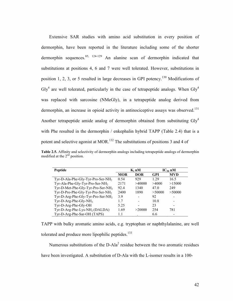
42
Extensive SAR studies with amino acid substitution in every position of
dermorphin, have been reported in the literature including some of the shorter
dermorphin sequences.85, 124-129 An alanine scan of dermorphin indicated that
substitutions at positions 4, 6 and 7 were well tolerated. However, substitutions in
position 1, 2, 3, or 5 resulted in large decreases in GPI potency.130 Modifications of
Gly4 are well tolerated, particularly in the case of tetrapeptide analogs. When Gly4
was replaced with sarcosine (NMeGly), in a tetrapeptide analog derived from
dermorphin, an increase in opioid activity in antinociceptive assays was observed.131
Another tetrapeptide amide analog of dermorphin obtained from substituting Gly4
with Phe resulted in the dermorphin / enkephalin hybrid TAPP (Table 2.4) that is a
potent and selective agonist at MOR.132 The substitutions of positions 3 and 4 of
Table 2.5. Affinity and selectivity of dermorphin analogs including tetrapeptide analogs of dermorphin modified at the 2nd position.
TAPP with bulky aromatic amino acids, e.g. tryptophan or naphthylalanine, are well
tolerated and produce more lipophilic peptides.133
Numerous substitutions of the D-Ala2 residue between the two aromatic residues
have been investigated. A substitution of D-Ala with the L-isomer results in a 100-
Ki nM IC50 nM Peptide MOR DOR GPI MVD
Tyr-D-Ala-Phe-Gly-Tyr-Pro-Ser-NH2 0.54 929 1.29 16.5 Tyr-Ala-Phe-Gly-Tyr-Pro-Ser-NH2 2171 >40000 >4000 >15000 Tyr-D-Met-Phe-Gly-Tyr-Pro-Ser-NH2 92.4 1340 47.0 249 Tyr-D-Pro-Phe-Gly-Tyr-Pro-Ser-NH2 2400 1890 >50000 >50000 Tyr-D-Arg-Phe-Gly-Tyr-Pro-Ser-NH2 3.9 - 92 - Tyr-D-Arg-Phe-Gly-NH2 1.7 - 10.8 - Tyr-D-Arg-Phe-Gly-OH 5.25 - 23 - Tyr-D-Arg-Phe-Lys-NH2 (DALDA) 1.69 >20000 254 781 Tyr-D-Arg-Phe-Sar-OH (TAPS) 1.1 - 6.6 -

43
fold decrease in MOR affinity and abolishes activity in the GPI (<0.1% the potency
of dermorphin, Table 2.5). Substitution of D-Ala2 in dermorphin with L-Tic (1,2,3,4-
tetrahydroisoquinoline-3-carboxylic acid), conformationally constrained analog of
Phe L-Tic resulted in a DOR antagonist.134 Similarly, substitution with D-Pro2
rendered the peptide nonselective and agonist activity was lost in the GPI assay
(Table 2.5).118 Substitution of D-Ala2 with D-Met reduced binding to the MOR,
whereas substitution with D-Arg resulted in analogs with either similar or in some
cases increased MOR affinity and analgesic potency.118, 131, 135 The tetrapeptide
analog TAPS (Table 2.5) was reported by Paakkari et al., to be a potent
antinociceptive agent and caused respiratory stimulation rather than depression that
was antagonized by naloxonazine- the putative MOR1 subtype antagonist.136 TAPS
was also shown to antagonize the respiratory depression caused by dermorphin. These
results have led to the proposal that TAPS is an agonist at the MOR1 subtype and an
antagonist at the MOR2 subtype in vivo.136 Based on Schwyzer’s proposal that MOR
are situated in an ionic membrane compartment,137 Schiller and coworkers
hypothesized that positively charged ligands would display MOR selectivity.132 This
hypothesis was supported by the results for the tetrapeptide dermorphin derivative
with a positively charged residue Lys in position 4 which exhibited increased MOR
selectivity.132 The [D-Arg2,Lys4]dermorphin analog DALDA (Table 2.5), which
contains a +3 net positive charge, also demonstrated superior MOR selectivity in
binding assays. Quarternization of the side chain amine of Lys4 in DALDA was well
tolerated and the resulting analogs retained potent in vivo antinociceptive activity in

44
the mouse writhing assay after s.c. administration.138 This antinociceptive effects
were significantly reduced by the quarternized antagonist N-methyllevallorphan. This
suggest that these analogs exhibit peripheral antinociceptive activity in this assay.138
Substitution of Tyr1 of DALDA by Dmt [(2,6-dimethyl) tyrosine] resulted in
analog with 10-fold higher affinities for both MOR and DOR139 and 200 fold higher
affinity for KOR.140 It was also found that in the rat tail flick test after i.t.
administration, Dmt1[DALDA] was 220 and 3,000 times more potent than DALDA
and morphine respectively. 141
2.4 Affinity Labels
Although a considerable amount of information is available from the SAR of
opioid ligands as well as from chimeric and site directed mutagenesis studies of
opioid receptors, determination of the details of binding and ligand interaction with
the receptor at the molecular level still remains a formidable challenge. To understand
interaction of opioid receptors with their ligands at the molecular level, other
approaches are needed. Affinity labels represent one such approach for probing the
structure of membrane-bound proteins which cannot be readily solved by
crystallography.142 Portoghese proposed that affinity labels bind to their receptors in a
two-step process (Figure 2.5).142 The first reversible step depends on the relative

45
Figure 2.5. An illustration of the two steps involved in covalent binding of an affinity label to its target. Taken from reference 143
affinity of the ligand for the target site, and the second step (Figure 2.5, receptor type
A) involves proper alignment of an electrophilic group on the affinity label with a
compatible, receptor-based nucleophile that is in close proximity. Because of the
second irreversible step, high receptor selectivity is theoretically possible, and affinity
labels can detect subtle differences in receptor-ligand interactions. As indicated in
Figure 2.5, in receptor type B the affinity label fails to undergo the second recognition
process since it does not have sufficient reactivity to bind to the nearby nucleophile.
Alternatively, if a reactive electrophile is too far away from the nucleophile on the
receptor (Figure 2.5, receptor type C), then the second irreversible binding will not
occur. Therefore, affinity labels undergoing irreversible binding to the receptor can be
highly useful in differentiating different opioid receptor types. Site specific

46
interactions can be obtained by determining the attachment point of an affinity label
to its receptor providing important information about the location and orientation of
opioid ligands.143
Depending upon the nature and chemical reactivity of the functionality, affinity
labels can be classified as electrophilic affinity labels, which are inherently reactive,
or photoaffinity labels which require photoactivation to achieve the reactive state. It
should be pointed out that a ligand with extremely high affinity but without the
capability of binding covalently can also be considered an affinity label; but a Kd
value of not greater than 1 X 10-12 M is required. 142
2.4.1 Photoaffinity Labels
Photoaffinity labels are functionalities that can be activated by a brief exposure to
light to generate a highly reactive intermediate species. Because of the high reactivity
of these photolyzed intermediates, they often bind indiscriminately to nearby
functionalities in the receptor binding site.142 Therefore, whether or not a
photoaffinity label will bind to a particular opioid receptor type is determined
primarily by its selectivity as a reversible ligand in the first recognition step. Some
examples of photoreactive intermediates are the nitrene from the azido functionality
and the carbene from diazo or diazirine functionalities (Figure 2.6).144
Figure 2.6. Precursors of reactive species in photoaffinity labels144

47
The first photoaffinity label for an opioid receptor was the [3H]norlevorphanol
derivative APL (Figure 2.7), prepared by Winter and Goldstein 145 APL exhibited
irreversible binding when it was photolyzed in the presence of the GPI or opioid
receptors in the mouse brain particulate fraction, but the binding was not effectively
blocked by levorphanol suggesting extensive non-specific binding. Later, when the
N-methyl quaternary derivative of APL, MAPL (Figure 2.7) was synthesized and
subsequently tested on mouse brain and GPI, MAPL showed some degree of
irreversible binding.146 Some other examples of photoaffinity labels for opioid
Figure 2.7. A: Photoaffinity labels for opioid receptors, B: Amino acid or acid derivative of photoaffinity labels used to characterize opioid receptors.

48
receptors are diazoketone and arylazide derivatives of fentanyl designed by
Maryanoff and coworkers147 (Figure 2.7). Later, Peers and coworkers reported the
synthesis of the nitro-azido derivative of 14β-aminomorphinone (NAM, Figure 2.7)
which is a full antagonist in both the GPI and MVD preparation. Although NAM
appears to bind selectively to MOR in binding studies, it is not suitable for labeling
opioid receptors because of its slow irreversible binding.148
There have been reports of several enkephalin-based photoaffinity label analogs
with an azido group as the photoaffinity functionality (Table 2.6). Amongst these,
only a few are selective for MOR. Some of the azido-containing photoaffinity labels
for MOR reported are a DAMGO-based peptide (Tyr-D-Ala-Gly-Me-Phe(pN3)-
Glyol, ~ 0.3 µM to irreversibly label 50% of MOR sites) prepared by Garbay-
Jaureguiberry and coworkers,149 and a photoaffinity label derivative of the
somatostatin based MOR selective antagonist CTAP (D-Phe-cyclo[Cys-(p-N3Phe)-D-
Trp-Lys-Thr-Pen]-Thr-NH2, IC50 = 48.6 nM) reported by Landis et al150 however, it
was not reported whether this analog bound irreversibly to MOR.
Table 2.6 Enkephalin-based photoaffinity labels for opioid receptors. Taken from reference 143.
Tyr-D-Ala-Gly-Phe-Met-Tyr-NH(CH2)2NH-C6H4(2-NO2,4-N3) Tyr-D-Ala-Gly-Phe-Leu-NHCH(CO2H)(CH2)4NH-C6H4(2-NO2,4-N3) Tyr-D-Ala-Gly-NH(CH2)3NH-C6H4(p-N3) Tyr-D-Ala-Gly-Phe-NH(CH2)3NH-C6H4(p-N3) Tyr-D-Ala-Gly-D / L-Phe(m-N3)-Leu-NH2 Tyr-D-Ala-Gly-Phe-Leu-NH(CH2)2NH-C6H4(2-NO2,4-N3) Tyr-D-Ala-Gly-Phe-Leu-NH(CH2)2NHCO(CH2)2NH-C6H4(2-NO2,4-N3) Tyr-D-Thr-Gly-Phe(p-N3)-Leu-NH2 Tyr-D-Ala-Gly-MePhe(p-N3)-Gly-ol

49
The main disadvantage of using the azido group as a photoaffinity label for
opioid receptors is that the short wavelength (~250 nm) of UV irradiation generally
used to generate the reactive species can inactivate opioid receptors.151 In order to
overcome this problem 4-azido-2-nitrobenzoic acid (ANB) or Bpa (p-benzoyl-
phenylalanine), Figure 2.7) can be used as the photoaffinity label group. These
functionalities shift the wavelength required for photolysis to longer wavelengths,
which may prevent opioid receptor inactivation; this was shown by Herblin and
coworkers when they reported synthesis and selective binding of a tetrapeptide
morpheceptin analog containing Bpa (Tyr-Pro-Phe-Bpa-NH2) to MOR.152 However
this analog showed only modest affinity for MOR (IC50 = 0.27 µM), and only 25% of
the receptor was inactivated upon photolysis in the presence of 2.8 µM of this
compound at a wavelength of 300-350 nm.152 The bulk of this amino acid (Bpa) may
interfere with receptor-ligand interactions which could limit its incorporation to
positions in the C-terminal sequence.
2.4.2 Electrophilic Affinity Labels
When the affinity label contains an electrophilic functionality, several factors may
affect the selectivity of such a label: 1) receptor affinity for the ligand, 2) selectivity
of ligand for the receptor 3) selectivity and chemical reactivity of the electrophile, and
4) proximity of the electrophile to a nearby nucleophile on the receptor. Since two
recognition steps are involved in irreversible binding, in some cases increased
receptor selectivity can be achieved, depending upon the reactivity and proximity of
an appropriate nucleophile in the receptor (see Figure 2.5). Therefore, an electrophilic

50
affinity label can be a useful tool to selectively label one opioid receptor type among
multiple types.
2.4.2.1 Small Molecule-Based Electrophilic Affinity Labels: β-Funaltrexamine
and Other Analogs
The first successful electrophilic affinity label for opioid receptors was prepared
by Portoghese and coworkers by incorporating a nitrogen mustard at the 6β position
of naltrexamine, resulting in β-chlornaltrexamine (β-CNA, Figure 2.8).153, 154 A
nitrogen mustard is a highly reactive electrophile, and as such this affinity label
bound irreversibly to all opioid receptor types. It should be noted that only one of the
chloroethyl groups is needed for the irreversible binding.155 Another example of
nitrogen mustard-containing opiate analogs in the literature, such as the oxymophone
analog β-chloroxymorphamine (β-COA).156, 157
In order to improve the receptor selectivity of opiate-derived affinity labels,
Portoghese and co-workers prepared the β-fumaramide derivative β-funaltrexamine
(β-FNA, Figure 2.8), which contains a less reactive electrophile, in place of the
nitrogen mustard at the 6β position of naltrexamine.158 The resulting compound
bound irreversibly to MOR where it acts an antagonist, whereas it is a reversible
agonist at KOR. Whether or not β-FNA binds irreversibly to DOR remains
unclear.159, 160 It was also shown that the configuration and orientation of the
fumaramide are important for irreversible binding to MOR. Neither the 6α analogue
of β-FNA nor the 6β-maleimide derivative with a cis double bond were capable of
irreversible binding to MOR.160

51
Figure 2.8. Small molecule electrophilic and reporter affinity labels for MOR
β-FNA was the first affinity label whose point of attachment to an opioid receptor
was successfully determined.52 Liu-Chen and coworkers used molecular biology and
protein isolation techniques to identify the attachment point. Initially, based on the
binding of [3H]β-FNA to MOR / KOR receptor chimeras, a region of MOR spanning
from the third intracellular loop to the C-terminus was determined to be essential for
irreversible binding.51 However, upon isolation and partial purification of the labeled
receptor, the point of attachment was found to be in the EL2-TM5 region (Figure

52
2.9). Subsequently, site directed mutagenesis of amino acid residues in this region
identified Lys233 as the attachment point (Figure 2.9).52 This finding of the
attachment point is significant since Lys233 is conserved in all three opioid receptor
types. It appears that the selectivity of β-FNA for MOR is due to the differences in
Figure 2.9. Location of the attachment point (Lys233) of β-FNA to MOR. Taken from reference 52
the tertiary structures of the receptors. This observation further reinforces the utility
of affinity labels as a direct approach to studying receptor-ligand interactions.
A number of 14β-amino substituted derivatives of naloxone and morphine were
prepared by Archer and coworkers.161-163 Reactive functionalities that were attached
to the 14β-amino group include bromoacetamide, thioglycolamide and cinnamoyl
groups. Among these, the p-nitro-substituted derivative with a 5β-methyl group
MET-CAMO (Figure 2.8) appeared to bind irreversibly to MOR.162, 163 This was also
the first N-methyl derivative reported with long-lasting MOR selective antagonist

53
activity with no agonist activity. Similarly, the p-chloro substituted derivative MET-
Cl-CAMO (Figure 2.8) also bound irreversibly to MOR and was a long-lasting MOR
selective antagonist.164
Other examples of MOR selective affinity labels are hydrazone derivatives of the
6-ketone of naloxone, naltrexone and oxymophone described by Pasternak and
coworkers,165-167 and the etonitazene derivative BIT designed by Rice and co-workers
(Figure 2.8). 168, 169
Portoghese and coworkers reported the design of ‘reporter affinity labels’ by
attaching an o-phthaladehyde group to the opioid antagonists naltreaxamine and 6’-
and 7’- aminonaltrindole.170-173 This unique approach was based on the reaction of a
o-phthaladehyde with an amine and thiol (from Lys and Cys side chains in the
receptor), resulting in a fluorescent isoindole; detection of a fluorescence indicates
that a covalent reaction has occurred. Recently, the attachment points of the reporter
affinity label naltrexamine naphthalene dialdehyde derivative NNA (Figure 2.8) to
MOR was determined using site-directed mutagenesis.174 Lys233 and the adjacent
Cys235 of TM5 were determined to be the residues involved in isoindole formation,
as mutation of either of these residues prevented the generation of the fluorescent
product. Although this method allows the determination of attachment points without
isolating the labeled receptor, this approach is limited by the requirement of two
specific nucleophilic receptor residues in close proximity to one another and the
reactive functionality on the ligand.

54
2.4.2.2 Peptide-Based Electrophilic Affinity Labels
Although a number of nonpeptide affinity labels for opioid receptors have been
reported in the literature, peptide-based affinity labels have been limited to mainly
photoaffinity labels (section 2.4.1). Since endogenous ligands for opioid receptors are
peptides, it is important to understand the interactions between peptides and opioid
receptors at the molecular level. Peptide-based electrophilic affinity labels present a
useful class of pharmacological tools which can provide valuable information
regarding the binding of opioid peptides to their receptors.
The first peptide-based electrophilic affinity labels were reported by Pelton and
workers in 1980.175 The C-terminal chloromethyl ketone derivatives of leucine-
enkephalin (Tyr-Ala-Gly-Phe-Leu-CH2Cl) (DLECK) and its D-Ala2 analog [D-
Ala2,Leu5]enkephalin (DALECK) were prepared as potential affinity labels. Although
the compounds exhibited high potency at the opioid receptors in the GPI, the analogs
failed to show any irreversible activity in this tissue.175 Szucs et al. later
reinvestigated the biological effects of these compounds and reported 50%
irreversible blockade of [3H]naloxone binding to opioid receptor sites in rat brain
membranes by both peptides.176 The triated derivative of DALECK was later
prepared by Newman and Barnard and the preferential binding of this peptide to
MOR was demonstrated. Moreover, they were able to determine the molecular weight
of MOR (58 KDa) based on detection of the labeled receptor on a gel. However, the
attachment point on MOR could not be determined.177 There have been a few other
examples of electrophilic affinity labels reported for MOR. Benyhe and coworkers

55
reported the chloromethyl ketone analog) of DAMGO (Tyr-D-Ala-Gly-(Me)Phe-
CH2Cl) which resulted in concentration-dependent irreversible inhibition of
[3H]naloxone binding with an IC50 of 1-5 µM. Shimohigashi and coworkers in 1992
reported the use of the 3-nitro-2-pyridinesulphenyl (Npys) group in a enkephalin and
morphiceptin analogs. Among these, the enkephalin analog (Tyr-D-Ala-Gly-Phe-
Leu(CH2SNpys) produced irreversible inhibition of [3H]DAMGO binding in a
concentration-dependent manner. The concentration required to label half of the
receptors was 19 nM.178
Research in our group has focused on designing and synthesizing peptidic affinity
labels for MOR based on endomorphins, dermorphin or DAMGO as the parent
ligand. These ligands were modified by incorporating either a bromoacetamide or an
isothiocyanate functionality as the electrophile (Table 2.7). The affinities and
selectivity of these peptides were evaluated in radioligand binding assays using
Chinese hamster ovary (CHO) cells expressing MOR and DOR.
In the endomorphin series of analogs, the para positions of either Phe3 or Phe4
was modified by incorporation of an amino functionality, which was then further
derivatized to yield the bromoacetamide and isothiocyanate analogs.179 Although the
analogs retained selectivity for MOR, they all exhibited lower affinities than
endomorphin (IC50 = 4.2 nM) in binding experiments. The highest affinity analog in
this series was [Phe(p-NHCOCH2Br)4]endomorphin (IC50 = 158 nM).179

56
Table 2.7 Progress towards developing peptide-based affinity labels for MOR: analogs of endomorphin, DAMGO and dermorphin.
Parent peptide Affinity Label Modifications
Effect on MOR Affinity (IC50) Compared to Parent Peptide
Endomorphin Tyr-Pro-Phe(p-X)-PheNH2 IC50 = 4.20 ± 0.07 nM for X = H
X = -NCS, -NHCOCH2Br
Affinity decreased >1000 fold179
Tyr-Pro-Phe-Phe(p-X)NH2 IC50 = 4.20 ± 0.07 nM for X = H
X = -NCS, -NHCOCH2Br
Affinity decreased 40-70 fold179
DAMGO Tyr-D-Ala-Gly-(Me)Phe(p-X)-Gly-ol IC50 = 0.51 ± 0.1 nM for X = H
X = -NCS, -NHCOCH2Br
Affinity decreased >1000 fold180
Dermorphin Tyr-D-Ala-Phe(p-X)-Gly-Tyr-Pro-SerNH2 IC50 = 0.72 ± 0.07 nM for X = H
X = -NCS, -NHCOCH2Br
Affinity decreased >1000 fold181
[Phe5]dermorphin Tyr-D-Ala-Phe-Gly-Phe(p-X)-Pro-SerNH2 IC50 = 1.89 ± 0.2 nM for X = H
X = -NCS, -NHCOCH2Br
X= -NCS, affinity decreased >200 fold X = -NHCOCH2Br, IC50= 27.7 ± 3.5 nM Did not exhibit wash-resistant inhibition of binding181
[Phe5,Lys7]dermorphin Tyr-D-Ala-Phe-Gly-Phe(p-X)-Pro-LysNH2 IC50 = 1.04 ± 0.05 nM for X = H
X = -NCS, -NHCOCH2Br
X= -NCS, IC50= 92.8 ± 17.0 X= -NHCOCH2Br, IC50 = 15.1 ± 2.4 nM Did not exhibit resistant inhibition of binding to MOR181
57
Following the same design strategy, analogs of DAMGO were also prepared by
modifying the para position of NMePhe4. However, in every instance MOR affinity
decreased over 1000 fold.180
Analogs of dermorphin and [Lys7]dermorphin were designed by modifying both
the ‘message’ and ‘address’ regions of the peptides.181 Introduction of an electrophile
in the ‘message’ region (the para position of Phe3) decreased MOR affinity by >1000
fold, whereas changes in ‘address’ region in both dermorphin and [Lys7]dermorphin
were well tolerated. [Phe(p-NHCOCH2Br)5]- and [Phe(p-
NHCOCH2Br)5,Lys7]dermorphin showed high affinity for MOR (IC50 = 27.7 and
15.1 nM, respectively). However, none of the analogs in either the dermorphin or
[Lys7]dermorphin series showed wash-resistant inhibition of binding, and hence they
were not affinity labels for MOR.181
2.5 Significance: Peptide vs Small Molecule-Based Affinity Labels
Since pain relief is mediated mainly through MOR, it is important to understand
the interactions between MOR ligands and the receptor. The endogenous ligands of
the opioid receptors are peptides, and studies of chimeric opioid receptors and site-
directed mutagenesis suggest that peptide ligands may interact differently with opioid
receptors than non-peptide ligands.22 Therefore, information about interactions of
peptide ligands with opioid receptors is complimentary to that obtained for non-
peptide ligands. Moreover, information obtained from chimeric and site-directed
mutagenesis studies is complicated by the potential alteration of the secondary and /
or tertiary structure of receptor protein. Electrophilic affinity labels, which interact in

58
a two-step non-equilibrium manner, can provide direct information about ligand
binding to the receptor at the molecular level. Hence, peptide-based affinity labels can
be utilized as valuable pharmacological tools to provide information about the
binding of these ligands to MOR.
2.6 Bibliography
1. Miotto, K. M., K.; Evans, C.J. Moelcular characterization of opioid receptors. In
The Pharmacology of Opioid Peptides, Tseung, L. F., Ed. Harwood academic
publishers: Singapore, 1995; pp 57-58.
2. Beckett, A. H.; Casy, A. F. Synthetic analgesics: stereochemical considerations. J.
Pharm. Pharmacol. 1954, 6, 986-1001.
3. Portoghese, P. S. A new concept on the mode of interaction of narcotic analgesics
with receptors. J. Med. Chem. 1965, 8, 609-616.
4. Martin, W. R.; Eades, C. G.; Thompson, J. A.; Huppler, R. E.; Gilbert, P. E. The
effects of morphine- and nalorphine- like drugs in the nondependent and
morphine-dependent chronic spinal dog. J. Pharmacol. Exp. Ther. 1976, 197,
517-532.
5. Pert, C. B.; Snyder, S. H. Opiate receptor: demonstration in nervous tissue.
Science 1973, 179, 1011-1014.

59
6. Terenius, L. Stereospecific interaction between narcotic analgesics and a synaptic
plasm a membrane fraction of rat cerebral cortex. Acta. Pharmacol. Toxicol.
(Copenh) 1973, 32, 317-320.
7. Simon, E. J.; Hiller, J. M.; Edelman, I. Stereospecific binding of the potent
narcotic analgesic (3H) Etorphine to rat-brain homogenate. Proc. Natl. Acad. Sci.
U. S. A. 1973, 70, 1947-1949.
8. Gilbert, P. E.; Martin, W. R. The effects of morphine and nalorphine-like drugs in
the nondependent, morphine-dependent and cyclazocine-dependent chronic spinal
dog. J. Pharmacol. Exp. Ther. 1976, 198, 66-82.
9. Chen, Y.; Mestek, A.; Liu, J.; Hurley, J. A.; Yu, L. Molecular cloning and
functional expression of a mu-opioid receptor from rat brain. Mol. Pharmacol.
1993, 44, 8-12.
10. Yasuda, K.; Raynor, K.; Kong, H.; Breder, C. D.; Takeda, J.; Reisine, T.; Bell, G.
I. Cloning and functional comparison of kappa and delta opioid receptors from
mouse brain. Proc. Natl. Acad. Sci. U. S. A. 1993, 90, 6736-6740.
11. Mollereau, C.; Parmentier, M.; Mailleux, P.; Butour, J. L.; Moisand, C.; Chalon,
P.; Caput, D.; Vassart, G.; Meunier, J. C. ORL1, a novel member of the opioid
receptor family. Cloning, functional expression and localization. FEBS Lett. 1994,
341, 33-38.
12. Meunier, J. C.; Mollereau, C.; Toll, L.; Suaudeau, C.; Moisand, C.; Alvinerie, P.;
Butour, J. L.; Guillemot, J. C.; Ferrara, P.; Monsarrat, B.; et al. Isolation and

60
structure of the endogenous agonist of opioid receptor-like ORL1 receptor.
Nature 1995, 377, 532-535.
13. Pasternak, G. W. Insights into mu opioid pharmacology the role of mu opioid
receptor subtypes. Life Sci. 2001, 68, 2213-2219.
14. Jordan, B. A.; Devi, L. A. G-protein-coupled receptor heterodimerization
modulates receptor function. Nature 1999, 399, 697-700.
15. Evans, C. J.; Keith, D. E., Jr.; Morrison, H.; Magendzo, K.; Edwards, R. H.
Cloning of a delta opioid receptor by functional expression. Science 1992, 258,
1952-1955.
16. Kieffer, B. L.; Befort, K.; Gaveriaux-Ruff, C.; Hirth, C. G. The delta-opioid
receptor: isolation of a cDNA by expression cloning and pharmacological
characterization. Proc. Natl. Acad. Sci. U. S. A. 1992, 89, 12048-12052.
17. Minami, M.; Toya, T.; Katao, Y.; Maekawa, K.; Nakamura, S.; Onogi, T.;
Kaneko, S.; Satoh, M. Cloning and expression of a cDNA for the rat kappa-opioid
receptor. FEBS Lett. 1993, 329, 291-295.
18. Fukuda, K.; Kato, S.; Mori, K.; Nishi, M.; Takeshima, H. Primary structures and
expression from cDNAs of rat opioid receptor delta- and mu-subtypes. FEBS Lett.
1993, 327, 311-314.
19. Kieffer, B. L. Recent advances in molecular recognition and signal transduction
of active peptides: receptors for opioid peptides. Cell. Mol. Neurobiol. 1995, 15,
615-635.

61
20. Knapp, R. J.; Malatynska, E.; Collins, N.; Fang, L.; Wang, J. Y.; Hruby, V. J.;
Roeske, W. R.; Yamamura, H. I. Molecular biology and pharmacology of cloned
opioid receptors. FASEB J. 1995, 9, 516-525.
21. Chen, Y.; Mestek, A.; Liu, J.; Yu, L. Molecular cloning of a rat kappa opioid
receptor reveals sequence similarities to the mu and delta opioid receptors.
Biochem. J. 1993, 295 ( Pt 3), 625-628.
22. Law, P. Y.; Wong, Y. H.; Loh, H. H. Mutational analysis of the structure and
function of opioid receptors. Biopolymers 1999, 51, 440-455.
23. http://www.opioid.umn.edu/.
24. Cleary, J. F. Cancer pain management. Cancer Control 2000, 7, 120-131.
25. Committee. The use of opioids for the treatment of chronic pain. A consensus
statement from the American Academy of Pain Medicine and the American Pain
Society. Clin. J. Pain 1997, 13, 6-8.
26. O'Callaghan, J. P. Evolution of a rational use of opioids in chronic pain. Eur. J.
Pain 2001, 5 Suppl A, 21-26.
27. Aldrich, J. V.; Vigil-Cruz, S. C. Narcotic analgesics. In Burger's Medicinal
Chemistry and Drug Discovery, Abraham, D. J., Ed. John Wiley and Sons: New
York, 2003; Vol. 6, pp 329-481.
28. Gutstein, H. B.; Akil, H. Opioid analgesics. In Goodman and Gilman's The
Pharmacological Basis of Therapeutics, 10th ed.; Hardman, J.; Limbird, L.;
Gilman, A., Eds. McGraw-Hill: New York, 2001; pp 569-619.

62
29. Mansour, A.; Lewis, M. E.; Khachaturian, H.; Akil, H.; Watson, S. J.
Pharmacological and anatomical evidence of selective mu, delta, and kappa
opioid receptor binding in rat brain. Brain Res. 1986, 399, 69-79.
30. Kuhar, M. J.; De Souza, E. B.; Unnerstall, J. R. Neurotransmitter receptor
mapping by autoradiography and other methods. Annu. Rev. Neurosci. 1986, 9,
27-59.
31. Mansour, A.; Khachaturian, H.; Lewis, M. E.; Akil, H.; Watson, S. J. Anatomy of
CNS opioid receptors. Trends Neurosci. 1988, 11, 308-314.
32. Sharif, N. A.; Hughes, J. Discrete mapping of brain Mu and delta opioid receptors
using selective peptides: quantitative autoradiography, species differences and
comparison with kappa receptors. Peptides 1989, 10, 499-522.
33. Tempel, A.; Zukin, R. S. Neuroanatomical patterns of the mu, delta, and kappa
opioid receptors of rat brain as determined by quantitative in vitro
autoradiography. Proc. Natl. Acad. Sci. U. S. A. 1987, 84, 4308-4312.
34. Hawkins, K. N.; Knapp, R. J.; Gehlert, D. R.; Lui, G. K.; Yamamura, M. S.;
Roeske, L. C.; Hruby, V. J.; Yamamura, H. I. Quantitative autoradiography of
[3H]CTOP binding to mu opioid receptors in rat brain. Life Sci. 1988, 42, 2541-
2551.
35. Kuhar, M. J. D. S., E. B. Receptor autoradiography as an aid in explaining drug
action. In Imaging Drug Action in the Brain, D., E., Ed. Boca Ration: CRC press:
London, 1993; pp 49-60.

63
36. Knapp, R. J. V., L.K.; Yamaura, H.I. Selective Ligands for mu and delta opioid
receptors. In The Pharmacology of Opioid Peptides, Tseung, L. F., Ed. Harwood
Academic Publishers: Wisconsin, 1993; pp 1-27.
37. Chaturvedi, K.; Christoffers, K. H.; Singh, K.; Howells, R. D. Structure and
regulation of opioid receptors. Biopolymers 2000, 55, 334-346.
38. Kong, H.; Raynor, K.; Yasuda, K.; Moe, S. T.; Portoghese, P. S.; Bell, G. I.;
Reisine, T. A single residue, aspartic acid 95, in the delta opioid receptor specifies
selective high affinity agonist binding. J. Biol. Chem. 1993, 268, 23055-23058.
39. Surratt, C. K.; Johnson, P. S.; Moriwaki, A.; Seidleck, B. K.; Blaschak, C. J.;
Wang, J. B.; Uhl, G. R. -mu opiate receptor. Charged transmembrane domain
amino acids are critical for agonist recognition and intrinsic activity. J. Biol.
Chem. 1994, 269, 20548-20553.
40. Wang, J. B.; Johnson, P. S.; Wu, J. M.; Wang, W. F.; Uhl, G. R. Human kappa
opiate receptor second extracellular loop elevates dynorphin's affinity for human
mu/kappa chimeras. J. Biol. Chem. 1994, 269, 25966-25969.
41. Fukuda, K.; Kato, S.; Mori, K. Location of regions of the opioid receptor involved
in selective agonist binding. J. Biol. Chem. 1995, 270, 6702-6709.
42. Onogi, T.; Minami, M.; Katao, Y.; Nakagawa, T.; Aoki, Y.; Toya, T.; Katsumata,
S.; Satoh, M. DAMGO, a mu-opioid receptor selective agonist, distinguishes
between mu- and delta-opioid receptors around their first extracellular loops.
FEBS Lett. 1995, 357, 93-97.

64
43. Xue, J. C.; Chen, C.; Zhu, J.; Kunapuli, S.; DeRiel, J. K.; Yu, L.; Liu-Chen, L. Y.
Differential binding domains of peptide and non-peptide ligands in the cloned rat
kappa opioid receptor. J. Biol. Chem. 1994, 269, 30195-30199.
44. Zhu, J.; Xue, J. C.; Law, P. Y.; Claude, P. A.; Luo, L. Y.; Yin, J.; Chen, C.; Liu-
Chen, L. Y. The region in the mu opioid receptor conferring selectivity for
sufentanil over the delta receptor is different from that over the kappa receptor.
FEBS Lett. 1996, 384, 198-202.
45. Xue, J. C.; Chen, C.; Zhu, J.; Kunapuli, S. P.; de Riel, J. K.; Yu, L.; Liu-Chen, L.
Y. The third extracellular loop of the mu opioid receptor is important for agonist
selectivity. J. Biol. Chem. 1995, 270, 12977-12979.
46. Seki, T.; Minami, M.; Nakagawa, T.; Ienaga, Y.; Morisada, A.; Satoh, M.
DAMGO recognizes four residues in the third extracellular loop to discriminate
between mu- and kappa-opioid receptors. Eur. J. Pharmacol. 1998, 350, 301-310.
47. Varga, E. V.; Li, X.; Stropova, D.; Zalewska, T.; Landsman, R. S.; Knapp, R. J.;
Malatynska, E.; Kawai, K.; Mizusura, A.; Nagase, H.; Calderon, S. N.; Rice, K.;
Hruby, V. J.; Roeske, W. R.; Yamamura, H. I. The third extracellular loop of the
human delta-opioid receptor determines the selectivity of delta-opioid agonists.
Mol. Pharmacol. 1996, 50, 1619-1624.
48. Meng, F.; Ueda, Y.; Hoversten, M. T.; Thompson, R. C.; Taylor, L.; Watson, S.
J.; Akil, H. Mapping the receptor domains critical for the binding selectivity of
delta-opioid receptor ligands. Eur. J. Pharmacol. 1996, 311, 285-292.

65
49. Mansour, A.; Taylor, L. P.; Fine, J. L.; Thompson, R. C.; Hoversten, M. T.;
Mosberg, H. I.; Watson, S. J.; Akil, H. Key residues defining the mu-opioid
receptor binding pocket: a site-directed mutagenesis study. J. Neurochem. 1997,
68, 344-353.
50. Spivak, C. E.; Beglan, C. L.; Seidleck, B. K.; Hirshbein, L. D.; Blaschak, C. J.;
Uhl, G. R.; Surratt, C. K. Naloxone activation of mu-opioid receptors mutated at a
histidine residue lining the opioid binding cavity. Mol. Pharmacol. 1997, 52, 983-
992.
51. Chen, C.; Xue, J. C.; Zhu, J.; Chen, Y. W.; Kunapuli, S.; Kim de Riel, J.; Yu, L.;
Liu-Chen, L. Y. Characterization of irreversible binding of beta-funaltrexamine to
the cloned rat mu opioid receptor. J. Biol. Chem. 1995, 270, 17866-17870.
52. Chen, C.; Yin, J.; Riel, J. K.; DesJarlais, R. L.; Raveglia, L. F.; Zhu, J.; Liu-Chen,
L. Y. Determination of the amino acid residue involved in [3H]beta-
funaltrexamine covalent binding in the cloned rat mu-opioid receptor. J. Biol.
Chem. 1996, 271, 21422-21429.
53. Palczewski, K.; Kumasaka, T.; Hori, T.; Behnke, C. A.; Motoshima, H.; Fox, B.
A.; Le Trong, I.; Teller, D. C.; Okada, T.; Stenkamp, R. E.; Yamamoto, M.;
Miyano, M. Crystal structure of rhodopsin: A G protein-coupled receptor. Science
2000, 289, 739-745.
54. Scheerer, P.; Park, J. H.; Hildebrand, P. W.; Kim, Y. J.; Krauss, N.; Choe, H. W.;
Hofmann, K. P.; Ernst, O. P. Crystal structure of opsin in its G-protein-interacting
conformation. Nature 2008, 455, 497-502.

66
55. Rasmussen, S. G.; Choi, H. J.; Rosenbaum, D. M.; Kobilka, T. S.; Thian, F. S.;
Edwards, P. C.; Burghammer, M.; Ratnala, V. R.; Sanishvili, R.; Fischetti, R. F.;
Schertler, G. F.; Weis, W. I.; Kobilka, B. K. Crystal structure of the human beta2
adrenergic G-protein-coupled receptor. Nature 2007, 450, 383-387.
56. Cherezov, V.; Rosenbaum, D. M.; Hanson, M. A.; Rasmussen, S. G.; Thian, F. S.;
Kobilka, T. S.; Choi, H. J.; Kuhn, P.; Weis, W. I.; Kobilka, B. K.; Stevens, R. C.
High-resolution crystal structure of an engineered human beta2-adrenergic G
protein-coupled receptor. Science 2007, 318, 1258-1265.
57. Pogozheva, I. D.; Przydzial, M. J.; Mosberg, H. I. Homology modeling of opioid
receptor-ligand complexes using experimental constraints. AAPS J. 2005, 7,
E434-448.
58. Decaillot, F. M.; Befort, K.; Filliol, D.; Yue, S.; Walker, P.; Kieffer, B. L. Opioid
receptor random mutagenesis reveals a mechanism for G protein-coupled receptor
activation. Nat. Struct. Biol. 2003, 10, 629-636.
59. Bissantz, C.; Bernard, P.; Hibert, M.; Rognan, D. Protein-based virtual screening
of chemical databases. II. Are homology models of G-Protein Coupled Receptors
suitable targets? Proteins 2003, 50, 5-25.
60. Horn, F.; Bettler, E.; Oliveira, L.; Campagne, F.; Cohen, F. E.; Vriend, G.
GPCRDB information system for G protein-coupled receptors. Nucleic. Acids.
Res. 2003, 31, 294-297.
61. Huang, P.; Li, J.; Chen, C.; Visiers, I.; Weinstein, H.; Liu-Chen, L. Y. Functional
role of a conserved motif in TM6 of the rat mu opioid receptor: constitutively

67
active and inactive receptors result from substitutions of Thr6.34(279) with Lys
and Asp. Biochemistry 2001, 40, 13501-13509.
62. Zhang, Y.; Sham, Y. Y.; Rajamani, R.; Gao, J.; Portoghese, P. S. Homology
modeling and molecular dynamics simulations of the mu opioid receptor in a
membrane-aqueous system. ChemBioChem. 2005, 6, 853-859.
63. McFadyen, I. J.; Sobczyk-Kojiro, K.; Schaefer, M. J.; Ho, J. C.; Omnaas, J. R.;
Mosberg, H. I.; Traynor, J. R. Tetrapeptide derivatives of [D-Pen2,D-Pen5]-
enkephalin (DPDPE) lacking an N-terminal tyrosine residue are agonists at the
mu-opioid receptor. J. Pharmacol. Exp. Ther. 2000, 295, 960-966.
64. Fowler, C. B.; Pogozheva, I. D.; Lomize, A. L.; LeVine, H., 3rd; Mosberg, H. I.
Complex of an active mu-opioid receptor with a cyclic peptide agonist modeled
from experimental constraints. Biochemistry 2004, 43, 15796-15810.
65. Fowler, C. B.; Pogozheva, I. D.; LeVine, H., 3rd; Mosberg, H. I. Refinement of a
homology model of the mu-opioid receptor using distance constraints from
intrinsic and engineered zinc-binding sites. Biochemistry 2004, 43, 8700-8710.
66. Baneres, J. L.; Martin, A.; Hullot, P.; Girard, J. P.; Rossi, J. C.; Parello, J.
Structure-based analysis of GPCR function: conformational adaptation of both
agonist and receptor upon leukotriene B4 binding to recombinant BLT1. J. Mol.
Biol. 2003, 329, 801-814.
67. Chabre, M.; Breton, J. Orientation of aromatic residues in rhodopsin. Rotation of
one tryptophan upon the meta I to meta II transition afer illumination. Photochem.
Photobiol. 1979, 30, 295-299.
68
68. Li, J.; Huang, P.; Chen, C.; de Riel, J. K.; Weinstein, H.; Liu-Chen, L. Y.
Constitutive activation of the mu opioid receptor by mutation of D3.49(164), but
not D3.32(147): D3.49(164) is critical for stabilization of the inactive form of the
receptor and for its expression. Biochemistry 2001, 40, 12039-12050.
69. Xu, W.; Sanz, A.; Pardo, L.; Liu-Chen, L. Y. Activation of the mu opioid receptor
involves conformational rearrangements of multiple transmembrane domains.
Biochemistry 2008, 47, 10576-10586.
70. Eswar, N.; John, B.; Mirkovic, N.; Fiser, A.; Ilyin, V. A.; Pieper, U.; Stuart, A.
C.; Marti-Renom, M. A.; Madhusudhan, M. S.; Yerkovich, B.; Sali, A. Tools for
comparative protein structure modeling and analysis. Nucleic. Acids. Res. 2003,
31, 3375-3380.
71. John, B.; Sali, A. Comparative protein structure modeling by iterative alignment,
model building and model assessment. Nucleic. Acids. Res. 2003, 31, 3982-3992.
72. Shacham, S.; Topf, M.; Avisar, N.; Glaser, F.; Marantz, Y.; Bar-Haim, S.;
Noiman, S.; Naor, Z.; Becker, O. M. Modeling the 3D structure of GPCRs from
sequence. Med. Res. Rev. 2001, 21, 472-483.
73. Gates, M.; Tschudi, G. The synthesis of morphine. J. Am. Chem. Soc. 1952, 52,
1109-1110.
74. Gates, M.; Tschudi, G. The synthesis of morphine. J. Am. Chem. Soc. 1956, 78,
1380-1393.

69
75. Rapoport, H.; Lavigne, J. B. Stereochemical Studies in the Morphine Series. The
Relative Configuration at Carbons Thirteen and Fourteen1. J. Am. Chem. Soc.
1953, 75, 5329-5334.
76. Bentley, K. W.; Cardwell, H. M. E. The morphine-the baine group of alkaloids.
V. The absolute stereochemistry of the morphine, benzylisoquinoline, aporphine,
and tetrahydroberberine alkaloids. J. Chem. Soc. 1955, 3252-3260.
77. Casy, A. F.; Dewar, G. H. The Steric Factor in Medicinal Chemistry.
Dissymmetric Probes of Pharmacological receptors. In Plenum: NewYork, 1993;
pp 429-501.
78. Fairbairn, J. W.; Helliwell, K. Papaver bracteatum Lindley: thebaine content in
relation to plant development. J. Pharm. Pharmacol. 1977, 29, 65-69.
79. Corbett, A. D.; Paterson, S. J.; Kosterlitz, H. W. Selectivity of Ligands for Opioid
receptors. In Opioids I, Handbook of Experimental Pharmacology, Herz, A.; Akil,
H.; Simon, E. J., Eds. Springer-Verlag: Berlin, 1993; Vol. 104/I, pp 645-679.
80. Hughes, J.; Smith, T. W.; Kosterlitz, H. W.; Fothergill, L. A.; Morgan, B. A.;
Morris, H. R. Identification of two related pentapeptides from the brain with
potent opiate agonist activity. Nature 1975, 258, 577-580.
81. Cox, B. M.; Opheim, K. E.; Teschemacher, H.; Goldstein, A. A peptide-like
substance from pituitary that acts like morphine. 2. Purification and properties.
Life Sci. 1975, 16, 1777-1782.

70
82. Teschemacher, H.; Opheim, K. E.; Cox, B. M.; Goldstein, A. A peptide-like
substance from pituitary that acts like morphine. I. Isolation. Life Sci. 1975, 16,
1771-1775.
83. Li, C. H.; Chung, D. Isolation and structure of an untriakontapeptide with opiate
activity from camel pituitary glands. Proc. Natl. Acad. Sci. U. S. A. 1976, 73,
1145-1148.
84. Chavkin, C.; Goldstein, A. Specific receptor for the opioid peptide dynorphin:
structure--activity relationships. Proc. Natl. Acad. Sci. U. S. A. 1981, 78, 6543-
6547.
85. Erspamer, V. The opioid peptides of the amphibian skin. Int. J. Dev. Neurosci.
1992, 10, 3-30.
86. Zadina, J. E.; Hackler, L.; Ge, L. J.; Kastin, A. J. A potent and selective
endogenous agonist for the mu-opiate receptor. Nature 1997, 386, 499-502.
87. Zadina, J. E.; Martin-Schild, S.; Gerall, A. A.; Kastin, A. J.; Hackler, L.; Ge, L. J.;
Zhang, X. Endomorphins: novel endogenous mu-opiate receptor agonists in
regions of high mu-opiate receptor density. Ann. N. Y. Acad. Sci. 1999, 897, 136-
144.
88. Erspamer, V.; Melchiorri, P.; Falconieri-Erspamer, G.; Negri, L.; Corsi, R.;
Severini, C.; Barra, D.; Simmaco, M.; Kreil, G. Deltorphins: a family of naturally
occurring peptides with high affinity and selectivity for delta opioid binding sites.
Proc. Natl. Acad. Sci. U. S. A. 1989, 86, 5188-5192.

71
89. Hruby, V. J.; Agnes, R. S. Conformation-activity relationships of opioid peptides
with selective activities at opioid receptors. Biopolymers 1999, 51, 391-410.
90. Naqvi, T.; Haq, W.; Mathur, K. B. Structure-activity relationship studies of
dynorphin A and related peptides. Peptides 1998, 19, 1277-1292.
91. Goldstein, A.; Tachibana, S.; Lowney, L. I.; Hunkapiller, M.; Hood, L.
Dynorphin-(1-13), an extraordinarily potent opioid peptide. Proc. Natl. Acad. Sci.
U. S. A. 1979, 76, 6666-6670.
92. Morley, J. S. Structure-activity relationships of enkephalin-like peptides. Annu.
Rev. Pharmacol. Toxicol. 1980, 20, 81-110.
93. Udenfriend, S.; Meienhofer, J. Opioid Peptides: Biology, Chemistry, and
Genetics. Academic Press: Orlando, FL, 1984; Vol. 6.
94. Hruby, V. J.; Gehrig, C. A. Recent developments in the design of receptor
specific opioid peptides. Med. Res. Rev. 1989, 9, 343-401.
95. Schiller, P. W. Development of receptor-specific opioid peptide analogues. Prog.
Med. Chem. 1991, 28, 301-340.
96. Quirion, R.; Kiso, Y.; Pert, C. B. Syndyphalin SD-25: a highly selective ligand for
mu opiate receptors. FEBS Lett. 1982, 141, 203-206.
97. Shuman, R. T. H., M. D.; Woods, J. H.; Gessellchen, P. Peptides: Chemistry,
Structure, Biology. In Rivier, J. E. M., G. R., Ed. ESCOM: Leiden, The
Netherlands, 1990; pp 326-628.
98. DiMaio, J.; Nguyen, T. M.; Lemieux, C.; Schiller, P. W. Synthesis and
pharmacological characterization in vitro of cyclic enkephalin analogues: effect of

72
conformational constraints on opiate receptor selectivity. J. Med. Chem. 1982, 25,
1432-1438.
99. Handa, B. K.; Land, A. C.; Lord, J. A.; Morgan, B. A.; Rance, M. J.; Smith, C. F.
Analogues of beta-LPH61-64 possessing selective agonist activity at mu-opiate
receptors. Eur. J. Pharmacol. 1981, 70, 531-540.
100. Kiso, Y.; Yamaguchi, M.; Akita, T.; Moritoki, H.; Takei, M.; Nakamura, H.
Super-active enkephalin analogues. Simple tripeptide hydroxyalkylamides exhibit
surprisingly high and long-lasting opioid activities. Naturwissenschaften 1981,
68, 210-212.
101. Audigier, Y.; Mazarguil, H.; Gout, R.; Cros, J. Structure-activity relationships of
enkephalin analogs at opiate and enkephalin receptors: correlation with analgesia.
Eur. J. Pharmacol. 1980, 63, 35-46.
102. Fournie-Zaluski, M. C.; Gacel, G.; Maigret, B.; Premilat, S.; Roques, B. P.
Structural requirements for specific recognition of mu or delta opiate receptors.
Mol. Pharmacol. 1981, 20, 484-491.
103. Roques, B. P.; Gacel, G.; Fournie-Zaluski, M. C.; Senault, B.; Lecomte, J. M.
Demonstration of the crucial role of the phenylalanine moiety in enkephalin
analogues for differential recognition of the mu- and delta-receptors. Eur. J.
Pharmacol. 1979, 60, 109-110.
104. DiMaio, J.; Schiller, P. W. A cyclic enkephalin analog with high in vitro opiate
activity. Proc. Natl. Acad. Sci. U. S. A. 1980, 77, 7162-7166.

73
105. Schiller, P. W.; DiMaio, J. Opiate receptor subclasses differ in their
conformational requirements. Nature 1982, 297, 74-76.
106. Maigret, B.; Fournie-Zaluski, M. C.; Roques, B.; Premilat, S. Proposals for the
mu-active conformation of the enkephalin analog Tyr-cyclol(-Nγ-D-A2-bu-Gly-
Phe-Leu-). Mol. Pharmacol. 1986, 29, 314-320.
107. Mammi, N. J.; Hassan, M.; Goodman, M. Conformational analysis of a cyclic
enkephalin analog by proton NMR and computer simulations. J. Am. Chem. Soc.
1985, 107, 4008-4013.
108. Hall, D.; Pavitt, N. Conformation of cyclic analogs of enkephalin. III. Effect of
varying ring size. Biopolymers 1985, 24, 935-945.
109. Hall, D.; Pavitt, N. Conformation of a cyclic tetrapeptide related to an analog of
enkephalin. Biopolymers 1985, 23, 1441-1445.
110. Kessler, H.; Holzemann, G.; Zechel, C. Conformational analysis of cyclic
enkephalin analogs of the type Tyr-cyclo-(-Nω-Xxx-Gly-Phe-Leu-) Int. J. Pept.
Protein. Res. 1985, 25, 267-279.
111. Deeks, T.; Crooks, P. A.; Waigh, R. D. Synthesis and analgesic properties of two
leucine-enkephalin analogues containing a conformationally restrained N-terminal
tyrosine residue. J. Med. Chem. 1983, 26, 762-765.
112. Erspamer, V. Bioactive secretions of the Amphibian integument. In Amphibian
Biology, Hetawole, H., Ed. Surrey Beatty & Sons Publ.: 1994; pp 178-350.
113. Broccardo, M.; Erspamer, V.; Falconieri Erspamer, G.; Improta, G.; Linari, G.;
Melchiorri, P.; Montecucchi, P. C. Pharmacological data on dermorphins, a new

74
class of potent opioid peptides from amphibian skin. Br. J. Pharmacol. 1981, 73,
625-631.
114. Montecucchi, P. C.; de Castiglione, R.; Piani, S.; Gozzini, L.; Erspamer, V.
Amino acid composition and sequence of dermorphin, a novel opiate-like peptide
from the skin of Phyllomedusa sauvagei. Int. J. Pept. Protein. Res. 1981, 17, 275-
283.
115. Montecucchi, P. C.; de Castiglione, R.; Erspamer, V. Identification of dermorphin
and Hyp6-dermorphin in skin extracts of the Brazilian frog Phyllomedusa rhodei.
Int. J. Pept. Protein. Res. 1981, 17, 316-321.
116. Richter, K.; Egger, R.; Kreil, G. D-alanine in the frog skin peptide dermorphin is
derived from L-alanine in the precursor. Science 1987, 238, 200-202.
117. Richter, K.; Egger, R.; Negri, L.; Corsi, R.; Severini, C.; Kreil, G. cDNAs
encoding [D-Ala2]deltorphin precursors from skin of Phyllomedusa bicolor also
contain genetic information for three dermorphin-related opioid peptides. Proc.
Natl. Acad. Sci. U. S. A. 1990, 87, 4836-4839.
118. Melchiorri, P.; Negri, L. The dermorphin peptide family. Gen. Pharmacol. 1996,
27, 1099-1107.
119. Kreil, G. Peptides containing a D-amino acid from frogs and molluscs. J. Biol.
Chem. 1994, 269, 10967-10970.
120. Negri, L.; Erspamer, G. F.; Severini, C.; Potenza, R. L.; Melchiorri, P.; Erspamer,
V. Dermorphin-related peptides from the skin of Phyllomedusa bicolor and their
amidated analogs activate two mu opioid receptor subtypes that modulate

75
antinociception and catalepsy in the rat. Proc. Natl. Acad. Sci. U. S. A. 1992, 89,
7203-7207.
121. Ambo, A.; Sasaki, Y.; Suzuki, K. Synthesis of carboxyl-terminal extension
analogs of dermorphin and evaluation of their opioid receptor-binding and opioid
activities. Chem. Pharm. Bull. (Tokyo) 1994, 42, 888-891.
122. Negri, L.; Improta, G. Distribution and metabolism of dermorphin in rats.
Pharmacol. Res. Commun. 1984, 16, 1183-1191.
123. Lazarus, L. H.; Guglietta, A.; Wilson, W. E.; Irons, B. J.; de Castiglione, R.
Dimeric dermorphin analogues as mu-receptor probes on rat brain membranes.
Correlation between central mu-receptor potency and suppression of gastric acid
secretion. J. Biol. Chem. 1989, 264, 354-362.
124. Darlak, K. G., Z.; Salvadori, S.; Tomatis, R. Synthesis and biological activity of
some cyclic dermorphins. Pol. J. Chem. 1987, 62, 445-450.
125. de Castiglione, R.; Faoro, F.; Perseo, G.; Piani, S.; Santangelo, F.; Melchiorri, P.;
Falconieri Erspamer, G.; Erspamer, V.; Guglietta, A. Synthetic peptides related to
the dermorphins. I. Synthesis and biological activities of the shorter homologues
and of analogues of the heptapeptides. Peptides 1981, 2, 265-269.
126. Melchiorri, P.; Erspamer, G. F.; Erspamer, V.; Guglietta, A.; De Castiglione, R.;
Faoro, F.; Perseo, G.; Piani, S.; Santangelo, F. Synthetic peptides related to the
dermorphins. II. Synthesis and biological activities of new analogues. Peptides
1982, 3, 745-748.

76
127. Melchiorri, P.; Negri, L.; Falconieri-Erspamer, G.; Severini, C.; Corsi, R.; Soaje,
M.; Erspamer, V.; Barra, D. Structure-activity relationships of the delta-opioid-
selective agonists, deltorphins. Eur. J. Pharmacol. 1991, 195, 201-207.
128. Mosberg, H. I.; Heyl, D. L.; Haaseth, R. C.; Omnaas, J. R.; Medzihradsky, F.;
Smith, C. B. Cyclic dermorphin-like tetrapeptides with delta-opioid receptor
selectivity. 3. Effect of residue 3 modification on in vitro opioid activity. Mol.
Pharmacol. 1990, 38, 924-928.
129. Sivanandaiah, K. M.; Gurusiddappa, S.; Babu, V. V. Synthesis and biological
studies of dermorphin and its analogs substituted at positions 5 and 7. Int. J. Pept.
Protein. Res. 1989, 33, 463-467.
130. Darlak, K.; Grzonka, Z.; Krzascik, P.; Janicki, P.; Gumulka, S. W. Structure-
activity studies of dermorphin. The role of side chains of amino acid residues on
the biological activity of dermorphin. Peptides 1984, 5, 687-689.
131. Sasaki, Y.; Matsui, M.; Fujita, H.; Hosono, M.; Taguchi, M.; Suzuki, K.;
Sakurada, S.; Sato, T.; Sakurada, T.; Kisara, K. The analgesic activity of D-Arg2-
dermorphin and its N-terminal tetrapeptide analogs after subcutaneous
administration in mice. Neuropeptides 1985, 5, 391-394.
132. Schiller, P. W.; Nguyen, T. M.; Chung, N. N.; Lemieux, C. Dermorphin
analogues carrying an increased positive net charge in their "message" domain
display extremely high mu opioid receptor selectivity. J. Med. Chem. 1989, 32,
698-703.

77
133. Schiller, P. W.; Nguyen, T. M.-D.; Lemieux, C. Peptides. In Jung, G.; Bayer, E.,
Eds. DeGruyter: Berlin, 1988; p 613.
134. Tancredi, T.; Salvadori, S.; Amodeo, P.; Picone, D.; Lazarus, L. H.; Bryant, S. D.;
Guerrini, R.; Marzola, G.; Temussi, P. A. Conversion of enkephalin and
dermorphin into delta-selective opioid antagonists by single-residue substitution.
Eur. J. Biochem. 1994, 224, 241-247.
135. Kisara, K.; Sakurada, S.; Sakurada, T.; Sasaki, Y.; Sato, T.; Suzuki, K.;
Watanabe, H. Dermorphin analogues containing D-kyotorphin: structure-
antinociceptive relationships in mice. Br. J. Pharmacol. 1986, 87, 183-189.
136. Paakkari, P.; Paakkari, I.; Vonhof, S.; Feuerstein, G.; Siren, A. L. Dermorphin
analog Tyr-D-Arg2-Phe-sarcosine-induced opioid analgesia and respiratory
stimulation: the role of mu 1-receptors? J. Pharmacol. Exp. Ther. 1993, 266, 544-
550.
137. Schwyzer, R. Molecular mechanism of opioid receptor selection. Biochemistry
1986, 25, 6335-6342.
138. Sasaki, Y.; Watanabe, Y.; Ambo, A.; Suzuki, K. Synthesis and biological
properties of quaternized N-methylation analogs of D-Arg2-dermorphin
tetrapeptide. Bioorg. Med. Chem. Lett. 1994, 4, 2049-2054.
139. Schiller, P. W.; Nguyen, T. M.; Berezowska, I.; Dupuis, S.; Weltrowska, G.;
Chung, N. N.; Lemieux, C. Synthesis and in vitro opioid activity profiles of
DALDA analogues. Eur. J. Med. Chem. 2000, 35, 895-901.
78
140. Shimoyama, M.; Shimoyama, N.; Zhao, G. M.; Schiller, P. W.; Szeto, H. H.
Antinociceptive and respiratory effects of intrathecal H-Tyr-D-Arg-Phe-Lys-NH2
(DALDA) and [Dmt1] DALDA. J. Pharmacol. Exp. Ther. 2001, 297, 364-371.
141. Szeto, H. H.; Lovelace, J. L.; Fridland, G.; Soong, Y.; Fasolo, J.; Wu, D.;
Desiderio, D. M.; Schiller, P. W. In vivo pharmacokinetics of selective mu-opioid
peptide agonists. J. Pharmacol. Exp. Ther. 2001, 298, 57-61.
142. Takemori, A. E.; Portoghese, P. S. Affinity labels for opioid receptors. Annu. Rev.
Pharmacol. Toxicol. 1985, 25, 193-223.
143. Portoghese, P. S.; el Kouhen, R.; Law, P. Y.; Loh, H. H.; Le Bourdonnec, B.
Affinity labels as tools for the identification of opioid receptor recognition sites.
Farmaco 2001, 56, 191-196.
144. Kotzyba-Hibert, F.; Kapfer, I.; Goeldner, M. Recent Trends in Photoaffinity
Labeling. Angew. Chem. Int. Ed. Engl. 1995, 34, 1296-1312.
145. Winter, B. A.; Goldstein, A. A photochemical affinity-labelling reagent for the
opiate receptor(s). Mol. Pharmacol. 1972, 8, 601-611.
146. Schulz, R.; Goldstein, A. Irreversible alteration of opiate receptor function by a
photoaffinity labelling reagent. Life Sci. 1975, 16, 1843-1848.
147. Maryanoff, B. E.; Simon, E. J.; Gioannini, T.; Gorissen, H. Potential affinity
labels for the opiate receptor based on fentanyl and related compounds. J. Med.
Chem. 1982, 25, 913-919.
148. Peers, E. M.; Rance, M. J.; Barnard, E. A.; Haynes, A. S.; Smith, C. F.
Photoaffinity probes for opiate receptors: synthesis and properties of a nitro-

79
azido-derivative of 14-beta-aminomorphinone. Life Sci. 1983, 33 Suppl 1, 439-
442.
149. Garbay-Jaureguiberry, C.; Robichon, A.; Dauge, V.; Rossignol, P.; Roques, B. P.
Highly selective photoaffinity labeling of mu and delta opioid receptors. Proc.
Natl. Acad. Sci. U. S. A. 1984, 81, 7718-7722.
150. Landis, G.; Lui, G.; Shook, J. E.; Yamamura, H. I.; Burks, T. F.; Hruby, V. J.
Synthesis of highly mu and delta opioid receptor selective peptides containing a
photoaffinity group. J. Med. Chem. 1989, 32, 638-643.
151. Glasel, J. A.; Venn, R. F. The sensitivity of opiate receptors and ligands to short
wavelength ultraviolet light. Life Sci. 1981, 29, 221-228.
152. Herblin, W. F.; Kauer, J. C.; Tam, S. W. Photoinactivation of the mu opioid
receptor using a novel synthetic morphiceptin analog. Eur. J. Pharmacol. 1987,
139, 273-279.
153. Portoghese, P. S.; Larson, D. L.; Jiang, J. B.; Caruso, T. P.; Takemori, A. E.
Synthesis and pharmacologic characterization of an alkylating analogue
(chlornaltrexamine) of naltrexone with ultralong-lasting narcotic antagonist
properties. J. Med. Chem. 1979, 22, 168-173.
154. Portoghese, P. S.; Larson, D. L.; Jiang, J. B.; Takemori, A. E.; Caruso, T. P.
6beta-[N,N-Bis(2-chloroethyl)amino]-17-(cyclopropylmethyl)-4,5alpha-epoxy-
3,14-dihydroxymorphinan(chlornaltrexamine) a potent opioid receptor alkylating
agent with ultralong narcotic antagonist actitivty. J. Med. Chem. 1978, 21, 598-
599.

80
155. Schoenecker, J. W.; Takemori, A. E.; Portoghese, P. S. Opioid agonist and
antagonist activities of monofunctional nitrogen mustard analogues of beta-
chlornaltrexamine. J. Med. Chem. 1987, 30, 933-935.
156. Caruso, T. P.; Larson, D. L.; Portoghese, P. S.; Takemori, A. E. Pharmacological
studies with an alkylating narcotic agonist, chloroxymorphamine, and antagonist,
chlornaltrexamine. J. Pharmacol. Exp. Ther. 1980, 213, 539-544.
157. Caruso, T. P.; Takemori, A. E.; Larson, D. L.; Portoghese, P. S.
Chloroxymorphamine, and opioid receptor site-directed alkylating agent having
narcotic agonist activity. Science 1979, 204, 316-318.
158. Portoghese, P. S.; Larson, D. L.; Sayre, L. M.; Fries, D. S.; Takemori, A. E. A
novel opioid receptor site directed alkylating agent with irreversible narcotic
antagonistic and reversible agonistic activities. J. Med. Chem. 1980, 23, 233-234.
159. Hayes, A. G.; Sheehan, M. J.; Tyers, M. B. Determination of the receptor
selectivity of opioid agonists in the guinea-pig ileum and mouse vas deferens by
use of beta-funaltrexamine. Br. J. Pharmacol. 1985, 86, 899-904.
160. Sayre, L. M.; Larson, D. L.; Fries, D. S.; Takemori, A. E.; Portoghese, P. S.
Importance of C-6 chirality in conferring irreversible opioid antagonism to
naltrexone-derived affinity labels. J. Med. Chem. 1983, 26, 1229-1235.
161. Jiang, Q.; Sebastian, A.; Archer, S.; Bidlack, J. M. 5 β-Methyl-14 β-(p-
nitrocinnamoylamino)-7,8-dihydromorphinone: a long-lasting mu-opioid receptor
antagonist devoid of agonist properties. Eur. J. Pharmacol. 1993, 230, 129-130.

81
162. Jiang, Q.; Sebastian, A.; Archer, S.; Bidlack, J. M. 5 β-Methyl-14 β-(p-
nitrocinnamoylamino)-7,8-dihydromorphinone and its corresponding N-
cyclopropylmethyl analog, N-cyclopropylmethylnor-5 β-methyl-14 β-(p-
nitrocinnamoylamino)- 7,8-dihydromorphinone: mu-selective irreversible opioid
antagonists. J. Pharmacol. Exp. Ther. 1994, 268, 1107-1113.
163. Sebastian, A.; Bidlack, J. M.; Jiang, Q.; Deecher, D.; Teitler, M.; Glick, S. D.;
Archer, S. 14 β-[(p-nitrocinnamoyl)amino]morphinones, 14 β-[(p-
nitrocinnamoyl)amino]-7,8-dihydromorphinones, and their codeinone analogues:
synthesis and receptor activity. J. Med. Chem. 1993, 36, 3154-3160.
164. McLaughlin, J. P.; Sebastian, A.; Archer, S.; Bidlack, J. M. 14 beta-
Chlorocinnamoylamino derivatives of metopon: long-term mu-opioid receptor
antagonists. Eur. J. Pharmacol. 1997, 320, 121-129.
165. Hahn, E. F.; Carroll-Buatti, M.; Pasternak, G. W. Irreversible opiate agonists and
antagonists: the 14-hydroxydihydromorphinone azines. J. Neurosci. 1982, 2, 572-
576.
166. Pasternak, G. W.; Childers, S. R.; Snyder, S. H. Naloxazone, a long-acting opiate
antagonist: effects on analgesia in intact animals and on opiate receptor binding in
vitro. J. Pharmacol. Exp. Ther. 1980, 214, 455-462.
167. Pasternak, G. W.; Hahn, E. F. Long-acting opiate agonists and antagonists: 14-
hydroxydihydromorphinone hydrazones. J. Med. Chem. 1980, 23, 674-676.
168. Burke, T. R., Jr.; Bajwa, B. S.; Jacobson, A. E.; Rice, K. C.; Streaty, R. A.; Klee,
W. A. Probes for narcotic receptor mediated phenomena. 7. Synthesis and

82
pharmacological properties of irreversible ligands specific for mu or delta opiate
receptors. J. Med. Chem. 1984, 27, 1570-1574.
169. Rice, K. C.; Jacobson, A. E.; Burke, T. R., Jr.; Bajwa, B. S.; Streaty, R. A.; Klee,
W. A. Irreversible ligands with high selectivity toward delta and mu opiate
receptors. Science 1983, 220, 314-316.
170. Haris, S. P.; Zhang, Y.; Le Bourdonnec, B.; McCurdy, C. R.; Portoghese, P. S. O-
Naphthalenedicarboxaldehyde derivative of 7'-aminonaltrindole as a selective
delta-opioid receptor affinity label. J. Med. Chem. 2007, 50, 3392-3396.
171. Le Bourdonnec, B.; El Kouhen, R.; Lunzer, M. M.; Law, P. Y.; Loh, H. H.;
Portoghese, P. S. Reporter affinity labels: an o-phthalaldehyde derivative of beta-
naltrexamine as a fluorogenic ligand for opioid receptors. J. Med. Chem. 2000,
43, 2489-2492.
172. Le Bourdonnec, B.; El Kouhen, R.; Poda, G.; Law, P. Y.; Loh, H. H.; Ferguson,
D. M.; Portoghese, P. S. Covalently induced activation of the delta opioid
receptor by a fluorogenic affinity label, 7'-
(phthalaldehydecarboxamido)naltrindole (PNTI). J. Med. Chem. 2001, 44, 1017-
1020.
173. McCurdy, C. R.; Le Bourdonnec, B.; Metzger, T. G.; El Kouhen, R.; Zhang, Y.;
Law, P. Y.; Portoghese, P. S. Naphthalene dicarboxaldehyde as an electrophilic
fluorogenic moiety for affinity labeling: application to opioid receptor affinity
labels with greatly improved fluorogenic properties. J. Med. Chem. 2002, 45,
2887-2890.

83
174. Zhang, Y.; McCurdy, C. R.; Metzger, T. G.; Portoghese, P. S. Specific cross-
linking of Lys233 and Cys235 in the mu opioid receptor by a reporter affinity label.
Biochemistry 2005, 44, 2271-2275.
175. Pelton, J. T.; Johnston, R. B.; Balk, J. L.; Schmidt, C. J.; Roche, E. B. Synthesis
and biological activity of chloromethyl ketones of leucine enkephalin. Biochem.
Biophys. Res. Commun. 1980, 97, 1391-1398.
176. Szucs, M.; Benyhe, S.; Borsodi, A.; Wollemann, M.; Jancso, G.; Szecsi, J.;
Medzihradszky, K. Binding characteristics and analgesic activity of D-ALA2-
Leu5-enkephalin chloromethyl ketone. Life Sci. 1983, 32, 2777-2784.
177. Newman, E. L.; Barnard, E. A. Identification of an opioid receptor subunit
carrying the mu binding site. Biochemistry 1984, 23, 5385-5389.
178. Shimohigashi, Y.; Takada, K.; Sakamoto, H.; Matsumoto, H.; Yasunaga, T.;
Kondo, M.; Ohno, M. Discriminative affinity labelling of opioid receptors by
enkephalin and morphiceptin analogues containing 3-nitro-2-pyridinesulphenyl-
activated thiol residues. J. Chromatogr. 1992, 597, 425-428.
179. Choi, H.; Murray, T. F.; Aldrich, J. V. Synthesis and evaluation of potential
affinity labels derived from endomorphin-2. J. Pept. Res. 2003, 61, 58-62.
180. Wang, X. Doctoral Thesis. University of Kansas: 2007.
181. Choi, H.; Murray, T. F.; Aldrich, J. V. Dermorphin-based potential affinity labels
for mu-opioid receptors. J. Pept. Res. 2003, 61, 40-45.

84
Chapter 3.
Discovery of Dermorphin-Based Affinity Labels with
Subnanomolar Affinity for Mu Opioid Receptors
* The results described in this chapter are published in ‘Sinha, Bhaswati; Cao,
Zhengyu; Murray, Thomas F.; and Aldrich, Jane V. Discovery of Dermorphin-
Based Affinity Labels with Subnanomolar Affinity for Mu Opioid Receptors.
J.Med. Chem. 2009, accepted.’
* Note that each chapter has independent compound numbers.

85
3.1 Introduction Narcotic analgesics produce pain relief generally through activation of µ opioid
receptors (MOR), but the use of these analgesics is limited by their side effects,
namely respiratory depression, tolerance, constipation and physical dependence.1
Therefore, there is an ongoing need to develop novel analgesics with fewer side
effects. Understanding receptor-ligand interactions at the molecular level can
facilitate the design of novel opioid ligands. Since the cloning of the three major
opioid receptors, MOR, δ opioid receptors (DOR) and κ opioid receptors (KOR), in
the 1990s and determination of their sequences,2, 3 there have been considerable
advancements in understanding opioid receptor-ligand interactions. These studies
have utilized chimeric receptors (such as MOR/KOR chimeras, etc.) and site-directed
mutagenesis.4 Although these approaches have provided considerable information
regarding receptor-ligand interactions, interpreting the results can be complicated by
changes in the secondary and / or tertiary structures of the protein.4 Also while these
approaches provide information about which residues in the receptor may interact
with the ligand, they often do not provide information about what portions of the
ligand are involved in these interactions.
Currently, there is no high-resolution crystal structure available for any of the
opioid receptors. The only transmembrane receptor proteins in the GPCR family
whose crystal structures have been solved are the human β2 adrenergic receptor,5, 6
bovine rhodopsin in its dark state bound to 11-cis retinal,7, 8 and very recently a
crystal structure of rhodopsin in its G-protein interacting conformation.9 To date, all

86
of the computational models of opioid receptors were based on homology modeling
using crystal structures of rhodopsin bound to retinal. Very recently Xu and
coworkers have proposed several key features of monomer and homodimers forms of
MOR based on the crystal structure of ligand-free opsin.10 However, the main
disadvantage of these methods is the lack of amino acid sequence identity between
opioid receptors and rhodopsin. In order for this method of comparative modeling to
be reasonably accurate, 50% or higher identity between target and template protein is
desired,4 but comparison of opioid receptor sequences and rhodopsin indicate only
~20% identity of all residues and ~29% identity in the TM regions.4 Therefore,
automated homology modeling of rhodopsin and opioid receptors may generate many
errors, mostly from misalignment of sequences.11-13 One way to improve the accuracy
of such theoretical models is by utilizing adequate receptor specific (in this case
opioid receptor) experimental constraints. This can only be achieved through
understanding the interactions of opioid ligands with the receptors at the molecular
level.
Since pain relief is mediated mainly through MOR, it is important to understand
the interactions between MOR ligands and their receptor. The endogenous ligands of
the opioid receptors are peptides, and studies of chimeric opioid receptors and site-
directed mutagenesis suggest that peptide ligands may interact differently with opioid
receptors than non-peptide ligands.4 Therefore, structural information obtained from
interactions of peptide ligands with opioid receptors can be complimentary to that
obtained for nonpeptide ligands.
87
3.2 Background
Affinity labels, which are ligands that interact with their target in a non-
equilibrium manner,14 can provide detailed information about specific receptor-ligand
interactions,15, 16 and the information obtained from affinity labels can compliment
results obtained from molecular biology and computational methods. Affinity labels
can be either photoaffinity or electrophilic affinity labels. Among the electrophilic
affinity labels, the naltrexamine derivative β-funaltrexamine (β-FNA), a well studied
affinity label for MOR, was the first electrophilic affinity label (and one of only two
affinity labels17) for opioid receptors whose covalent attachment point (Lys233 in
MOR) has been successfully determined.16
Although a number of nonpeptide affinity labels for opioid receptors have been
reported in the literature,14 until recently peptide-based affinity labels have been
limited mostly to photoaffinity labels.14 An azido-containing photoaffinity label
derivative of DAMGO (Tyr-D-Ala-Gly-MePhe(pN3)-Gly-ol)18 and a Bpa (p-benzoyl-
L-phenylalanine)-containing tetrapeptide analog of morpheceptin19 are early examples
of peptide-based photoaffinity labels for MOR. A disadvantage of using azido-
containing photoaffinity labels is that short wavelength UV irradiation generally used
to generate the reactive species can inactivate opioid receptors.20 Alkylation of the
receptor by electrophilic affinity labels, on the other hand, depends on the selectivity
and chemical reactivity of the electrophile, and thus is not subject to the receptor
inactivation that can occur with photoaffinity labels. Examples of peptide-based
electrophilic affinity labels, selective for DOR, that have been reported include [D-

88
Ala2,Cys6]enkephalin (DALCE),21 the chloromethyl ketone of [D-
Ala2,Leu5]enkephalin (DALECK),22 and isothiocyanate and bromoacetamide-
containing TIPP (Tyr-Tic-Phe-Phe) derivatives discovered in our laboratory.23, 24, 25
There have been very few reports of electrophilic peptide-based affinity labels
selective for MOR. The chloromethyl ketone of [Tyr-D-Ala-Gly-
(NMe)Phe]enkephalin-1-4 (DAMK, IC50 = 1-5 µM for concentration dependent
irreversible inhibition of [3H]naloxone binding)26 and Tyr-D-Ala-Gly-Phe-
Leu(CH2S)Npys (Npys = 3-nitro-2-pyridinesulphenyl, IC50 = 19 nM for concentration
dependent irreversible inhibition of [3H]DAMGO binding)27 are the only examples of
peptide-based electrophilic affinity labels for MOR reported in the literature. Previous
attempts in our group to prepare affinity labels for MOR by incorporating an
electrophilic functionality such as a bromoacetamide or isothiocyanate at the para
position of either Phe3 or Phe4 in endomorphin-2 (Tyr-Pro-Phe-PheNH2) were
unsuccessful because the modified analogs exhibited large (40- to 80-fold) decreases
in MOR binding affinity compared to the parent peptide endomorphin-2.28
3.3 Dermorphin and Previous Dermorphin-Based Analogs
Dermorphin (Figure 3.1), an endogenous peptide from South American frog
skin,29 was selected as the parent ligand for further modification in the present study.
Dermorphin is a highly selective ligand with 100-fold higher affinity than morphine
for MOR.29 In 1981, Montecuchhi et al. and Brocardo et al. first identified
dermorphin in the skin of the South American frog Phyllomedusa sauvagei30, 31 and a

89
Figure 3.1. Structure of dermorphin
dermorphin analog with hydroxyproline (Hyp) in place of Pro6 from the skin of
Phyllomedusa rohdei.32 The characteristic feature of the frog skin peptides are their
N-terminal tripeptide Tyr-D-aa-Phe sequence, which constitutes the ‘message’
domain33 of these peptides (see Chapter 2.3.2.4.1 for details). This sequence is
distinct from the well established tetrapeptide message sequence Tyr-Gly-Gly-Phe for
the enkephalins and most of the mammalian opioid peptides.
The D-configuration at position 2 of dermorphin is critical for MOR binding and
opioid activity.29 Substitution of D-Ala with L-Ala results in a 100-fold decrease in
MOR affinity and GPI activity.29 There have been a number of reports of structure
activity relationship (SAR) studies on position 2 of dermorphin. It was found that
substituting D-Ala with a conformational constrained residue like D-Pro reduces the
peptide’s affinity for MOR by 5000 fold.29 However, replacement of D-Ala with a
larger amino acid (e.g. D-Arg) is well tolerated, and D-Arg-substituted dermorphin
analogs either retain, or in some cases exhibit increased, MOR affinity and analgesic
potency.29 Some of the [D-Arg2]dermorphin analogs are the tetrapeptide DALDA
H2NHN
NH
HN
NH
N
HO
O
O
O
O
O NHO O
NH2
OH
OH
Dermorphin: Tyr-D-Ala-Phe-Gly-Tyr-Pro-SerNH2
90
(Tyr-D-Arg-Phe-Lys-NH2)34 and TAPS (Tyr-D-Arg-Phe-Ser-OH)35 which exhibit
high affinity for MOR in rat brain and potent antinociception.
C-Terminal amidation among the naturally occurring dermorphins has been
found to have some beneficial effect on MOR affinity, when compared to C-terminal
acid peptides.29 This is thought to be due to the absence of repulsive interactions with
the negative charge on the C-terminal acid derivative and also to increased resistance
against carboxypeptidase-mediated cleavage.29
3.3.1 Dermorphin-Based Affinity Labels
In a previous effort by our group to design potent affinity labels selective for
MOR, the para position of either Phe3 in the ‘message’ sequence or a Phe in position
5 in the ‘address’ sequence of dermorphin were modified with either an
isothiocyanate or a bromoacetamide functionality as a reactive electrophile.36 These
substitutions, however, resulted in large decreases in MOR affinity. In fact, p-
isothiocyanate substitution on Phe3 was better tolerated by DOR, and the resulting
analog exhibited higher affinity for DOR than MOR (Table 3.1). Compared to Phe3
substitution, Phe5-substituted analogs were generally better tolerated by both MOR
and DOR (Table 3.1). Among the analogs [Phe(p-NHCOCH2Br)5]dermorphin
showed reasonable binding affinity (IC50 = 27.7 nM), and hence was examined for
wash-resistant inhibition of binding to MOR using [3H]DAMGO as the radioligand.
Unfortunately, this ligand failed to show any evidence of irreversible binding.36 In a
related series of analogs, Ser7 of dermorphin was replaced with Lys and the

91
Table 3.1 Dermorphin-based potential affinity labels reported previously.36
Peptide IC50 (nM)
MOR
IC50 (nM)
DOR
Dermorphin
0.72 ± 0.08 197 ± 28
Phe3 modifications
X1 = NH2, 41.5 ± 7.2 >10000
X1 = NCS, 650 ± 720 238 ± 42
X1 = NHCOCH2Br, 1140 ± 190 8040 ± 1050
Phe5 modifications
[Phe5]dermorphin
X1 = H,
1.89 ± 0.18 218 ± 14
X1 = NH2 2.79 ± 0.74 212 ± 34
X1 = NCS 159 ± 21.8 1430 ± 280
X1 = NHCOCH2Br 27.7 ± 3.5 373 ± 126
[Phe5,Lys7]dermorphin
X1 = NH2 1.04 ± 0.05 476 ± 73
X1 = NCS 92.8 ± 17.0 5760 ± 1390
X1 = NHCOCH2Br 15.1 ± 2.4 707 ± 142

92
corresponding Phe5-substituted analogs of [Phe5,Lys7]dermorphin showed
approximately 2-fold higher MOR affinity compared to the corresponding Ser7
analogs, like the Ser7 analogs [Phe(p-NHCOCH2Br)5,Lys7]dermorphin (IC50 = 15.1
nM, Table 3.1) did not appear to bind irreversibly to the receptor.36
3.4 Rationale for the Design of New Affinity Labels
In the present study, we chose an alternative location in the ‘message’ sequence,
position 2, to incorporate a reactive functionality. Larger D-amino acids are tolerated
at this position in peptides by MOR29 (see section 3.3 above), suggesting that
introduction of a functionality such as an affinity label into the side chain of this
residue would be tolerated by the receptor. In the present study, D-Ala at position 2
was replaced by either D-Orn or D-Lys. The free amine on the side chain of these
amino acids was used as a suitable handle to incorporate the electrophilic
bromoacetamide or isothiocyanate functionalities (Figure 3.2). This strategy also
permits varying the length of the amino acid side chain to optimize binding of the
affinity label to its receptor. For this series of analogs, [D-Orn(COCH3)2]- and [D-
Lys(COCH3)2]dermorphin served as reversible control peptides for the respective
series of compounds in the pharmacological assays.

93
Figure 3.2. Potential affinity label derivatives for MOR and the corresponding reversible control peptides based on the parent peptide dermorphin
3.5 Results and Discussion
The potential affinity labels (1, 2, 4 and 5) and the reversible controls (3 and 6)
were successfully prepared following a solid phase synthetic protocol developed
previously in our group (Scheme 3.1).25, 37 Purification of the analogs was carried out
by reversed phase preparative HPLC. All of the analogs (1-6) were obtained >97%
purity as determined by analytical HPLC (Table 3.2).
Each of the peptides 1-6 displayed subnanomolar to low nanomolar affinity for
MOR in in standard radioligand binding assays.38 Of the analogs prepared, 1, 2 and 4
exhibited the highest affinities for MOR (subnanomolar IC50 values); their affinities
were substantially higher than the corresponding Phe3-substituted analogs (IC50 = 40-
6050 nM).36 In addition, these three potential affinity labels exhibit equal (peptide 1)

94
or higher affinity (7 and 2 times higher for analogs 2 and 4, respectively) than the
Scheme 3.1: Fmoc-based solid phase synthesis of the peptide affinity labels based on the parent peptide dermorphin Table 3.2: Analytical data for dermorphin analogs 1-6
A 5-50% gradient of MeCN over 45 min at a flowrate of 1 mL / min was used. aSystem 1 = 0.1% trifluoroacetic acid (TFA) in aq. MeCN bSystem 2 = 0.09 M triethylammonium phosphate (TEAP) and MeCN
Compound no.
Dermorphin Analogs
System 1a tR /Purity (%)
System 2b
tR (min) / Purity (%) Calculated M+H+ Observed
M+H+
1 [D-Orn(=C=S)2] 22.7/ 100 20.86/97.6 888.3 888.3 2 [D-Orn(COCH2Br)2] 15.77/ 99.5 14.2/97.9 966.3, 968.3 966.3, 968.3 3 [D-Orn(COCH3)2] 12.37/ 100.0 10.99/100 888.4 888.4 4 [D-Lys(=C=S)2] 23.51/ 99.5 22.18/98.6 902.0 902.0 5 [D-Lys(COCH2Br)2] 16.88/ 100.0 23.69 /97.5 980.3, 982.3 980.3, 982.3 6 [D-Lys(COCH3)2] 13.67/ 100.0 12.27/100 902.4 902.4

95
Table 3.3: Binding affinities of dermorphin derivatives for MOR and DORa
aRelative to dermorphin (IC50 MOR-dermorphin / IC50 MOR-compound. [3H]DAMGO ([D-Ala2,MeNPhe4,glyol]enkephalin) and [3H]DPDPE (cyclo[D-Pen2,D-Pen5]enkephalin) were used as radioligands for MOR and DOR respectively. b IC50 (DOR)/IC50 (MOR). c From ref. 37 parent peptide dermorphin.
The two isothiocyanate-containing potential affinity labels in the two series (D-
Orn and D-Lys) exhibit similar binding affinities for MOR, while the affinity of the
bromoacetamide derivative 2 in the D-Orn series is 60-times higher than the
corresponding D-Lys derivative 5. Similarly, the acetylated control compound in the
D-Orn series, 3, exhibits significantly higher affinity than the corresponding control
compound 6 in the D-Lys series (Table 3.3). Clearly, the lengths of the side chain in
the D-Orn and D-Lys analogs as well as the identity of the attached functionality can
influence binding of the dermorphin analogs to MOR. The differences in the affinities
could be due to several factors, including steric and / or electronic properties that
affect the interactions of the side chain with the receptor binding site.
Comparing the affinities of these peptides for DOR, the isothiocyanate derivative
in the D-Orn series, 1, exhibits unexpected high affinity for DOR, 4 times higher than
the affinity of the corresponding analog 3 in the D-Lys series. The acetylated control
IC50 (nM ± SEM)
aRel. MOR affinity
Selectivityb
Dermorphin Analogs
MOR DOR 1 [D-Orn(=C=S)2] 0.81 ± 0.29 23.8 ±2.1 0.89 29 2 [D-Orn(COCH2Br)2] 0.11 ± 0.02 342 ± 20 6.54 3110 3 [D-Orn(COCH3)2] 4.25 ± 0.35 272 ± 23 0.17 64 4 [D-Lys(=C=S)2] 0.38 ± 0.08 97.1 ±4.9 1.89 255 5 [D-Lys(COCH2Br)2] 5.23 ± 2.31 382 ± 22 0.14 73 6 [D-Lys(COCH3)2] 29.8 ± 7.6 436 ± 34 0.02 15 Dermorphinc 0.72 ± 0.07 197 ± 28 1.0 274
96
compounds and bromoacetamide analogs in both series (compounds 2, 3, 5 and 6)
show much lower affinity for DOR compared to 1 and 4, and lower DOR affinity than
the parent peptide dermorphin; no other major differences in DOR affinities was
observed between the two series (Table 3.3).
Except for the isothiocyanate derivative 1, the D-Orn series of compounds are
more selective for MOR over DOR than the corresponding D-Lys compounds. The
potential affinity label derivative with the highest apparent selectivity is [D-
Orn(COCH2Br)2]dermorphin, 2, which exhibits >3000-fold difference in the IC50
values for MOR over DOR, 50-fold more selective than the reversible control
compound 3 and 11-fold more selective for MOR than the parent peptide dermorphin
(Table 3.3). In contrast, [D-Lys(COCH2Br)2]dermorphin, 5, exhibits 4-fold lower
selectivity for MOR compared to dermorphin due to the large decrease in MOR
affinity. For the isothiocyanate derivatives, however, the trend for selectivity is
reversed. The D-Orn(=C=S)2 derivative 1 is 9-fold less selective for MOR than
dermorphin and also 2-fold less selective than [D-Lys(=C=S)2]dermorphin, 4 (Table
3.3). The selectivities were calculated using IC50 values which are a function of
radioligand concentrations used; therefore comparisons of the selectivities for these
peptides to those reported in other studies should be made with caution.
Since all four potential affinity labels showed subnanomolar to nanomolar affinity
for MOR, they were examined to determine whether they may bind covalently to
MOR. Wash resistant inhibition of [3H]DAMGO binding by these four analogs, 1, 2,
4 and 5, at their IC50 values was determined according to the procedure described

97
previously.39 The acetylated derivatives 3 and 6 were included as reversible controls
to verify that the washing procedure completely removed noncovalently bound
compound. The washing procedure removed >80% of both reversible control
peptides. In the D-Orn series, [D-Orn(=C=S)2]dermorphin (1) at its IC50 value (0.43
nM) caused 40 ± 8% loss of [3H]DAMGO binding compared to control membranes
(P<0.001) even after extensive washing (Figure 3.3), suggesting that this peptide
bound covalently to a nearby nucleophile in the binding site of MOR.40
Figure 3.3.: (A) Wash-resistant inhibition of binding of [D-Orn2]dermorphin (1-3) and (B) [D-Lys2]dermorphin (4-6) derivatives. The concentration of the peptide in the incubations are indicated in parenthesis. *= p<0.05, ***=p<0.01 compared to control membranes
In contrast, although [D-Orn(COCH2Br)2]dermorphin (2) shows the highest MOR
affinity (IC50 = 0.11 nM) of all the compounds tested, this peptide did not exhibit
wash-resistant inhibition of binding to MOR at a concentration equal to its IC50 of
0.11 nM (Figure 3.3), but was effectively removed by the washing procedure.
However, when the wash-resistant inhibition of binding experiments were repeated at

98
higher concentrations (1 and 10 nM) this analog did show statistically significant
concentration-dependent wash-resistant inhibition of binding (P<0.001) compared to
control membranes (Figure 3.4). In the D-Lys series, both the bromoacetamide and
isothiocyanate derivatives exhibit statistically significant (P<0.001) inhibition of
[3H]DAMGO binding after extensive washing; the inhibition of [3H]DAMGO
Figure 3.4: Concentration-dependent wash-resistant inhibition of binding of dermorphin analogs: A: [D-Orn(-COCH2Br)2]dermorphin (2), B: [D-Lys(=C=S)2]dermorphin (4). ***=p<0.001 compared to control
binding by compounds 4 and 5 was 31 ± 2% for both compounds (Figure 3.3).
Moreover, compound 4 produced concentration-dependent wash-resistant inhibition
of [3H]DAMGO binding at higher concentrations of 4 and 40 nM (P<0.001, Figure
3.4).
Time dependent wash-resistant inhibition of binding of the affinity labels 1-4 will
be carried out in the near future. Since 1 exhibited considerable affinity for DOR
(IC50 = 24 nM, Table 3.3), wash-resistant inhibition of binding of 1 to DOR will also
be examined.
A B

99
3.6 Conclusions
In conclusion, we have successfully identified a series of dermorphin-based
affinity label analogs that show exceptionally high affinity (IC50 = 0.1-5 nM) for
MOR in standard binding assays. These analogs were designed by modifying position
2 of dermorphin, which is a new strategy for designing peptide-based affinity label
derivatives of opioid peptides that has not been previously reported. This resulted in a
substantial improvement in MOR binding affinity (between 10- to 100-fold)
compared to the previous dermorphin-based analogs synthesized in our laboratory in
which the para position of Phe3 or a Phe in position 5 of dermorphin or
[Lys7]dermorphin were modified.36 Three of the four potential affinity labels in the
present study show subnanomolar affinity for MOR, indicating the side chains in [D-
Orn(X)2]dermorphin (X= -COCH2Br or =C=S) and [D-Lys(=C=S)2]dermorphin are
well tolerated in the binding pocket of MOR. All four potential affinity labels (1, 2, 4
and 5) exhibit wash-resistant inhibition of binding to MOR. This suggests that these
compounds likely bind covalently to MOR.
Comparison of the binding affinities reported for previous MOR selective affinity
labels and the analogs prepared in the present study indicate that the dermorphin-
based affinity labels have substantially higher MOR affinity. Previously Tyr-D-Ala-
Gly-Phe-Leu(CH2S)Npys was reported to be the highest affinity (IC50 = 19 nM in
radioligand binding assays) peptide-based electrophilic affinity label for MOR,
although it lacked selectivity and also showed similar affinity for DOR (IC50 = 12 nM
in radioligand binding assays).27 Importantly, three of these four affinity labels, [D-

100
Orn(=C=S)2]- (1), [D-Orn(-COCH2Br)2]- (2) and [D-Lys(=C=S)2]dermophin (4)
appear to have higher affinity (approximately 3- to 20-fold) than the well-studied
nonpeptide MOR affinity label β-FNA (IC50 = 2.2 nM in radioligand binding
assays).15, 41 With the successful identification of the peptide-based electrophilic
affinity labels, the next step will be to use these affinity labels to study MOR.
3.7 Experimental
3.7.1 Solid Phase Peptide Synthesis (SPPS) of Dermorphin-Based Affinity
Labels.
Materials
The PAL-PEG-PS (peptide amide linker-polyethylene glyol-polystyrene) resin
and DIEA (N,N-diisopropylethylamine) were purchased from Applied Biosystems
(Foster City, CA). 1-Hydroxybenzotriazole (HOBt) and benzotriazole-1-yl-oxy-tris-
pyrrolidino-phosphonium hexafluorophosphate (PyBOP) were purchased from
Novabiochem (San Diego, CA). Piperidine was purchased from Aldrich Chemical
Company (Milwaukee, WI), and the standard Fmoc (9-fluorenylmethoxycarbony) -
protected L-amino acids were purchased from Novabiochem. Fmoc-D-Lys(Aloc)-OH
and Fmoc-D-Orn(Aloc)-OH were purchased from Bachem (San Carlos, CA).
Bromoacetic acid and 1,1’-thiocarbonyldiimidazole (TCD) were purchased from
Acros Organic (New Jersey). Phosphoric acid was purchased from ICN Biomedicals

101
Inc. (Aurora, OH). All HPLC-grade solvents (acetonitrile, N,N-dimethylformamide
(DMF), dichloromethane (DCM), and methanol), acetic acid, diethyl ether and
triethylamine used for peptide synthesis or HPLC were purchased from Fisher
Scientific Co. (St. Louis, MO). TFA was purchased from Pierce (Rockville, IL).
Synthesis of dermorphin analogs
The peptide analogs were synthesized by solid phase peptide synthesis according to
methods previously developed in our laboratory.37 Amino acids used for synthesis
were Fmoc protected except for the N-terminal Tyr which was Boc (tert-
butyloxycarbonyl) protected. The side chain protecting groups were tBu for Ser and
Tyr, and Aloc (allyloxycarbonyl) for D-Orn or D-Lys. Peptide synthesis was carried
out on a high load PAL-PEG-PS resin (0.38 to 0.40 mmol/g, Scheme 3.1).37 The
coupling reactions for the five C-terminal residues (Phe-Gly-Tyr-Pro-Ser) were
performed on a CS Bio (model CS336) automated peptide synthesizer generally for
2.5 h; the couplings generally used a 4-fold excess of amino acid with PyBOP and
HOBt (4 equiv each) as the activating reagents and DIEA (8 equiv) as the base in
DMF except where noted. Coupling of Fmoc-D-Lys(Aloc) and Fmoc-D-Orn(Aloc)
were performed manually for 2 h using a 2-fold excess of the amino acid with PyBOP
and HOBt (2 equiv each) and DIEA (4 equiv) in DMF. Completion of the reactions
was confirmed by either a negative ninhydrin test for coupling to a primary amine or
a negative chloranil test for coupling to a secondary amine. Fmoc deprotection was
achieved using 20% piperidine in DMF (2 x 20 min); the resin was then washed with

102
DCM:DMF (1:1, 10 min). The N-terminal amino acid Boc-Tyr(tBu)-OH was then
coupled manually (4-fold excess) using PyBop, HOBt (4-fold excess each) and DIEA
(8-fold excess) in DMF. After peptide chain assembly, the Aloc group was
deprotected using a catalytic amount of tetrakis triphenylphosphine palladium(0) (0.1
equiv) in DCM twice for 30 min, in the presence of phenyl silane (24 equiv) as a
scavenger for the allyl group.37, 42 After the deprotection, the resin was washed
extensively following a published procedure.37, 43 The resin (300 mg) was then
divided into three parts. One part (100 mg) was treated with a large excess (~20
equiv) of acetic anhydride in DMF (3-4 mL) for 40 min to obtain the reversible
control compound. To obtain the isothiocyanate derivative, the second part of the
resin (100 mg) was treated with thiocarbonyldiimidazole (TCD, 4 equiv) in a
minimum amount of DMF (3 mL) for 4 h. To obtain the bromoacetamide derivative
the remaining part of the resin (100 mg) was reacted with bromoacetic acid (10 equiv)
and N,N-diisopropylcarbodiimide (DIC, 8 equiv) overnight. The bromoacetic acid
was preactivated with DIC in a minimal amount of DMF (3 mL) and then this
mixture was added to the resin. The completion of these reactions was confirmed by a
negative ninhydrin test.
3.7.2 Cleavage from Resin Each part of the resin was then cleaved by treating with 95% TFA and 5% water
(total volume 1.5 mL) for 2 h, followed by filtration. For the acetylated reversible
controls, the peptide cleaved from the resin was diluted with 3-4 mL of 10% aqueous
acetic acid and then back extracted with ether (2 x 2 mL). The aqueous layer was

103
collected and lyophilized to give the crude peptide. For the affinity label peptides,
extraction with ether was not carried out. Instead, the TFA solution was concentrated
under nitrogen, diluted with 10% acetic acid (3-4 mL) and lyophilized to give the
crude products.
3.7.3 Purification and Analysis The crude peptides were purified by preparative reversed-phase HPLC (Shimadzu
LC-6AD system equipped with a Shimadzu SPD-10AVP detector) on a Vydac C18
column (10µ, 300 Å, 22 x 250 mm). For the affinity label analogs (1, 2, 4 and 5) the
purified fractions were immediately lyophilized to avoid potential degradation of the
affinity labels by water acting as a nucleophile. For purification, a linear gradient of
5-50% aqueous MeCN containing 0.1% TFA over 45 min at a flow rate of 20 mL/min
was used. The purification was monitored at 214 nm and 280 nm.
The purity of the final peptides was verified by analytical HPLC (Shimadzu LC-
10ATVP system equipped with a Shimadzu SPD-10AVP detector) with a Vydac C18
column (5µ, 300Å, 4.6 x 50 mm). The purity was evaluated in two solvent systems. A
linear gradient of 5-50% solvent B at a flow rate of 1 mL/min was used; the eluents
were monitored at 214 and 280 nm. For system 1, solvent A was 0.1% TFA in water
and solvent B was 0.1% TFA in MeCN. For system 2, solvent A was 0.09 M TEAP at
pH 2.5 and solvent B was MeCN.44 Molecular weights of the pure peptides were
verified by ESI-MS mass spectroscopy (Table 3.2) using a Waters LCT Premier time
of flight mass spectrometer.

104
3.7.4 Pharmacological Assays
The purified peptides were examined for affinity for MOR and DOR following
standard radioligand binding assays using [3H]DAMGO and [3H]DPDPE,
respectively, as the radioligands. Based on the binding affinities and selectivity
obtained from initial radioligand binding experiments, compounds 1-6 were then
further evaluated for wash-resistant inhibition of binding of [3H]DAMGO to MOR.
3.7.4.1 Radioligand Binding Assays
Radioligand binding assays for compounds 1-6 were performed using membranes
derived from Chinese hamster ovary (CHO) cells stably expressing MOR and DOR.
CHO cells were harvested at confluency in 25 mM Tris buffer (pH 7.4) and
centrifuged at 40,000g for 25 min. The pellets were resuspended using 25 mM Tris
buffer, pH 7.4. This procedure was repeated 3 times. Incubations were carried out in
triplicate with varying concentrations of peptides (0.1 nM to 10 µM) for 90 min at
room temperature using 1 nM [3H]DAMGO and 0.15 nM [3H]DPDPE as radioligands
for MOR and DOR, respectively. Binding assays were performed in the presence of
protease inhibitor cocktail (10 µM bestatin, 3 µM captopril and 50 µM L-leucyl-L-
leucine) and 3 mM Mg2+. To determine the nonspecific binding, 10 µM of unlabeled
DAMGO and DPDPE was used for MOR and DOR, respectively. The reactions were
stopped by filtration using a 48 or 96-well Brandel cell harvester. The filters were
incubated with scintillation cocktail (9.5 mL) with shaking for at least 6 h. The
radioactivity was then measured by scintillation counting for 5 min.
105
3.7.4.2 Wash-Resistant Inhibition of Binding Assays23 CHO cells stably expressing MOR were homogenized in 25 mM Tris buffer (30
mL), pH 7.4, using a Dounce glass tissue homogenizer (Pestle A, 10-12 times) and
centrifuged at 40,000 g for 25 min. The pellets were resuspended using 25 mM Tris
buffer, pH 7.4, and this procedure of centrifugation-resuspension was repeated for a
total of 3 times. The CHO membrane homogenates (9.9 mL at 200 µg/mL protein)
were preincubated in borosilicate glass tubes in the presence or absence of 100 µL of
experimental peptides (1-6) at the final concentrations indicated in Figures 3.3 and
3.4. Each tube was gently inverted 3 times every 15 min. After 90 min incubation, the
homogenates were centrifuged at 40,000 g for 15 min. The pellets were resuspended
in 25 mM Tris (8 mL) using a teflon / glass homogenizer. For each sample a separate
homogenizer was used to avoid cross-contamination. The centrifugation and
resuspension steps were repeated for a total of 5 washes. After the fifth
centrifugation, the pellets were homogenized in 25 mM Tris buffer, pH 7.4 (12 mL).
The radioligand binding assay was then performed as described above. The results are
expressed as percentage of control membranes that were preincubated in the absence
of compounds (Figures 3.3 and 3.4).
3.8 Bibliography
1. Aldrich, J. V.; Vigil-Cruz, S. C. Narcotic analgesics. In Burger's Medicinal
Chemistry and Drug Discovery, Abraham, D. J., Ed. John Wiley and Sons: New
York, 2003; Vol. 6, pp 329-481.

106
2. Chen, Y.; Mestek, A.; Liu, J.; Hurley, J. A.; Yu, L. Molecular cloning and
functional expression of a mu-opioid receptor from rat brain. Mol. Pharmacol.
1993, 44, 8-12.
3. Fukuda, K.; Kato, S.; Mori, K.; Nishi, M.; Takeshima, H. Primary structures and
expression from cDNAs of rat opioid receptor delta- and mu-subtypes. FEBS Lett.
1993, 327, 311-314.
4. Law, P. Y.; Wong, Y. H.; Loh, H. H. Mutational analysis of the structure and
function of opioid receptors. Biopolymers 1999, 51, 440-455.
5. Rasmussen, S. G.; Choi, H. J.; Rosenbaum, D. M.; Kobilka, T. S.; Thian, F. S.;
Edwards, P. C.; Burghammer, M.; Ratnala, V. R.; Sanishvili, R.; Fischetti, R. F.;
Schertler, G. F.; Weis, W. I.; Kobilka, B. K. Crystal structure of the human beta2
adrenergic G-protein-coupled receptor. Nature 2007, 450, 383-387.
6. Cherezov, V.; Rosenbaum, D. M.; Hanson, M. A.; Rasmussen, S. G.; Thian, F. S.;
Kobilka, T. S.; Choi, H. J.; Kuhn, P.; Weis, W. I.; Kobilka, B. K.; Stevens, R. C.
High-resolution crystal structure of an engineered human beta2-adrenergic G
protein-coupled receptor. Science 2007, 318, 1258-1265.
7. Palczewski, K.; Kumasaka, T.; Hori, T.; Behnke, C. A.; Motoshima, H.; Fox, B.
A.; Le Trong, I.; Teller, D. C.; Okada, T.; Stenkamp, R. E.; Yamamoto, M.;
Miyano, M. Crystal structure of rhodopsin: A G protein-coupled receptor. Science
2000, 289, 739-745.
8. Stenkamp, R. E.; Teller, D. C.; Palczewski, K. Crystal structure of rhodopsin: a
G-protein-coupled receptor. ChemBioChem 2002, 3, 963-967.
107
9. Scheerer, P.; Park, J. H.; Hildebrand, P. W.; Kim, Y. J.; Krauss, N.; Choe, H. W.;
Hofmann, K. P.; Ernst, O. P. Crystal structure of opsin in its G-protein-interacting
conformation. Nature 2008, 455, 497-502.
10. Xu, W.; Sanz, A.; Pardo, L.; Liu-Chen, L. Y. Activation of the mu opioid receptor
involves conformational rearrangements of multiple transmembrane domains.
Biochemistry 2008, 47, 10576-10586.
11. Eswar, N.; John, B.; Mirkovic, N.; Fiser, A.; Ilyin, V. A.; Pieper, U.; Stuart, A.
C.; Marti-Renom, M. A.; Madhusudhan, M. S.; Yerkovich, B.; Sali, A. Tools for
comparative protein structure modeling and analysis. Nucleic Acids Res. 2003, 31,
3375-3380.
12. John, B.; Sali, A. Comparative protein structure modeling by iterative alignment,
model building and model assessment. Nucleic Acids Res. 2003, 31, 3982-3992.
13. Shacham, S.; Topf, M.; Avisar, N.; Glaser, F.; Marantz, Y.; Bar-Haim, S.;
Noiman, S.; Naor, Z.; Becker, O. M. Modeling the 3D structure of GPCRs from
sequence. Med. Res. Rev. 2001, 21, 472-483.
14. Takemori, A. E.; Portoghese, P. S. Affinity labels for opioid receptors. Annu Rev
Pharmacol. Toxicol. 1985, 25, 193-223.
15. Chen, C.; Xue, J. C.; Zhu, J.; Chen, Y. W.; Kunapuli, S.; Kim de Riel, J.; Yu, L.;
Liu-Chen, L. Y. Characterization of irreversible binding of beta-funaltrexamine to
the cloned rat mu opioid receptor. J. Biol. Chem. 1995, 270, 17866-17870.
16. Chen, C.; Yin, J.; Riel, J. K.; DesJarlais, R. L.; Raveglia, L. F.; Zhu, J.; Liu-Chen,
L. Y. Determination of the amino acid residue involved in [3H]beta-

108
funaltrexamine covalent binding in the cloned rat mu-opioid receptor. J. Biol.
Chem. 1996, 271, 21422-21429.
17. Zhang, Y.; McCurdy, C. R.; Metzger, T. G.; Portoghese, P. S. Specific cross-
linking of Lys233 and Cys235 in the mu opioid receptor by a reporter affinity label.
Biochemistry 2005, 44, 2271-2275.
18. Garbay-Jaureguiberry, C.; Robichon, A.; Dauge, V.; Rossignol, P.; Roques, B. P.
Highly selective photoaffinity labeling of mu and delta opioid receptors. Proc.
Natl. Acad. Sci. U. S. A. 1984, 81, 7718-7722.
19. Herblin, W. F.; Kauer, J. C.; Tam, S. W. Photoinactivation of the mu opioid
receptor using a novel synthetic morphiceptin analog. Eur. J. Pharmacol. 1987,
139, 273-279.
20. Glasel, J. A.; Venn, R. F. The sensitivity of opiate receptors and ligands to short
wavelength ultraviolet light. Life Sci. 1981, 29, 221-228.
21. Bowen, W. D.; Hellewell, S. B.; Kelemen, M.; Huey, R.; Stewart, D. Affinity
labeling of delta-opiate receptors using [D-Ala2,Leu5,Cys6]enkephalin. Covalent
attachment via thiol-disulfide exchange. J. Biol. Chem. 1987, 262, 13434-13439.
22. Szucs, M.; Belcheva, M.; Simon, J.; Benyhe, S.; Toth, G.; Hepp, J.; Wollemann,
M.; Medzihradszky, K. Covalent labeling of opioid receptors with 3H-D-Ala2-
Leu5-enkephalin chloromethyl ketone. I. Binding characteristics in rat brain
membranes. Life. Sci. 1987, 41, 177-184.
23. Maeda, D. Y.; Berman, F.; Murray, T. F.; Aldrich, J. V. Synthesis and evaluation
of isothiocyanate-containing derivatives of the delta-opioid receptor antagonist

109
Tyr-Tic-Phe-Phe (TIPP) as potential affinity labels for delta-opioid receptors. J.
Med. Chem. 2000, 43, 5044-5049.
24. Kumar, V.; Murray, T. F.; Aldrich, J. V. Solid phase synthesis and evaluation of
Tyr-Tic-Phe-Phe(p-NHCOCH2Br) ([Phe(p-bromoacetamide)4]TIPP), a potent
affinity label for delta opioid receptors. J. Med. Chem. 2002, 45, 3820-3823.
25. Aldrich, J. V.; Kumar, V.; Dattachowdhury, B.; Peck, A. M.; Wang, X.; Murray,
T. F. Solid phase synthesis and application of labeled peptide derivatives: probes
of receptor-opioid peptide interactions. Int. J. Pept. Res. Ther. 2008, 14, 315-321.
26. Benyhe, S.; Hepp, J.; Simon, J.; Borsodi, A.; Medzihradszky, K.; Wollemann, M.
Tyr-D-Ala-Gly-(Me)Phe-chloromethyl ketone: a mu specific affinity label for the
opioid receptor. Neuropeptides 1987, 9, 225-235.
27. Shimohigashi, Y.; Takada, K.; Sakamoto, H.; Matsumoto, H.; Yasunaga, T.;
Kondo, M.; Ohno, M. Discriminative affinity labelling of opioid receptors by
enkephalin and morphiceptin analogues containing 3-nitro-2-pyridinesulphenyl-
activated thiol residues. J. Chromatogr. 1992, 597, 425-428.
28. Choi, H.; Murray, T. F.; Aldrich, J. V. Synthesis and evaluation of potential
affinity labels derived from endomorphin-2. J. Pept. Res. 2003, 61, 58-62.
29. Melchiorri, P.; Negri, L. The dermorphin peptide family. Gen. Pharmacol. 1996,
27, 1099-1107.
30. Broccardo, M.; Erspamer, V.; Falconieri Erspamer, G.; Improta, G.; Linari, G.;
Melchiorri, P.; Montecucchi, P. C. Pharmacological data on dermorphins, a new

110
class of potent opioid peptides from amphibian skin. Br. J. Pharmacol. 1981, 73,
625-631.
31. Montecucchi, P. C.; de Castiglione, R.; Piani, S.; Gozzini, L.; Erspamer, V.
Amino acid composition and sequence of dermorphin, a novel opiate-like peptide
from the skin of Phyllomedusa sauvagei. Int. J. Pept. Protein. Res. 1981, 17, 275-
283.
32. Montecucchi, P. C.; de Castiglione, R.; Erspamer, V. Identification of dermorphin
and Hyp6-dermorphin in skin extracts of the Brazilian frog Phyllomedusa rhodei.
Int. J. Pept. Protein. Res. 1981, 17, 316-321.
33. Chavkin, C.; Goldstein, A. Specific receptor for the opioid peptide dynorphin:
structure--activity relationships. Proc. Natl. Acad. Sci. U. S. A. 1981, 78, 6543-
6547.
34. Sasaki, Y.; Watanabe, Y.; Ambo, A.; Suzuki, K. Synthesis and biological
properties of quaternized N-methylation analogs of D-Arg2-dermorphin
tetrapeptide. Bioorg. Med. Chem. Lett. 1994, 4, 2049-2054.
35. Sato, T.; Sakurada, S.; Sakurada, T.; Furuta, S.; Chaki, K.; Kisara, K.; Sasaki, Y.;
Suzuki, K. Opioid activities of D-Arg2-substituted tetrapeptides. J. Pharmacol.
Exp. Ther. 1987, 242, 654-659.
36. Choi, H.; Murray, T. F.; Aldrich, J. V. Dermorphin-based potential affinity labels
for mu-opioid receptors. J. Pept. Res. 2003, 61, 40-45.
111
37. Leelasvatanakij, L.; Aldrich, J. V. A solid-phase synthetic strategy for the
preparation of peptide-based affinity labels: synthesis of dynorphin A analogs. J.
Pept. Res. 2000, 56, 80-87.
38. Arttamangkul, S.; Ishmael, J. E.; Murray, T. F.; Grandy, D. K.; DeLander, G. E.;
Kieffer, B. L.; Aldrich, J. V. Synthesis and opioid activity of conformationally
constrained dynorphin A analogues. 2. Conformational constraint in the "address"
sequence. J. Med. Chem. 1997, 40, 1211-1218.
39. Maeda, D. Y.; Ishmael, J. E.; Murray, T. F.; Aldrich, J. V. Synthesis and
evaluation of N,N-dialkyl enkephalin-based affinity labels for delta opioid
receptors. J. Med. Chem. 2000, 43, 3941-3948.
40. Sinha, B.; Cao, Z.; Murray, T. F.; Aldrich, J. V. Discovery of dermorphin-based
affinity labels with subnanomolar affinity for mu opioid receptors. J. Med. Chem.
2009, Accepted.
41. Tam, S. W.; Liu-Chen, L. Y. Reversible and irreversible binding of beta-
funaltrexamine to mu, delta and kappa opioid receptors in guinea pig brain
membranes. J. Pharmacol. Exp. Ther. 1986, 239, 351-357.
42. Balse-Srinivasan, P.; Grieco, P.; Cai, M.; Trivedi, D.; Hruby, V. J. Structure-
activity relationships of novel cyclic alpha-MSH/beta-MSH hybrid analogues that
lead to potent and selective ligands for the human MC3R and human MC5R. J.
Med. Chem. 2003, 46, 3728-3733.

112
43. Kates, S. A.; Daniels, S. B.; Albericio, F. Automated allyl cleavage for
continuous-flow synthesis of cyclic and branched peptides. Anal. Biochem. 1993,
212, 303-310.
44. Corran, P. H. Reverse-phase chromatography of proteins. In HPLC of
Macromolecules, second ed.; Oliver, R. W. A., Ed. Oxford University Press: New
York, 1998; p 122.
113
Chapter 4.
Synthesis and Evaluation of DAMGO-Based Affinity Labels for
MOR and Discovery of an Unexpected Side Reaction
* Note that each chapter has independent compound numbers.

114
4.1 Introduction
There are three main classes of opioid receptors: µ, δ and κ, which belong to the
superfamily of G-protein coupled receptors (GPCR).1 Among these, µ opioid
receptors (MOR) have been the main target of opioid analgesics.2 Although the
clinically important opioid analgesic agents, e.g. morphine, methadone, fentanyl and
related drugs, produce pain relief through activation of MOR, these agents are also
associated with severe side effects, namely respiratory depression, constipation,
tolerance and physical dependence.1
In order to develop potent analgesics with less severe side effects it is imperative
to understand the interaction of opioid ligands with their receptors at the molecular
level. Morphine, the prototypical MOR agonist, and the related analogs are non
peptide in nature, but the endogenous ligands for MOR, which were identified after
successful characterization of multiple opioid receptors in the 1970s, were found to
be peptides.3, 4 The endogenous mammalian opioid peptides for MOR are the
enkephalins, β-endorphin,5 Met-enkephalin-Arg-Phe,6 endomorphin-I and
endomorphin-II.7, 8 Since different ligands (peptide vs nonpeptide) may interact
differently with receptors,9 information obtained from the interactions of peptides
with receptors can be complimentary to that obtained about the interactions of non-
peptidic ligands.
Currently, there is no high-resolution crystal structure available for any of the
opioid receptors. The only transmembrane receptor proteins in the GPCR family

115
whose crystal structures have been solved are the human β2 adrenergic receptor,10, 11
rhodopsin in its dark state bound to 11-cis retinal,12 and very recently the crystal
structure of rhodopsin in its G-protein interacting conformation.13 To date, all of the
computational models of opioid receptors were based on homology modeling starting
from crystal structure of rhodopsin bound to retinal. However, the main disadvantage
of this method is the lack of sequence homology between amino acid sequences in
opioid receptors and rhodopsin. Therefore, homology modeling of opioid receptors
based on rhodopsin may generate a number of errors, mostly from misalignment of
sequences.14-16 One way to improve the accuracy of such theoretical models is by
utilizing receptor specific (in this case opioid receptor) experimental constraints.
Since the cloning of the three major opioid receptors in the 1990s and
determination of their sequences,17, 18 there have been considerable advancements in
understanding opioid receptor-ligand interactions. The study of chimeric receptors
and receptors containing point mutations has revealed the complexity of receptor-
ligand interactions, including differences between the interactions of the same ligand
with different receptors as well as differences in interactions of different ligands with
the same receptors.9 These data should be interpreted with caution, as changes in the
primary sequence of a receptor could have significant effects on the secondary and /
or tertiary structure of the receptor protein which in turn can affect the affinities of
various ligands.9 Use of a more direct pharmacological approach could avoid this
potential problem.

116
Affinity labels, compounds that bind to their receptors in a non-equilibrium
manner, can provide specific information based on the attachment point of the ligands
to their receptors19 which can then be used as ‘anchor points’ to evaluate and improve
current computational models for receptor-ligand interactions. Therefore, affinity
labels can be utilized as complimentary pharmacological tools to existing molecular
biology techniques and computational models. An affinity label, as described in
chapter 2, consists of two different structural elements: an affinity core that binds to
the binding site in the receptor, and a reactive group (i.e. an electrophillic group or
photoaffinity label) to bind covalently to the receptor. In the first step, the affinity
label binds to the receptor in a reversible manner. In the second step, depending upon
the reactivity of the affinity label and its proximity to a nearby functionality on to the
receptor, the affinity label may undergo irreversible binding to the receptor. This
second step can provide additional selectivity to the ligand for a given receptor.19
Among electrophiles, Michael acceptors, halomethylketones and isothiocyanate have
been commonly used.19 A number of non-peptide based affinity labels for opioid
receptors have been reported in the literature. Among these β-FNA (β-
funaltrexamine), a fumarate methyl ester derivative of naltrexone, has been one of the
most extensively used affinity label derivative for opioid receptors.20 This is the first
of only two affinity labels whose point of attachment to any opioid receptor (Lys233
in TM 5) has been identified.21, 22 Portoghese and co-workers recently identified the
attachment points of a reporter affinity label, an analog of naltrexamine containing a
fluorogenic naphthalene dialdehyde moiety, to Lys233 and Cys235 of MOR.23

117
As mentioned earlier, because of potential differences in the interactions of
peptide and non-peptides with opioid receptors,9 complimentary information can be
obtained from peptide-based affinity labels. However, peptide-based affinity labels
have mostly been limited to photoaffinity labels.19 Early examples of peptide-based
photoaffinity labels for MOR are an azido containing analog of DAMGO (Tyr-D-
Ala-Gly-MePhe(pN3)-Gly-ol)24 and a tetrapeptide analog of morpheceptin containing
Bpa (p-benzoyl-L-phenylalanine).25 However, a disadvantage of using azido
containing photoaffinity labels is that the short wavelength UV irradiation generally
used to generate the reactive species can inactivate opioid receptors.26 Alkylation of
the receptor by electrophilic affinity labels, on the other hand, depends on the
selectivity and chemical reactivity of the electrophile, and the receptors are not
subjected to the photoinactivation that can occur with photoaffinity labels. Examples
of peptide-based electrophilic affinity labels, selective for δ opioid receptor (DOR),
that have been reported include [D-Ala2,Cys6]enkephalin (DALCE),27 the
chloromethyl ketone of [D-Ala2,Leu5]enkephalin (DALECK),28 and isothiocyanate
and bromoacetamide-containing TIPP (Tyr-Tic-Phe-Phe) derivatives discovered in
our laboratory.29-31 There have been very few reports of electrophilic peptide-based
affinity labels selective for MOR. The chloromethyl ketone of Tyr-D-Ala-Gly-
(NMe)Phe (DAMK, IC50 = 1-5 µM for dose-dependent irreversible inhibition of
[3H]naloxone binding)32 and Tyr-D-Ala-Gly-Phe-Leu(CH2SNpys) (Npys = 3-nitro-2-
pyridinesulphenyl, IC50 = 19 nM for irreversible inhibition of [3H]DAMGO

118
binding)33 are the only examples of peptide-based electrophilic affinity labels for
MOR reported in the literature.
4.2 DAMGO-Based Affinity Labels: Design Strategy
Continuing our effort to design new, selective and potent peptide-based affinity
labels for MOR, we chose DAMGO, a highly potent and selective agonist for MOR,34
as a parent ligand for further modification (Figure 4.1). DAMGO is 200-fold selective
for MOR over DOR and shows negligible affinity for kappa opioid receptors
(KOR).34 DAMGO was identified from a series of analogs based on the
Figure 4.1. DAMGO (Tyr-D-Ala-Gly-NMePhe glyol)
tetrapeptide Tyr-D-Ala-Gly-MePhe-OH34 which was identified earlier by
modification of enkephalin (Tyr-Gly-Gly-Phe-Leu(Met)-OH).34 The substitution of a
D-amino acid in position 2 of the enkephalin resulted in enzymatically stable
analogs.35 Because of its high affinity and selectivity for MOR, DAMGO is routinely
used to characterize MOR and thus represents an attractive lead ligand to design
affinity labels. There has been only one report of a photoaffinity label derivative of
DAMGO in the literature. Tyr-D-Ala-Gly-NMePhe(p-N3)glyol is a DAMGO-based

119
photoaffinity label with similar selectivity as DAMGO for MOR but with lower
affinity than the parent compound (Ki = 25 nM).24 A higher concentration of the
photoaffinity probe (0.3 µM) was required to label 50% of the MOR population,
making the labeling experiments very challenging.24 Moreover, the UV wavelength
(254 nm) used for photoactivation of the receptor bound ligand could potentially
inactivate the opioid receptor.26
As an alternative strategy, electrophillic affinity labels were designed by
incorporating either a bromoacetamide or an isothiocyanate functionality at the para
position of NMePhe4 by another member in our research group (Figure 4.2). However
Figure 4.2. Previously designed DAMGO-based affinity labels
the modified analogs exhibited >1000-fold decrease in MOR affinity, and thus this
modification was not tolerated by MOR at all (Table 4.1).36
Table 4.1 Binding affinity of previously prepared DAMGO affinity labels.
DAMGO Analogs MOR IC50 ± SEM (nM)
DAMGO 0.51 ± 0.1
[N-MePhe(p-NH2)4]DAMGO 160 ± 35
[N-MePhe(p-NCS)4]DAMGO 2490 ± 1070
[N-MePhe(p-NHCOCH2Br)4]DAMGO 1210 ± 52

120
With the success of the highly selective and high-affinity dermorphin-based
electrophillic affinity labels (Chapter 3), we utilized the same design strategy to
develop DAMGO-based affinity labels for MOR. Therefore, we substituted D-Orn or
D-Lys in position 2 of DAMGO (D-Ala2) and attached a bromoacetamide or an
isothiocyanate as the electrophilic functionality to the side chain amine of the D-
amino acid. The acetylated analogs served as reversible controls for each series
(Figure 4.3).
Figure 4.3. Potential affinity labels for MOR and the corresponding reversible control peptides based on the parent peptide DAMGO
4.3 Results and Discussion
The synthesis of the DAMGO-based potential affinity labels was carried out on
3,4-dihydro-2H-pyran-2-ylmethoxymethyl polystyrene (DHP-HM) resin following
the Fmoc solid phase peptide synthetic strategy developed previously in our
laboratory.37

121
4.3.1 Loading of Fmoc-Gly-ol onto the DHP-HM Resin and Synthesis of
DAMGO Analogs
To introduce the glyol functionality into the proposed DAMGO derivatives, the
first step of the synthesis (Figure 4.4) involved loading of Fmoc-Glyol onto the DHP-
HM resin in the presence of pyridinium p-toluene sulfonate (PPTS) in dichloroethane
Figure 4.4. Loading of Fmoc-Glyol onto the DHP-HM resin.
(DCE).38 The percentage loading was determined by Fmoc quantitation which is
based on measuring the absorbance of the dibenzofulvene adduct formed by
deprotection of the Fmoc group from the resin following treatment with piperidine.
The loading was 91%, which is within acceptable limits.
With the Fmoc-Glyol loaded DHP-HM resin in hand, the syntheses of the
proposed DAMGO derivatives (Scheme 4.1) were then carried out according to the
solid phase peptide synthesis protocol described in Chapter 3 (section 3.7.1).

122
Scheme 4.1 Synthetic scheme for DAMGO analogs.
4.3.2 Side Reaction: Formation of Cyclic O-Alkyl Thiocarbamates
Although the synthesis and purification of the acetamide and bromoacetamide
derivatives 2, 3, 5 and 6 proceeded smoothly (Table 4.2), analyses of the
isothiocyanate containing DAMGO analogs revealed a side reaction. The analysis of
purified fractions from the attempted synthesis of 4, [D-Lys(=C=S)2]DAMGO, by
HPLC and ESI-mass spectrometry revealed an unexpected result: two pure fractions
Fmoc
1.Deblock (Piperidine/DMF 1:4)2.Fmoc-AA-OH/PyBOP/HOBt/DIEA 1:1:1:2
Boc-Tyr-D-AAa-Gly-MePhe-Glyol
Alloc
tBu Pd(PPh3)4 (0.1 equiv)PhSiH3 (24 equiv) in DCM, 30 min twice
n
N
NN
N
S
, DMF1.
2. Cleavageb
BrOH
O, DIC, DMF1.
2. Cleavageb
1. (CH3CO)2O2. Cleavageb
Tyr-D-AA(=C=S)-Gly-MePhe-Glyol Tyr- D-AA(COCH2Br)-Gly-MePhe-Glyol
aD-AA: D-Orn or D-LysbCleavage: 95% TFA, 5% water
O
Boc-Tyr-D-AAa-Gly-MePhe-Glyol
tBu
Tyr-D-AA(COCH3)-Gly-MePhe-Glyol
1. D-AA = D-Orn4. D-AA = D-Lys
2. D-AA = D-Orn5. D-AA = D-Lys3. D-AA = D-Orn
6. D-AA = D-Lys

123
had the same molecular weight ([D-Lys(=C=S)2]DAMGO, 613.2), but the fractions
differed in their HPLC retention times by ~8 min (Table 4.3 and Figure 4.5). A
similar patterns in HPLC and mass spectra were seen in the case of the attempted
synthesis of [D-Orn(=C=S)2]DAMGO, 1. Therefore, we investigated the side reaction
by characterizing fractions A and B of 4.
Table 4.2 Analytical data for DAMGO analogs 2, 3, 5 and 6
5-50% gradient of MeCN over 45 min at a flowrate of 1 mL/min were used. aSystem 1 = 0.1% TFA in aq. MeCN bSystem 2 = 0.09 M TEAP and MeCN cDue to isotopes of Br
Table 4.3 HPLC retention times and molecular weights of two pure fractions (A and B) obtained from the attempted synthesis of [D-Lys(=C=S)2 ]DAMGO, 4
aSystem 1 = 0.1% TFA in aq. MeCN
Compound no.
DAMGO
Analogs
System 1a
tR /Purity(%)
System 2b
tR (min) / Purity (%)
Calculated M+H+ Observed
M+H+
2 [D-Orn(COCH2Br)2]- 14.05/ 98.4 10.03/ 98.0 677.2 677.2, 679.2c
3 [D-Orn(COCH3)2]- 8.85/ 100.0 6.88/ 98.0 599.3 599.3
5 [D-Lys(COCH2Br)2]- 13.14/ 97.4 11.15/ 97.0 691.2 691.2,693.2c
6 [D-Lys(COCH3)2]- 10.04/ 97.8 8.35/ 85.3 613.7 613.3
Fraction System 1a (tR)
Observed M+H+
A 8.72 613.2
B 15.35 613.2

124
Minutes0 5 10 15 20 25 30 35 40
mVo
lts
0
50
100
mV
olts
0
50
100
8.72
15.5
2
Minutes0 5 10 15 20 25 30 35 40
mVo
lts
0
100
200
300
mVo
lts
0
100
200
300
15.3
5
Figure 4.5. HPLC spectra of fractions A (top) and B (bottom) obtained during purification of the products obtained from the attempted synthesis of [D-Lys(=C=S)2]DAMGO, 4, in a 15-50% gradient of MeCN containing 0.1% TFA over 45 min at a flowrate of 1 mL/min.
When the fractions were investigated the following day, the MS/MS of both
fractions were identical, suggesting that one of the samples could have degraded to
yield the second product. Subsequent analytical HPLC using the same gradient as
above showed a major peak at 8.72 min for both fractions, indicating that fraction B
had converted to A. Based on these results, it was proposed that the linear [D-
Lys(=C=S)2]DAMGO reacted to yield a cyclic analog.
Fr A
Fr B

125
4.3.2.1 Proposed Side Reaction:
Based on the hypothesis mentioned above, the following possible side products
were proposed (Figure 4.6):39
Figure 4.6. Reactions for the formation of two possible cyclic O-alkyl thiocarbamates. Here [D-Lys(=C=S)2]DAMGO is shown as the example.
In reaction (a) (Figure 4.6), the C-terminal glyol functionality acting as a
nucleophile attacks the electrophilic carbon of the isothiocyanate functionality,
resulting in the cyclic O-alkyl thiocarbamate I. Alternately, in reaction (b), the phenol
of the N-terminal Tyr could attack the carbon in the isothiocyanate to form cyclic
thiocarbamate II. Both I and II and the linear analog have the same molecular weight
(613.2). Dermorphin analogs containing an isothiocyanate functionality ([D-
Orn(=C=S)2]dermorphin and [D-Lys(=C=S)2]dermorphin Chapter 3) which have a
similar N-terminal sequence did not show any side reactions involving formation of a

126
cyclic thiocarbamate. Based on this reaction (a) seems to be the one which is more
likely to have occurred. To identify the compounds, the peptides were characterized
by Fourier transform infrared (FTIR) and nuclear magnetic resonance (NMR)
spectroscopy.39
4.3.2.2 Characterization of the Side Products: FTIR of Fr A and Fr B
If either of the proposed reactions (Figure 4.6) occurs, then one of the fractions A
or B would show IR stretching for the isothiocyanate functionality and the other
would not because of the formation of the cyclic product (either I or II, Figure 4.6).
Figure 4.7 shows the IR spectra of fractions A and B, analyzed as KBr pellets. The IR
spectra of fraction A did not show any bands characteristic of an isothiocyanate
functionality (around 2100-2200 cm-1), whereas fraction B has a distinct band around
2189 cm-1 (Figure 4.7) which is characteristic of the isothiocyanate functionality.
These results are consistent with our hypothesis that a thiocarbamate cyclic product
was formed which was isolated as fraction A.39

127
Figure 4.7. IR spectra of fractions A (top) and B (bottom) obtained from the attempted synthesis of [D-Lys(=C=S)2]DAMGO, 4. Fraction B shows isothiocyanate stretching at around 2100-2190 cm-1 (indicated by arrow), whereas fraction A does not.
4.3.2.3 Characterization of the Products by Proton NMR
Next, we used NMR to determine which of the two proposed products (Figure
4.6.) were formed from the cyclization reaction. Table 4.4 shows the chemical shifts
of relevant functional groups. As can be seen from the table, both fractions showed a
singlet at 9.35 ppm corresponding to the phenol functionality of the N-terminal Tyr.
5 0 01 0 0 01 5 0 02 0 0 02 5 0 03 0 0 03 5 0 04 0 0 01 / c m
5
7 . 5
1 0
1 2 . 5
1 5
1 7 . 5
2 0
2 2 . 5
2 5
% T
3276
.83
2939
.31
2680
.87
2607
.58
2189
.06
2117
.69
1670
.24
1203
.50
1135
.99
1033
.77
F T IR M e a s u re m e n t
5 0 01 0 0 01 5 0 02 0 0 02 5 0 03 0 0 03 5 0 04 0 0 01 / c m
7 . 5
1 5
2 2 . 5
3 0
3 7 . 5
4 5
% T
3234
.40
2960
.53
2885
.31
234
3.35
1677
.95
1647
.10
1205
.43
1137
.92
F T IR M e a s u re m e n t
Fr B
-N=C=S
Fr A

128
This result rules out the possibility of reaction (b) (Figure 4.6) since there would be
no Tyr phenol proton in this potential side product. Fraction A did not show any peak
Table 4.4 1H NMR data of fractions A and B obtained from the attempted synthesis of [D-Lys(=C=S)2]DAMGO, 4
Fraction δ (ppm) Corresponding Functional Groupa
No broad peak around 3.4 No glyol (OH) A
9.35 (singlet) Tyr phenol
3.4 (broad) Glyol B
9.35 (singlet) Tyr phenol
aexchanged upon treatment with D2O
for the glyol hydroxyl group (around 3.4 ppm), suggesting that this functionality was
not involved in the side reaction; and hence the identity of the cyclic O-alkyl
thiocarbamate in fraction A is structure I (Figure 4.7). Fraction B showed a broad
band around 3.4 ppm (Table 4.4), characteristic of an aliphatic hydroxyl group, i.e
from the glyol functionality, indicating that fraction B is the desired linear
isothiocyanate containing peptide. As expected for hydroxyl groups, the peaks at 3.4
and 9.35 were exchangeable with D2O.
4.3.3 Radioligand Binding and Wash-Resistant Inhibition of Binding Assays
Since compounds 1 and 4 resulted in unstable side products, these two
compounds were not subjected to radioligand binding assays. The other four
DAMGO analogs 2, 3, 5 and 6 were tested for their binding affinity to both MOR and
DOR, stably expressed on CHO cells, following the standard procedure. These results
are shown in Table 4.5
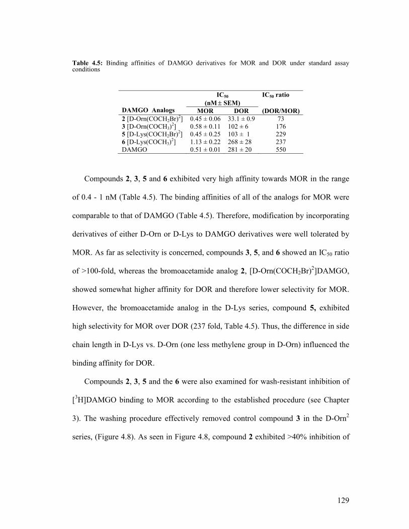
129
Table 4.5: Binding affinities of DAMGO derivatives for MOR and DOR under standard assay conditions
Compounds 2, 3, 5 and 6 exhibited very high affinity towards MOR in the range
of 0.4 - 1 nM (Table 4.5). The binding affinities of all of the analogs for MOR were
comparable to that of DAMGO (Table 4.5). Therefore, modification by incorporating
derivatives of either D-Orn or D-Lys to DAMGO derivatives were well tolerated by
MOR. As far as selectivity is concerned, compounds 3, 5, and 6 showed an IC50 ratio
of >100-fold, whereas the bromoacetamide analog 2, [D-Orn(COCH2Br)2]DAMGO,
showed somewhat higher affinity for DOR and therefore lower selectivity for MOR.
However, the bromoacetamide analog in the D-Lys series, compound 5, exhibited
high selectivity for MOR over DOR (237 fold, Table 4.5). Thus, the difference in side
chain length in D-Lys vs. D-Orn (one less methylene group in D-Orn) influenced the
binding affinity for DOR.
Compounds 2, 3, 5 and the 6 were also examined for wash-resistant inhibition of
[3H]DAMGO binding to MOR according to the established procedure (see Chapter
3). The washing procedure effectively removed control compound 3 in the D-Orn2
series, (Figure 4.8). As seen in Figure 4.8, compound 2 exhibited >40% inhibition of
IC50
(nM ± SEM) IC50 ratio
DAMGO Analogs MOR DOR (DOR/MOR) 2 [D-Orn(COCH2Br)2] 0.45 ± 0.06 33.1 ± 0.9 73 3 [D-Orn(COCH3)2] 0.58 ± 0.11 102 ± 6 176 5 [D-Lys(COCH2Br)2] 0.45 ± 0.25 103 ± 1 229 6 [D-Lys(COCH3)2] 1.13 ± 0.22 268 ± 28 237 DAMGO 0.51 ± 0.01 281 ± 20 550

130
[3H]DAMGO binding to MOR compared to untreated control membranes. This
suggests that compound 2 may interact with MOR in a non-equilibrium manner, and
0
20
40
60
80
100
120
Control 3 (0.58 nM) 2 (0.45 nM)
Compound Name
Perc
enra
ge [3
H]D
AMG
O B
ound
Figure 4.8. Wash-resistant inhibition of binding of [D-Orn2]DAMGO analogs 2 and 3. The membranes were incubated with the peptides at the concentrations indicated in parentheses.
thus be an affinity label for MOR. However, the corresponding analog 5 in the D-Lys
series [D-Lys(COCH2Br)2]DAMGO, 5, was removed by washing (Figure 4.9).
Therefore, 5 may not appear to be affinity label for MOR. It is important to note that
the small difference in the side chain of the amino acid at the 2nd position (D-Orn in
the case of 2 and D-Lys in the case of 5) could have a significant effect on covalent
binding to opioid receptors. The discovery of this new DAMGO-based electrophilic
affinity label 2 demonstrated a successful strategy for developing peptide-based
affinity labels through fine tuning the position of the affinity label to facilitate
alkylation at the binding site in MOR.

131
0
20
40
60
80
100
120
Control 6 (0.93 nM) 5 (0.45 nM)
Compound Name
Perc
enta
ge [3
H]D
AM
GO
Bou
nd
Figure 4.9. Wash-resistant inhibition of binding by [D-Lys2]DAMGO analogs: 5 and 6
4.3.4: Overcoming the Side Reaction: Design and Synthesis of DAMGA ([D-
Ala2,N-MePhe4,Gly5]enkephalinamide) Analogs.
Since the DAMGO-based isothiocyanate analogs [D-Orn(=C=S)2]DAMGO, 2,
and [D-Lys(=C=S)2]DAMGO, 4, resulted in the formation of cyclic O-alkyl
thiocarbamate derivatives, these isothiocyanate analogs was not tested for binding to
MOR. As discussed earlier, characterization of the linear peptide and cyclic side
product revealed that the C-terminal glyol functionality participated in the formation
of the cyclic product. Therefore, in order to overcome the side reaction, we modified
the C-terminal glyol functionality.
The C-terminal glyol functionality was replaced by the glycyl amide functionality
which can not participate in the cyclization. The bromoacetamide and the reversible
acetylated control compounds for both [D-Orn2]- and[ D-Lys2]DAMGA were also

132
prepared, as indicated in Figure 4.10, to compare their affinities for MOR with the
previously prepared DAMGO analogs.
H2NHN
NH
NNH
HO
O (CH2)nNHX
O
O
OOH
n = 3 (D-Orn)= 4 (D-Lys)
X = =C=S= -COCH2Br= -COCH3
H2NHN
NH
NNH
HO
O (CH2)nNHX
O
O
ONH2
O
DAMGO Analogs
DAMGA Analogs
Figure 4.10. Modifications incorporated in the DAMGA analogs

133
derivatives of DAMGO was successfully overcome by replacing the glyol with a
glycylamide. The isothiocyanate analogs 7 and 10 (Table 4.6) were successfully
synthesized and purified without any formation of side product.
Fmoc-NH-PAL-PEG-PS
1.Deblock (Piperidine/DMF 1:4)2.Fmoc-AA-OH/PyBOP/HOBt/DIEA 1:1:1:2
Boc-Tyr-D-AAa-Gly-MePhe-Gly-NH-PAL-PEG-PS
Alloc
tBu Pd(PPh3)4 (0.1 equiv)PhSiH3 (24 equiv) in DCM , 30 mins twice
n
N
NN
N
S
, DMF1.
2. Cleavageb
BrOH
O, DIC, DMF1.
2. Cleavageb
1. (CH3CO)2O2. Cleavageb
Tyr-D-AA(=C=S)-Gly-MePhe-Gly-NH2 Tyr- D-AA(COCH2Br)-Gly-MePhe-GlyNH2
aD-AA: D-Orn or D-LysbCleavage: 95% TFA, 5% Water
Boc-Tyr-D-AAa-Gly-MePhe-Gly-NH-PAL-PEG-PS
tBu
Tyr-D-AA(COCH3)-Gly-MePhe-GlyNH2
Scheme 4.2. Synthesis of DAMGA analogs
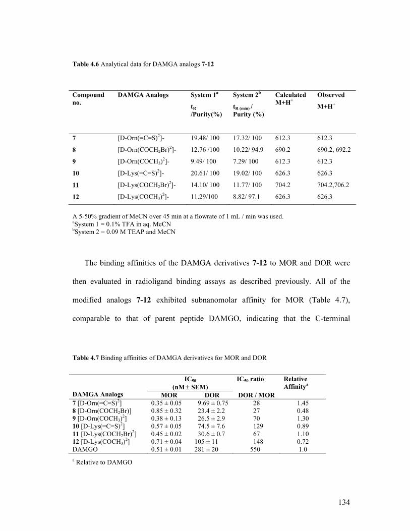
134
Table 4.6 Analytical data for DAMGA analogs 7-12
A 5-50% gradient of MeCN over 45 min at a flowrate of 1 mL / min was used. aSystem 1 = 0.1% TFA in aq. MeCN bSystem 2 = 0.09 M TEAP and MeCN
The binding affinities of the DAMGA derivatives 7-12 to MOR and DOR were
then evaluated in radioligand binding assays as described previously. All of the
modified analogs 7-12 exhibited subnanomolar affinity for MOR (Table 4.7),
comparable to that of parent peptide DAMGO, indicating that the C-terminal
Table 4.7 Binding affinities of DAMGA derivatives for MOR and DOR
a Relative to DAMGO
Compound no.
DAMGA Analogs System 1a
tR /Purity(%)
System 2b
tR (min) / Purity (%)
Calculated M+H+
Observed
M+H+
7 [D-Orn(=C=S)2]- 19.48/ 100 17.32/ 100 612.3 612.3
8 [D-Orn(COCH2Br)2]- 12.76 /100 10.22/ 94.9 690.2 690.2, 692.2
9 [D-Orn(COCH3)2]- 9.49/ 100 7.29/ 100 612.3 612.3
10 [D-Lys(=C=S)2]- 20.61/ 100 19.02/ 100 626.3 626.3
11 [D-Lys(COCH2Br)2]- 14.10/ 100 11.77/ 100 704.2 704.2,706.2
12 [D-Lys(COCH3)2]- 11.29/100 8.82/ 97.1 626.3 626.3
IC50
(nM ± SEM) IC50 ratio
DAMGA Analogs MOR DOR DOR / MOR
Relative Affinitya
7 [D-Orn(=C=S)2] 0.35 ± 0.05 9.69 ± 0.75 28 1.45 8 [D-Orn(COCH2Br)] 0.85 ± 0.32 23.4 ± 2.2 27 0.48 9 [D-Orn(COCH3)2] 0.38 ± 0.13 26.5 ± 2.9 70 1.30 10 [D-Lys(=C=S)2] 0.57 ± 0.05 74.5 ± 7.6 129 0.89 11 [D-Lys(COCH2Br)2] 0.45 ± 0.02 30.6 ± 0.7 67 1.10 12 [D-Lys(COCH3)2] 0.71 ± 0.04 105 ± 11 148 0.72 DAMGO 0.51 ± 0.01 281 ± 20 550 1.0

135
replacement by the alcohol to amide was well tolerated by MOR. However, most of
the DAMGA analogs (7-9 and 11) showed considerable affinity for DOR as well, in
the range of 10-30 nM (Table 4.7). All of the analogs show significantly higher
affinity for DOR when compared to DAMGO (IC50 = 281 nM). Therefore, the C-
terminal glyol functionality plays an important role in conferring selectivity for MOR
by decreasing affinity for DOR. The [D-Lys2]DAMGA series of analogs, however,
showed greater selectivity for MOR compared to the [D-Orn2]DAMGA series of
analogs. The loss in selectivity for MOR resulting from the replacement of glyol with
the glycylamide functionality appears to be counterbalanced to some extent by the
extra methylene unit in the side chain of D-Lys in the case of 10 (IC50 for DOR = 74
nM) and 12 (IC50 for DOR = 105 nM) but not 11.
0
20
40
60
80
100
120
Control 12 (0.62 nM) 11 (0.46 nM) 10 (0.47 nM)
Compound Name
Perc
enta
ge [3
H] D
AM
GO
Bou
nd
Figure 4.11. Wash-resistant inhibition of binding by [D-Lys2]DAMGA analogs 10 and 11
The DAMGA analogs were also evaluated for wash-resistant inhibition of binding
to MOR according to the established protocol described previously. Among the [D-

136
Lys2]DAMGA analogs, as seen in Figure 4.11, the bromoacetamide analog 11 ([D-
Lys(COCH2Br)2]DAMGA) exhibited 44% wash-resistant inhibition of [3H]DAMGO
binding compared to untreated control membranes. This suggests that this compound
could be binding irreversibly to MOR and thus be an affinity label. The reversible
compound, 12, did not exhibit any wash-resistant inhibition of binding, confirming
the efficiency of the washing procedures in these experiments. The isothiocyanate
analog, 10, however, was removed from the membrane by the washing procedure
(>90% [3H]DAMGO binding in presence of 12 at its IC50 concentration, Figure 4.11).
Although 11 appears to exhibit irreversible binding to MOR at its IC50 value (0.46
nM), its lower selectivity for MOR over DOR (DOR / MOR = 67, Table 4.7) could be
an impediment in utilizing 11 as an affinity label for MOR at higher concentrations.
Wash-resistant inhibition of [3H]DPDPE binding of 11 to DOR needs to be carried
out to determine whether 11 may interact irreversibly with DOR. For the [D-
Orn2]DAMGA analogs, in initial wash-resistant inhibition of binding experiments the
reversible control compound 9 was not removed by the washing procedure, and
therefore the washing procedure may need to be modified and these experiments will
need to be repeated.
4.4 Conclusions
A new series of affinity label analogs were successfully prepared using Fmoc-
based solid phase synthesis procedure by replacing the D-Ala2 of DAMGO with
either D-Orn2 or D-Lys2 and attaching either a bromoacetamide (–COCH2Br) or an
isothiocyanate (=C=S) as an electrophillic functionality to the side chain amine of

137
either D-Orn2 or D-Lys2. During the purification of both [D-Orn(=C=S)]2DAMGO, 1,
and [D-Lys(=C=S)2]DAMGO, 4, the formation of an intramolecular cyclic O-alkyl
thiocarbamate side product occurred. The side reaction was verified by
characterization of the products obtained from the attempted synthesis of 4 by HPLC,
MS, FT-IR and NMR. The data obtained from the spectroscopic analysis of the side
product supports an intramolecular attack of the C-terminal glyol on the
isothiocyanate in [D-Orn(=C=S)2]DAMGO or [D-Lys(=C=S)2]DAMGO with the
formation of the cyclic O-alkyl thiocarbamates. The bromoacetamide derivatives (2
and 5) as well as the reversible controls (3 and 6) were successfully synthesized,
purified and evaluated for their binding affinities for MOR. The bromoacetamide
analogs 2 and 5 exhibited subnanomolar binding affinities (IC50 = 0.45 nM) and
selectivity for MOR over DOR (IC50 ratio 75-230). Interestingly only 2 showed wash-
resistant inhibition of binding (>40% at its IC50 concentration) of [3H]DAMGO to
MOR while 5 failed to inhibit binding to MOR in a wash-resistant manner, suggesting
that only 2 binds irreversibly to MOR.
The formation of the cyclic O-alkyl thiocarbamate side product from the
isothiocyanate derivatives was successfully overcome by replacing the C-terminal
glyol functionality of DAMGO with a glycyl amide. The corresponding DAMGA
series of affinity labels were successfully synthesized and examined for receptor
affinity as well as wash-resistant inhibition of binding. The bromoacetamide
derivative, 11 bound to MOR in a wash resistant manner. However the isothiocyanate

138
analog, 12, failed to demonstrate wash-resistant inhibition of binding at its IC50
concentration.
Although all of the DAMGA analogs exhibited subnanomolar binding affinity
(IC50 = 0.3-0.8 nM) to MOR in initial radioligand binding experiments, most of the
DAMGA analogs (7-9 and 11) also had nanomolar affinity for DOR (IC50 = 10-30
nM). Therefore, the C-terminal glyol functionality plays an important role in
imparting selectivity for MOR over DOR, and replacing it with a glycylamide
resulted in significant decreases in MOR selectivity.
4.5 Experimental
The DAMGO-based affinity label analogs were prepared using solid phase
peptide synthesis methodology described in the previous chapter (Chapter 3, section
3.7.1). The differences between the two synthetic schemes were in the choice of resin
and loading of Fmoc-glyol onto the DHP-HM resin.
Materials
The DHP-HM resin (3,4-dihydro-2H-pyran-2ylmethoxymethyl polystyrene, 100-
200 mesh) was obtained from Novabiochem (San Diego, CA). Fmoc-Gly-ol was
obtained from AnaSpec Inc (Fermont, CA). Pyridinium p-toluenesulfonate (PPTS)
was obtained from Sigma-Aldrich (Saint Louis, MO). The Fmoc-protected amino
acids, the remaining chemicals and the HPLC grade solvents were obtained from the
sources listed in Chapter 3 (section 3.7.1).

139
Synthesis of DAMGO-based analogs
4.5.1 Loading of Fmoc-Glyol on to DHP-HM Resin and SSPS of DAMGO
Derivatives (Figure 4.4) 38
The first step in the synthesis of the DAMGO-based analogs (Figure 4.3) was
loading of Fmoc-Gly-ol onto the 3,4-dihydro-2H-pyran-2-ylmethoxymethyl
polystyrene (DHP-HM) resin (1.3 mmol/ g). The required amount of resin (670 mg)
was swollen in dichloroethane (DCE, 6 mL) for 1 h. Fmoc-Glyol (740 mg, 3 equiv)
and PPTS (327 mg, 1.5 equiv) were added to the swollen resin and the mixture stirred
at 80°C for 48 h. After 48 h the resin was drained, washed with DCM (4 x 1 min),
DMF (4 x 1 min) and DCM (3 x1 min) and dried under vacuum overnight.
The loading of the resin was determined by quantitative analysis of the Fmoc
group deprotection.40 The deprotection reaction rapidly generates dibenzovulvene,
which is scavenged by piperidine to afford an adduct that absorbs at 302 nm. The
resin substitution was calculated according to equation 1
Substitution in mmol/ g = (A * V * 103)/ 7800 * W (1)
where A= Absorbance
V= Volume (10 mL)
W = Weight of the sample resin.
For the Fmoc deprotection and quantitation reaction, three different samples were
analyzed. These were Fmoc-PAL-PEG-PS resin (3-4 mg) of known substitution (0.38
mmol/g), a blank and three samples of DHP-HM resin (3-4 mg each) following

140
loading of Fmoc-Glyol as described above. In a 10 mL volumetric flask DCM (0.4
mL) and piperidine (0.4 mL) were added to the resin and the mixture was kept for 30
min at room temperature. Then, methanol (1.6 mL) was added to each sample and the
solutions diluted to 10 mL with DCM. The resulting samples were then filtered and
the absorbance of each sample recorded at 302 nm.
The substitution of DHP-HM resin was determined to be 0.87 mmol/ g following
this protocol; based on the theoretical substitution (0.95 mmol/ g), and the percent
loading was 91%. Following determination of the resin loading, ~650 mg of this resin
was used to synthesize the analogs 1-6 (100 mg for each analog) according to the
solid phase peptide synthesis procedure described previously (Chapter 3, section
3.7.1).
4.5.2 Solid Phase Peptide Synthesis of the DAMGA Derivatives
The synthesis of the DAMGA derivatives (7-12) started with 500 mg of Fmoc-
PAL-PEG-PS (0.38 mmol/g) resin. The Fmoc solid phase peptide synthesis procedure
described earlier (Chapter 3, section 3.7.1) was used to synthesize all of the analogs.
4.5.3 Cleavage from Resin The affinity label peptides (1, 2, 4, 5, 7, 8, 10, and 11) were cleaved from
resin using 95% TFA and 5% water, as described previously in Chapter 3.7.2, and the
peptides lyophilized to give the crude products. Owing to their lower hydrophobicity,
the reversible controls (3, 6, 9, and 12) were diluted with 3-4 mL of 10% aqueous
acetic acid and then back extracted with ether (2 x 2 mL). The aqueous layers were
collected and lyophilized to give the crude peptides.
141
4.5.4 Purification and Analysis
The crude peptides were purified by reversed phase preparative HPLC as
described in Chapter 3 (section 3.7.3) using a linear gradient of 5-50% aqueous
MeCN containing 0.1% TFA over 45 min at a flow rate of 20 mL/min. After
purification of the affinity label analogs (1, 2, 4, 5, 7, 8, 10 and 11) the fractions were
collected and immediately lyophilized to avoid potential degradation of the affinity
label analogues by water. The fractions were then analyzed by analytical HPLC and
ESI-mass spectrometry according to the protocol described in Chapter 3 (section
3.7.3). For 3, 6, 9 and 12 the pure fractions were then combined and lyophilized to get
the pure peptide. For 2, 5, 7, 8, 10 and 11 the pure lyophilized fractions were
combined and re-lyophilized to get the pure peptides. The purity of the final peptides
were verified in analytical HPLC using two different systems: 0.1% TFA in aq.
MeCN and 0.09 M TEAP and MeCN
4.5.5 Instrumentation
The FTIR spectra of KBr pellets were collected over a range of 500 to 4000 cm-1
on a Shimadzu FTIR-8400S spectrophotometer. Proton NMR spectra were recorded
on a Bruker 500 MHz spectrophotometer in d6 –DMSO.
4.5.6 Pharmacological Assays:
The radioligand binding and wash-resistant inhibition of binding of all of the
analogs (2, 3, 5, 6 and 7-12) were carried out with Chinese hamster ovary (CHO)
cells expressing MOR and DOR according to procedures described earlier (Chapter 3,

142
section 3.7.4). [3H]DAMGO and [3H]DPDPE were used as the radioligands for MOR
and DOR, respectively.
4.6 Bibliography
1. Aldrich, J. V.; Vigil-Cruz, S. C. Narcotic analgesics. In Burger's Medicinal
Chemistry and Drug Discovery, Abraham, D. J., Ed. John Wiley and Sons: New
York, 2003; Vol. 6, pp 329-481.
2. Kieffer, B. L. Opioids: first lessons from knockout mice. Trends. Pharmacol. Sci.
1999, 20, 19-26.
3. Simon, E. J.; Hiller, J. M.; Edelman, I. Stereospecific binding of the potent
narcotic analgesic (3H) Etorphine to rat-brain homogenate. Proc. Natl. Acad. Sci.
U. S. A. 1973, 70, 1947-1949.
4. Lord, J. A.; Waterfield, A. A.; Hughes, J.; Kosterlitz, H. W. Endogenous opioid
peptides: multiple agonists and receptors. Nature 1977, 267, 495-499.
5. Eipper, B. A.; Mains, R. E. Further analysis of post-translational processing of
beta-endorphin in rat intermediate pituitary. J. Biol. Chem. 1981, 256, 5689-5695.
6. Stern, A. S.; Lewis, R. V.; Kimura, S.; Rossier, J.; Gerber, L. D.; Brink, L.; Stein,
S.; Udenfriend, S. Isolation of the opioid heptapeptide Met-enkephalin-
[Arg6,Phe7] from bovine adrenal medullary granules and striatum. Proc. Natl.
Acad. Sci. U. S. A. 1979, 76, 6680-6683.
7. Zadina, J. E.; Hackler, L.; Ge, L. J.; Kastin, A. J. A potent and selective
endogenous agonist for the mu-opiate receptor. Nature 1997, 386, 499-502.

143
8. Zadina, J. E.; Martin-Schild, S.; Gerall, A. A.; Kastin, A. J.; Hackler, L.; Ge, L. J.;
Zhang, X. Endomorphins: novel endogenous mu-opiate receptor agonists in
regions of high mu-opiate receptor density. Ann. N. Y. Acad. Sci. 1999, 897, 136-
144.
9. Law, P. Y.; Wong, Y. H.; Loh, H. H. Mutational analysis of the structure and
function of opioid receptors. Biopolymers 1999, 51, 440-455.
10. Rasmussen, S. G.; Choi, H. J.; Rosenbaum, D. M.; Kobilka, T. S.; Thian, F. S.;
Edwards, P. C.; Burghammer, M.; Ratnala, V. R.; Sanishvili, R.; Fischetti, R. F.;
Schertler, G. F.; Weis, W. I.; Kobilka, B. K. Crystal structure of the human beta2
adrenergic G-protein-coupled receptor. Nature 2007, 450, 383-387.
11. Cherezov, V.; Rosenbaum, D. M.; Hanson, M. A.; Rasmussen, S. G.; Thian, F. S.;
Kobilka, T. S.; Choi, H. J.; Kuhn, P.; Weis, W. I.; Kobilka, B. K.; Stevens, R. C.
High-resolution crystal structure of an engineered human beta2-adrenergic G
protein-coupled receptor. Science 2007, 318, 1258-1265.
12. Palczewski, K.; Kumasaka, T.; Hori, T.; Behnke, C. A.; Motoshima, H.; Fox, B.
A.; Le Trong, I.; Teller, D. C.; Okada, T.; Stenkamp, R. E.; Yamamoto, M.;
Miyano, M. Crystal structure of rhodopsin: A G protein-coupled receptor. Science
2000, 289, 739-745.
13. Scheerer, P.; Park, J. H.; Hildebrand, P. W.; Kim, Y. J.; Krauss, N.; Choe, H. W.;
Hofmann, K. P.; Ernst, O. P. Crystal structure of opsin in its G-protein-interacting
conformation. Nature 2008, 455, 497-502.

144
14. Eswar, N.; John, B.; Mirkovic, N.; Fiser, A.; Ilyin, V. A.; Pieper, U.; Stuart, A.
C.; Marti-Renom, M. A.; Madhusudhan, M. S.; Yerkovich, B.; Sali, A. Tools for
comparative protein structure modeling and analysis. Nucleic Acids Res. 2003, 31,
3375-3380.
15. John, B.; Sali, A. Comparative protein structure modeling by iterative alignment,
model building and model assessment. Nucleic Acids Res. 2003, 31, 3982-3992.
16. Shacham, S.; Topf, M.; Avisar, N.; Glaser, F.; Marantz, Y.; Bar-Haim, S.;
Noiman, S.; Naor, Z.; Becker, O. M. Modeling the 3D structure of GPCRs from
sequence. Med. Res. Rev. 2001, 21, 472-483.
17. Chen, Y.; Mestek, A.; Liu, J.; Hurley, J. A.; Yu, L. Molecular cloning and
functional expression of a mu-opioid receptor from rat brain. Mol. Pharmacol.
1993, 44, 8-12.
18. Fukuda, K.; Kato, S.; Mori, K.; Nishi, M.; Takeshima, H. Primary structures and
expression from cDNAs of rat opioid receptor delta- and mu-subtypes. FEBS Lett.
1993, 327, 311-314.
19. Takemori, A. E.; Portoghese, P. S. Affinity labels for opioid receptors. Annu. Rev.
Pharmacol. Toxicol. 1985, 25, 193-223.
20. Portoghese, P. S.; Larson, D. L.; Sayre, L. M.; Fries, D. S.; Takemori, A. E. A
novel opioid receptor site directed alkylating agent with irreversible narcotic
antagonistic and reversible agonistic activities. J. Med. Chem. 1980, 23, 233-234.

145
21. Chen, C.; Xue, J. C.; Zhu, J.; Chen, Y. W.; Kunapuli, S.; Kim de Riel, J.; Yu, L.;
Liu-Chen, L. Y. Characterization of irreversible binding of beta-funaltrexamine to
the cloned rat mu opioid receptor. J. Biol. Chem. 1995, 270, 17866-17870.
22. Chen, C.; Yin, J.; Riel, J. K.; DesJarlais, R. L.; Raveglia, L. F.; Zhu, J.; Liu-Chen,
L. Y. Determination of the amino acid residue involved in [3H]beta-
funaltrexamine covalent binding in the cloned rat mu-opioid receptor. J. Biol.
Chem. 1996, 271, 21422-21429.
23. Zhang, Y.; McCurdy, C. R.; Metzger, T. G.; Portoghese, P. S. Specific cross-
linking of Lys233 and Cys235 in the mu opioid receptor by a reporter affinity label.
Biochemistry 2005, 44, 2271-2275.
24. Garbay-Jaureguiberry, C.; Robichon, A.; Dauge, V.; Rossignol, P.; Roques, B. P.
Highly selective photoaffinity labeling of mu and delta opioid receptors. Proc.
Natl. Acad. Sci. U. S. A. 1984, 81, 7718-7722.
25. Herblin, W. F.; Kauer, J. C.; Tam, S. W. Photoinactivation of the mu opioid
receptor using a novel synthetic morphiceptin analog. Eur. J. Pharmacol. 1987,
139, 273-279.
26. Glasel, J. A.; Venn, R. F. The sensitivity of opiate receptors and ligands to short
wavelength ultraviolet light. Life Sci. 1981, 29, 221-228.
27. Bowen, W. D.; Hellewell, S. B.; Kelemen, M.; Huey, R.; Stewart, D. Affinity
labeling of delta-opiate receptors using [D-Ala2,Leu5,Cys6]enkephalin. Covalent
attachment via thiol-disulfide exchange. J. Biol. Chem. 1987, 262, 13434-13439.

146
28. Szucs, M.; Belcheva, M.; Simon, J.; Benyhe, S.; Toth, G.; Hepp, J.; Wollemann,
M.; Medzihradszky, K. Covalent labeling of opioid receptors with 3H-D-Ala2-
Leu5-enkephalin chloromethyl ketone. I. Binding characteristics in rat brain
membranes. Life Sci. 1987, 41, 177-184.
29. Maeda, D. Y.; Berman, F.; Murray, T. F.; Aldrich, J. V. Synthesis and evaluation
of isothiocyanate-containing derivatives of the delta-opioid receptor antagonist
Tyr-Tic-Phe-Phe (TIPP) as potential affinity labels for delta-opioid receptors. J.
Med. Chem. 2000, 43, 5044-5049.
30. Kumar, V.; Murray, T. F.; Aldrich, J. V. Solid phase synthesis and evaluation of
Tyr-Tic-Phe-Phe(p-NHCOCH2Br) ([Phe(p-bromoacetamide)4]TIPP), a potent
affinity label for delta opioid receptors. J. Med. Chem. 2002, 45, 3820-3823.
31. Aldrich, J. V.; Kumar, V.; Dattachowdhury, B.; Peck, A. M.; Wang, X.; Murray,
T. F. Solid phase synthesis and application of labeled peptide derivatives: probes
of receptor-opioid peptide interactions. Int. J. Pept. Res. Ther. 2008, 14, 315-321.
32. Benyhe, S.; Hepp, J.; Simon, J.; Borsodi, A.; Medzihradszky, K.; Wollemann, M.
Tyr-D-Ala-Gly-(Me)Phe-chloromethyl ketone: a mu specific affinity label for the
opioid receptor. Neuropeptides 1987, 9, 225-235.
33. Shimohigashi, Y.; Takada, K.; Sakamoto, H.; Matsumoto, H.; Yasunaga, T.;
Kondo, M.; Ohno, M. Discriminative affinity labelling of opioid receptors by
enkephalin and morphiceptin analogues containing 3-nitro-2-pyridinesulphenyl-
activated thiol residues. J. Chromatogr. 1992, 597, 425-428.

147
34. Handa, B. K.; Land, A. C.; Lord, J. A.; Morgan, B. A.; Rance, M. J.; Smith, C. F.
Analogues of beta-LPH61-64 possessing selective agonist activity at mu-opiate
receptors. Eur. J. Pharmacol. 1981, 70, 531-540.
35. Pert, C. B.; Pert, A.; Chang, J. K.; Fong, B. T. (D-Ala2)-Met-enkephalinamide: a
potent, long-lasting synthetic pentapeptide analgesic. Science 1976, 194, 330-332.
36. Wang, X. Doctoral Thesis. University of Kansas: 2007.
37. Leelasvatanakij, L.; Aldrich, J. V. A solid-phase synthetic strategy for the
preparation of peptide-based affinity labels: synthesis of dynorphin A analogs. J.
Pept. Res. 2000, 56, 80-87.
38. Thompson, L. A.; Ellman, J. A. Straightforward and general method for coupling
alcohols to solid supports. . Tetrahedron Lett. 1994, 35, 9333-9336.
39. Dattachowdhury, B.; Murray, T. F.; Aldrich, J. V. The synthesis of DAMGO-
based potential affinity labels with high mu opioid receptor affinity and the
formation of cyclic O-alkyl thiocarbamates. Adv. Exp. Med. Biol. 2009, 611, 265-
266.
40. Chan, W. C.; White, P. D. Fmoc Solid Phase Peptide Synthesis - A Practical
Approach. Hames, B. D., Ed. Oxford University Press: New York, 2004; pp 29-
30.

148
Chapter 5.
Design, Synthesis and Evaluation of a Dermorphin-Based
Multifunctional Affinity Label Probe for Mu Opioid Receptors
* Note that each chapter has compound numbers.

149
5.1 Introduction
The cloning of the opioid receptors in the early 1990s1-5 led to significant
advancements in understanding opioid receptor structures and receptor-ligand
interactions at the molecular level.
Affinity labels, ligands that bind to their target receptors in an irreversible
manner,6 have been useful biochemical tools to study opioid receptors. There have
been reports in the literature of a large number of affinity labels, mostly nonpeptide
ligands, for characterizing different opioid receptor types.6, 7 Nonpeptide affinity
labels for opioid receptors have been predominantly electrophilic affinity labels. β-
Chlornaltrexamine (β-CNA),8 labels all three opioid receptors, and β-funaltrexamine
(β-FNA) alkylates µ opioid receptors (MOR).9
β-FNA, a MOR selective antagonist, was the first affinity label for opioid receptor
whose point of attachment to its receptor was successfully determined by Liu-Chen
and coworkers using molecular biology and protein isolation techniques. Initially,
based on the binding of [3H]β-FNA to MOR / κ opioid receptor (KOR) receptor
chimeras, a region of MOR spanning from the third intracellular loop to the C-
terminus was determined to be essential for irreversible binding.10 However, upon
isolation and partial purification of the labeled receptor, the point of attachment was
found to be in the extracellular loop 2 (EL)-transmembrane 5 (TM5) region.
Subsequently, site directed mutagenesis of amino acid residues in this region
identified Lys233, a conserved residues in all three opioid receptors, as the
attachment point.11 This illustrates the challenges and limitations of studying

150
receptor-ligand interactions and further underscores the importance of obtaining
direct experimental evidence pertaining to receptor-ligand interactions. The only
other affinity label whose point of attachment has been determined is the ‘reporter
affinity label’ naltrexamine naphthalene dialdehyde derivative NNA where the
attachment points to MOR were evaluated using site-directed mutagenesis by
Portoghese and coworkers.12 Lys233 and the adjacent Cys235 at the top of TM5 were
determined to be the residues involved in formation of a fluorescent isoindole with
NNA indicating covalent binding of the reporter affinity label. Mutation of either of
these residues resulted in the loss of fluorescence. Although this method allows the
determination of attachment points without isolating the labeled receptor, a critical
limitation to this approach is the requirement for two specific nucleophilic receptor
residues, Lys and Cys, in close proximity to one another to form the fluorescent
product.
Peptide-based affinity labels, on the other hand, have been predominantly
photoaffinity labels, e.g. the azide derivatives of several enkephalin analogs.6, 7 The
main disadvantage of using the azido group as a photoaffinity label for opioid
receptors is that the shorter UV wavelength (254 nm) generally used to generate the
reactive species can inactivate opioid receptors.13 Reports of peptide-based
electrophilic affinity labels in literature have been limited. Prior to isothiocyanate
derivatives of opioid peptides as electrophilic affinity labels designed in our group,14-
16 opioid peptide derivatives containing electrophilic affinity labels were limited to
chloromethylketone derivatives of the enkephalins,17, 18 the cysteine-containing

151
enkephalin analog of DALCE ([D-Ala2,Leu5Cys6]enkephalin Tyr-D-Ala-Gly-Phe-
Leu-CysOH)19 and enkephalin analogs containing melphanan (Mel),20 the nitrogen
mustard derivative of p-aminophenylalanine.
Since pain relief is mediated mainly through MOR, it is important to understand
the interactions between MOR ligands and their receptor. The endogenous ligands of
the opioid receptors are peptides, and studies of chimeric opioid receptors and site-
directed mutagenesis suggest that peptide ligands may interact differently with opioid
receptors than nonpeptide ligands.21 Therefore, structural information obtained from
interactions of peptide ligands with opioid receptors can be complimentary to that
obtained for nonpeptide ligands.
There have been very few reports of electrophilic peptide-based affinity labels
selective for MOR. The chloromethyl ketone of Tyr-D-Ala-Gly-(NMe)Phen (DAMK,
IC50 = 1-5 µM for dose-dependent irreversible inhibition of [3H]naloxone binding)22
and Tyr-D-Ala-Gly-Phe-Leu(CH2SNpys) (Npys = 3-nitro-2-pyridinesulphenyl, IC50 =
19 nM for dose-dependent irreversible inhibition of [3H]DAMGO binding)23 are the
only examples of peptide-based electrophilic affinity labels for MOR reported in the
literature. Previous attempts in our group to prepare affinity labels for MOR by
incorporating an electrophilic functionality such as a bromoacetamide or
isothiocyanate at the para position of either Phe3 or Phe4 in endomorphin-2 (Tyr-Pro-
Phe-PheNH2) were unsuccessful because the modified analogs exhibited large (40- to
80-fold) decreases in MOR binding affinity compared to the parent peptide
endomorphin-2.24 Earlier attempts were also made to identify electrophilic affinity

152
labels based on dermorphin, an endogenous heptapeptide (Tyr-D-Ala-Phe-Gly-Tyr-
Pro-SerNH2) derived from South American frog skin that is very potent and highly
selective for MOR.25 Previously, the para position of Phe3 or a Phe in position 5 of
dermorphin and [Lys7]dermorphin were modified to introduce an electrophilic
functionality, i.e. a bromoacetamide or isothiocyanate group.26 Modification in the
‘message’ domain (Phe3) resulted in a >1000-fold decrease in MOR affinity. While
modification of a Phe in position 5 in the ‘address’ domain of dermorphin and
[Lys7]dermorphin was well tolerated and the peptides retained nanomolar affinity for
MOR, none of these modified analogs exhibited wash-resistant inhibition of binding
to MOR, and therefore are not affinity labels for these receptors.26
In a continued effort to develop MOR selective affinity labels, we have recently
discovered dermorphin-based affinity label analogs that show exceptionally high
affinity (IC50 = 0.1-5 nM) for MOR. These analogs were designed by modifying
position 2 of dermorphin by incorporating D-Orn or D-Lys and attaching a
bromoacetamide or an isothiocyanate as the electrophilic functionality (see Chapter 3
for details), which is a new strategy for designing peptide-based affinity label
derivatives of opioid peptides that has not been previously reported. This resulted in a
substantial improvement in binding affinity (between 10- to 100-fold) compared to
the previous dermorphin-based analogs synthesized in our laboratory.26 Importantly,
three of these four affinity labels, [D-Orn(=C=S)2]-, [D-Orn(-COCH2Br)2]- and [D-
Lys(=C=S)2]dermophin appear to have higher affinity (approximately 3-20 fold) than
the well-studied nonpeptide MOR affinity label β-FNA (IC50 = 2.2 nM in standard

153
binding assays)10, 27 and exhibit wash-resistant inhibition of binding at very low (≤ 1
nM) concentrations equal to their IC50 values.28 To our knowledge, these peptides are the highest
affinity peptide-based affinity labels for MOR reported to date.
The long range goal of this project is to determine the attachment point of a
peptide-based affinity label to MOR. In this project, [D-Lys(=C=S)2]dermorphin, one of
the recently discovered dermorphin-based MOR affinity label (see Chapter 3), was
selected as the lead peptide for designing a multifunctional probe with the goal of
identifying the attachment point of this peptide to MOR. This ligand was selected due
to its high affinity (IC50 = 0.38 nM), selectivity (IC50 ratio DOR/MOR = 250) and its
ability to bind to MOR in a wash-resistant manner (see Chapter 3).
Peptides, due to their polymeric nature, provide definite advantages over
nonpeptides in isolation studies of affinity labeled receptors. For example, appropriate
residues in the peptide can be used to attach a purification tag such as biotin or d-
desthiobiotin (DSB). This may then be used to assist in receptor enrichment via
affinity purification (e.g., with streptavidin for biotinylated peptides). A biotinylated
derivative of β-endorphin has been used to purify MOR.29 Opioid receptors are
transmembrane in nature are expressed at very low concentration in different cell
lines.21 Although MOR has been expressed in Escherichia coli30 and insect cell
lines,31, 32 the quantities obtained from such expression were not enough to carry out
spectroscopic studies.33 Affinity purification via d-desthiobiotin-streptavdin
interaction would enrich the available receptors. The lower affinity of the biotin
precursor DSB for the biotin binding proteins,34 e.g., streptavidin, compared to biotin
154
offers a distinct advantage over biotin. It is very difficult to dissociate biotinylated
compounds from streptavidin due to the extremely high affinity of biotin for
streptavidin (Kd = 4 x 10-14 M),35 which further complicates the analysis of labeled
receptors. This problem can be overcome by using DSB. Although there is no
agreement in the literature on how much lower the affinity of DSB is compared to
biotin, it is believed to be at least several orders of magnitude.36-40
To aid in the detection of the labeled receptor in microscopy experiments, a
fluorescent group incorporated into our lead affinity label peptide [D-
Lys(=C=S)2]dermorphin would be very useful. Previously, fluorescent derivatives
(Alexa 488 or BIODIPY) attached to either the Lys side chain in [Lys7]dermorphin41
or the C-terminus of dermorphin42 have been reported to be well tolerated by MOR.
Hence, we expect attachment of a fluorescent label to our dermorphin-based affinity
label peptide will also be well tolerated. To minimize nonspecific interactions with
the labeled receptor a hydrophilic fluorescent group, i.e. 5-carboxyrhodamine B or
Oregon Green, was selected. These fluorophores are also insensitive to pH in the
physiological range43 which minimizes possible alterations of the spectrofluorometric
properties of the fluorophores with any change in pH (e.g. inside the cell) during
microscopy experiments.

155
5.2 Design of a Dermorphin-Based Multifunctional Affinity Label: Design Strategy To develop a multifunctional probe for MOR, the lead peptide [D-
Lys(=C=S)2]dermorphin was further modified to incorporate DSB as a purification
tag, Oregon Green or 5-carboxyrhodamine B as a fluorescent tag, and poly(ethylene
glycol) (PEG)-like linkers to decrease the hydrophobicity of the peptide (Figure 5.1).
Figure 5.2 shows the design of the dermorphin-based multifunctional affinity label
peptide for MOR.
Figure 5.1. The fluorescent and purification tags and the PEG-like linker for incorporation into multifunctional dermorphin derivatives

156
Figure 5.2. Design of dermorphin-based multifunctional affinity labels for MOR The lead peptide [D-Lys(=C=S)2]dermorphin was extended at the C-terminus by two
additional Lys residues. The Lys residues were separated from each other and the
peptide by hydrophilic (PEG)-like linkers. The Lys residues were used as handles to
incorporate the purification and fluorescent tags attached to the side chain amines of
these residues (Figure 5.2). The fluorescent group was attached to the Lys closest to
the C-terminus to prevent potential interference with receptor binding. A shorter β-
alanine linker was incorporated at the C-terminus for attachment to the resin.

157
5.3 Results and Discussion
5.3.1 Synthesis of the Multifunctional [D-Lys(=C=S)2]dermorphin Derivative
A solid phase synthetic methodology was developed to selectively incorporate
three different labels - a fluorescent label (Oregon Green or 5-carboxyrhodamine B),
a purification tag (DSB) and the affinity label (isothiocyanate group) into the peptide.
The choice of the protecting groups for the three different Lys side chain amines in
the peptide was critical to the success of this strategy. The side chain protecting
chosen for the synthesis of the multilabeled peptides were Mtt (4-methyltrityl), ivDde
(1-(4,4-dimethyl-2,6-dioxocyclohex-1-ylidene)-3-methylbutyl) and Aloc
(allyloxycarbonyl) for the Lys residues from the C- to the N- terminus. The protecting
groups for the other amino acids remained the same as before (see Chapter 3). The
structure of the fully protected peptide intermediate on the resin is shown in Figure
5.3.
Figure 5.3 Structure of the fully protected dermorphin intermediate.

158
Fmoc-PAL-PEG-PS
Fmoc solid phase peptide synthesis
Boc-Tyr-D-Lys-Phe-Gly-Tyr-Pro-Ser-L-Lys-L-Lys- -Ala-NH-PEG-PS
ivDdeAloc
1. 2% hydrazine, DMF2. DSB/PyBOP/HOBt/DIEA (3:3:3:6) in DMF3. 3% TFA, DCM4. Oregon Green
or 5-carboxyrhodamine B/ PyBOP/ HOBt/ DIEA (1:1:1:2)5. Pd(PPh3)4, PhSiH3 (Aloc deprotection)
tBu
1. Acetic anhydride, DMF2. 95% TFA, 5% H2O (cleavage) 1.Thiocarbonyldiimidazole, DMF
2. 95% TFA, 5% H2O (cleavage)
Tyr-D-Lys-Phe-Gly-Tyr-Pro-Ser-L-Lys-L-Lys- -Ala-NH2
NHCOCH3
Tyr-D-Lys-Phe-Gly-Tyr-Pro-Ser-L-Lys-L-Lys- -Ala-NH2
L= PEG-like linker
Reversible Control Affinity Label
NH
OO
O
O
(ivDde deprotection)
(Mtt deprotection)
tBu MtttBu
Boc-Tyr-D-Lys-Phe-Gly-Tyr-Pro-Ser-L-Lys-L-Lys- -Ala-NH-PEG-PS
tBu tBu tBu DSB
DSB N=C=S DSB
Mtt:
ivDde:OO
Aloc: O
O
RX = Oregon Green or
5-carboxyrhodamine B
R = 1 Oregon Green3 5-carboxyrhodamine B
R
R = 2 Oregon Green4 5-carboxyrhodamine B
R
1.Deblock (Piperidine/DMF 1:4)2.Fmoc-AA-OH/PyBOP/HOBt/DIEA 1:1:1:2
n
Scheme 5.1 Solid phase synthetic strategy for dermorphin-based multilfunctional peptides.
The standard Fmoc solid phase synthetic procedure (Scheme 5.1) was followed to
assemble the entire peptide chain on the PAL-PEG-PS resin with the side chain
protecting groups as shown in Figure 5.3. At this stage, ivDde was selectively
deprotected from the second Lys residue from the C-terminus by treating the peptide
on the resin with 2% hydrazine44 in the presence of allyl alcohol. Allyl alcohol was
included in the reaction to prevent reduction of the Aloc group.45 The free amine thus
generated was then reacted with DSB in the presence of PyBOP and HOBt to

159
incorporate the purification tag. Next the resin was treated with 3% TFA in the
presence of TIPS (triisopropylsilane) to remove the Mtt group from the Lys residue
closest to the C-terminus. The free amine thus obtained was then treated with the
fluorophore Oregon Green 488 5-carboxylic acid to incorporate the fluorescent tag.
Residual amine groups were acetylated afterwards using acetic anhydride.
Since the cost of commercially available single isomer fluorescent dyes is very
high ($130/ 5 mg for Oregon Green), only one equivalent of this reagent was used for
incorporation of Oregon Green. Alternatively, 5-carboxyrhodamine B was also used
as the fluorescent tag. Although the single isomers of carboxyrhodamine are also very
expensive ($160/10 mg), the sodium salt of a mixture of 5- and 6-carboxyrhodamine
B is quite inexpensive. It is commercially available as a 20% aqueous solution
($12.60/kg, Abbey Color, Philadelphia, PA) and is marketed as Rhodamine WT (for
water tracer).46 Acidic workup of Rhodamine WT with HCl precipitates a mixture of
the 5- and 6-carboxy isomers which are easily separated by preparative HPLC;46 the
resulting individual isomers were characterized by analytical HPLC, ESI-MS and
NMR. However, sample preparation for purification initially met with some
difficulty, as a mixture of the carboxyrhodamine B isomers was not readily soluble in
a mixture of MeCN/H2O. After trying different solvents, a 50:50 mixture of
isopropanol and H2O turned out to be the most suitable solvent; 1 mL of this mixture
was used to dissolve 40 mg of the sample. Thus, 80 mg of the mixture of
carboxyrhodamine B isomers could be purified at a time, resulting in ~20 mg of pure

160
Minutes0 5 10 15 20 25 30 35 40 45
mVo
lts
0
50
100
mVo
lts
0
50
100
Figure 5.4. Separation of 5- and 6-carboxy isomers of rhodamine B in 20-50% MeCN containing 0.1% TFA over 60 mins at a flow rate of 1 mL/min.
5-carboxyrhodamine B. 5- and 6-carboxy isomers. The 6-carboxy isomer elutes first
from the HPLC column, followed by the 5-carboxy isomer as verified by 1H NMR
spectroscopy. Similar to the coupling of Oregon Green to the multilabeled peptide, 1
equiv of 5-carboxyrhodamine B was coupled to the free amine side chain of Lys
obtained after deprotection of the Mtt group with 3% TFA. The reaction was run
overnight in the dark to prevent possible degradation of the fluorophore in the
presence of light. Residual amine groups were acetylated afterwards using acetic
anhydride. The rest of the synthesis was also carried out in the dark for the reason
described above. After the coupling of 5-carboxyrhodamine B the rest of the
synthesis (Aloc deprotection and incorporation of the affinity label) was carried out
following the established procedures47 (Scheme 5.1). The final cleavage of the crude
peptides was carried out in 95% TFA and 5% H2O; extraction with ether was avoided
due to their high hydrophobicities.
5-carboxyrhodamine B 6-carboxyrhodamine B

161
Minutes0 5 10 15 20 25 30 35 40 45 50
mVo
lts
0
25
50
75
mV
olts
0
25
50
75
Minutes0 5 10 15 20 25 30 35 40 45 50
mVo
lts
0
20
40
60
mV
olts
0
20
40
60
Figure 5.5. HPLC spectra of the crude multilabeled peptide. A. HPLC after coupling of 5-carboxyrhodamine and Aloc deprotection. B. Multilabeled peptide 3 after sequential coupling of 5-carboxyrhodamine, Aloc deprotection and incorporation of the isothiocyanate functionality. A 10-60% gradient of aqueous MeCN containing 0.1% TFA over 50 min at a flow rate of 1 mL/min was used for HPLC analysis.
Figure 5.5 shows the HPLC spectra of the crude multilabeled peptide and an
intermediate. The crude peptides were successfully purified by preparative HPLC; the
purity of all four peptides 1-4 (>97%) was determined by analytical HPLC in two
different solvent systems and molecular weights were verified by ESI-MS (Table
5.1).
280 nm A
280 nm B

162
Table 5.1 Analytical data for multilabeled dermorphin analogs 1-4
A 15-50% gradient of MeCN over 35 min at a flowrate of 1 mL/min was used. bSystem 2 = 0.09 M TEAP (triethylamine phosphate) and MeCN aSystem 1 = 0.1% TFA in aqueous. MeCN
5.3.2 Results from Preliminary Microscopy Experiments
To demonstrate possible binding of the multilabeled fluorescent peptide 1
containing Oregon Green as the fluorophore to MOR, initial microscopic experiments
were carried out utilizing SH-SY5Y cells, a human neuroblastoma cell line, stably
expressing MOR and DOR.48 Panel A in Figure 5.6 shows the fluorescence obtained
after incubating the SH-SY5Y cells with 400 nM of compound 1 for 90 min at 4° C
followed by extensive washing of the cells. In panel C, the cells were first incubated
with 400 nM of 1 at 4o C for 90 min followed by incubation with 10 µM of naloxone,
which is an antagonist with high affinity for MOR,7 for 30 min. The fluorescence due
to the peptide did not appear to decrease as shown in panel C (Figure 5.6, indicated
ESI-MS (m/z) Multilabeled Dermorphin Analogs
System 1a
tR /Purity(%)
System 2b
tR /Purity(%)
Calculated Observed
1 [D-Lys(=C=S)2]Oregon Green 21.16/ 100 17.78/ 100 [M+3H]3+ = 703.9 [M+2H]2+ = 1055.5
[M+3H]3+ = 703.9 [M+2H]2+ = 1055.5
2 [D-Lys(COCH3)2]Oregon Green 15.61/ 97.1 12.92/ 100 [M+3H]3+ = 703.9 [M+2H]2+ = 1055.5
[M+3H]3+ = 703.9 [M+2H]2+ = 1055.5
3 [D-Lys(=C=S)2]5-carboxyrhodamine 24.87/ 100.0
20.91/ 99.8 [M+3H]3+ = 729.0 [M+2H]2+ = 1093.1
[M+3H]3+ = 729.0 [M+2H]2+ = 1093.1
4 [D-Lys(COCH3)2]5-carboxyrhodamine 19.95/ 97.4 16.17/ 99.0 [M+3H]3+ = 729.0 [M+2H]2+ = 1093.0
[M+3H]3+ = 728.7 [M+2H]2+ = 1093.1

163
by arrows), indicating that naloxone could not displace the fluorescent peptide from
Figure 5.6. Labeling of SH-SY5Y cells by the [D-Lys(N=C=S)2dermorphin derivative 1 containing Oregon Green. A: Labeling of cells with the 400 nM of the fluorescent peptide 1 alone at 4o C for 90 min (indicated by arrows). B: Protection experiment in which cells were first incubated with naloxone (10 µM) for 30 min followed by labeling with 1 at 4o C for 90 min. C: Labeling of cells with the peptide (indicated by arrows) for 90 min, followed by incubation with naloxone. D: Cells not incubated with peptide or naloxone for 90 min. Cells were extensively washed following incubation. The fluorescence of Oregon Green was measured following excitation at 494 nm and emission at 531 nm.
C D
A B

164
MOR and suggesting that the peptide may bind irreversibly to MOR. To verify
whether the fluorescent peptide 1 and naloxone are interacting with the same
receptor, a protection experiment was carried out, where cells were first treated with
10 µM of naloxone for 30 min followed by a treatment with 400 nM of 1 for 90 min.
As can be seen in panel B of Figure 5.6 minimal fluorescence above background was
observed consistent with peptide 1 and naloxone interacting with the same receptor.
To detect any auto-fluorescence of the SH-SY5Y cells in the labeling experiments,
cells were treated with buffer only (panel D of Figure 5.6). Therefore the results
obtained from this preliminary microscopy experiment suggest labeling of MOR by
the dermorphin-based multifunctional peptide 1. The inability of naloxone to displace
the fluorescent peptide from MOR further suggests that the peptide binds irreversibly
to MOR, and thus demonstrates the utility of this approach.
5.4. Conclusions
A multifunctional affinity label peptide was designed by modifying [D-
Lys(=C=S)2]dermorphin which exhibits high affinity (IC50 = 0.38 nM), selectivity
and wash resistant inhibition of binding to MOR (Chapter 3). The design involved
incorporating additional C-terminal Lys residues as handles to attach a purification
tag and fluorescent tags to aid in receptor isolation and detection of the labeled
receptor, respectively. A solid-phase synthetic methodology utilizing selective
deprotection of different protecting groups on the side chain amines of Lys residues
permitted the incorporation of multiple labels (Oregon Green or 5-carboxyrhodamine
B and DSB) to yield the multilabeled dermorphin derivatives. Preliminary

165
microscopy experiments examining the interaction of the fluorescent peptide 1 with
MOR on SH-SY5Y cells suggests irreversible binding of the multifunctional affinity
label dermorphin derivative, and thus demonstrates the utility of this approach.
Radioligand binding assays of 1-4 will be carried out in the near future to determine
the affinity and confirm the wash-resistant inhibition of binding of the multilabeled
[D-Lys(=C=S)2]dermorphin derivatives. Additional microscopic experiments will
also be performed to verify the results of the initial studies, to evaluate the effects of
different ligands on fluorescent labeling and also to study receptor trafficking.
5.5 Experimental 5.5.1 Synthesis of Dermorphin-based Multifunctional Affinity Label
The dermorphin-based multifunctional affinity label derivatives were prepared by
solid phase peptide synthesis methodology described in Chapter 3. The methodology
for selective incorporation of different functional tags (purification and fluorescent
tags) is described below.
Materials
The Fmoc-mini-PEG linker (2-(2-(2-aminoethoxy)ethoxy)acetic acid) was purchased
from Peptides International (Louisville, KY). Fmoc-βAla-OH, Fmoc-Lys(Mtt)-OH
and Fmoc-Lys(ivDde)-OH were purchased from Novabiochem (San Diego, CA).
Oregon Green 488, Rhodamine WT and d-desthiobiotin were obtained from
Molecular Probes (Eugene, OR), Abbey Color (Philadelphia, PA) and Sigma-Aldrich

166
(St. Louis, MO), respectively. Hydrazine was obtained from Sigma-Aldrich (St.
Louis, MO). The remaining chemicals, Fmoc protected amino acids, Fmoc-PAL-
PEG-PS resin, and HPLC grade solvents were obtained from sources listed in Chapter
3.
Synthesis of dermorphin-based multifunctional affinity labels
In addition to the Fmoc-protected amino acids mentioned in Chapter 3, Fmoc-βAla-
OH, Fmoc-Lys(Mtt)-OH, Fmoc-mini-PEG-linker and Fmoc-Lys(ivDde)-OH were
used in the synthesis. The synthesis started with Fmoc-PAL-PEG-PS resin (300 mg,
0.19–0.21 mmol/g). Following Fmoc deprotection of the resin using piperidine and
DMF (1:4), Fmoc-βAla-OH, Fmoc-Lys(Mtt)-OH, Fmoc-mini-PEG-linker and Fmoc-
Lys(ivDde)-OH were sequentially coupled following the Fmoc solid phase peptide
synthesis procedures described in Chapter 3 (Scheme 5.1). The peptide chain was
then further elongated with Fmoc-mini-PEG-linker and the fully protected
dermorphin peptide sequence assembled on the resin as per the protocol in Chapter 3.
The linkers, Ser7, Pro6 and the N-terminal Tyr1 were coupled twice and required
overnight reaction for the second coupling. The completion of the couplings of amino
acids and linker were monitored by the ninhydrin test for coupling to a primary amine
and by the chloranil test if coupled to a secondary amine.
After assembly of the entire peptide chain on the resin, the ivDde protecting group
from the C-terminal Lys was selectively removed by treatment with 2% hydrazine in
DMF twice for 10 min. The resin was then washed with DCM/DMF (1:1, 10 x 1
min). DSB was then coupled to the resulting primary amine with PyBOP, and HOBt

167
as the coupling agents and DIEA as base. DSB (3 equiv) was first dissolved in a
minimum amount of DMSO: DMF (1:1). PyBOP (3 equiv) and HOBt (3 equiv) were
dissolved in a a minimum amount of DMSO: DMF (1:1) separately and the solution
then added to the DSB followed by DIEA (6 equiv). The mixture was then added to
the resin and the reaction run overnight. The completion of the coupling was verified
by a negative ninhydrin test. Next the Mtt group from the second Lys from the C-
terminus was selectively deprotected by treatment with 3% TFA and 1% TIPS in
DCM for 30 mins. The resin was then washed with DCM/DMF (1:1, 10 x 1 min).
The resultant free amine was reacted with Oregon Green 488, PyBOP, HOBt and
DIEA (1:1:1:2) overnight in the dark. Residual amine groups were acetylated
afterwards using acetic anhydride. Cleavage of an aliquot (~15 mg) and analysis by
HPLC and ESI-MS were carried out to verify the product. At this stage, the Aloc
group of the side chain of D-Lys2 was deprotected with a catalytic amount of
Pd(PPh3)4 plus phenyl silane according to the procedure described previously.47, 49
The resin containing the free amine on the side chain of D-Lys2 was then divided into
two parts (100 mg each). One part was treated with a large excess of acetic anhydride
(20 equiv) in DMF to obtain the acetylated control compound. The second part was
reacted with thiocarbonyl diimidazole (4 equiv) in DMF to yield the desired
isothiocyanate affinity label as per the protocol described earlier in Chapter 3.
To synthesize the dermorphin-based multifuctional affinity label containing 5-
carboxyrhodamine B, the 5-isomer was isolated from the mixture of the two isomers
(5- and 6-carboxyrhodamine B) in Rhodamine WT (see section 5.5.4 below). The

168
peptide was synthesized following the same procedure described above. Aliquot
analysis by HPLC (Figure 5.5) and ESI-MS was carried out after reaction of 5-
carboxyrhodamine B as described above to verify the product. Aloc deprotection and
subsequent synthesis of the reversible control and affinity label were carried out
following the protocol described above.
It is important to note that all synthetic steps following incorporation of the
fluorescent functionalities Oregon Green or 5-carboxyrhodamine B were carried out
in the dark to prevent possible degradation of the fluorophore.
5.5.2 Cleavage from Resin
The peptides were cleaved from the resin with 95% TFA and 5% H2O for 2 h in
the dark. Due to the considerable hydrophobicties of these peptides ether extraction
was not carried out. Instead, following filtration of the resin excess TFA was removed
by evaporation. The resulting peptide was diluted 10 fold with 10% aqueous acetic
acid and lyophilized to get the crude products.
5.5.3 Purification and Analysis
The crude peptides (1-4, 30-40 mg each) were purified by reversed phase
preparative HPLC as described in chapter 3 using a linear gradient of 15-70%
aqueous MeCN containing 0.1% TFA over 55 min at a flow rate of 20 mL/min. The
samples were prepared by dissolving the crude peptides in 1:9 MeCN: H2O. After
purification of the affinity label analogs (1 and 3) the fractions were collected and
immediately lyophilized to avoid potential degradation of the affinity label. The
fractions were then analyzed by analytical HPLC and ESI-mass spectrometry

169
according to the protocol described in chapter 3 (section 3.5.3). For the affinity labels
1 and 3 the pure lyophilized fractions were dissolved, combined and re-lyophilized to
give the pure peptides. For the reversible control peptides 2 and 4, the pure fractions
were combined and lyophilized to give the pure peptides. The purity of the final
peptides (1-4) was verified by analytical HPLC using two different solvent systems:
0.1% TFA in aqueous MeCN and 0.09 M TEAP (triethylamine phosphate) and
MeCN (see Table 5.1).
5.5.4 Separation of the Isomers from Rhodamine WT.
Rhodamine WT (Abby Color) was obtained as the sodium salt as a 20% aqueous
solution. Acidification of Rhodamine WT (25 mL) by dropwise addition of 2 equiv of
3 M HCl precipitated a mixture of the 5- and 6-carboxy isomers. The precipitate was
lyophilized after adding a few mL of water to give the crude product. The retention
times of the two isomers were 19.01 and 25.37 min by analytical HPLC (20-50%
gradient of aq MeCN containing 0.1% TFA over 60 min). For purification of 5-
carboxyrhodamine B from the mixture, 40 mg of the crude sample was dissolved per
mL of isopropanol/water in the ration of 1:1. The solution was then sonicated and
centrifuged for 10 min. The purification was carried out by reversed phase
preparative HPLC using 25-55% aqueous MeCN (containing 0.1% TFA) over 60
min. The fractions for the two isomers were collected separately, lyophilized and
analyzed by HPLC and ESI-MS. The identities of the isomers were determined using
proton NMR analysis in deuterated methanol of each pure isomer. The chemical
shifts (ppm) of the protons shown in Figure 5.7 for the two isomers of
170
carboxyrhodamine B are as follows: for 5-carboxyrhodamine B, the chemical shifts
were δ 8.63 (H1), δ 8.32 (H2), δ 7.54 (H3) and for 6-carboxyrhodamine B, the
chemical shifts were δ 7.18 (H1), δ 7.15 (H2) and δ 6.73 (H3).50 Based on the NMR,
the peaks from HPLC were assigned as 6-carboxyrhodamine (tR = 19.01 min) and 5-
carboxyrhodamine B (tR = 25.37 min). From 80 mgs of crude Rhodamine B, 20 mg of
pure 5-carboxyrhodamine B was
Figure 5.7. Isomers of carboxyrhodamine B
obtained. The purity of 5-carboxyrhodamine B was verified by analytical HPLC using
two different solvent systems: 0.1% TFA in aqueous MeCN (20-50% over 30 min, tR
= 11.92 min, purity = 97.8%) and 0.1% TFA in aqueous MeOH (tR = 23.25 min,
purity = 98.6%). The molecular weight of 5-carboxyrhodamine B was verified by
ESI-MS mass spectroscopy (m/z = 487.88 for [M+H]+1) using a Waters LCT Premier
time of flight mass spectrometer.

171
5.5.5 Microscopy Experiments
SH-SY5Y cells stably expressing MOR and DOR were purchased from ATCC
(American Type Culture Collection). SH-SY5Y cells were seeded onto a 384 well
plate in phenol-free RPMI media containing 10% FBS (fetal bovine serum) and high
glucose (4500 mg/L), and the cells incubated for 36 hours. Following incubation, the
cells were washed with PBS (phosphate buffered saline) and the plate was centrifuged
for 5 min at 1000 rpm using a Sorvall centrifuge to make certain that the SH-SY5Y
cells were attached to the plate surface.
After centrifugation, a group of cells was treated with the affinity label (1) (400
nM) for 90 min at 4° C. For protection experiments, another group of cells was first
treated with naloxone (10 µM) for 30 min at 4° C followed by treatment with 1 (400
nM) for 90 minutes at 4° C. A third group of cells was treated with 1 for 90 min at 4o
C followed by naloxone incubation (10 µM) for 30 min at 4° C, and finally a fourth
group of cells was treated with only PBS for 90 min at 4° C. Following all of the
treatments, all groups of cells were washed with ice-cold PBS 5 times and the plate
was kept on ice until microscopy was performed.
Images of each group of cells were then taken by epifluorescence microscopy
using 494 nm as the excitation wavelength and 531 nm as the emission wavelength
appropriate for Oregon Green. Imaging was performed on a custom-built spinning
disk confocol microscope (Intelligent Imaging Innovations, Denver CO, CSU-10-
Based) using an Olympus IX-81 inverted fluorescence microscope frame laser

172
appropriate for excitation of Oregon Green. Cells were imaged using a Hamamatsu
Back-Thinned EMCCD (512 x 512) for epifluoroscence using an appropriately
matched emission filter and a high speed (<8 ms) emission filter wheel.
5.6 Bibliography
1. Chen, Y.; Mestek, A.; Liu, J.; Hurley, J. A.; Yu, L. Molecular cloning and
functional expression of a mu-opioid receptor from rat brain. Mol. Pharmacol.
1993, 44, 8-12.
2. Evans, C. J.; Keith, D. E., Jr.; Morrison, H.; Magendzo, K.; Edwards, R. H.
Cloning of a delta opioid receptor by functional expression. Science 1992, 258,
1952-1955.
3. Fukuda, K.; Kato, S.; Mori, K.; Nishi, M.; Takeshima, H. Primary structures and
expression from cDNAs of rat opioid receptor delta- and mu-subtypes. FEBS Lett.
1993, 327, 311-314.
4. Kieffer, B. L.; Befort, K.; Gaveriaux-Ruff, C.; Hirth, C. G. The delta-opioid
receptor: isolation of a cDNA by expression cloning and pharmacological
characterization. Proc. Natl. Acad. Sci. U. S. A. 1992, 89, 12048-12052.
5. Minami, M.; Toya, T.; Katao, Y.; Maekawa, K.; Nakamura, S.; Onogi, T.;
Kaneko, S.; Satoh, M. Cloning and expression of a cDNA for the rat kappa-opioid
receptor. FEBS Lett. 1993, 329, 291-295.
6. Takemori, A. E.; Portoghese, P. S. Affinity labels for opioid receptors. Annu. Rev.
Pharmacol. Toxicol. 1985, 25, 193-223.

173
7. Aldrich, J. V.; Vigil-Cruz, S. C. Narcotic analgesics. In Burger's Medicinal
Chemistry and Drug Discovery, Abraham, D. J., Ed. John Wiley and Sons: New
York, 2003; Vol. 6, pp 329-481.
8. Portoghese, P. S.; Larson, D. L.; Jiang, J. B.; Caruso, T. P.; Takemori, A. E.
Synthesis and pharmacologic characterization of an alkylating analogue
(chlornaltrexamine) of naltrexone with ultralong-lasting narcotic antagonist
properties. J. Med. Chem. 1979, 22, 168-173.
9. Portoghese, P. S.; Larson, D. L.; Sayre, L. M.; Fries, D. S.; Takemori, A. E. A
novel opioid receptor site directed alkylating agent with irreversible narcotic
antagonistic and reversible agonistic activities. J. Med. Chem. 1980, 23, 233-234.
10. Chen, C.; Xue, J. C.; Zhu, J.; Chen, Y. W.; Kunapuli, S.; Kim de Riel, J.; Yu, L.;
Liu-Chen, L. Y. Characterization of irreversible binding of beta-funaltrexamine to
the cloned rat mu opioid receptor. J. Biol. Chem. 1995, 270, 17866-17870.
11. Chen, C.; Yin, J.; Riel, J. K.; DesJarlais, R. L.; Raveglia, L. F.; Zhu, J.; Liu-Chen,
L. Y. Determination of the amino acid residue involved in [3H]beta-
funaltrexamine covalent binding in the cloned rat mu-opioid receptor. J. Biol.
Chem. 1996, 271, 21422-21429.
12. Zhang, Y.; McCurdy, C. R.; Metzger, T. G.; Portoghese, P. S. Specific cross-
linking of Lys233 and Cys235 in the mu opioid receptor by a reporter affinity label.
Biochemistry 2005, 44, 2271-2275.
13. Glasel, J. A.; Venn, R. F. The sensitivity of opiate receptors and ligands to short
wavelength ultraviolet light. Life Sci. 1981, 29, 221-228.

174
14. Maeda, D. Y.; Ishmael, J. E.; Murray, T. F.; Aldrich, J. V. Synthesis and
evaluation of N,N-dialkyl enkephalin-based affinity labels for delta opioid
receptors. J. Med. Chem. 2000, 43, 3941-3948.
15. Maeda, D. Y.; Berman, F.; Murray, T. F.; Aldrich, J. V. Synthesis and evaluation
of isothiocyanate-containing derivatives of the delta-opioid receptor antagonist
Tyr-Tic-Phe-Phe (TIPP) as potential affinity labels for delta-opioid receptors. J.
Med. Chem. 2000, 43, 5044-5049.
16. Kumar, V.; Murray, T. F.; Aldrich, J. V. Solid phase synthesis and evaluation of
Tyr-Tic-Phe-Phe(p-NHCOCH2Br) ([Phe(p-bromoacetamide)4]TIPP), a potent
affinity label for delta opioid receptors. J. Med. Chem. 2002, 45, 3820-3823.
17. Szucs, M.; Benyhe, S.; Borsodi, A.; Wollemann, M.; Jancso, G.; Szecsi, J.;
Medzihradszky, K. Binding characteristics and analgesic activity of D-Ala2-Leu5-
enkephalin chloromethyl ketone. Life Sci. 1983, 32, 2777-2784.
18. Szucs, M.; Belcheva, M.; Simon, J.; Benyhe, S.; Toth, G.; Hepp, J.; Wollemann,
M.; Medzihradszky, K. Covalent labeling of opioid receptors with 3H-D-Ala2-
Leu5-enkephalin chloromethyl ketone. I. Binding characteristics in rat brain
membranes. Life Sci. 1987, 41, 177-184.
19. Bowen, W. D.; Hellewell, S. B.; Kelemen, M.; Huey, R.; Stewart, D. Affinity
labeling of delta-opiate receptors using [D-Ala2,Leu5,Cys6]enkephalin. Covalent
attachment via thiol-disulfide exchange. J. Biol. Chem. 1987, 262, 13434-13439.
20. Szucs, M.; Di Gleria, K.; Medzihradszky, K. A new potential affinity label for the
opiate receptor. Life Sci. 1983, 33 Suppl 1, 435-438.

175
21. Law, P. Y.; Wong, Y. H.; Loh, H. H. Mutational analysis of the structure and
function of opioid receptors. Biopolymers 1999, 51, 440-455.
22. Benyhe, S.; Hepp, J.; Simon, J.; Borsodi, A.; Medzihradszky, K.; Wollemann, M.
Tyr-D-Ala-Gly-(Me)Phe-chloromethyl ketone: a mu specific affinity label for the
opioid receptor. Neuropeptides 1987, 9, 225-235.
23. Shimohigashi, Y.; Takada, K.; Sakamoto, H.; Matsumoto, H.; Yasunaga, T.;
Kondo, M.; Ohno, M. Discriminative affinity labelling of opioid receptors by
enkephalin and morphiceptin analogues containing 3-nitro-2-pyridinesulphenyl-
activated thiol residues. J. Chromatogr. 1992, 597, 425-428.
24. Choi, H.; Murray, T. F.; Aldrich, J. V. Synthesis and evaluation of potential
affinity labels derived from endomorphin-2. J. Pept. Res. 2003, 61, 58-62.
25. Melchiorri, P.; Negri, L. The dermorphin peptide family. Gen. Pharmacol. 1996,
27, 1099-1107.
26. Choi, H.; Murray, T. F.; Aldrich, J. V. Dermorphin-based potential affinity labels
for mu-opioid receptors. J. Pept. Res. 2003, 61, 40-45.
27. Tam, S. W.; Liu-Chen, L. Y. Reversible and irreversible binding of beta-
funaltrexamine to mu, delta and kappa opioid receptors in guinea pig brain
membranes. J. Pharmacol. Exp. Ther. 1986, 239, 351-357.
28. Sinha, B.; Cao, Z.; Murray, T. F.; Aldrich, J. V. Discovery of dermorphin-based
affinity labels with subnanomolar affinity for mu opioid receptors. J. Med. Chem.
2009, Accepted.

176
29. Eppler, C. M.; Hulmes, J. D.; Wang, J. B.; Johnson, B.; Corbett, M.; Luthin, D.
R.; Uhl, G. R.; Linden, J. Purification and partial amino acid sequence of a mu
opioid receptor from rat brain. J. Biol. Chem. 1993, 268, 26447-26451.
30. Stanasila, L.; Massotte, D.; Kieffer, B. L.; Pattus, F. Expression of delta, kappa
and mu human opioid receptors in Escherichia coli and reconstitution of the high-
affinity state for agonist with heterotrimeric G proteins. Eur. J. Biochem. 1999,
260, 430-438.
31. Massotte, D.; Baroche, L.; Simonin, F.; Yu, L.; Kieffer, B.; Pattus, F.
Characterization of delta, kappa, and mu human opioid receptors overexpressed in
baculovirus-infected insect cells. J. Biol. Chem. 1997, 272, 19987-19992.
32. Kempf, J.; Snook, L. A.; Vonesch, J. L.; Dahms, T. E.; Pattus, F.; Massotte, D.
Expression of the human mu opioid receptor in a stable Sf9 cell line. J.
Biotechnol. 2002, 95, 181-187.
33. Kerman, A.; Ananthanarayanan, V. S. Expression and spectroscopic
characterization of a large fragment of the mu-opioid receptor. Biochim. Biophys.
Acta. 2005, 1747, 133-140.
34. Hirsch, J. D.; Eslamizar, L.; Filanoski, B. J.; Malekzadeh, N.; Haugland, R. P.;
Beechem, J. M. Easily reversible desthiobiotin binding to streptavidin, avidin, and
other biotin-binding proteins: uses for protein labeling, detection, and isolation.
Anal. Biochem. 2002, 308, 343-357.

177
35. Holmberg, A.; Blomstergren, A.; Nord, O.; Lukacs, M.; Lundeberg, J.; Uhlen, M.
The biotin-streptavidin interaction can be reversibly broken using water at
elevated temperatures. Electrophoresis 2005, 26, 501-510.
36. Florin, E. L.; Moy, V. T.; Gaub, H. E. Adhesion forces between individual ligand-
receptor pairs. Science 1994, 264, 415-417.
37. Hofmann, K.; Wood, S. W.; Brinton, C. C.; Montibeller, J. A.; Finn, F. M.
Iminobiotin affinity columns and their application to retrieval of streptavidin.
Proc. Natl. Acad. Sci. U. S. A. 1980, 77, 4666-4668.
38. Kuhn, B.; Kollman, P. A. Binding of a diverse set of ligands to avidin and
streptavidin: an accurate quantitative prediction of their relative affinities by a
combination of molecular mechanics and continuum solvent models. J. Med.
Chem. 2000, 43, 3786-3791.
39. Torreggiani, A.; Fini, G. The binding of biotin analogues by streptavidin: a
Raman spectroscopic study. Biospectroscopy 1998, 4, 197-208.
40. Welch, W.; Ruppert, J.; Jain, A. N. Hammerhead: fast, fully automated docking of
flexible ligands to protein binding sites. Chem. Biol. 1996, 3, 449-462.
41. Arttamangkul, S.; Alvarez-Maubecin, V.; Thomas, G.; Williams, J. T.; Grandy,
D. K. Binding and internalization of fluorescent opioid peptide conjugates in
living cells. Mol. Pharmacol. 2000, 58, 1570-1580.
42. Gaudriault, G.; Nouel, D.; Dal Farra, C.; Beaudet, A.; Vincent, J. P. Receptor-
induced internalization of selective peptidic mu and delta opioid ligands. J. Biol.
Chem. 1997, 272, 2880-2888.

178
43. Haugland, R. P. Invitrogen The Handbook: A Guide to Fluorescent Probes and
Labelling Technologies. 2005; p 61-67.
44. Chhabra, S. R.; Hothi, B.; Evans, D. J.; White, P. D.; Bycroft, B. W.; Chan, W. C.
An appraisal of new variants of Dde amine protecting group for solid phase
peptide synthesis. Tetrahedron Lett. 1998, 39, 1603-1606.
45. Rohwedder, B.; Mutti, Y.; Dumy, P.; Mutter, M. Hydrazinolysis of Dde:
Complete Orthogonality with Aloc Protecting Groups 1. Tettrahedron Lett. 1998,
39, 1175-1178.
46. Kruger, R. G.; Dostal, P.; McCafferty, D. G. An economical and preparative
orthogonal solid phase synthesis of fluorescein and rhodamine derivatized
peptides: FRET substrates for the Staphylococcus aureus sortase SrtA
transpeptidase reaction. Chem. Commun. (Camb) 2002, 2092-2093.
47. Leelasvatanakij, L.; Aldrich, J. V. A solid-phase synthetic strategy for the
preparation of peptide-based affinity labels: synthesis of dynorphin A analogs. J.
Pept. Res. 2000, 56, 80-87.
48. Prather, P. L.; Tsai, A. W.; Law, P. Y. Mu and delta opioid receptor
desensitization in undifferentiated human neuroblastoma SHSY5Y cells. J.
Pharmacol. Exp. Ther. 1994, 270, 177-184.
49. Balse-Srinivasan, P.; Grieco, P.; Cai, M.; Trivedi, D.; Hruby, V. J. Structure-
activity relationships of novel cyclic alpha-MSH/beta-MSH hybrid analogues that
lead to potent and selective ligands for the human MC3R and human MC5R. J.
Med. Chem. 2003, 46, 3728-3733.

179
50. Vasudevan, D.; Fimmen, R. L.; Francisco, A. B. Tracer-grade rhodamine WT:
structure of constituent isomers and their sorption behavior. Environ. Sci.
Technol. 2001, 35, 4089-4096.

180
Chapter 6.
Conclusions and Future Work

181
6.1 Introduction
The objective of this dissertation work was to design, and synthesize peptide-
based affinity labels for µ opioid receptors (MOR). Affinity labels, compounds that
bind to their target receptor in an irreversible manner, can be very useful tools to
study receptor-ligand interactions.1 Since the endogenous ligands for opioid receptors
are peptides, information obtained from such peptide-based affinity labels could
provide valuable insights that can facilitate the development of novel therapeutic
agents.
Towards this goal, dermorphin and DAMGO were selected as parent peptides in
this research for developing affinity labels, including multifunctional affinity labels,
for MOR. The methodology, synthesis and results obtained from these studies have
been described in detail in Chapters 3, 4 and 5. Here the important conclusions and
significance of the projects will be summarized and future work described.
6.2 Conclusions from Research Projects
6.2.1 Project 1
Discovery of Dermorphin-Based Affinity Labels with Subnanomolar Affinity for
Mu Opioid Receptors (Chapter 3)
A series of affinity label analogs with high affinity and selectivity for MOR were
discovered by substituting D-Ala2 of dermorphin (Tyr-D-Ala-Phe-Gly-Tyr-Pro-
SerNH2) with either D-Orn2 or D-Lys2 and attaching either an isothiocyanate or a
bromoacetamide onto the side chain amine of these two modified analogs.2, 3 All four

182
potential affinity label derivatives exhibited very high affinity (0.1-5 nM, Table 6.1)
for MOR in standard radioligand binding assays. This was a substantial improvement
in binding affinity (between 10- to 100-fold) compared to the previous dermorphin-
based analogs synthesized in our laboratory in which the para position of Phe3 or a
Phe in position 5 of dermorphin or [Lys7]dermorphin were modified.4 Futhermore,
three of these four potential affinity labels showed subnanomolar binding affinity
(IC50 < 1 nM) indicating that these modifications are well tolerated in the binding
pocket of MOR. From the differences in the binding affinities observed in the case of
the bromoacetamide derivatives in the D-Orn vs. D-Lys series of derivatives (Table
6.1), it appears that the different lengths of the side chains in D-Lys and D-Orn as
well as the identity of the attached functionality play important roles in determining
the affinities of these dermorphin analogs for MOR.
Table 6.1: Binding affinities of dermorphin derivatives for MOR and DOR
IC50 (nM ± SEM)
aRel. MOR affinity
IC50 ratio
Dermorphin Analogs
MOR DOR DOR/MOR 1 [D-Orn(=C=S)2] 0.81 ± 0.29 23.8 ±2.1 0.89 29 2 [D-Orn(COCH2Br)2] 0.11 ± 0.02 342 ± 20 6.54 3110 3 [D-Orn(COCH3)2] 4.25 ± 0.35 272 ± 23 0.17 64 4 [D-Lys(=C=S)2] 0.38 ± 0.08 97.1 ±4.9 1.89 255 5 [D-Lys(COCH2Br)2] 5.23 ± 2.31 382 ± 22 0.14 73 6 [D-Lys(COCH3)2] 29.8 ± 7.6 436 ± 34 0.02 15 Dermorphinb 0.72 ± 0.07 197 ± 28 1.0 274 a Relative to dermorphin. b From ref. 4.
In the wash-resistant inhibition of binding experiments, all four potential affinity
labels exhibit 30-40 % inhibition of [3H]DAMGO in a wash-resistant manner, three
of them (1, 4 and 5) at concentrations equal to their IC50 values. One analog, 2,

183
required higher concentration (1 nM) than its IC50 value (0.11 nM) to exhibit wash-
resistant inhibition of binding to MOR. These results indicate these four modified
analogs of dermorphin are electrophilic affinity labels that likely bind irreversibly to
MOR. In addition, analogs 2 and 4 also exhibited concentration-dependent wash
resistant inhibition of binding when evaluated at concentrations higher than their IC50
values.
To the best of our knowledge, the peptide-based affinity labels discovered in the
present study have the highest binding affinity for MOR among the peptide-based
affinity labels reported in the literature. These peptides exhibit 4- to 190-fold higher
affinity compared to the Tyr-D-Ala-Gly-Phe-Leu(CH2SNpys), reported to date to
have the highest affinity for MOR (IC50 = 19 nM in standard radioligand binding
assays).5
6.2.2 Project 2
Synthesis and Evaluation of DAMGO-Based Affinity Labels for MOR and
Discovery of an Unexpected Side Reaction (Chapter 4)
The objective of this project was to design, synthesize and evaluate DAMGO
(Tyr-D-Ala-Gly-MePhe-Gly-ol)-based affinity labels for MOR. With the successful
discovery of dermorphin-based affinity labels described above (Project 1), the same
design strategy was applied to prepare DAMGO-based affinity labels. This would
serve two purposes: 1) To establish whether this design strategy, i.e. attachment of an
affinity label group to the side chain amine of a D-amino acid (e.g. D-Orn or D-Lys)
at the 2nd position of these peptide ligands, could be a general approach for
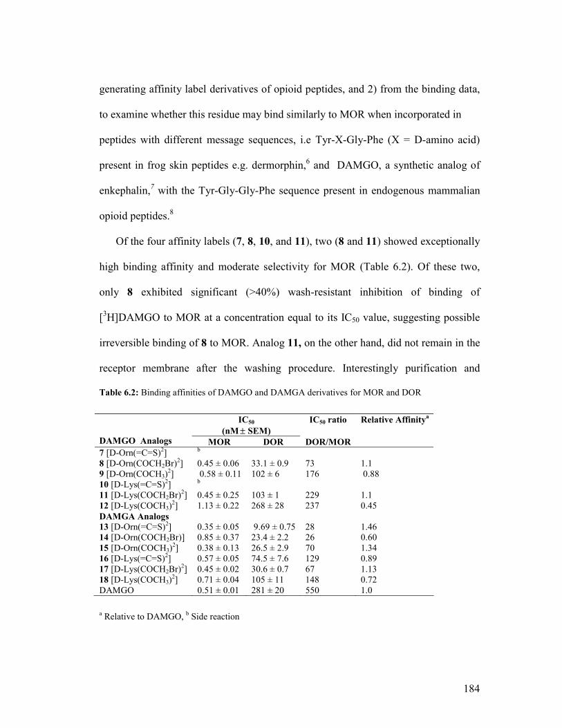
184
generating affinity label derivatives of opioid peptides, and 2) from the binding data,
to examine whether this residue may bind similarly to MOR when incorporated in
peptides with different message sequences, i.e Tyr-X-Gly-Phe (X = D-amino acid)
present in frog skin peptides e.g. dermorphin,6 and DAMGO, a synthetic analog of
enkephalin,7 with the Tyr-Gly-Gly-Phe sequence present in endogenous mammalian
opioid peptides.8
Of the four affinity labels (7, 8, 10, and 11), two (8 and 11) showed exceptionally
high binding affinity and moderate selectivity for MOR (Table 6.2). Of these two,
only 8 exhibited significant (>40%) wash-resistant inhibition of binding of
[3H]DAMGO to MOR at a concentration equal to its IC50 value, suggesting possible
irreversible binding of 8 to MOR. Analog 11, on the other hand, did not remain in the
receptor membrane after the washing procedure. Interestingly purification and
Table 6.2: Binding affinities of DAMGO and DAMGA derivatives for MOR and DOR
a Relative to DAMGO, b Side reaction
IC50
(nM ± SEM) IC50 ratio
Relative Affinitya
DAMGO Analogs MOR DOR DOR/MOR 7 [D-Orn(=C=S)2] b
8 [D-Orn(COCH2Br)2] 0.45 ± 0.06 33.1 ± 0.9 73 1.1 9 [D-Orn(COCH3)2] 0.58 ± 0.11 102 ± 6 176 0.88 10 [D-Lys(=C=S)2] b
11 [D-Lys(COCH2Br)2] 0.45 ± 0.25 103 ± 1 229 1.1 12 [D-Lys(COCH3)2] 1.13 ± 0.22 268 ± 28 237 0.45 DAMGA Analogs 13 [D-Orn(=C=S)2] 0.35 ± 0.05 9.69 ± 0.75 28 1.46 14 [D-Orn(COCH2Br)] 0.85 ± 0.37 23.4 ± 2.2 26 0.60 15 [D-Orn(COCH3)2] 0.38 ± 0.13 26.5 ± 2.9 70 1.34 16 [D-Lys(=C=S)2] 0.57 ± 0.05 74.5 ± 7.6 129 0.89 17 [D-Lys(COCH2Br)2] 0.45 ± 0.02 30.6 ± 0.7 67 1.13 18 [D-Lys(COCH3)2] 0.71 ± 0.04 105 ± 11 148 0.72 DAMGO 0.51 ± 0.01 281 ± 20 550 1.0

185
analysis of the isothiocyanate-containing analogs (7 and 10) revealed an unexpected
side reaction with the formation of cyclic O-alkyl thiocarbamates. This side reaction
involves the C-terminal gly-ol functionality of DAMGO acting as a nucleophile and
attacking the electrophilic isothiocyanate functionality attached to the side chain of
either D-Orn or D-Lys. This leads to the formation of the cyclic O-alkyl
thiocarbamate derivative (Figure 6.1).9
H2N CHCCH2
O HN CHC
O HN CHC
H
ON CHC
CH2
HN
O
OH
CH3
OH
H2N CHCCH2
O
OH
HN CHC
HN
HN CHC
H
ON CHCH3
OHN
CH2
OC
SMW: 613.2 MW: 613.2
NC
S
O
Figure 6.1. Cyclization reaction leading to the cyclic O-alkyl thiocarbamate side product. Reaction of [D-Lys(=C=S)2]DAMGO is shown as the example.
The design of the affinity label derivatives was then modified to overcome the
formation of this side product by replacing the C-terminal glyol group by a
glycylamide functionality to give [D-Ala2,NMePhe4,Gly5]enkephalinamide
(DAMGA). The new series of analogs (13-18, Table 6.2) containing the
isothiocyanate and bromoacetamide functionalities were then successfully prepared
and evaluated for their binding affinities to MOR. As shown in Table 6.2, although
the new series of analogs maintain extremely high affinity (IC50 = 0.3-0.8 nM) for
MOR, the selectivity of most of these ligands for MOR dropped significantly (3- to 4-

186
fold) compared to the DAMGO derivatives. This suggests that the C-terminal glyol
functionality plays an important role in conferring selectivity for MOR, as replacing it
with the glycylamide functionality resulted in increased affinity for DOR.
The present study successfully established a general approach for designing
affinity labels based on MOR-selective peptide ligands containing a D-amino acid at
position 2. The modifications were well tolerated and the resulting peptides showed
very high affinity for MOR. One of the DAMGO-based analogs, 8, showed >40%
wash-resistant inhibition of binding of [3H]DAMGO to MOR at concentration equal
to its IC50 value (0.45 nM), suggesting irreversible binding of this analog to MOR. Of
the [D-Lys2]DAMGA series of analogs (16 – 18), 17 exhibited 44% wash-resistant
inhibition of binding at concentration equal to its IC50 value suggesting this peptide
may interact irreversibly with MOR. The wash-resistant inhibition of binding of [D-
Orn2]DAMGA derivatives (13 – 15) will be carried out soon.
6.2.3 Project 3
Design, Synthesis and Evaluation of a Dermorphin-Based Multifunctional
Affinity Label Probe for Mu Opioid Receptors (Chapter 5)
With the long range goal of identifying the point of attachment of a peptide-based
affinity label to MOR, the objective of this project was to design and synthesize a
MOR selective affinity label peptide as a multifunctional probe which would
facilitate the process of receptor isolation and identification of the attachment point of
the affinity label to MOR.
187
Towards this goal, a MOR-selective peptide-based affinity label [D-
Lys(=C=S)2]dermorphin (Project 1, Chapter 3, compound 4) was chosen as the lead
peptide for incorporating additional functionalities, namely d-desthiobiotin (DSB) as
a purification tag, and Oregon Green or 5-carboxyrhodamine B as a fluorescent tag
(Figure 6.2) to facilitate receptor isolation and aid in visualizing the labeled receptor,
respectively. This particular analog was chosen due to its high binding affinity (IC50 =
0.38 nM), selectivity (DOR/MOR = 250) and wash-resistant inhibition of binding to
NH
OHN
HO
O
N
OHN
OH
NH
OHN
ONH
OH2N
HO
X
OO N
HO
OHN
O
O
NH
NH2
OO
NHO
HN NH
O
O
HOOC
O
F F
OH
NHO
HN
O
O
HOOC
N N
O
Purification Tag
Fluorescent Tagor
X= =C=S (affinity label)= -COCH3 (reversible control)
PEG-like Linkers
O
Figure 6.2. The design of the dermorphin-based multifunctional affinity label for MOR
MOR in a concentration-dependent manner (see Chapter 3). The design of this
multifunctional affinity label probe included additional Lys residues that served as
handles to incorporate the additional tags. The purification tag and the fluorescent tag
188
were separated from each other and from the peptide by a hydrophilic poly(ethylene
glycol) (PEG)-like linker to increase the water solubility of the peptide and decrease
nonspecific binding.
The synthetic methodology to prepare this multifunctional affinity label peptide
on a solid support involved choosing protecting groups for the three different Lys side
chain amines present in the multilabel peptide so that each protecting group could be
selectively removed and the appropriate label incorporated without affecting the other
protecting groups present in this peptide. Employing this synthetic strategy the
multilabeled analogs were successfully synthesized and obtained in >97% purity
based on analytical HPLC following purification.
With the successful synthesis of the multifunctional affinity label peptides,
preliminary microscopic experiments were performed by labeling SH-SY5Y cells that
stably express MOR with the multilabeled [D-Lys(=C=S)2]dermorphin derivative
containing Oregon Green (Compound 1, Chapter 5). Preliminary microscopic results
obtained by labeling SH-SY5Y cells with 1, including protection and displacement
experiments with the MOR antagonist naloxone, suggest irreversible binding of 1 to
MOR. Thus, the multifunctional probe was successfully used to label and visualize
MOR, demonstrating the utility of this approach.
6.3 Future Work
The multilabeled affinity label peptide containing Oregon Green or 5-
carboxyrhodamine B will first be evaluated for MOR affinity and selectivity in
standard radioligand binding assays using CHO cells stably expressing MOR and
189
DOR to determine their affinities relative to parent affinity label derivative of
dermorphin (compound 4, Chapter 3). The wash-resistant inhibition of binding by the
multifunctional peptides (compound 1 and 3, Chapter 5) will be examined to optimize
concentration, and incubation time.10 To ensure effective removal of noncovalently
bound compound and to assess efficiency of the washing procedure, reversible
control multilabeled peptides (2 and 4, Chapter 5) will also be evaluated for wash-
resistant inhibition of binding. Optimizing the washing procedure is particularly
important since addition of DSB and fluorescent group could make difficult to
remove noncovalently bound peptide from MOR.
The microscopy experiments with the multifunctional peptide described in
Chapter 5 will be repeated using CHO cells expressing MOR. Wash-resistant
fluorescent labeling of cells with the peptides 1 and 3 (Chapter 5) will be evaluated
using confocal microscopy. Like the wash-resistant inhibition of binding assay,
parallel experiments will also be performed with the reversible control compounds (2
and 4, Chapter 5) to evaluate the effectiveness of the washing procedure.
The long range goal of this project is to identify the attachment point of the
peptide-based affinity label to MOR. After the binding affinities are determined in
radioligand binding assays and wash-resistant inhibition of binding and wash-
resistant fluorescent labeling evaluated as described above, receptor isolation studies
will be undertaken to determine the attachment point of the multifunctional peptide to
MOR. The CHO cells stably expressing MOR will be labeled with the fluorescent
peptide at an appropriate concentration. Epitope tagged MOR will be used for such
190
isolation experiments and immunoprecipitation of the labeled receptor using an
appropriate antibody against the epitope will also be included in the isolation
procedure. Since the peptide contains a fluorescent label, SDS-PAGE of the labeled
receptor followed by fluorescent imaging would help identify the desired band. The
labeled receptor band thus isolated could then be enzymatically cleaved and the
labeled receptor fragment could be further isolated from other unlabeled fragments
through binding of the DSB group to streptavidin11 and the unlabeled fragments
removed by washing. The isolated labeled fragments will then be analyzed by
MALDI-TOF-MS (matrix-assisted laser desorption/ionization-time of flight-mass
spectrometry) to attempt to determine the attachment point.11-13 The optimum
conditions (compatibility with detergents, concentration, incubation time, elution
efficiency from the streptavidin bound peptides, etc) will be first established based on
detailed model studies with the multilabeled peptides based on literature procedures
used in the purification of MOR.14 For these studies deuterated multilabeled peptides
would be prepared to assist detection labeled fragments by MALDI-TOF-MS.15
Another important application of the multilabeled fluorescent peptide will be to
study receptor internalization. After labeling MOR expressed on CHO cells with
dermorphin-based fluorescent peptides at a low temperature (4° C), internalization
can be monitored by confocol microscopy following raising the temperature to 35°
C.16 Since dermorphin is an agonist, the multilabeled derivative would be expected to
result in receptor internalization.

191
6.4 Bibliography
1. Takemori, A. E.; Portoghese, P. S. Affinity labels for opioid receptors. Annu. Rev.
Pharmacol. Toxicol. 1985, 25, 193-223.
2. Leelasvatanakij, L.; Aldrich, J. V. A solid-phase synthetic strategy for the
preparation of peptide-based affinity labels: synthesis of dynorphin A analogs. J.
Pept. Res. 2000, 56, 80-87.
3. Sinha, B.; Cao, Z.; Murray, T. F.; Aldrich, J. V. Discovery of dermorphin-based
affinity labels with subnanomolar affinity for mu opioid receptors. J. Med. Chem.
2009, Accepted.
4. Choi, H.; Murray, T. F.; Aldrich, J. V. Dermorphin-based potential affinity labels
for mu-opioid receptors. J. Pept. Res. 2003, 61, 40-45.
5. Shimohigashi, Y.; Takada, K.; Sakamoto, H.; Matsumoto, H.; Yasunaga, T.;
Kondo, M.; Ohno, M. Discriminative affinity labelling of opioid receptors by
enkephalin and morphiceptin analogues containing 3-nitro-2-pyridinesulphenyl-
activated thiol residues. J. Chromatogr. 1992, 597, 425-428.
6. Melchiorri, P.; Negri, L. The dermorphin peptide family. Gen. Pharmacol. 1996,
27, 1099-1107.
7. Handa, B. K.; Land, A. C.; Lord, J. A.; Morgan, B. A.; Rance, M. J.; Smith, C. F.
Analogues of beta-LPH61-64 possessing selective agonist activity at mu-opiate
receptors. Eur. J. Pharmacol. 1981, 70, 531-540.

192
8. Aldrich, J. V.; Vigil-Cruz, S. C. Narcotic analgesics. In Burger's Medicinal
Chemistry and Drug Discovery, Abraham, D. J., Ed. John Wiley and Sons: New
York, 2003; Vol. 6, pp 329-481.
9. Dattachowdhury, B.; Murray, T. F.; Aldrich, J. V. The synthesis of DAMGO-
based potential affinity labels with high mu opioid receptor affinity and the
formation of cyclic O-alkyl thiocarbamates. Adv. Exp. Med. Biol. 2009, 611, 265-
266.
10. Liu-Chen, L. Y.; Li, S. X.; Tallarida, R. J. Studies on kinetics of [3H]beta-
funaltrexamine binding to mu opioid receptor. Mol. Pharmacol. 1990, 37, 243-
250.
11. Girault, S.; Sagan, S.; Bolbach, G.; Lavielle, S.; Chassaing, G. The use of
photolabelled peptides to localize the substance-P-binding site in the human
neurokinin-1 tachykinin receptor. Eur. J. Biochem. 1996, 240, 215-222.
12. Sachon, E.; Tasseau, O.; Lavielle, S.; Sagan, S.; Bolbach, G. Isotope and affinity
tags in photoreactive substance P analogues to identify the covalent linkage
within the NK-1 receptor by MALDI-TOF analysis. Anal. Chem. 2003, 75, 6536-
6543.
13. Girault, S.; Chassaing, G.; Blais, J. C.; Brunot, A.; Bolbach, G. Coupling of
MALDI-TOF mass analysis to the separation of biotinylated peptides by magnetic
streptavidin beads. Anal. Chem. 1996, 68, 2122-2126.

193
14. Christoffers, K. H.; Li, H.; Keenan, S. M.; Howells, R. D. Purification and mass
spectrometric analysis of the mu opioid receptor. Brain Res. Mol. Brain Res.
2003, 118, 119-131.
15. Wang, X. Doctoral Thesis. University of Kansas: 2007.
16. Arttamangkul, S.; Alvarez-Maubecin, V.; Thomas, G.; Williams, J. T.; Grandy,
D. K. Binding and internalization of fluorescent opioid peptide conjugates in
living cells. Mol. Pharmacol. 2000, 58, 1570-1580.
Top Related