


Languages
Pages
Legal


Copyright WILEY-VCH Verlag GmbH & Co. KGaA, 69469 Weinheim, Germany, 2019.
Supporting Information
for Advanced Optical Materials, DOI: 10.1002/adom.201900260
Arbitrary Manipulation of Light Intensity by BilayerAluminum Metasurfaces
Zhancheng Li, Wenwei Liu, Hua Cheng, Duk-Yong Choi,Shuqi Chen,* and Jianguo Tian

1
Copyright WILEY-VCH Verlag GmbH & Co. KGaA, 69469 Weinheim, Germany, 2019.
Supporting Information
Arbitrary Manipulation of Light Intensity by Bilayer Aluminum Metasurfaces
Zhancheng Li, Wenwei Liu, Hua Cheng, Duk-Yong Choi, Shuqi Chen* and Jianguo Tian
S1. The Wave-Transfer Matrix Method
The implementation of broadband polarization-selective transmission in the proposed
bilayer metasurfaces is mainly attributed to the interference between the multiple reflections
and the direct reflection. To verify this interpretation, we utilized the wave-transfer matrix
method to make an analysis.[S1-S3] For a planar interface between two media i and j (whether
with or without a nano structures), the forward and backward propagating waves can be
related by utilizing a 44 wave-transfer Matrix Mji while a 44 propagation matrix Pi can be
used for a homogeneous medium i. The forward and backward propagating waves at the two
ends of a multilayered medium can be related by the overall wave-transfer matrix M of the
multilayered medium that can be treated as the matrix product of each part: M=MnM2M1,
where the element 1,2,…,N are numbered along the wave propagating direction. For the
proposed bilayer metasurfaces, the overall wave-transfer matrix M can be expressed as:
M=MdcPcMcbPbMba, as shown in Figure S1(a). We simulated the coefficients of the scattering
matrix Sji of each interface firstly, and then we calculated the corresponding wave-transfer
matrix Mji. Finally, we calculated the overall wave-transfer matrix M of the proposed bilayer
metasurfaces, retrieved the coefficients of the overall scattering matrix S and calculated the
transmission intensities. Figure S1(b) shows the simulated and calculated results of the
modular square tij = |Tij|2 of the transmission coefficients Txx and Tyy. Results show that the
calculated results are in good agreement with the simulated one. The slight difference is
owing to the neglecting of nanorod thickness that will introduced a tiny error in the

2
calculation of overall transfer matrix M. We also calculated the transmittance of the proposed
bilayer metasurface under illumination with different polarization angles. As shown in Figure
S1(c), the calculated results are also in good agreement with the simulated one (Figure 2b).
Moreover, we also calculated the relationship between txx and the number of the nanorod layer,
as shown in Figure S1(d). The results show that the working bandwidth is expanded
dramatically in the proposed bilayer design compared to the single layer one and the working
bandwidth can be further expanded with the increasing of the nanorod layer. It is worth
mentioned that, even the working bandwidth can be further expanded with the increasing of
the nanorod layer, the increasing percentage decrease dramatically. The further expanded of
the working bandwidth can be realized by directly stacking two bilayer designs with adjacent
working bandwidth instead of simply increasing the nanorod layer.

3
Supplementary figures
Figure S1. (a) Schematic of the multiple reflections in the proposed bilayer metasurfaces. (b)
The simulated and calculated results of the modular square tij = |Tij|2 of the transmission
coefficients Txx and Tyy. (c) The calculated results of the transmittance under linearly polarized
illumination with different polarization angles. (d) The calculated results of txx for
metasurfaces with different numbers of nanorod layer (layer 2).

4
Figure S2. Simulated results of txx and tyy under illuminations with different incident angles
(as the inset shows), the transmission ratio (tyy- txx)/ tyy in (c) is calculated with the simulated
results in (a) and (b) while the transmission ratio in (f) is calculated with the simulated results
in (d) and (e).
Figure S3. Simulated results of txx and tyy for bilayer metasurfaces with different
misalignment distances along (a) (b) x-axis, (c) (d) y-axis and (e) (f) both x- and y-axis and
different misalignment angle along (g) (h) z-axis.

5
Figure S4. The experimental measurement results of transmittance under linearly polarized
illuminations with different polarization angles. (a) Metasurfaces with single layer nanorods
and (b) metasurfaces with bilayer nanorods.
Figure S5. The experimental measurement results of the polarization-encoded images under
linearly polarized illumination with different polarization angles. Results confirm that the
polarization-encoded image 1 is single outputs and the polarization-encoded image 2 is dual
outputs.

6
Figure S6. (a) The captured images of the QR code for optical anti-counterfeiting under
linearly polarized illumination with different polarization angles. (b) The captured images of
the QR code for optical anti-counterfeiting under illumination with different polarization
states and wavelengths (the bottom row of images are captured under x-polarized illumination
with an analyzer in the y direction).

7
Figure S7. The sample fabrication process of the designed bilayer metasurfaces.
Figure S8. SEM image of the fabricated periodic bilayer metasurface for the verification of
the misalignment between the two layers. The orientation angle 𝜃 of the nanorods is equal to
90°. In order to see the aluminum nanorods in the upper layer clearly, the SU-8 polymer with
thickness approximately equal to 150 nm was etched by oxygen plasma. Then, a very thin
platinum was coated on the surface of the sample. However, due to small atomic contrast
between Al nanorods and SU-8 and smooth morphology, the aluminum nanorods in the
bottom layer could not be seen in the SEM. The white dots in the SEM image come from the
etch residue of SU-8 (SU-8 contains antimony which could not be removed by oxygen
plasma). To verify the misalignment between the two layers, we removed part of the SU-8
upon the bottom aluminum nanorods by using mechanical method. The etch residues are
smaller in the bottom layer because the remained SU-8 is thin after the mechanical removing.
8
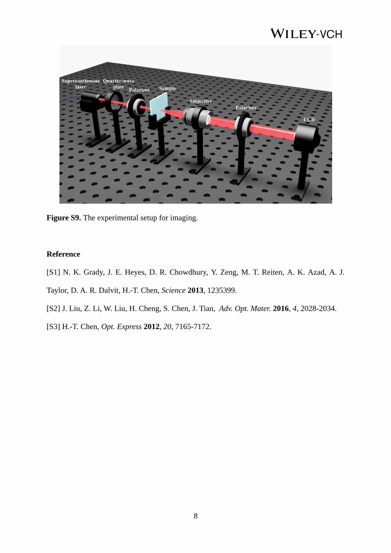
Figure S9. The experimental setup for imaging.
Reference
[S1] N. K. Grady, J. E. Heyes, D. R. Chowdhury, Y. Zeng, M. T. Reiten, A. K. Azad, A. J.
Taylor, D. A. R. Dalvit, H.-T. Chen, Science 2013, 1235399.
[S2] J. Liu, Z. Li, W. Liu, H. Cheng, S. Chen, J. Tian, Adv. Opt. Mater. 2016, 4, 2028-2034.
[S3] H.-T. Chen, Opt. Express 2012, 20, 7165-7172.
Top Related