


Languages
Pages
Legal

Loughborough UniversityInstitutional Repository
Cool-temperature-mediatedactivation of phospholipaseC-[gamma]2 in the humanhereditary disease PLAID
This item was submitted to Loughborough University's Institutional Repositoryby the/an author.
Citation: SCHADE, A. ... et al., 2016. Cool-temperature-mediated activationof phospholipase C-[gamma]2 in the human hereditary disease PLAID. CellularSignalling, 28 (9), pp. 12371251.
Additional Information:
• This paper was accepted for publication in the journal Cellu-lar Signalling and the definitive published version is available athttp://dx.doi.org/10.1016/j.cellsig.2016.05.010.
Metadata Record: https://dspace.lboro.ac.uk/2134/21459
Version: Accepted for publication
Publisher: c© Elsevier
Rights: This work is made available according to the conditions of the Cre-ative Commons Attribution-NonCommercial-NoDerivatives 4.0 International(CC BY-NC-ND 4.0) licence. Full details of this licence are available at:https://creativecommons.org/licenses/by-nc-nd/4.0/
Please cite the published version.
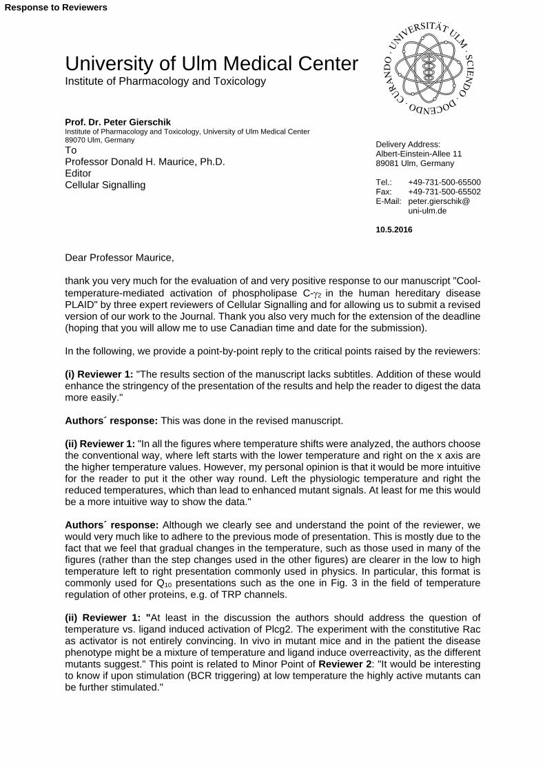
University of Ulm Medical Center Institute of Pharmacology and Toxicology Prof. Dr. Peter Gierschik Institute of Pharmacology and Toxicology, University of Ulm Medical Center 89070 Ulm, Germany
To Professor Donald H. Maurice, Ph.D. Editor Cellular Signalling Dear Professor Maurice, thank you very much for the evaluation of and very positive response to our manuscript "Cool-temperature-mediated activation of phospholipase C-2 in the human hereditary disease PLAID" by three expert reviewers of Cellular Signalling and for allowing us to submit a revised version of our work to the Journal. Thank you also very much for the extension of the deadline (hoping that you will allow me to use Canadian time and date for the submission). In the following, we provide a point-by-point reply to the critical points raised by the reviewers: (i) Reviewer 1: "The results section of the manuscript lacks subtitles. Addition of these would enhance the stringency of the presentation of the results and help the reader to digest the data more easily." Authors´ response: This was done in the revised manuscript. (ii) Reviewer 1: "In all the figures where temperature shifts were analyzed, the authors choose the conventional way, where left starts with the lower temperature and right on the x axis are the higher temperature values. However, my personal opinion is that it would be more intuitive for the reader to put it the other way round. Left the physiologic temperature and right the reduced temperatures, which than lead to enhanced mutant signals. At least for me this would be a more intuitive way to show the data." Authors´ response: Although we clearly see and understand the point of the reviewer, we would very much like to adhere to the previous mode of presentation. This is mostly due to the fact that we feel that gradual changes in the temperature, such as those used in many of the figures (rather than the step changes used in the other figures) are clearer in the low to high temperature left to right presentation commonly used in physics. In particular, this format is commonly used for Q10 presentations such as the one in Fig. 3 in the field of temperature regulation of other proteins, e.g. of TRP channels. (ii) Reviewer 1: "At least in the discussion the authors should address the question of temperature vs. ligand induced activation of Plcg2. The experiment with the constitutive Rac as activator is not entirely convincing. In vivo in mutant mice and in the patient the disease phenotype might be a mixture of temperature and ligand induce overreactivity, as the different mutants suggest." This point is related to Minor Point of Reviewer 2: "It would be interesting to know if upon stimulation (BCR triggering) at low temperature the highly active mutants can be further stimulated."
Delivery Address: Albert-Einstein-Allee 11 89081 Ulm, Germany Tel.: +49-731-500-65500 Fax: +49-731-500-65502 E-Mail: peter.gierschik@ uni-ulm.de 10.5.2016
Response to Reviewers

2
Authors´ response: We have followed these important suggestions as much as we could possibly do in the COS-7 cell context, by analyzing the activation of exogenous wild-type versus20-22 and 19 mutant PLC2 by EGF receptors endogenously present in these cells. The results, which are shown in Fig. 11 of the revised manuscript, are intriguing because they imply that that the mutant PLC2 enzymes are resistant to stimulation by EGFR activation, both at 31 oC and 37 oC. This is behavior is in striking contrast to that of wild-type PLC2 with approx. 12-fold stimulation by EFGR activation at both 31 oC and 37 oC. Although activation of PLC2 in COS-7 by activated EGFR differs from its activation in B lymphocytes by antigen-activated BCR or in other cells by other receptor tyrosine kinases, these results are similar to the loss of function effects seen in cells of PLAID patients and could provide hints to the mechanistic basis of the apparently disparate effects of the PLAID mutations, gain and loss of PLC2 function. (iii) Reviewer 1: "The octamer peptide PCI is fully conserved in PLC<gamma>1 and mediates cold sensitivity in PLC<gamma>2. Which effect causes its deletion in PLC<gamma>1." Authors´ response: We have not specifically addressed this question. However, since the PCI peptide is contained in the region deleted in the PLC1 mutant "20-22", we would predict that that the functions of PLC1PCI would be similar to those of PLC120-22 (cf. Fig. 4A), such as the functions of PLC2PCI resemble those of PLC220-22 (cf. Fig. 4B, left). However, we do not feel that the octamer mediates cold sensitivity in PLC2, since it is maintained in PLC219, which is nevertheless sensitive to activation by cooling (cf. Fig. 8B). (iv) Reviewer 1: "The authors could provide data on the mobilization of Ca2+ upon the temperature shift. This would strengthen the data on IP3 measurements." Authors´ response: We appreciate this suggestion of the reviewer. However, in other studies that we have performed on PLC2, we have always seen a close correlation between inositol phosphate formation and increase in cytosolic Ca2+, at least upon acute activation of the enzyme. In the chronic activation setting used in most of the experiments shown here, we would anticipate technical difficulties in observing reliable changes in [Ca2+]i for the PLC2 PLAID mutants. (v) Reviewer 3: "Attempts were made to provide a structural rational for the activation mediated by cool temperatures. The main limitation here is that the work relies on indirect experiments that measure enzyme activity of PLCgamma2 variants in transfected cells without any further support from other approaches. Conceptual schemes are helpful but experimental limitations have to be pointed out. This should be emphasized in discussion." Authors´ response: This was explicitly done on p. 19 of the revised manuscript. (vi) Reviewer 3: "Several observations deserve some more emphasis even if they can not be easily explained at present. These include: -The data suggesting that PLCgamma1 variants harboring deletions are not regulated by cool temperatures and that regions corresponding to deletions in PLAID have a bigger role in auto-inhibition in this enzyme. -The data related to Ali5 mutation and activation by cool temperatures. This mutation is not in the region affected by PLAID, actually not in the regulatory region at all. Some link to the PH domain has been suggested but this is mechanistically unclear. Unexpected behavior of Ali5 variant on its own and the distinct position of the mutation need to be pointed out. -The fact that many deletions in PLCgamma2 SH region are further activated by cool temperatures." Authors´ response: All three points have been addressed in the text of the revised manuscript on pp. 11, 13 (top paragraph), and 13 (bottom paragraph). (vii) Reviewer 3: "It should be stated that assumption (or expectation) has been made that observations based on COS cell transfections will be relevant for endogenous expression levels in B-cells."

3
Authors´ response: This was done in the revised manuscript on p. 20 in the context of discussing the resistance of the two deletion mutants to activated EGFR in COS-7 cells [cf. (ii)]. An additional point not requested by the reviewers was the examination of the requirement of the spPH domain per se for cool temperature regulation of PLC2. We felt that this experiment needed to be done and think that its results should be reported in this manuscript (now in Fig. 10B) to avoid misinterpretations of the role of spPH in mediating the enzyme´s response to cooling. The results are intriguing: while spPH exerts a striking regulatory role within the PLAID PLC2 mutant PLC219, it is not required for the cool temperature response of further truncated mutants, such as the bipartite mutant made up of fragments X and Y. We would be happy, if you allowed us to show this data and an accordingly revised version of the model now shown in Fig. 10C-H. In summary, we believe that we have carefully considered and taken care of most, if not all of the referees' critical points. We sincerely hope that these changes make our work acceptable for publication in Cellular Signalling. Needless to say, we are extremely grateful to all three reviewers for their expert opinions and invaluable and important advice on the manuscript. Yours sincerely,
Peter Gierschik

1 2 3 4 5 6 7 8 9 10 11 12 13 14 15 16 17 18 19 20 21 22 23 24 25 26 27 28 29 30 31 32 33 34 35 36 37 38 39 40 41 42 43 44 45 46 47 48 49 50 51 52 53 54 55 56 57 58 59 60 61 62 63 64 65
1
Cool-temperature-mediated activation of phospholipase C-2
in the human hereditary disease PLAID
Anja Schadea, Claudia Walliser
a, Martin Wist
a,
Jennifer Haasa, Petra Vatter
a, Johann M. Kraus
b, Davide Filingeri
c,1,
George Havenithc, Hans A. Kestler
d, Joshua D. Milner
e, Peter Gierschik
a,*
aInstitute of Pharmacology and Toxicology, Ulm University Medical Center, 89070 Ulm, Germany
bMedical Systems Biology, Ulm University, 89081 Ulm, Germany
cEnvironmental Ergonomics Research Centre, Loughborough University, Leicestershire, LE11 3TU,
United Kingdom
dFriedrich Schiller University Jena and Leibniz Institute for Age Research, Fritz Lipmann Institute,
07745 Jena, Germany
eAllergic Inflammation Unit, Laboratory of Allergic Diseases, NIAID, NIH, Bethesda, MD 20892,
U.S.A.
Abbreviations: PLC, inositol-phospholipid-specific phospholipase C; SH2, Src homology domain
2; SH3, Src homology domain 3; PH, pleckstrin homology domain; spPH, split PH domain; PCI,
phospholipase C inhibitor peptide; Rac, Ras-related C3 botulinum toxin substrate; BCR, B cell
receptor; SA, specific array; PLAID, PLC2-associated antibody deficiency and immune
dysregulation; TRP, transient receptor potential; PtdIns, phosphatidylinositol; PtdIns(4)P,
phosphatidylinositol 4-phosphate; PtdIns(4,5)P2, phosphatidylinositol 4,5-bisphosphate; Co., control;
aa, amino acid
* Corresponding author at: Institute of Pharmacology and Toxicology, Ulm University Medical
Center, Albert-Einstein-Allee 11, 89081 Ulm, Germany
E-mail address: [email protected] (P. Gierschik).
1 Present address: Center for Environmental Design Research, University of California, Berkeley,
CA 94720-1839, U.S.A.
*ManuscriptClick here to view linked References

1 2 3 4 5 6 7 8 9 10 11 12 13 14 15 16 17 18 19 20 21 22 23 24 25 26 27 28 29 30 31 32 33 34 35 36 37 38 39 40 41 42 43 44 45 46 47 48 49 50 51 52 53 54 55 56 57 58 59 60 61 62 63 64 65
2
A B S T R A C T
Deletions in the gene encoding signal-transducing inositol phospholipid-specific phospholipase
C-2 (PLC2) are associated with the novel human hereditary disease PLAID (PLC2-associated
antibody deficiency and immune dysregulation). PLAID is characterized by a rather puzzling
concurrence of augmented and diminished functions of the immune system, such as cold urticaria
triggered by only minimal decreases in temperature, autoimmunity, and immunodeficiency.
Understanding of the functional effects of the genomic alterations at the level of the affected enzyme,
PLC2, is currently lacking. PLC2 is critically involved in coupling various cell surface receptors to
regulation of important functions of immune cells such as mast cells, B cells, monocytes/macrophages,
and neutrophils. PLC2 is unique by carrying three Src (SH) and one split pleckstrin homology domain
(spPH) between the two catalytic subdomains (spPHn-SH2n-SH2c-SH3-spPHc). Prevailing evidence
suggests that activation of PLC2 is primarily due to loss of SH-region-mediated autoinhibition and/or
enhanced plasma membrane translocation. Here, we show that the two PLAID PLC2 mutants lacking
portions of the SH region are strongly (> 100-fold), rapidly, and reversibly activated by cooling by
only a few degrees. We found that the mechanism(s) underlying PLC2 PLAID mutant activation by
cool temperatures is distinct from a mere loss of SH-region-mediated autoinhibition and dependent on
both the integrity and the pliability of the spPH domain. The results suggest a new mechanism of
PLC activation with unique thermodynamic features and assign a novel regulatory role to its spPH
domain. Involvement of this mechanism in other human disease states associated with cooling such as
exertional asthma and certain acute coronary events appears an intriguing possibility.
Keywords: Phospholipase C-2; Inositol phospholipid; Rac2 GTPase; Split PH domain;
Autoinhibition; Cold temperature sensitivity

1 2 3 4 5 6 7 8 9 10 11 12 13 14 15 16 17 18 19 20 21 22 23 24 25 26 27 28 29 30 31 32 33 34 35 36 37 38 39 40 41 42 43 44 45 46 47 48 49 50 51 52 53 54 55 56 57 58 59 60 61 62 63 64 65
3
1. Introduction
Inositol-phospholipid-specific phospholipases C (PLCs) catalyse the formation of inositol 1,4,5-
trisphosphate and diacylglycerol, and, at the same time, decrease the local or general plasma
membrane abundance of their substrate, phosphatidylinositol 4,5-bisphosphate (PtdInsP2) [1]. The
latter three molecules are important mediators of cellular signaling. An enormous variety of cell
surface receptors regulates important cellular functions utilizing PLCs, ranging from G-protein-
coupled receptors over certain ion channels to many transmembrane non-enzymes and enzymes, e.g.
receptor tyrosine kinases. The mammalian PLCs are divided into six subfamilies, , , , , , and .
Analyses of PLC crystal structures have revealed that the catalytic mechanism of PLCs is well
conserved between all PLC family members. Their activation by cell surface receptors involves both
translocation of the soluble PLC enzymes to the plasma membrane, i.e. the site of their phospholipid
substrate(s), and removal of intramolecular autoinhibition [2-5]. However, certain findings suggest
further, still unknown regulatory mechanisms of PLC isozyme activation [1].
The two members of the PLC subfamily, PLC1 and PLC2, control functions represented in
many, if not all cell types, such as cell growth and differentiation, as well as migratory responses, but
are also involved in highly specialized tasks [6]. Examples of the latter are those regulated by PLC2 in
cells of the immune system. PLC1 and PLC2 are activated by receptor and nonreceptor tyrosine
kinases; PLC2 is also activated by Rac GTPases [7]. In B lymphocytes, this interaction amplifies B-
cell-receptor-mediated Ca2+
signalling [8]. The structures of the two PLC isozymes are unique in that
the two catalytic subdomains X and Y are separated by a modular assembly comprising a split PH
domain (spPHn and spPHc), two SH2 (SH2n and SH2c), and one SH3 domain. The whole assembly
(spPHn-SH2n-SH2c-SH3-spPHc) is also referred to as specific array (SA). Studies on isolated SA
structures showed that the split PH domains of PLC1 and PLC2 do not alter their three-dimensional
shapes upon insertion of the entire PLC1 SH2n-SH2c-SH3 region between the two PLC1 spPH
halves and upon peptide ligand binding to the insertion [9] or, in case of spPH of PLC2, upon its
interaction with activated Rac2 [10]. These findings suggested that the PLC split PH domain is a
more rigid, conformationally stiff element of SA and that it mediates PLC2 activation mainly by
allowing Rac2 to translocate the enzyme to the plasma membrane. Recent evidence suggests that the
SH2c domain is a major determinant of PLC1 autoinhibition and that activation of the enzyme by
tyrosine phosphorylation at a site immediately downstream of the domain (Y783
) proceeds by
competition of the phosphorylated peptide with a so far unidentified site on the catalytic XY TIM
barrel for binding to SH2c [11,12].

1 2 3 4 5 6 7 8 9 10 11 12 13 14 15 16 17 18 19 20 21 22 23 24 25 26 27 28 29 30 31 32 33 34 35 36 37 38 39 40 41 42 43 44 45 46 47 48 49 50 51 52 53 54 55 56 57 58 59 60 61 62 63 64 65
4
Alterations of the primary structures of PLC1 and PLC2 are involved in disease, both in
humans and in animal models. Thus, point mutations in the human PLCG1 gene have been linked to
secondary, radiation-associated angiosarcoma [13] and to cutaneous T cell lymphoma [14]. Two
mouse models of autoimmunity and autoinflammation, designated Ali5 and Ali14, have been
described, which are caused by gain-of-function point mutations of PLC2, D993
G and Y495
C,
respectively [15-17]. The Ali5 mutation also gives rise to platelet hyperreactivity and a prothrombotic
phenotype in mice [18]. Recently, deletion of exon 19 or exons 20-22 of the human PLCG2 gene has
been shown to cause a novel human hereditary disease characterized by cold urticaria,
immunodeficiency, and autoimmunity, designated PLAID for PLC2-associated antibody deficiency
and immune dysregulation [19]. In affected individuals, only very subtle skin cooling, such as the one
caused by a single tear rolling down the cheek at room temperature, causes urticarial wheals and flares
within one minute [20]. A related, but distinct human disease, predominantly characterized by
autoinflammation and designated APLAID for autoinflammatory PLAID, is caused by a gain-of-
function point mutation, S707
Y, located in SH2c of PLC2 ([21], cf. [22], for a more comprehensive
review). Although some effects of decreasing temperature on functions downstream of PLC2 have
been documented in cells from PLAID patients [19], understanding of cool temperature regulation of
the enzyme that is affected by the deletion mutations at first hand, PLC2, is currently lacking. Here,
we show that the two PLC2 mutants identified in PLAID patients, PLC219 and PLC220-22 (Fig.
1A), are exquisitely sensitive to cooling and that the magnitude of the response is unprecedented in
that it goes far beyond those previously observed for many other signaling proteins sensitive to
temperature changes, such as the transient receptor potential (TRP) cation channels [23]. The results
suggest that PLAID PLC2 mutants are activated by only minute decreases in temperature by a novel
mechanism that is primarily mediated by the split PH domain and distinct from a loss of
autoinhibition.
2. Material and methods
2.1. Material
The mouse monoclonal antibody 9B11 reactive against the c-Myc epitope (EQKLISEEDL) and
the polyclonal antiserum reactive against human PLC2 raised in rabbits (sc-407) were obtained from
Cell Signaling Technology and Santa Cruz, respectively. The mouse monoclonal antibody AC-15
reactive against -actin (A1978), human epidermal growth factor (E9644), and cycloheximide
(C7698) were obtained from Sigma.
1 2 3 4 5 6 7 8 9 10 11 12 13 14 15 16 17 18 19 20 21 22 23 24 25 26 27 28 29 30 31 32 33 34 35 36 37 38 39 40 41 42 43 44 45 46 47 48 49 50 51 52 53 54 55 56 57 58 59 60 61 62 63 64 65
5
2.2. Construction of vectors
The construction of complementary DNAs encoding c-Myc-epitope-tagged human PLC1 (1291
aa, accession number ABB84466), human PLC2 (1265 aa, accession number NP_002652), and the
spPH domain chimera PLC2-PH11 is described in [24]. The cDNAs of PLC219 (deletion of exon
19, aa 646-685), PLC219-PH12, PLC219-PH21, PLC219-PH11 [with one or both portions of
the PLC2 spPH domain (aa 468-513 and aa 849-914, respectively) replaced in PLC219 by the
corresponding regions of PLC1 (aa 482-527 and aa 872-937, respectively)], PLC1"19" [deletion of
PLC1 residues corresponding to residues 646 to 685 of PLC2 (aa 668-707)], PLC2SH (deletion of
the SH2n-SH2c-SH3 region, aa 515-840), PLC2SH2c (deletion of the C-terminal SH2 domain, aa
639-766), and PLC2PCI (aa 726-733) were constructed by in vitro mutagenesis using the
QuikChange II XL Site-Directed Mutagenesis Kit (200521, Agilent Technologies). The deletion of
exons 20-22 in PLC2 (PLC220-22, aa 686-806), of the PLC1 residues corresponding to residues
686 to 806 of PLC2 (aa 708-828; PLC1"20-22")], and of the PLC2 specific array (PLC2SA, aa
476-908) was performed using the PCR overlap extension method. The introduction of point
mutations was performed by in vitro mutagenesis using the QuikChange II XL Site-Directed
Mutagenesis Kit according to the manufacturer’s instructions. For the insertion of 2 SH domains into
PLC2SH, a linker containing an AvrII restriction site (GCCCTAGG, AvrII site underlined) was
introduced into the deletion site of PLC2SH. The SH domains (SH2n, aa 515-638, SH2n, aa 637-
747, SH3, aa 767-839, SH2n-SH2c, aa 515-747, SH2c-SH3, aa 637-840 were amplified with primers
containing an AvrII restriction site on either end and inserted into PLC2SH by restriction and
ligation. The linker introduced five additional residues in positions 515-519 of the protein. There were
no functional differences between the PLC2SH mutants with and without these residues.
Complementary DNAs encoding PLC2-XPHn
(aa 1-514) and PLC2-YPHc
(aa 841-1265) were
amplified by PCR. Both fragments were c-Myc-tagged at their C-termini. DNAs encoding PLC2-X
(aa 1-470) and PLC2-Y (aa 914-1265) were amplified by PCR. PLC2-X and PLC2-Y were c-Myc-
tagged at their N- and C-termini, respectively. The primer sequences and PCR protocols are available
from the authors upon request.
2.3. Cell culture and transfection
COS-7 cells were maintained at 37 °C in a humidified atmosphere of 90 % air and 10 % CO2 in
Dulbecco’s modified Eagle’s medium (41965-039, Gibco) supplemented with 10 % (v/v) fetal calf
serum (10270-106, Gibco) and 2 mM glutamine, 100 units/ml penicillin, and 100 µg/ml streptomycin
(all from PAA Laboratories, Cölbe, Germany). Prior to transfection, COS-7 cells were seeded into 24-
well plates at a density of 0.75 x 10⁵ cells/well, and grown for 24 h in 0.5 ml of medium/well. For

1 2 3 4 5 6 7 8 9 10 11 12 13 14 15 16 17 18 19 20 21 22 23 24 25 26 27 28 29 30 31 32 33 34 35 36 37 38 39 40 41 42 43 44 45 46 47 48 49 50 51 52 53 54 55 56 57 58 59 60 61 62 63 64 65
6
transfection, plasmid DNA (500 ng/well) was diluted in 50 µl jetPRIME® buffer and 1 µl of
jetPRIME® Reagent (114-15, Polyplus Transfection, Illkirch, France) was added according to the
manufacturer’s instructions. The total amount of DNA was maintained constant by adding empty
vector. Four h after the addition of the DNA-jetPRIME® complexes to the dishes, the medium was
replaced by fresh medium, and the cells were incubated for a further 20 h at 37 °C and 10 % CO2.
2.4. Radiolabeling of inositol phospholipids and analysis of inositol phosphate formation
Twenty four hours after transfection, COS-7 cells were washed once with 0.3 ml/well of
Dulbecco’s PBS (PAA Laboratories) and then supplied with 0.2 ml/well of Dulbecco’s modified
Eagle’s medium containing supplements as described under Cell culture and transfection, and
additionally supplemented with 25 mM HEPES and 2 mM sodium pyruvate (both from PAA
Laboratories) to maintain the pH of the medium [25], 2.5 µCi/ml myo-[2-3H]inositol
(NET1156005MC, Perkin-Elmer), and 10 mM LiCl. The cells were incubated for 20 h in this medium
in individual incubation chambers in ambient atmosphere at temperatures ranging from 25 oC to 39
oC,
washed once with 0.2 ml/well of Dulbecco’s PBS, and then lysed by addition of 0.2 ml/well of 10 mM
ice-cold formic acid. After keeping the samples for 30 min at 4 °C, 0.3 ml/well of 10 mM NH4OH was
added for neutralization, and the sample was centrifuged for 5 min at 20,000 x g. The supernatants
were loaded onto columns containing 0.5 ml of Dowex® 1 x 8-200 ion exchange resin (217425,
Sigma) that had been converted to the formate form and equilibrated with H2O. The columns were
washed once with 3 ml of H2O and twice with 3.5 ml each of 60 mM sodium formate and 5 mM
sodium tetraborate. Inositol phosphates were eluted with 3 ml of 1 M ammonium formate and 100 mM
formic acid. The eluate was supplemented with 15 ml of scintillation fluid (Quicksafe A, 1008000,
Zinsser Analytic, Frankfurt, Germany) and the radioactivity was quantified by liquid scintillation
counting.
2.5. Construction of individual incubation chambers
Chambers allowing the temperature-controlled incubation of individual 24-well tissue culture
plates (92424; TPP, Switzerland) were custom assembled using 15 x 12 x 23 cm Styrofoam containers
(Schaumaplast, Reilingen, Germany) equipped with circuits made up of one Velleman VM148
thermostat control module (190655-62) and one Dallas DS18S20-55 temperature sensor with digital
output (176168-62) to control two serially connected heating foils (532878-62; all from Conrad
Electronic, http://www.conrad.de) to be placed on either side of the tissue culture plate during
incubation. To allow for additional external, analogous control of the temperature, a 10 mm hole
was drilled into the wall of the container and the tissue culture plate to allow insertion of a
thermometer into one well. Power supply to up to 8 individual chambers was through a TDK-Lambda

1 2 3 4 5 6 7 8 9 10 11 12 13 14 15 16 17 18 19 20 21 22 23 24 25 26 27 28 29 30 31 32 33 34 35 36 37 38 39 40 41 42 43 44 45 46 47 48 49 50 51 52 53 54 55 56 57 58 59 60 61 62 63 64 65
7
LS75-12 AC/DC converter unit (511823-62; Conrad). A construction guidance including a circuit
diagram is available from the authors on request.
2.6. Inositol phospholipid analysis
Inositol phospholipids were extracted from transfected cells and analyzed as before [17]. COS-7
cells were grown and radiolabeled in 6-well plates. At the end of the radiolabeling procedure, 10 µl of
the medium supernatant were placed in a scintillation vial with 3 ml of Quicksafe A liquid scintillator
(Zinsser Analytic) and the radioactivity was quantified by liquid scintillation counting. The
radiolabeled cells were lysed by addition of 1.2 ml of 4.5 % (v/v) perchloric acid. After incubating the
samples for 30 min on ice, they were scraped into 1.5 ml reaction tubes and centrifuged at 4 °C for 20
min at 3,700 × g. Supernatants and pellets were separated. The pellets were resuspended in 100 l of
water and 375 l of chloroform/methanol/HCl (100:200:15) was added. The samples were vortexed,
and an additional 125 l of chloroform and 125 l of 0.1 M HCl were added. After further vortexing,
the samples were centrifuged at room temperature for 10 min at 700 × g. Fifty l each of the lower,
chloroformic phase containing the inositol phospholipids were subjected to liquid scintillation
counting as described above.
2.7. Determination of the 10-degree temperature coefficients
According to Hille [26], the 10-degree temperature coefficient, Q10, of a biological process can be
calculated for an arbitrary temperature interval T from
Using and
, where Ti and Tref are the individual and a reference temperatures
and Ai and Aref are the individual and a reference activity, this equation can be rewritten to
Upon plotting the Ti vs. the
, the Q10 value(s) can be calculated from the slopes of the linear
portions of the resultant graphs.

1 2 3 4 5 6 7 8 9 10 11 12 13 14 15 16 17 18 19 20 21 22 23 24 25 26 27 28 29 30 31 32 33 34 35 36 37 38 39 40 41 42 43 44 45 46 47 48 49 50 51 52 53 54 55 56 57 58 59 60 61 62 63 64 65
8
2.8. Miscellaneous
Curve fitting was done using GraphPad Prism, version 4.03 (GraphPad Software, San Diego, CA,
USA). In Fig. 3, the global curve fitting procedure was applied, where the extra sum of squares F-test
is employed to determine whether the best-fit values of the two parameters differ between data sets.
The simpler model was selected unless the P value was less than 0.05. The propagation of errors was
calculated as outlined in [27]. The various PLC isoforms were tested in this study multiple times in
many different combinations, including many that are not presented in this work. The experiments
specifically shown herein were repeated two to three times throughout. Data from representative
experiments are presented as means ± standard error of triplicate determinations. Control curves were
done in Figs. 4B and 7 to confirm that results shown in different panels of the same figure are
comparable. The intensities of immunoreactive bands on Western blots corresponding to PLC2
isozymes carrying a c-Myc epitope were within the range allowing semiquantitative comparisons, as
shown by control experiments using purified c-Myc-tagged wild-type PLC2. Samples to be analyzed
by Western blotting were taken, quasi as a fourth replicate, from the same plate as and immediately
adjacent to the samples taken in triplicate for functional analysis. Using this protocol and paying
meticulous attention to experimental detail, we have not experienced variations in gel loading of these
samples (cf. Fig. 5B).
3. Results
3.1. The PLC2 deletion mutants PLC2D19 and PLC2D20-22 are specifically activated by cool
temperatures
PLC219 and PLC220-22 were expressed in COS-7 cells at 37 oC and the cells were then
radiolabeled with [3H]inositol at temperatures ranging from 39
oC to 25
oC, followed by measurement
of inositol phosphate formation. Fig. 1B shows that the two PLC2 deletion mutants caused slight
increases in inositol phosphate formation in comparison to the wild-type enzyme at 37 oC. Consistent
with earlier results [19], these increases were approximately 2.8- and 3.6-fold for PLC219 and
PLC220-22, respectively, relative to the increase over basal activity observed for wild-type PLC2.
Much more strikingly, however, there was a marked stimulation of inositol phosphate formation when
cells expressing either deletion mutant were incubated at only slightly lower temperatures. In both
cases, there was a biphasic stimulatory response with declining temperatures, with a maximum at 31
oC and a gradual reduction upon further cooling to 25
oC. There was only a modest monophasic
decrease in inositol phosphate formation in cells expressing wild-type PLC2 and in control cells when
the temperature was reduced from 39 oC to 25
oC. Strikingly, at 31
oC, the absolute increase in inositol
phosphate formation in cells expressing PLC219 and PLC220-22 over basal activity of mock-
1 2 3 4 5 6 7 8 9 10 11 12 13 14 15 16 17 18 19 20 21 22 23 24 25 26 27 28 29 30 31 32 33 34 35 36 37 38 39 40 41 42 43 44 45 46 47 48 49 50 51 52 53 54 55 56 57 58 59 60 61 62 63 64 65
9
transfected control cells was enhanced approx. 480 ± 91- and 430 ± 84-fold (means ± SEM) relative to
the increase over basal inositol phosphate formation observed in cells expressing wild-type PLC2.
Fig. 1C shows that the expression of wild-type PLC2 decreased with decreasing incubation
temperature, while the expression of both PLC219 and PLC220-22 took slight increases at
intermediate temperatures ranging from 35 oC to 31
oC and from 35
oC to 25
oC, respectively. While
reduced expression of wild-type PLC2 at lower temperatures may explain, at least in part, the
monophasic decrease in inositol phosphate formation by this enzyme, the limited magnitude of the
changes observed in Fig. 1C for PLC219 and PLC220-22 argues against a critical role of
fluctuating enzyme expression in the marked changes of inositol phosphate formation enzyme activity
evident in Fig. 1B.
3.2. The activation of PLC219 and PLC220-22 by subphysiological temperatures occurs after
protein synthesis and is reversible.
The influence of a decrease in incubation temperature on the abundance of inositol phospholipids
in mock-transfected COS-7 cells and cells expressing either wild-type or 19 mutant PLC2 is shown
in Figs. 1D and E. PLC is capable of hydrolyzing PtdIns, PtdIns(4)P, PtdIns(4,5)P2 [28]. There was
no increase in the abundance of PLC substrate inositol phospholipids upon cooling. Instead, a
monophasic loss was apparent in all three cases upon decreasing the temperature to 25 oC to
approximately 13 % of the levels observed at 37 oC. This behavior was largely independent of whether
or not wild-type PLC2 or PLC219 was expressed in the cells. While this loss may reflect slower
synthesis of the substrate phospholipids at lower temperatures and may explain some of the decline in
inositol phosphate formation by PLC219 and PLC220-22 at temperatures below 31 oC (cf. Fig.
1B), it argues against increased substrate availability causing the dramatic stimulation of the PLC2
deletion mutants upon cooling from 37 oC to 31
oC (cf. Fig. 1B).
The next experiment was designed to examine whether stimulation of PLC219 and PLC220-
22 by cool temperatures occurs before or after synthesis of the mutant PLC2 proteins. To this end,
cells already containing either wild-type PLC2, PLC219, or PLC220-22 (as a result of a 24-h-
transfection period at 37 oC) were treated for a further 20 h (i.e. during the [
3H]inositol radiolabeling
phase) with or without cooling from 37 oC or 31
oC, both in the absence and in the presence of 100
g/ml cycloheximide. Using this protocol, we expected that cycloheximide would block the
stimulatory effect of cooling on inositol phospholipid hydrolysis by the mutants, if cooling were to be
effective prior to or during recombinant protein synthesis. Fig. 2A shows that this was not the case.
Thus, cooling to 31 oC caused marked increases in inositol phosphate formation, regardless of whether
cycloheximide was absent or present during the second phase. Fig. 2B shows that, both at 37 oC and

1 2 3 4 5 6 7 8 9 10 11 12 13 14 15 16 17 18 19 20 21 22 23 24 25 26 27 28 29 30 31 32 33 34 35 36 37 38 39 40 41 42 43 44 45 46 47 48 49 50 51 52 53 54 55 56 57 58 59 60 61 62 63 64 65
10
at 31 oC, cycloheximide prevented the increase in abundance of wild-type and deletion mutant PLC2
during the second phase (in comparison to the control samples obtained after the initial phase),
indicating that cycloheximide was in fact effective as a protein biosynthesis inhibitor in this
experiment. Taken together, these results indicate that the stimulatory effect of subphysiologic
temperatures on deletion mutant PLC2 activity occurs after protein synthesis.
To examine whether the stimulatory effects of the 19 and 20-22 deletions on PLC2 activity
were reversible, wild-type PLC2, PLC219, and PLC220-22 were incubated for the last four hours
of the transfection protocol, i.e. in the absence of radiolabeled inositol phospholipid substrate, at either
37 oC or 31
oC and then radiolabeled with [
3H]inositol at one of the two temperatures. Fig. 2D shows
that the three different incubation protocols had little, if any, effect on the expression of each of the
three enzymes. As shown in Fig. 2C, preincubation of the three enzymes at 31 oC had no effect on
their ability to promote inositol phosphate formation. Only when cells were incubated at 31 oC in the
presence of [3H]inositol, enhanced inositol phosphate formation was evident by the two deletion
mutants, in contrast to wild-type PLC2. A time-course of the changes of inositol phosphate formation
by wild-type and mutant PLC2 upon changes in incubation temperature from 37 oC to 31
oC and vice
versa is shown in Figs. 2E and 2F. Both deletion mutants were activated with no apparent lag time
with cooling to 31 oC. However, deactivation of both enzymes by warming to 37
oC required a lag
time of approximately 10 min to come into effect. Thus, activation of PLC219 and PLC220-22 by
cool temperatures is a rapid and slowly, but fully reversible process.
The determination of the 10 oC temperature coefficient (Q10) value, widely used to characterize
the regulation of TRP channels by temperature [23], for wild-type PLC2, PLC219, and PLC220-
22 is shown in Fig. 3. While only a single linear component with a Q10 value of 4.6 was evident for
wild-type PLC2 between 39 oC and 25
oC, both PLC219 and PLC220-22 displayed two separate
phases of opposite signs and markedly distinct Q10 values. Specifically, while Q10 was as high as 6745
for the marked increase in activity between 39 oC and 33
oC, it was lower by more than three orders of
magnitude and not different, by sign and by magnitude, from the Q10 value describing the decrease in
activity of the wild-type enzyme, 4.6, between 31 oC and 25
oC. Thus, it appears that the major
functional consequence of the two deletions is their increased activity upon cooling from 37 oC to
approximately 31 oC. The decrease in activity observed at temperatures below 31
oC appears to be a
property inherently present in the remaining elements of wild-type PLC2. The Q10 values of
PLC219 and PLC220-22 exceed those reported for other temperature-regulated proteins, such as
thermoTRPs (with values ranging from 10 to > 100 [23]). However, higher Q10 values are not without
precedence. For example, the step of heat damage to the development of the posterior crossvein of
Drosophila that is most sensitive to increasing temperature showed a Q10 of about 360 [29].
1 2 3 4 5 6 7 8 9 10 11 12 13 14 15 16 17 18 19 20 21 22 23 24 25 26 27 28 29 30 31 32 33 34 35 36 37 38 39 40 41 42 43 44 45 46 47 48 49 50 51 52 53 54 55 56 57 58 59 60 61 62 63 64 65
11
3.3. Cool-temperature-mediated activation of PLC2D19 and PLC2D20-22 is distinct from loss of
SH-region-mediated autoinhibition.
The structural organization of PLC2 between its two catalytic subdomains X and Y is very
similar to that of its close relative PLC1 [6]. To address the question, whether deletion in PLC1 of
residues corresponding to those encoded by exons 19 and 20-22 of PLCG2 has similar functional
consequences, the relevant mutants of human PLC1 were produced. Since the human PLCG1 and
PLCG2 genes exhibit distinct patterns of genomic organization, the deletions in PLC1 do not
correspond to specific exons in PLCG1. Hence, the mutants are designated PLC1"19" and
PLC1"20-22". Fig. 4A shows that both PLC1"19" and PLC1"20-22", in contrast to their PLC2
counterparts, were already highly constitutively active at 37 oC (54- and 37-fold increases in inositol
phosphate formation, respectively, in comparison to the increase caused by wild-type PLC1 relative to
control). Furthermore, only minor changes in their activity were observed upon reduction of the
incubation temperature from 37 oC to 27
oC. There were only minor changes in inositol phosphate
formation by wild-type PLC1. Fig. S1A shows that there were minor, if any changes in protein
expression of the PLC enzymes with decreasing temperatures, except for PLC1"20-22" and
PLC220-22, which showed increased expression. It seems possible on the basis of these findings
that the regions of PLC1 corresponding to the regions deleted in PLC2 in PLAID exert a stronger
autoinhibitory influence on PLC1 than their PLC2 counterparts on PLC2 and that this difference
blunts the stimulatory response of PLC1"19" and PLC1"20-22" to cooling. This possibility
notwithstanding, the results indicate that the temperature sensitivities of PLC1 and PLC2 carrying
deletions corresponding to exons 19 and 20-22 of PLCG2 are distinct.
The region encoded by exons 20 to 22 of PLCG2 contains an octapeptide (YRKMRLRY) at the
end of SH2c that is absolutely conserved in PLC1 and has previously been shown to mediate
inhibition of PLC2 in trans and in cis [30,31]. To determine whether deletion of the octamer,
designated PCI (for phospholipase C inhibitor), is sufficient to mediate sensitivity of PLC2 to cool
temperatures, the deletion mutant PLC2PCI was functionally compared to PLC220-22 and wild-
type PLC2 (Fig. 4B, left panel). In addition, three other mutants carrying deletions known to promote
constitutive activity of PLC2 at 37 oC, PLC2SA, PLC2SH, and PLC2SH2c, were characterized
(Fig. 4C, right panel). PLC2PCI was analyzed again in the latter experiment as a control to ensure
that the results shown in the two panels of Fig. 4B are comparable. Figs. 4B and S1B show that
PLC2PCI shared most features of its temperature sensitivity with PLC220-22 and, by extension,
with PLC219 (cf. Fig. 1B): only a limited enhancement of its activity at 37 oC, a marked activation
upon lowering the incubation temperature from 37 oC to 31
oC, and a decrease in activity at

1 2 3 4 5 6 7 8 9 10 11 12 13 14 15 16 17 18 19 20 21 22 23 24 25 26 27 28 29 30 31 32 33 34 35 36 37 38 39 40 41 42 43 44 45 46 47 48 49 50 51 52 53 54 55 56 57 58 59 60 61 62 63 64 65
12
temperatures below 31 oC. In contrast, PLC2SA, PLC2SH, and PLC2SH2c, although exhibiting
constitutive activity at 37 oC, showed an only modest, further increase in activity with cooling, which
was monophasic for PLC2SH2c and only vaguely biphasic for PLC2SA and PLC2SH.
At first sight, the constitutive activities of the mutants PLC2SA, PLC2SH, and PLC2SH2c
observed at 37 oC in Fig. 4B, right panel, appeared rather limited in comparison to the constitutive
activities reported earlier for similar mutants of PLC1 and PLC2 [31,32]. Therefore, the activities at
37 oC of wild-type PLC2, PLC219, PLC220-22, PLC2PCI, PLC2SH2c, PLC2SH, and
PLC2SA were compared as a function of their expression levels in a more comprehensive analysis
(Figs. 5A and B). The results show that, taking the relative abundance of the PLC2 variants into
account (Fig. 5B), PLC2PCI, PLC2SA, PLC2SH2c, and PLC2SH exhibited higher
constitutive activity at 37 oC than PLC219 and PLC220-22.
The fact that either of the two PLC2 mutants, PLC219 and PLC220-22, were activated by
cool temperatures to a similar degree raised the question as to the effect of a combined deletion of the
residues encoded by exons 19 through 22. Fig. 6 shows that, at 37 oC, the compound deletion mutant
PLC219-22 displayed a markedly enhanced activity in comparison to either PLC219 or
PLC220-22 alone, showing only marginal enhancements of their basal activities. The stimulatory
effect of cooling to 31 oC was assayed at two expression levels of the deletion mutants. At both
expression levels, the activity of PLC219-22 at 31 oC was much higher than the sum of the activities
of PLC219 and PLC220-22. Hence, at both temperatures, 37 oC and 31
oC, and at the expression
levels tested in this experiment, the deletions of PLCG2 exons 19 and 20-22 synergized to promote
activation of PLC2.
3.4. The mouse PLC2 mutant Ali5 is also sensitive to activation by cool temperatures.
The symptoms observed in the mouse Ali5 and Ali14 disease models bear resemblance to the
PLAID phenotype in some, but not in all respects [15,16,19,20]. In transfected COS-7 cells, the
corresponding PLC2 mutants, PLC2Ali5
and PLC2Ali14
displayed only slightly enhanced basal activity
at 37 oC, whereas the compound mutant, PLC2
Ali5/Ali14 clearly showed constitutively enhanced activity
[17]. In Ali5 mice, autoinflammatory dermatitis commenced in the superficial layers of the paws and
ears, i.e. in cool body regions [15]. This distribution resembles those of cutaneous granulomatous
lesions in PLAID patients [19]. We therefore compared the effect of cool temperatures on the activity
of the mutants PLC2Ali5
, PLC2
Ali14, and the compound mutant PLC2
Ali5/Ali14. Figs. 7, left panel, and
S2A show that all three mutants exhibited moderately enhanced activity at 37 oC (PLC2
Ali5/Ali14 >

1 2 3 4 5 6 7 8 9 10 11 12 13 14 15 16 17 18 19 20 21 22 23 24 25 26 27 28 29 30 31 32 33 34 35 36 37 38 39 40 41 42 43 44 45 46 47 48 49 50 51 52 53 54 55 56 57 58 59 60 61 62 63 64 65
13
PLC2Ali5
> PLC2Ali14
) as described before [17]. Importantly, however, the three mutants displayed
considerable differences in their sensitivities to cool temperatures, both in quantitative and in
qualitative terms. Thus, while the response of PLC2Ali5/Ali14
to decreasing temperatures closely
resembled the pattern observed for PLC220-22 (Figs. 7, center panel, and S2B), PLC2Ali14
showed
only a minor, monophasic increase in activity, which was opposite to the decrease observed for wild-
type PLC2 (Figs. 7, right panel, and S2C). PLC2Ali5
displayed an intermediate phenotype,
nonetheless showing an almost 7-fold activation upon cooling from 37 oC to 31
oC (Fig. 7, right
panel). These results are surprising since the Plcg2 mutations underlying the Ali5 and Ali14
phenotypes cause point mutations at positions outside of the SH2n-SH2c-SH3 region. Perhaps, similar
structural and, hence, functional alterations are brought about by distinct mutations of the enzyme.
Nevertheless, the results suggest that some of the lesions noticed in the affected animals, such as
inflammation in superficial skin layers, may in fact be caused by further activation of PLC2Ali5
by
cool temperatures. An intriguing question to be clarified by future investigation is whether PLC2
activated by other means, e.g. by other mutations or by tyrosine phosphorylation, are also sensitive to
cold temperatures.
A more comprehensive comparison of the effects of the various constituents of the SH2n-SH2c-
SH3-spPHc region, alone or in combination, on PLC2 constitutive activity and sensitivity to cool
temperatures is shown in Fig. 8A. Unlike observed previously [31,32], deletion of SH3 also caused an
increase in basal activity, which was even slightly higher (approximately 4.1-fold) than that monitored
for deletion of SH2c (2.6-fold), but lower than that for deletion of all three SH domains
(approximately 7.5-fold). The increases for the variants lacking two SH domains, PLC2SH2nSH2c,
PLC2SH2cSH3, and PLC2SH2nSH3, were approximately 3.6-, 1.9-, and 2.4-fold, respectively.
While deletion of SH2n did not change the temperature sensitivity of PLC2 relative to the wild-type
enzyme and deletion of all three SH domains resulted in a PLC2 mutant largely insensitive to cool
temperature, all other deletion mutants were clearly sensitive. Maximal sensitivity (approximately
6.1-fold) was observed for PLC2SH2cSH3, followed, in that order, by SH2nSH3, SH2nSH2c,
SH2c,and SH3. Note that the degrees of stimulation by cool temperatures was by far lower for
these mutants than the degrees observed for the PLAID PLC2 mutants 19 and 20-22 (cf. Fig. 1).
Fig. 8B shows a schematic representation of the deletions within the SH2n-SH2c-SH3 region (aa 515-
840).

1 2 3 4 5 6 7 8 9 10 11 12 13 14 15 16 17 18 19 20 21 22 23 24 25 26 27 28 29 30 31 32 33 34 35 36 37 38 39 40 41 42 43 44 45 46 47 48 49 50 51 52 53 54 55 56 57 58 59 60 61 62 63 64 65
14
3.5. Cool-temperature-triggered activation of PLC2 deletion mutants is controlled by, but does not
necessarily require the split PH domain.
PLC2 is activated by tyrosine phosphorylation and by activated Rac GTPases. The experiment
shown in Fig. 9A was designed to determine the effect of decreasing temperatures on the ability of
constitutively active Rac2G12V
to activate PLC219 in comparison to wild-type PLC2. At 37 oC,
Rac2G12V
caused similar enhancements of the activity of both enzymes (Figs. 9A, left panel, and S3A).
Lowering the incubation temperature, however, took a very different effect on Rac2G12V
-mediated
activation of the two enzymes. While only minor changes were observed for wild-type PLC2, there
was a progressive loss of the stimulatory effect of Rac2G12V
on PLC219. This loss is unlikely to be
due to exhaustion of the inositol phospholipids substrate at lower temperatures, since PLC219 is
well capable of producing even higher levels of inositol phosphates when expressed at higher density
(Fig. 9A, right panel, and S3B). Likewise, it was unlikely that Rac2G12V
became limiting at low
temperatures, since stimulation of wild-type PLC2 was well retained even at the lowest temperature,
27 oC.
The activation of PLC2 by Rac is mediated by the internal, split PH domain of the enzyme
[10,24]. The interdependence of the stimulatory effects of Rac2G12V
and cooling evident in Figs. 9A
and S3 prompted us to determine the role of this domain in PLC2 deletion mutant activation by
cooling. Alanine replacement in spPH of PLC2 of W899
, which is conserved in all PH domains [33]
has previously been shown to result in a loss of PLC2 stimulation by Rac, but not by tyrosine
phosphorylation [31]. Figs. 9B and S4A show that the W899
A replacement completely abrogated the
response of the enzyme to cool temperature activation. Furthermore, replacement of one or the other
half of the split PH domain of PLC2 by the corresponding portions of PLC1, previously shown to
block activation of the mutant enzymes by Rac, but to take no effect on their catalytic activity [24],
also eliminated enzyme activation by cool temperatures. In contrast, replacement of both PH domains
halves, which does not convey Rac sensitivity to PLC2 [24], fully rescued the response of PLC2 to
cooling. These findings indicate that there is important structural interaction between the SH2n-SH2c-
SH3 region and the surrounding sPH sequence. Two residues of the N-terminal half of the PLC1
spPH domain, Y509 and F510 (cf. Fig. 9C), have been shown to be important for the ability of spPHn
to cooperate with SH2c to maintain PLC1 in an inactive conformation [17,34]. To examine the role of
this site in the cool temperature response of PLC2, the two residues were substituted by alanine
residues in PLC219 carrying the entire spPH of PLC1 (PLC219-PH11), thus generating the
mutant PLC219-PH11-Y509
A/F510
A. Consistent with earlier findings [17,34], there was a slight
increase in basal activity of the latter mutant in comparison to PLC219-PH11. Remarkably,

1 2 3 4 5 6 7 8 9 10 11 12 13 14 15 16 17 18 19 20 21 22 23 24 25 26 27 28 29 30 31 32 33 34 35 36 37 38 39 40 41 42 43 44 45 46 47 48 49 50 51 52 53 54 55 56 57 58 59 60 61 62 63 64 65
15
however, and in striking contrast to PLC219-PH11, there was no response of the mutant to cooling.
Y509
and F510
of PLC1 corresponds to Y495
and C496
of PLC2. Most interestingly, alanine replacement
of either one of the latter residues within the PLC219 (carrying its own, authentic spPH domain),
caused a distinct loss of the enzyme´s response to cooling, replacement of both Y495
and C496
led to an
almost complete loss of this response (Figs. 9D and S4B). These results indicate that a functional split
PH domain, not necessarily the one genuinely contained in PLC2, is required with the context of the
PLAID PLC2 deletion mutants to mediate their activation by cool temperatures.
The observation that PLC2SH, a mutant lacking the entire SH2n-SH2c-SH3 region, displays
enhanced basal activity, but is largely insensitive to activation by cold is remarkable, because this
mutant lacks all constituents thought to mediate autoinhibition of the enzyme (Fig. 8B). On the one
hand, this finding clearly suggests that the activation pattern of the two PLAID mutants, PLC219
and PLC220-22, is distinct from a mere loss of autoinhibition, and indicates, by extension, that it is
based on a specific molecular mechanism(s). On the other hand, the observation is rather puzzling,
because cold activation appears to be, at the same time, dependent on and independent of specific
constituents of the SH2n-SH2c-SH3 region. However, given the importance of the split PH domain for
cold activation of PLC2 emerging from the experiments shown in Fig. 9B and D, the possibility
remained that activation by cold temperatures is sterically and/or conformationally restricted in the
mutant PLC2SH by the (artificial) covalent linkage between the two halves of the split PH domain
(cf. Fig. 9C). To examine this possibility, we took advantage of earlier findings suggesting that PLC
isozymes can be expressed as two independent polypeptide fragments containing either of the two
catalytic subdomains, X and Y [35,36]. Thus, PLC2SH was expressed either as a single polypeptide
and as two separate chains, encompassing either of the two catalytic subdomains XPHn
and YPHc
, either
one containing the corresponding half of the split PH domain, followed by functional analysis at 37 oC
and 31 oC. Figs. 10A and S5 show that expression of the N-terminal or the C-terminal half of PLC2
did not lead to enhanced inositol phosphate formation. In marked contrast, when both halves, XPHn
and
YPHc
, were coexpressed, basal inositol phosphate formation at 37 oC was markedly enhanced. More
importantly, however, decreasing the incubation temperature to 31 oC caused a substantial
enhancement of PLC activity, in striking contrast to cells expressing the SH deletion mutant of
PLC2, where no change in activity was observed. These results strongly suggest that the structural
element(s) mediating cold activation of PLAID PLC2 mutants reside outside the SH2n-SH2c-SH3
region and that its responsiveness to cool temperatures is blocked by covalent linkage between the two
portions of the PLC2 split PH domain.
To examine the requirement of the split PH domain in itself for cool-temperature-mediated
activation of coexpressed XPHn
and YPHc
, the coding regions of the split PH domain halves were

1 2 3 4 5 6 7 8 9 10 11 12 13 14 15 16 17 18 19 20 21 22 23 24 25 26 27 28 29 30 31 32 33 34 35 36 37 38 39 40 41 42 43 44 45 46 47 48 49 50 51 52 53 54 55 56 57 58 59 60 61 62 63 64 65
16
removed from the two fragments, resulting in fragments X and Y. The two fragments were expressed
alone or together and functionally analyzed at 31 oC or 37
oC. Most intriguingly, in contrast to cells
expressing either X or Y alone, cells coexpressing fragments X and Y displayed a marked
enhancement of inositol phosphate formation by lowering the incubation temperature to 31 oC (Fig.
10B). This enhancement was similar in extent as the one observed for PLC220-22 (approximately
32-fold versus 41-fold). Thus, although the split PH domain appears to control the cool temperature
sensitivity of the PLAID PLC2 mutants, it is not required per se for cool- temperature-mediated
activation of the enzyme´s core constituents. The expression of wild-type and 20-22 mutant PLC2 as
well as fragments X and Y was similar at 31 oC and 37
oC, respectively (Fig. S6).
3.5. The PLAID PLC2 mutants are resistant to activation by EGF receptors endogenously expressed
in COS-7 cells.
The rather puzzling concurrence of gain and apparent loss of function of PLC2 in certain PLAID
patient cells [19,37] prompted us to determine the effects of the two PLAID PLC2 mutations on the
response of the mutant enzymes to tyrosine kinase receptor activation. To this end, we made use of
EGF receptor tyrosine kinases endogenously expressed in COS-7 cells and capable of both regulating
several endogenous signalling functions and activating exogenous PLC2 [38,39,17] and examined
their ability to stimulate PLC2-mediated inositol phosphate formation. Fig. 11 shows that addition of
100 ng/ml of EGF caused similar (approximately 12-fold) increases in wild-type PLC2 activity at 37
oC and 31
oC. In contrast, neither PLC220-22 nor PLC219 were affected by addition of EGF,
regardless of whether the radiolabeling of cells and their treatment with EGF was done at 37 oC or 31
oC. The inability of EGF to mediate PLC2 PLAID mutant stimulation at 37
oC was not due to lack of
available enzyme substrate, since the two mutants were markedly sensitive to activation by cooling to
31 oC. Additional experiments (not shown) revealed that substrate depletion did also not occur in the
presence of the mutant PLC2 enzymes at 31 oC. Fig. S7 shows that the functional changes depicted in
Fig. 11 were not due to changes of wild-type or mutant PLC2 expression.
4. Discussion
The results presented in this work show that deletion of residues encoded by exons 19 and 20-22
from PLC2 causes both a mild constitutive activation of inositol phosphate formation at 37 oC and a
marked, several-hundred-fold enhancement of this activity in response to very small decreases in
temperature upon expression of the mutant enzymes in intact cells. These functional responses are
qualitatively and quantitatively similar, if not identical for both types of mutants. While the former
changes are consistent with those reported earlier for transfected cells, the latter are much higher than

1 2 3 4 5 6 7 8 9 10 11 12 13 14 15 16 17 18 19 20 21 22 23 24 25 26 27 28 29 30 31 32 33 34 35 36 37 38 39 40 41 42 43 44 45 46 47 48 49 50 51 52 53 54 55 56 57 58 59 60 61 62 63 64 65
17
the small increases in [Ca2+
]i observed when peripheral blood B cells of PLAID patients are exposed
to cold. In the latter case, differences to normal B cells were only observed at temperatures below 29
oC, not exceeding a maximum of an about two-fold increase at 21
oC [19]. The results shown here
suggest that the primary defect of the mutant enzymes, i.e. sensitivity to stimulation by cool
temperatures, emerges at temperatures only a few degrees below the normal body temperature. These
findings are in agreement with clinical findings on patients with a deletion of exons 20-22, where only
very subtle cooling, such as the one caused by a single tear rolling down the cheek of an affected
family member at room temperature caused symptoms within one minute [20]. In additional
experiments (not shown), we found that application of single drops of water (60 l) to human skin caused
a transient, evaporative-heat-loss-mediated decrease in skin temperature by about 6 oC. Of note, even at an
ambient, comfortable temperature of 27 oC, the temperature in many cutaneous and subcutaneous
regions is already somewhat lower than the esophageal core temperature of 37 oC [40], such that only
minimal further temperature decreases by evaporative heat loss may suffice to cause maximal PLC2
activation. The considerable decrease of the stimulatory effect observed at temperatures below 31 oC
(Fig. 3) may explain, together with the decrease in inositol phospholipid synthesis (Fig. 1D) - why
PLAID patients are typically negative in the cold stimulation time test (CSTT), involving cooling with
ice, and upon cold-water immersion [19,20]. Under these conditions, the cooled tissues may reside at
the enzyme-activating temperatures only very transiently, i.e. too shortly for allowing effective
activation mutant PLC2. The time course of PLC2 deletion mutant activation and its reversible
nature closely matches the clinical observation on PLAID patients of an immediate onset of
inflammatory symptoms upon evaporative skin cooling and a somewhat slower resolution of the
symptoms developing over the course of 30 minutes of rewarming at room temperature [20]. Mouse
PLC2 carrying a gain-of-function mutation, D993G, in the catalytic region, PLC2Ali5
, and causing
spontaneous autoinflammation and autoimmunity shows a similar, albeit less dramatic response to
cold temperatures. This suggests that some of the lesions noticed in the affected animals, such as
inflammation in superficial skin layers, may in fact be caused by further activation of PLC2Ali5
by
cool temperatures. An intriguing question to be clarified by future investigation is whether PLC2
activated by other means, e.g. by other mutations [21] or by tyrosine phosphorylation, are also
sensitive to cold temperatures.
Three mechanisms, not necessarily mutually exclusive, should be considered to explain the
sensitivity of the PLC2 mutants to cold temperatures in intact cells. First, other cold-sensitive
molecules endogenously present in COS-7 cells - and, possibly, in mast cells - might indirectly cause
activation of the mutants, e.g. via a soluble mediator or via altered protein-protein-interaction patterns.
Second, the mutants may be specifically enabled to sense temperature-mediated changes in the
physical properties of the plasma membrane phospholipid bilayer containing the enzyme´s

1 2 3 4 5 6 7 8 9 10 11 12 13 14 15 16 17 18 19 20 21 22 23 24 25 26 27 28 29 30 31 32 33 34 35 36 37 38 39 40 41 42 43 44 45 46 47 48 49 50 51 52 53 54 55 56 57 58 59 60 61 62 63 64 65
18
substrate(s). The third possibility is that an intrinsic conformational change is induced in mutant
PLC2 by temperatures between 37 oC and 31
oC. With regard to the first possibility, we would like to
point out that, among the known cold-temperature-sensitive molecules, TRPA1, TRPM8, and TRPC5,
the former two responded to cold temperatures only within lower temperature ranges [23] and
endogenous expression of the latter two was not evident in COS-7 cells [39-44]. While we cannot at
present formally exclude the second mechanism, we note that the phase transition temperature (Tm) of
dipalmitoylphosphatidyl-choline (the most representative lipid in model membrane studies) in
artificial phospholipid membrane vesicles is 41 oC [45] and thus outside the range of temperatures
mediating activation of the PLC2 mutants. However, the behavior of native plasma membranes may
be different. More experimentation is required to elucidate the exact nature of the interplay among the
mechanisms potentially involved in cool-temperature-mediated activation of PLAID PLC2 mutants
under conditions of physiological PtdIns(4,5)P2 presentation as a substrate to PLC2 in native plasma
membranes of intact cells.
If the structural element mediating, directly or indirectly, the sensitivity of PLC2 deletion
mutants to cool temperatures in intact cells resides on the enzyme itself, the simplest interpretation of
the results would be that removal of the portions encoded by exons 19 or 20-22 causes a loss of an
autoinhibition, since the two regions overlap with regions identified before [11,12,31,32] or in this
study (cf. Fig. 8) to be autoinhibitory at 37 oC. However, the results shown in Fig. 8 suggest that
constitutive activity at 37 oC and stimulation by cool temperatures are not necessarily correlated with
each other. While this would not formally exclude a loss of autoinhibition, acquisition of an
autostimulatory mechanism sensitive to decreasing temperatures appears as an alternative explanation.
At first glance, a structural element residing in the SH domain region of PLC2 would appear a likely
candidate for triggering such a stimulation. However, the deletion experiments shown in Fig. 8A
revealed that none of the constituents of the entire SH2n-SH2c-SH3 region is specifically required for
enzyme activation by cool temperatures and that even a deletion mutant lacking both of the short
partial repeats shared between the two SH2 domains (PLC2SH2nSH2c) is activated by incubation at
31 oC.
Thus, the possibility remains that cool-temperature-sensitivity in intact cells resides either in the
split PH domain or in the remainder of the enzyme, and that this sensitivity is specifically kept in
check by either the intact, native SH region, by its SH2c-SH3 portion, or by a covalent linkage
between the two split PH domain halves. At first sight, the fact that the cool-temperature-sensitivity is
lost when non-homonymous split PH domain halves are present in PLC2, when the tertiary structure
of the PH domain is disrupted by the W899
A mutation [17], or when two residues of its N-terminal
half, Y509
and F510
, are replaced by alanine residues, supports an important regulatory role of the split

1 2 3 4 5 6 7 8 9 10 11 12 13 14 15 16 17 18 19 20 21 22 23 24 25 26 27 28 29 30 31 32 33 34 35 36 37 38 39 40 41 42 43 44 45 46 47 48 49 50 51 52 53 54 55 56 57 58 59 60 61 62 63 64 65
19
PH domain. The latter residues have recently been mapped, by NMR titration analysis, to the interface
of spPH and SH2c and suggested to be involved in SH2c-mediated autoinhibition [11]. The fact that
removal of the two residues is also effective in the absence of almost the entire SH2c domain in
PLC219 is difficult to reconcile with an important role of a spPH-SH2c interaction in cool-
temperature regulation of mutant PLC2. However, chemical shift perturbations observed in NMR
experiments probing protein-protein-interactions may imply structural reorientation of residues upon
binding rather than direct involvement in the interaction surface [46]. This and the weak interaction
between the two domains observed before [32] is consistent with additional functions of these two
spPH domain residues. Interestingly, Y509
of PLC1 corresponds to Y495
of PLC2, which is mutated to
C495
in PLC2Ali14
and the double mutant PLC2Ali5/Ali14
. The considerable stimulatory effect of the
Ali14 mutation on the stimulation of PLC2Ali5
by cool temperature (cf. Fig. 7, left panel) suggests
there may loss- and gain-of function mutations in this locus with regard to cool-temperature sensitivity
of PLC2. Collectively, the results discussed up until this point suggest a new regulatory role of the
PLC2 split PH domain in mutant enzyme activation by cool temperatures. However, and quite
surprisingly, the results shown in Fig. 10B strongly suggest that the split PH domain is not required as
such for cool-temperature-mediated activation of mutant PLC2.
A model summarizing and schematically conceptualizing the responses of the PLAID PLC2 and
their further truncated variants to cooling from 37 oC to 31
oC is shown in Fig. 10C-H. We would like
to point out that these schematic views are limited by the fact that they solely rely on measurements of
PLC2 enzyme activity and that they await confirmation by direct structural analysis or by other types
of independent experimental evidence. Nevertheless, at least for the PLAID PLC2 mutants, our results
suggest a dynamic, regulatory role of spPH in controlling the cool temperature response of these
mutants. It is tempting to speculate that the interaction of spPH with activated Rac is lost during that
response (cf. Fig. 9A). Of interest, the spPH domains of PLC have been suggested before to undergo
dynamic changes leading to formation of intermolecular PH domains regulating agonist-mediated Ca2+
entry into cells [47-49], although this has remained somewhat controversial [9].
The marked sensitivity of the two PLAID PLC2 mutants to cool temperatures may explain the
gain-of-function symptoms observed in PLAID patients such as cold urticaria [19,20] and skin
granulomas [50]. They are likely to be caused by cold-temperature-mediated activation of PLC2 in
cutaneous mast cells, neutrophils, and monocytes, where the enzyme plays important roles in FcR-,
integrin-, and FcR-mediated inflammatory skin reactions [51,52]. Symptomatic allergic and
autoimmune disease, occurring in 56 % and 26 % of PLAID patients and potentially precipitated by
enhanced basal (37 oC) or cool-temperature-mediated activation of PLC2 in mast and other immune
cells, may also fall into this category. The latter may include B cells and dendritic cells. Loss-of-
1 2 3 4 5 6 7 8 9 10 11 12 13 14 15 16 17 18 19 20 21 22 23 24 25 26 27 28 29 30 31 32 33 34 35 36 37 38 39 40 41 42 43 44 45 46 47 48 49 50 51 52 53 54 55 56 57 58 59 60 61 62 63 64 65
20
function symptoms, such as antibody deficiency, recurrent sinopulmonary infection, and symptoms
resembling certain forms of common variable immunodeficiency [53], detected in 75 %, 44 %, and 11
%, respectively, of PLAID patients, are more difficult to explain. However, at least in the heterologous
expression system used here, the two PLAID mutants appeared to be resistant to stimulation by
activation of endogenous EGF receptors, both at cool (31 oC) and physiologic (37
oC) temperatures
(Fig. 11). These results suggest that resistance to receptor stimulation may, at least in part, be an
intrinsic property of the PLAID mutant enzymes themselves or of components endogenously present
in COS-7 cells. This is an important issue, since we expect, but certainly need to prove, that
observations reported herein in transfected COS-7 cells will be relevant for, e.g., B cells with
endogenous PLC2 expression. As for the B cell context, recent results suggest that the SH2c domain
of PLC2 plays a critical role in stabilizing the early BCR signaling complex, such that PLC2 mutants
lacking a functional cSH2 domain such as PLC219 and PLC220-22 may act in a dominant-
negative manner to prevent the formation of stable, signaling-competent BCR clusters consisting of
Syk, BLNK, Btk, and PLC2 [37]. Furthermore, association of BCR with the inhibitor of Ca2+
signaling, Cbl, was dysregulated in B cells expressing PLC220-22. These results notwithstanding, it
is well known that constant antigen receptor occupancy and signaling, including an increased basal
concentration of intracellular Ca2+
([Ca2+
]ibasal
) mediates maintenance of B cell anergy, which is
characterized by the persistence in the periphery of B cells unresponsive to immunogen [54-58]. A
defect leading to impaired BCR-induced increases in [Ca2+
]i has been described for type Ia patients
with common variable immunodeficiency (Freiburg classification) and shown to be associated with a
reduction in IgD-IgM
- CD27
+ class-switched memory B cells, hypogammaglobulinemia, and
autoimmune dysregulation [59], all of which are observed in PLAID patients. In normal B cells,
anergy is rapidly reversed after dissociation of self antigen, e.g. by using hapten competition, and
these cells regain antigen responsiveness [52]. In PLAID B cells, [Ca2+
]ibasal
would be expected to be
elevated, either chronically due to constitutively enhanced PLC2 mutant activity at 37 oC or
intermittently during passage through cool body regions of the patient, causing reversible reductions of
B cell responsiveness. Some of the trans-inhibitory mechanisms observed in anergic B cells, inhibiting
the signaling of G-protein-coupled chemoattractant receptors [59], may be operative even in other
immune cells, such as neutrophils, potentially contributing to the loss-of-immune-response
phenotype, e.g. reduced directed migration, observed in PLAID patients [50]. Anergic mechanisms
are also in effect in human mast cells in response to persistent cell activation, changing the cells´ set
points for further activation [60]. These mechanisms could explain why cold urticaria is not generally
observed in PLAID patients in all regions where the temperature on the skin should be low enough to
cause mutant PLC2 activation [37]. In human skin, most mast cells are present immediately below the
dermo-epidermal junction [61] and are, hence, exposed to temperatures very similar to those

1 2 3 4 5 6 7 8 9 10 11 12 13 14 15 16 17 18 19 20 21 22 23 24 25 26 27 28 29 30 31 32 33 34 35 36 37 38 39 40 41 42 43 44 45 46 47 48 49 50 51 52 53 54 55 56 57 58 59 60 61 62 63 64 65
21
prevailing on the skin. Small negative deviations from the set point temperature developing over short
time periods may be the relevant trigger of cold urticarial lesions in PLAID patients.
An interesting question raised by the dramatic functional consequence of the PLCG2 deletions
observed in PLAID patients is whether the genomic regions encompassing exons 19 through 22 are
subject to alternative processing of pre-messenger RNA, leading to exclusion of residues encoded by
exons 19 and/or 20-22 from the mature PLC2 protein. This appears as an important issue, since
almost 90 % of human genes undergo alternative splicing with a minor isoform frequency of 15 % or
more, with variations, intraindividually, between tissues and between individuals [62]. Alternative
RNA splicing has been shown to be pervasive across immune system lineages [63]. Intriguingly, one
of the two changes observed for PLC2-encoding mRNA in mouse CD19+ B cells is skipping of exons
20-22. The marked similarity between the genomic organization of the mouse Plcg2 and the human
PLCG2 genes raises the possibility that this alteration may also exist in humans to convey cool-
temperature-sensitivity to PLC2 in certain cell types and/or particular individuals. Cold environmental
temperatures are associated with a number of human disease states, including, e.g., exercise-induced
asthma and acute cold-induced coronary events [64,65]. Cold-mediated activation of variant PLC2 in
these states, e.g. in mast cells, appears as an intriguing possibility.
4. Conclusions
(i) The two human PLAID PLC2 mutants PLC219 and PLC220-22 are strongly (> 100-fold),
rapidly, and reversibly activated in intact cells by cooling the cells by only a few degrees. (ii) The
underlying mechanism(s) is distinct from a mere loss of SH-region-mediated autoinhibition and
dependent on both the integrity and the pliability of the spPH domain. (iii) The results suggest a new
mechanism of PLC activation with unique thermodynamic features and assign a novel regulatory role
to its spPH domain.
Supplementary data to this article can be found online.

1 2 3 4 5 6 7 8 9 10 11 12 13 14 15 16 17 18 19 20 21 22 23 24 25 26 27 28 29 30 31 32 33 34 35 36 37 38 39 40 41 42 43 44 45 46 47 48 49 50 51 52 53 54 55 56 57 58 59 60 61 62 63 64 65
22
Funding
Work in P.G.’s laboratory is funded by grants from the Deutsche Forschungsgemeinschaft (DFG)
(SFB 1074, TP A8). The contribution of J.K. and H.K. was supported by project Z1 of SFB 1074. A.S.
was a fellow of the DFG Research Training Group GRK1041 and the International Graduate School in
Molecular Medicine Ulm (IGradU) funded within the Excellence Initiative of the German Federal and
State Governments.
Author contributions
A.S., C.W., M.W., J.H., P.V., J.K., D.F., H.K., and G.H. performed the experiments and analyzed
the data. P.G. provided overall direction and wrote the manuscript with input from J.M. and the other
authors.
Conflict of interest
The authors declare that they have no conflicts of interest with the contents of this article.
Acknowledgements
The expert technical assistance of Susanne Gierschik and Norbert Zanker is greatly appreciated.
Special gratitude goes to Armin Buehler for custom designing and constructing the incubation
chambers. We are grateful to Drs. Bertil Hille, Tibor Rohacs, Eleonora Zakharian, Hal M. Hoffman,
and Nicholas G. Kounis, and to Elisabeth Hermkes for very helpful discussions.

1 2 3 4 5 6 7 8 9 10 11 12 13 14 15 16 17 18 19 20 21 22 23 24 25 26 27 28 29 30 31 32 33 34 35 36 37 38 39 40 41 42 43 44 45 46 47 48 49 50 51 52 53 54 55 56 57 58 59 60 61 62 63 64 65
23
References
1. G. Kadamur, E.M. Ross, Mammalian phospholipase C, Annu. Rev. Physiol. 75 (2013) 127-154.
2. M.R. Jezyk, J.T. Snyder, S. Gershberg, D.K. Worthylake, T.K. Harden, J. Sondek, Crystal
structure of Rac1 bound to its effector phospholipase C-2, Nat. Struct. Mol. Biol. 13 (2006)
1135-1140.
3. S.N. Hicks, M.R. Jezyk, S. Gershburg, J.P. Seifert, T.K. Harden, J. Sondek, General and versatile
autoinhibition of PLC isozymes, Mol. Cell 31 (2008) 383-394.
4. G.L. Waldo, T.K. Ricks, S.N. Hicks, M.L. Cheever, T. Kawano, K. Tsuboi, X. Wang, C. Montell,
T. Kozasa, J. Sondek, T.K. Harden, Kinetic scaffolding mediated by a phospholipase C- and Gq
signaling complex, Science 330 (2010) 974-980.
5. A.M. Lyon, V.M. Tesmer, V.D. Dhamsania, D.M. Thal, J. Gutierrez, S. Chowdhury, K.C.
Suddala, J.K. Northup, J.J. Tesmer, An autoinhibitory helix in the C-terminal region of
phospholipase C- mediates Gq activation, Nat. Struct. Mol. Biol. 18 (2011) 999-1005.
6. J.I. Wilde, S.P. Watson, Regulation of phospholipase C isoforms in haematopoietic cells: why
one, not the other? Cell. Signal. 13 (2001) 691-701.
7. T. Piechulek, T. Rehlen, C. Walliser, P. Vatter, B. Moepps, P. Gierschik, Isozyme-specific
stimulation of phospholipase C-2 by Rac GTPases, J. Biol. Chem. 280 (2005) 38923-38931.
8. C. Walliser, K. Tron, K. Clauss, O. Gutman, A.Y. Kobitski, M. Retlich, A. Schade, C. Rocker,
Y.I. Henis, G.U. Nienhaus, P. Gierschik, Rac-mediated stimulation of phospholipase C-2
amplifies B cell receptor-induced calcium signaling, J. Biol. Chem. (2015)
9. W. Wen, J. Yan, M. Zhang, Structural characterization of the split pleckstrin homology domain in
phospholipase C-1 and its interaction with TRPC3, J. Biol. Chem. 281 (2006) 12060-12068.
10. T.D. Bunney, O. Opaleye, S.M. Roe, P. Vatter, R.W. Baxendale, C. Walliser, K.L. Everett, M.B.
Josephs, C. Christow, F. Rodrigues-Lima, P. Gierschik, L.H. Pearl, M. Katan, Structural insights
into formation of an active signaling complex between Rac and phospholipase C-2, Mol. Cell 34
(2009) 223-233.
11. T.D. Bunney, D. Esposito, C. Mas-Droux, E. Lamber, R.W. Baxendale, M. Martins, A. Cole, D.
Svergun, P.C. Driscoll, M. Katan, Structural and functional integration of the PLC interaction
domains critical for regulatory mechanisms and signaling deregulation, Structure 20 (2012) 2062-
2075.
12. N. Hajicek, T.H. Charpentier, J.R. Rush, T.K. Harden, J. Sondek, Autoinhibition and
phosphorylation-induced activation of phospholipase C- isozymes, Biochemistry 52 (2013)
4810-4819.
13. S. Behjati, P.S. Tarpey, H. Sheldon, I. Martincorena, L.P. Van, G. Gundem, D.C. Wedge, M.
Ramakrishna, S.L. Cooke, N. Pillay, H.K. Vollan, E. Papaemmanuil, H. Koss, T.D. Bunney, C.
Hardy, O.R. Joseph, S. Martin, L. Mudie, A. Butler, J.W. Teague, M. Patil, G. Steers, Y. Cao, C.

1 2 3 4 5 6 7 8 9 10 11 12 13 14 15 16 17 18 19 20 21 22 23 24 25 26 27 28 29 30 31 32 33 34 35 36 37 38 39 40 41 42 43 44 45 46 47 48 49 50 51 52 53 54 55 56 57 58 59 60 61 62 63 64 65
24
Gumbs, D. Ingram, A.J. Lazar, L. Little, H. Mahadeshwar, A. Protopopov, G.A. Al Sannaa, S.
Seth, X. Song, J. Tang, J. Zhang, V. Ravi, K.E. Torres, B. Khatri, D. Halai, I. Roxanis, D.
Baumhoer, R. Tirabosco, M.F. Amary, C. Boshoff, U. McDermott, M. Katan, M.R. Stratton, P.A.
Futreal, A.M. Flanagan, A. Harris, P.J. Campbell, Recurrent PTPRB and PLCG1 mutations in
angiosarcoma, Nat. Genet. 46 (2014) 376-379.
14. J.P. Vaqué, G. Gomez-Lopez, V. Monsalvez, I. Varela, N. Martinez, C. Perez, O. Dominguez, O.
Grana, J.L. Rodriguez-Peralto, S.M. Rodriguez-Pinilla, C. Gonzalez-Vela, M. Rubio-Camarillo,
E. Martin-Sanchez, D.G. Pisano, E. Papadavid, T. Papadaki, L. Requena, J.A. Garcia-Marco, M.
Mendez, M. Provencio, M. Hospital, D. Suarez-Massa, C. Postigo, S.D. San, M. Lopez-Hoyos,
P.L. Ortiz-Romero, M.A. Piris, M. Sanchez-Beato, PLCG1 mutations in cutaneous T-cell
lymphomas, Blood 123 (2014) 2034-2043.
15. P. Yu, R. Constien, N. Dear, M. Katan, P. Hanke, T.D. Bunney, S. Kunder, L. Quintanilla-
Martinez, U. Huffstadt, A. Schroder, N.P. Jones, T. Peters, H. Fuchs, M.H. de Angelis, M. Nehls,
J. Grosse, P. Wabnitz, T.P. Meyer, K. Yasuda, M. Schiemann, C. Schneider-Fresenius, W. Jagla,
A. Russ, A. Popp, M. Josephs, A. Marquardt, J. Laufs, C. Schmittwolf, H. Wagner, K. Pfeffer,
G.C. Mudde, Autoimmunity and inflammation due to a gain-of-function mutation in
phospholipase C2 that specifically increases external Ca2+
entry, Immunity 22 (2005) 451-465.
16. K. Abe, H. Fuchs, A. Boersma, W. Hans, P. Yu, S. Kalaydjiev, M. Klaften, T. Adler, J. Calzada-
Wack, I. Mossbrugger, B. Rathkolb, J. Rozman, C. Prehn, M. Maraslioglu, Y. Kametani, S.
Shimada, J. Adamski, D.H. Busch, I. Esposito, M. Klingenspor, E. Wolf, W. Wurst, V. Gailus-
Durner, M. Katan, S. Marschall, D. Soewarto, S. Wagner, M.H. de Angelis, A novel N-ethyl-N-
nitrosourea-induced mutation in Phospholipase C2 causes inflammatory arthritis, metabolic
defects, and male infertility in vitro in a murine model, Arthritis Rheum. 63 (2011) 1301-1311.
17. K.L. Everett, T.D. Bunney, Y. Yoon, F. Rodrigues-Lima, R. Harris, P.C. Driscoll, K. Abe, H.
Fuchs, M.H. de Angelis, P. Yu, W. Cho, M. Katan, Characterization of phospholipase C
enzymes with gain-of-function mutations, J. Biol. Chem. 284 (2009) 23083-23093.
18. M. Elvers, R. Pozgaj, I. Pleines, F. May, M.J. Kuijpers, J.M. Heemskerk, P. Yu, B. Nieswandt,
Platelet hyperreactivity and a prothrombotic phenotype in mice with a gain-of-function mutation
in phospholipase C2, J. Thromb. Haemost. 8 (2010) 1353-1363.
19. M.J. Ombrello, E.F. Remmers, G. Sun, A.F. Freeman, S. Datta, P. Torabi-Parizi, N. Subramanian,
T.D. Bunney, R.W. Baxendale, M.S. Martins, N. Romberg, H. Komarow, I. Aksentijevich, H.S.
Kim, J. Ho, G. Cruse, M.Y. Jung, A.M. Gilfillan, D.D. Metcalfe, C. Nelson, M. O'Brien, L.
Wisch, K. Stone, D.C. Douek, C. Gandhi, A.A. Wanderer, H. Lee, S.F. Nelson, K.V. Shianna,
E.T. Cirulli, D.B. Goldstein, E.O. Long, S. Moir, E. Meffre, S.M. Holland, D.L. Kastner, M.
Katan, H.M. Hoffman, J.D. Milner, Cold urticaria, immunodeficiency, and autoimmunity related
to PLCG2 deletions, N. Engl. J. Med. 366 (2012) 330-338.
20. C. Gandhi, C. Healy, A.A. Wanderer, H.M. Hoffman, Familial atypical cold urticaria: description
of a new hereditary disease, J. Allergy Clin. Immunol. 124 (2009) 1245-1250.
21. Q. Zhou, G.S. Lee, J. Brady, S. Datta, M. Katan, A. Sheikh, M.S. Martins, T.D. Bunney, B.H.
Santich, S. Moir, D.B. Kuhns, D.A. Priel, A. Ombrello, D. Stone, M.J. Ombrello, J. Khan, J.D.
Milner, D.L. Kastner, I. Aksentijevich, A hypermorphic missense mutation in PLCG2, encoding
phospholipase C2, causes a dominantly inherited autoinflammatory disease with
immunodeficiency, Am. J. Hum. Genet. 91 (2012) 713-720.
22. B. Boisson, P. Quartier, J.L. Casanova, Immunological loss-of-function due to genetic gain-of-
function in humans: autosomal dominance of the third kind, Curr. Opin. Immunol. 32 (2015) 90-
105.

1 2 3 4 5 6 7 8 9 10 11 12 13 14 15 16 17 18 19 20 21 22 23 24 25 26 27 28 29 30 31 32 33 34 35 36 37 38 39 40 41 42 43 44 45 46 47 48 49 50 51 52 53 54 55 56 57 58 59 60 61 62 63 64 65
25
23. D.E. Clapham, C. Miller, A thermodynamic framework for understanding temperature sensing by
transient receptor potential (TRP) channels, Proc. Natl. Acad. Sci. U. S. A. 108 (2011) 19492-
19497.
24. C. Walliser, M. Retlich, R. Harris, K.L. Everett, M.B. Josephs, P. Vatter, D. Esposito, P.C.
Driscoll, M. Katan, P. Gierschik, T.D. Bunney, Rac regulates its effector phospholipase C2
through interaction with a split pleckstrin homology domain, J. Biol. Chem. 283 (2008) 30351-
30362.
25. G.T. Spierenburg, F.T. Oerlemans, J.P. van Laarhoven, C.H. de Bruyn, Phototoxicity of N-2-
hydroxyethylpiperazine-N'-2-ethanesulfonic acid-buffered culture media for human leukemic cell
lines, Cancer Res. 44 (1984) 2253-2254.
26. B. Hille, Ion Channels of Excitable Membranes, third ed., Sinauer Associates, Sunderland, USA,
2001.
27. S.L. Meyer, Propagation of error and least squares, in: Data Analysis for Scientists and
Engineers, Peer Management Consultants, Evanston, USA, 1975, pp. 39-48.
28. S.B. Lee, A.K. Rao, K.H. Lee, X. Yang, Y.S. Bae, S.G. Rhee, Decreased expression of
phospholipase C-2 isozyme in human platelets with impaired function, Blood 88 (1996) 1684-
1691.
29. R. Milkman, B. Hille, Analysis of some temperature effects on Drosophila pupae, Biol. Bull. 131
(1966) 331-345.
30. Y. Homma, T. Takenawa, Inhibitory effect of src homology (SH) 2/SH3 fragments of
phospholipase C- on the catalytic activity of phospholipase C isoforms: identification of a novel
phospholipase C inhibitor region, J. Biol. Chem. 267 (1992) 21844-21849.
31. K.L. Everett, A. Buehler, T.D. Bunney, A. Margineanu, R.W. Baxendale, P. Vatter, M. Retlich,
C. Walliser, H.B. Manning, M.A. Neil, C. Dunsby, P.M. French, P. Gierschik, M. Katan,
Membrane environment exerts an important influence on Rac-mediated activation of
phospholipase C2, Mol. Cell. Biol. 31 (2011) 1240-1251.
32. A. Gresset, S.N. Hicks, T.K. Harden, J. Sondek, Mechanism of phosphorylation-induced
activation of phospholipase C- isozymes, J. Biol. Chem. 285 (2010) 35836-35847.
33. P.J. Parker, B.A. Hemmings, P. Gierschik, PH domains and phospholipases - a meaningful
relationship? Trends Biochem. Sci. 19 (1994) 54-55.
33. K. DeBell, L. Graham, I. Reischl, C. Serrano, E. Bonvini, B. Rellahan, Intramolecular regulation
of phospholipase C-1 by its C-terminal Src homology 2 domain, Mol. Cell. Biol. 27 (2007) 854-
863.
34. D.A. Horstman, K. DeStefano, G. Carpenter, Enhanced phospholipase C-1 activity produced by
association of independently expressed X and Y domain polypeptides, Proc. Natl. Acad. Sci. U.
S. A. 93 (1996) 7518-7521.
36. W. Zhang, E.J. Neer, Reassembly of phospholipase C-2 from separated domains: analysis of
basal and G protein-stimulated activities, J. Biol. Chem. 276 (2001) 2503-2508.
37. J. Wang, H. Sohn, G. Sun, J.D. Milner, S.K. Pierce, The autoinhibitory C-terminal SH2 domain
of phospholipase C-2 stabilizes B cell receptor signalosome assembly, Sci. Signal. 7 (2014)
ra89.

1 2 3 4 5 6 7 8 9 10 11 12 13 14 15 16 17 18 19 20 21 22 23 24 25 26 27 28 29 30 31 32 33 34 35 36 37 38 39 40 41 42 43 44 45 46 47 48 49 50 51 52 53 54 55 56 57 58 59 60 61 62 63 64 65
26
38. O.A. Coso, M. Chiariello, J.C. Yu, H. Teramoto, P. Crespo, N. Xu, T. Miki, J.S. Gutkind, The
small GTP-binding proteins Rac1 and Cdc42 regulate the activity of the JNK/SAPK signaling
pathway, Cell 81 (1995) 1137-1146.
39. M. Matsuda, H.F. Paterson, R. Rodriguez, A.C. Fensome, M.V. Ellis, K. Swann, M. Katan, Real
time fluorescence imaging of PLC gamma translocation and its interaction with the epidermal
growth factor receptor, J. Cell Biol. 153 (2001) 599-612.
40. P. Webb, Temperatures of skin, subcutaneous tissue, muscle and core in resting men in cold,
comfortable and hot conditions, Eur. J. Appl. Physiol. Occup. Physiol. 64 (1992) 471-476.
41. A. Riccio, A.D. Medhurst, C. Mattei, R.E. Kelsell, A.R. Calver, A.D. Randall, C.D. Benham,
M.N. Pangalos, mRNA distribution analysis of human TRPC family in CNS and peripheral
tissues, Mol. Brain Res. 109 (2002) 95-104.
42. P. Varnai, B. Thyagarajan, T. Rohacs, T. Balla, Rapidly inducible changes in phosphatidylinositol
4,5-bisphosphate levels influence multiple regulatory functions of the lipid in intact living cells, J.
Cell Biol. 175 (2006) 377-382.
43. B.T. Kawasaki, Y. Liao, L. Birnbaumer, Role of Src in C3 transient receptor potential channel
function and evidence for a heterogeneous makeup of receptor- and store-operated Ca2+
entry
channels, Proc. Natl. Acad. Sci. U. S. A. 103 (2006) 335-340.
44. F. Mahieu, G. Owsianik, L. Verbert, A. Janssens, S.H. De, B. Nilius, T. Voets, TRPM8-
independent menthol-induced Ca2+
release from endoplasmic reticulum and Golgi, J. Biol. Chem.
282 (2007) 3325-3336.
45. J.R. Silvius, Thermotropic phase transitions of pure lipids in model membranes and their
modulation by membrane proteins, in: J.C. Jost, O.H. Griffith (Eds.), Lipid-Protein Interactions,
John Wiley and Sons, New York, USA, 1982, pp. 239-281.
46. M.R. O'Connell, R. Gamsjaeger, J.P. Mackay, The structural analysis of protein-protein
interactions by NMR spectroscopy, Proteomics 9 (2009) 5224-5232.
47. R.L. Patterson, D.B. van Rossum, D.L. Ford, K.J. Hurt, S.S. Bae, P.G. Suh, T. Kurosaki, S.H.
Snyder, D.L. Gill, Phospholipase C- is required for agonist-induced Ca2+
entry, Cell 111 (2002)
529-541.
48. D.B. van Rossum, R.L. Patterson, S. Sharma, R.K. Barrow, M. Kornberg, D.L. Gill, S.H. Snyder,
Phospholipase C1 controls surface expression of TRPC3 through an intermolecular PH domain,
Nature 434 (2005) 99-104.
49. G. Caraveo, D.B. van Rossum, R.L. Patterson, S.H. Snyder, S. Desiderio, Action of TFII-I
outside the nucleus as an inhibitor of agonist-induced calcium entry, Science 314 (2006) 122-125.
50. O.M. Aderibigbe, D.L. Priel, C.C. Lee, M.J. Ombrello, V.H. Prajapati, M.G. Liang, J.J. Lyons,
D.B. Kuhns, E.W. Cowen, J.D. Milner, Distinct cutaneous manifestations and cold-induced
leukocyte activation associated with PLCG2 mutations, JAMA Dermatol. 151 (2015) 627-634.
51. R. Wen, S.T. Jou, Y. Chen, A. Hoffmeyer, D. Wang, Phospholipase C2 is essential for specific
functions of FcR and FcR, J. Immunol. 169 (2002) 6743-6752.

1 2 3 4 5 6 7 8 9 10 11 12 13 14 15 16 17 18 19 20 21 22 23 24 25 26 27 28 29 30 31 32 33 34 35 36 37 38 39 40 41 42 43 44 45 46 47 48 49 50 51 52 53 54 55 56 57 58 59 60 61 62 63 64 65
27
52. Z. Jakus, E. Simon, D. Frommhold, M. Sperandio, A. Mocsai, Critical role of phospholipase C2
in integrin and Fc receptor-mediated neutrophil functions and the effector phase of autoimmune
arthritis, J. Exp. Med. 206 (2009) 577-593.
53. U. Salzer, K. Warnatz, H.H. Peter, Common variable immunodeficiency - an update, Arthritis
Res. Ther. 14 (2012) 223.
54. S.B. Gauld, R.J. Benschop, K.T. Merrell, J.C. Cambier, Maintenance of B cell anergy requires
constant antigen receptor occupancy and signaling, Nat. Immunol. 6 (2005) 1160-1167.
55. J.I. Healy, R.E. Dolmetsch, L.A. Timmerman, J.G. Cyster, M.L. Thomas, G.R. Crabtree, R.S.
Lewis, C.C. Goodnow, Different nuclear signals are activated by the B cell receptor during
positive versus negative signaling, Immunity. 6 (1997) 419-428.
56. R.J. Benschop, K. Aviszus, X. Zhang, T. Manser, J.C. Cambier, L.J. Wysocki, Activation and
anergy in bone marrow B cells of a novel immunoglobulin transgenic mouse that is both hapten
specific and autoreactive, Immunity 14 (2001) 33-43.
57. J. Zikherman, R. Parameswaran, A. Weiss, Endogenous antigen tunes the responsiveness of naive
B cells but not T cells, Nature 489 (2012) 160-164.
58. C. Foerster, N. Voelxen, M. Rakhmanov, B. Keller, S. Gutenberger, S. Goldacker, J. Thiel, S.
Feske, H.H. Peter, K. Warnatz, B cell receptor-mediated calcium signaling is impaired in B
lymphocytes of type Ia patients with common variable immunodeficiency, J. Immunol. 184
(2010) 7305-7313.
59. A. Brauweiler, K. Merrell, S.B. Gauld, J.C. Cambier, Acute and chronic exposure of immature B
cells to antigen leads to impaired homing and SHIP1-dependent reduction in stromal cell-derived
factor-1 responsiveness, J. Immunol. 178 (2007) 3353-3357.
60 D.W. MacGlashan, Jr., Self-termination/anergic mechanisms in human basophils and mast cells,
Int. Arch. Allergy Immunol. 150 (2009) 109-121.
61. T. Cowen, P. Trigg, R.A. Eady, Distribution of mast cells in human dermis: development of a
mapping technique, Br. J. Dermatol. 100 (1979) 635-640.
62. E.T. Wang, R. Sandberg, S. Luo, I. Khrebtukova, L. Zhang, C. Mayr, S.F. Kingsmore, G.P.
Schroth, C.B. Burge, Alternative isoform regulation in human tissue transcriptomes, Nature 456
(2008) 470-476.
63. A. Ergun, G. Doran, J.C. Costello, H.H. Paik, J.J. Collins, D. Mathis, C. Benoist, Differential
splicing across immune system lineages, Proc. Natl. Acad. Sci. U. S. A. 110 (2013) 14324-14329.
64. K.-H. Carlsen, K.C.L. Carlsen, Exercise-induced asthma, Paediatr. Respir. Rev. 3 (2002) 154-
160.
65. P.D. Thompson, B.A. Franklin, G.J. Balady, S.N. Blair, D. Corrado, N.A. Estes, III, J.E. Fulton,
N.F. Gordon, W.L. Haskell, M.S. Link, B.J. Maron, M.A. Mittleman, A. Pelliccia, N.K. Wenger,
S.N. Willich, F. Costa, Exercise and acute cardiovascular events placing the risks into
perspective: a scientific statement from the American Heart Association Council on Nutrition,
Physical Activity, and Metabolism and the Council on Clinical Cardiology, Circulation 115
(2007) 2358-2368.
66. S.L. Meyer, Propagation of error and least squares, in: Data Analysis for Scientists and
Engineers, Peer Management Consultants, Evanston, USA, 1975, pp. 39-48.

1 2 3 4 5 6 7 8 9 10 11 12 13 14 15 16 17 18 19 20 21 22 23 24 25 26 27 28 29 30 31 32 33 34 35 36 37 38 39 40 41 42 43 44 45 46 47 48 49 50 51 52 53 54 55 56 57 58 59 60 61 62 63 64 65
28
Figure legends
Fig. 1. The PLC2 deletion mutants PLC219 and PLC220-22 are specifically activated by cool
temperatures. (A) Domain organization of the PLC isoforms. The positions of the two deletions 19
and 20-22 are indicated. aa, amino acids. (B) COS-7 cells were transfected with 500 ng each per well
of either empty vector (), or vector encoding either wild-type PLC2 (), PLC219 (), or
PLC220-22 (). Twenty-four hours after transfection, the cells were incubated for 20 h with myo-
[2-3H]inositol at the indicated temperatures and inositol phosphate formation was then determined.
The levels of inositol phosphate formation at 37 oC are shown in expanded scale on the right vertical
axis (open symbols). (C) Expression of wild-type and mutant PLC2 isozymes in the experiment shown
in Fig. 1B. Cells from one well each were washed once with 0.2 ml of Dulbecco’s PBS and then lysed
by addition of 100 µl of SDS-PAGE sample preparation buffer. The samples were subjected to SDS-
PAGE and immunoblotting was performed using an antibody reactive against the c-Myc epitope on
PLC2. (D) The activation of PLC2 deletion mutants by cool temperatures is not explained by
changes in cellular levels of inositol phospholipids. COS-7 cells were transfected with 2 µg each per
well of empty vector (Control), vector encoding wild-type PLC2 (WT), or PLC219 (19). Twenty-
four hours after transfection, the cells were incubated for a further 20 h in individual incubation
chambers with myo-[2-3H]inositol at the indicated temperatures. The amount of [
3H]inositol present in
the culture medium after radiolabeling of the cells () and the cellular formation of inositol
phospholipids () was then determined as described in Experimental Procedures. The data was
analyzed by non-linear least square curve fitting to a polynominal second order (quadratic) equation.
(E) The expression of wild-type PLC2 and PLC219 was examined by subjecting cells from one well
each to analysis by SDS-PAGE and immunoblotting using antibodies reactive against the c-Myc
epitope.
Fig. 2. The activation of PLC219 and PLC220-22 by subphysiological temperatures occurs after
protein synthesis and is reversible. (A) COS-7 cells were transfected with 500 ng each per well of
either vector encoding wild-type PLC2, PLC219, or PLC220-22. Twenty-four hours after
transfection, the cells were incubated for a further 20 h at 31 °C or 37 °C in individual incubation
chambers with myo-[2-3H]inositol, as indicated in the absence or presence of 100 µg/ml
cycloheximide (CHX). (B) Expression of wild-type and mutant PLC2 isozymes in the experiment
shown in panel A. The control samples (Co.) were taken at the end of the initial 24-h-transfection
phase. Cells from one well each were analyzed by SDS-PAGE and immunoblotting using an antibody

1 2 3 4 5 6 7 8 9 10 11 12 13 14 15 16 17 18 19 20 21 22 23 24 25 26 27 28 29 30 31 32 33 34 35 36 37 38 39 40 41 42 43 44 45 46 47 48 49 50 51 52 53 54 55 56 57 58 59 60 61 62 63 64 65
29
reactive against the c-Myc epitope. We note that despite its effect on PLC2 protein abundance,
cycloheximide had only a minor effect on the increased formation of inositol phosphates by the two
deletion enzymes at 31 oC versus 37
oC (panel A). We think that this is due to the fact that the upper
limits of available phospholipid substrate were reached in this experiment. Hence, the levels inositol
phosphate formation in the samples containing the deletion mutants assayed at 31 oC may have
exceeded those determined in panel A under conditions of unlimited substrate supply. (C) COS-7
cells were transfected with 500 ng each per well of either vector encoding wild-type PLC2,
PLC219, or PLC220-22. Twenty hours after transfection the cells were pre-incubated for four
hours in individual incubation chambers at 31 °C (31→31; 31→37) or 37 °C (37→37). The cells were
then incubated for another 20 h with myo-[2-3H]inositol in individual incubation chambers at 31 °C
(31→31) or 37 °C (31→37;37→37). (D) Expression of wild-type and mutant PLC2 isozymes in the
experiment shown in panel C. (E) Time course. COS-7 cells were transfected with 500 ng each per
well of either vector encoding wild-type PLC2 (), PLC219 (), or PLC220-22 (). Cells were
transfected and radiolabeled and inositol phosphate formation was then determined. At the indicated
time points, the cells were shifted from 37 °C to 31 °C and back to 37 °C, respectively. Open symbols,
incubation at 37 °C throughout. (F) Expression of wild-type and mutant PLC2 isozymes in the
experiment shown in panel E. Cells from one well each were analyzed by SDS-PAGE and
immunoblotting using an antibody reactive against the c-Myc epitope.
Fig. 3. Determination of the 10-degree temperature coefficients, Q10. The data shown in Fig. 1B on
the cool temperature responses of cells expressing wild-type PLC2 (WT), PLC219 (19), and
PLC220-22 (20-22) was taken to determine the Q10 values of these responses as detailed in
Experimental Procedures. The individual temperatures Ti were plotted against
, with the
maximum activity of PLC219 at 31 oC chosen as the reference activity Aref and reference
temperature, Tref, respectively. The data of the linear components was analyzed by non-linear least
square curve fitting to a polynominal first order (straight line) equation. The slopes of the curves of
PLC219 and PLC220-22 were not significantly different by global curve fitting using shared
parameters from each other and, between 25 °C and 31 °C, from the slope obtained for wild-type
PLC2 (P = 0.9356 and P = 0.2121, respectively).
Fig. 4. Cool-temperature-mediated activation of PLC219 and PLC220-22 is distinct from loss of
SH-region-mediated autoinhibition. (A) The temperature sensitivities of the PLAID PLC2 deletion
mutants are unique. COS-7 cells were transfected with 500 ng each per well of vector encoding either
wild-type PLC1 (), PLC219(), PLC220-22() or mutants of PLC1 carrying deletions
corresponding to those in PLC2 and referred to as PLC1"19" () and PLC1"" (). (B)

1 2 3 4 5 6 7 8 9 10 11 12 13 14 15 16 17 18 19 20 21 22 23 24 25 26 27 28 29 30 31 32 33 34 35 36 37 38 39 40 41 42 43 44 45 46 47 48 49 50 51 52 53 54 55 56 57 58 59 60 61 62 63 64 65
30
The effects of deletions within the SH2n-SH2c-SH3 region on cool temperature regulation of PLC2
are specific. COS-7 cells were transfected with 50 ng each per well of vector encoding either wild-
type PLC2 (), PLC220-22 (), or PLC2PCI () (left panel), or with 10 ng each per well of
vector encoding either wild-type PLC2SA (), PLC2SH (), or PLC2SH2C (open diamonds), or
50 ng each per well of vector encoding PLC2PCI () as a control (right panel). The very similar
responses of cells expressing PLC2PCI shows that the results shown in the two panels are
comparable.
Fig. 5. Basal activities of wild-type and mutant PLC2 isozymes at 37 oC. (A) COS-7 cells were
transfected with either 500 ng each per well of empty vector (Co., control) or increasing amounts (10
ng, 150 ng, and 500 ng) of vector encoding wild-type PLC2 (WT), PLC219 (19), PLC220-22
(20-22), PLC2PCI (PCI), PLC2SH2c (SH2c), PLC2SH (SH), or PLC2SA (SA).
Twenty-four hours after transfection, the cells were incubated for 20 h at 37 °C with myo-[2-
3H]inositol and inositol phosphate formation was then determined. (B) Expression of wild-type and
mutant PLC2 isozymes in the experiment shown in panel A. Cells from one well each were analyzed
by SDS-PAGE and immunoblotting using antibodies reactive against the c-Myc epitope (upper panel)
or -actin (lower panel).
Fig. 6. The deletions of PLCG2 exons 19 and 20-22 synergize to promote basal and cool-temperature-
mediated activation of PLC2. Left panel, COS-7 cells were transfected as indicated at the abscissa
with 500 ng each per well of either vector encoding wild-type PLC2 (WT), or 500 ng, 10 ng, or 20 ng
of vector encoding PLC219 (19), PLC220-22 (20-22), or PLC219-22 (19-22). Twenty-four
hours after transfection, the cells were incubated for a further 20 h, as indicated at the abscissa at 31
°C or 37 °C, in individual incubation chambers with myo-[2-3H]inositol. Inositol phosphate formation
was then determined as described under Experimental Procedures. The significance of differences
between means ± standard errors was assessed by using the unpaired t test with two-tailed P values
with or without Welch´s correction as appropriate, as contained in GraphPad InStat®, version 3.10.
Two effects are said to be synergistic, if the effect of two components tested in combination is
statistically significantly higher than the sum (+) of the individual effects (**, 0.001 < P < 0.01; ***, P
< 0.001). Right panel, COS-7 cells were transfected with 500 ng of vector encoding wild-type PLC2
(WT), PLC219 (19), PLC220-22 (20-22), or PLC219-22 (19-22). Twenty-four hours after
transfection, the cells were incubated for a further 20 h. Cells from one well each were then analyzed
by immunoblotting using an antibody reactive against the c-Myc epitope.
Fig. 7. Effect of cool temperatures on the activities of PLC2Ali5
, PLC2Ali14
and PLC2Ali5/14
. Left panel,
COS-7 cells were transfected with 500 ng each per well of either empty control vector, or vector

1 2 3 4 5 6 7 8 9 10 11 12 13 14 15 16 17 18 19 20 21 22 23 24 25 26 27 28 29 30 31 32 33 34 35 36 37 38 39 40 41 42 43 44 45 46 47 48 49 50 51 52 53 54 55 56 57 58 59 60 61 62 63 64 65
31
encoding either wild-type PLC2, PLC2Ali5
, PLC2Ali14
, or PLC2Ali5/Ali14
. Twenty-four hours after
transfection, the cells were incubated for 20 h in individual incubation chambers with myo-[2-
3H]inositol at 31°C or 37°C. Middle and right panel, COS-7 cells were transfected as indicated with
500 ng each per well of vector encoding either wild-type PLC2 (), PLC220-22 (), PLC2Ali5
(),
PLC2Ali14
(), or PLC2Ali5/Ali14
(). Twenty-four hours after transfection, the cells were incubated for
20 h in individual incubation chambers with myo-[2-3H]inositol at the indicated temperatures. In the
right panel, cells that had been transfected with vector encoding PLC2Ali5/Ali14
were also analyzed as a
control. The response of these cells was very similar, both in qualitative and quantitative terms (not
shown), to the response of the PLC2Ali5/Ali14
-expressing cells shown in the left and in the center panel,
indicating that the results shown in the three panels are comparable.
Fig. 8. Functional effects of deletions within the SH2n-SH2c-SH3 region. (A) COS-7 cells were
transfected as indicated with 500 ng each per well of empty control vector or vector encoding wild-
type PLC2, or with 10 ng each per well of vector encoding PLC2SH2n, PLC2SH2c, PLC2SH3,
PLC2SH2nSH2c, PLC2SH2cSH3, PLC2SH2nSH3, or PLC2SH. Twenty-four hours after
transfection, the cells were incubated for a further 20 h in individual incubation chambers at 37 °C or
31 °C. (B) Schematic representation of the deletions within the SH2n-SH2c-SH3 region (aa 515-840).
The positions of the two partial repeats detected within the two SH2 domains
(KDGTFLVR/RDGAFLIR and GRVQHCRI/GKVKHCRI; SH2n/SH2c, identical residues
underlined) and of the various deletions used in this study are indicated.
Fig. 9. Cool-temperature-triggered activation of PLAID PLC2 deletion mutants is mediated by the
split PH domain. (A) Activation of PLC219 by Rac2G12V
is diminished at cool temperatures. Left
panel, COS-7 cells were cotransfected with 500 ng each per well of vector encoding either wild-type
PLC2 (,) or 100 ng of vector encoding PLC219 (,) together with 25 ng per well of either
empty vector (,) or vector encoding Rac2G12V
(,). There was no effect on inositol phosphate
formation upon expression of Rac2G12V
in the absence of PLC2 isozymes (not shown). Right panel,
COS-7 cells were transfected with either 500 ng per well of vector encoding wild-type PLC2 () or
increasing amounts of vector encoding PLC219 (). (B) The split PH domain mediates activation of
PLC219 by cold. COS-7 cells were transfected with 500 ng each per well of vector encoding either
wild-type PLC2, PLC219, PLC219W899A
, PLC219-PH12, PLC219-PH21, PLC219-PH11,
PLC2-PH11, or PLC219-PH11Y509A/F510A
. (C) Three-dimensional structures of the split PH domains
of PLC1 [2FJL [9]] and PLC2 [2W2X [10]], as analyzed and visualized using the PyMOL Molecular
Graphics System. The positions of the residues mutated in (B) and the site of insertion of the SH2n-
SH2c-SH3 region in wild-type PLC2, covalently linked in PLC2SH are indicated. (D) A functional

1 2 3 4 5 6 7 8 9 10 11 12 13 14 15 16 17 18 19 20 21 22 23 24 25 26 27 28 29 30 31 32 33 34 35 36 37 38 39 40 41 42 43 44 45 46 47 48 49 50 51 52 53 54 55 56 57 58 59 60 61 62 63 64 65
32
spPH domain is required for the temperature sensitivity of PLC219. COS-7 cells were transfected
with 500 ng each per well of vector encoding either wild-type PLC2, PLC2Y495A
, PLC2C496A
,
PLC2Y495A/C496A
, PLC219, PLC219Y495A
, PLC219C496A
, or PLC219Y495A/C496A
.
Fig. 10. Cool temperature sensitivity of PLC2SH mutant is restored by expression of the enzyme as
two separate chains. (A) COS-7 cells were transfected with 500 ng each per well of either empty
vector, 500 ng of vector encoding wild-type PLC2, 10 ng of vector encoding PLC2SH, or 250 ng of
vector encoding PLC2-XPHn
or PLC2-YPHc
(both encompassing the respective halves of the split PH
domain), or cotransfected with 250 ng of vector encoding PLC2-XPHn
together with 250 ng of vector
encoding PLC2-YPHc
. (B) The split PH domain is not essential for the cool temperature sensitivity of
PLC2 expressed as two separate chains. COS-7 cells were transfected with 500 ng each per well of
either empty vector (Co.), or vector encoding wild-type PLC2 (WT), PLC2-X (X), or PLC2-Y (Y)
(the latter two lacking the respective halves of the split PH domain) (all four vectors at 500 ng per
well), or cotransfected with 100 ng each per well of vectors encoding PLC2-X or PLC2-Y.
PLC220-22 (20-22) (500 ng vector per well) was analysed for comparison. (C-H) Model of the
activation of PLAID PLC2 mutants, exemplified by PLC220-22, and their deletion mutants by cool
temperature. (C) Wild-type PLC2 is autoinhibited in its basal state by constituent(s) of its SH domain
region and does not respond to cooling (-). (D) The 20-22 deletion within the SH2n-SH2c-SH3
region causes a reorientation of the SH domain region, allowing the enzyme to be markedly activated
(++) by cool temperatures in a process depending on the integrity of spPH. Only minor functional
changes are caused by the deletion at 37 oC (not shown). (E) Covalent linkage of the two PH domain
halves in the constitutively active (+) deletion mutant PLC2SA prevents further activation by
cooling. (F) Disengaging the two spPH halves from the tight and stable covalent linkage allows cool
temperature activation of the bipartite enzyme (++). (G and H) Coexpression of fragments X and Y
yields enzyme that is constitutively active at 37 oC (at suitable expression levels, not shown) (+).
Cooling to 31 oC results in marked further activation (++). Note that cool temperature activation of the
bipartite PLC2 mutants occurs in the absence of any of the SH2n-SHc-SH3 constituents (F and H).
Fig. 11. The PLC2 deletion mutants 20-22 and 19 are resistant to stimulation by EGF. (A) COS-7
cells were transfected with vectors encoding either wild-type PLC2 (WT) (500 ng per well),
PLC220-22 (20-22), or PLC219 (19) (both at 100 ng per well). Eighteen hours after
transfection, the cells were incubated for a further 24 h as indicated at either 31 oC or 37
oC with myo-
[2-3H]inositol and 10 mM LiCl in the absence of serum and then treated for 60 min at the same
temperatures in the presence of 10 mM LiCl with 100 ng/ml EGF, followed by determination of

1 2 3 4 5 6 7 8 9 10 11 12 13 14 15 16 17 18 19 20 21 22 23 24 25 26 27 28 29 30 31 32 33 34 35 36 37 38 39 40 41 42 43 44 45 46 47 48 49 50 51 52 53 54 55 56 57 58 59 60 61 62 63 64 65
33
inositol phosphate formation. Background inositol phosphate formation in response to addition of
EGF was determined in parallel on cells transfected with empty vector and subtracted from the
individual values, with appropriate consideration of error propagation [66]. Additional experiments
showed that the stimulatory effect of EGF on wild-type PLC2 activity was concentration-dependent
with half-maximal and maximal effects at approximately 10 ng/ml and 50 ng/ml, respectively, and was
almost completely blocked (- 95 %) by the EGFR inhibitor cetuximab (not shown).

!
" #
$ %
& #
&'
$
()
!
*
*
*
*
*
" #$ %& #&' $
()
!
+ %
!
%
+
, -
, -
)
Figure(s)

!
!"
#!
$%
&!
!
' '!!# $
% &!

! "
! " #
$
% #
!
! " # ! "

6 6 6
6
6
6
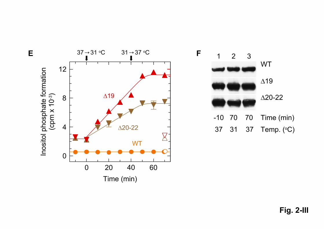
6 6
!
"
!

!

!
" " " ! " # !

!
" # " $
" $

!
"
# $ % & ' "
((
((
((
((
((
((
((


!
"
#$
%
&$
&'
%
(
)

! "
#
$
!"
#
" %
&
! "


! ! !
! !
!
!
!
!
! !

! " # $
%
&
'
'
( )
' &
*
+
6
( )
#
+
#
%
#+
%# ,
%
#
+
#
%
#+
%# ,
%
#
- - -- - - - - -
'


! "
#
$

! "

SUPPLEMENTARY MATERIAL
Cool-temperature-mediated activation of phospholipase C-2
in the human hereditary disease PLAID
Anja Schade, Claudia Walliser, Martin Wist, Jennifer Haas, Petra Vatter, Johann M. Kraus,
Davide Filingeri, George Havenith, Hans A. Kestler, Joshua D. Milner, Peter Gierschik
Supplementary figures S1-S5 Fig. S1. Expression of wild-type and mutant PLC isozymes in Figs. 4A and 4B
Fig. S2. Expression of wild-type and mutant PLC isozymes in Fig. 5
Fig. S3. Expression of wild-type and mutant PLC isozymes in Fig. 9A
Fig. S4. Expression of wild-type and mutant PLC isozymes in Figs. 9B and 9D
Fig. S5. Expression of wild-type and mutant PLC2 isozymes and portions thereof in Fig. 10A
Fig. S6. Expression of wild-type and mutant PLC2 isozymes and portions thereof in Fig. 10A
Fig. S7. Expression of wild-type and mutant PLC2 isozymes in Fig. 11

Supplementary Fig. S1
Fig. S1. (A) Expression of wild-type and mutant PLC1 and PLC2 isozymes in the experiment
shown in Fig. 4A. (B) Expression of wild-type and mutant PLC2 isozymes in the experiment
shown in Fig. 4B. Cells from one well each were washed once with 0.2 ml of Dulbecco’s PBS
and then lysed by addition of 100 µl of SDS-PAGE sample preparation buffer. The samples
were subjected to SDS-PAGE and immunoblotting was performed using an antibody reactive
against the c-Myc epitope on PLC1 or PLC2.
Δ
Δ
Δ
Δ
Δ
Δ
γ
γ ! Δ !
γ ! Δ !
γ Δ
γ Δ

Supplementary Fig. S2
Fig. S2. Expression of wild-type and mutant PLC2 isozymes in the experiment shown in Fig.
5, left panel, (A), center panel, (B), and right panel, (C). Cells from one well each were
analyzed by SDS-PAGE and immunoblotting using an antibody reactive against the c-Myc
epitope.
" # $ " #
Δ
" #
" #
%
&
' "
"#
"#
"# $ "#

Supplementary Fig. S3
Fig. S3. Expression of wild-type and mutant PLC2 isozymes in the experiment shown in Figs.
9A, left panel, (A) and 9A, right panel, (B). Cells from one well each were analyzed by SDS-
PAGE and immunoblotting using an antibody reactive against the c-Myc epitope.
Δ
&
( ) ' * $ + " "
Δ
Δ , - . /
, - . /

Supplementary Fig. S4
Fig. S4. Expression of wild-type and mutant PLC2 isozymes in the experiment shown in Figs.
9B (A) and 9D (B). Cells from one well each were analyzed by SDS-PAGE and immunoblotting
using an antibody reactive against the c-Myc epitope.
&
Δ
Δ &
Δ
Δ
Δ
Δ
0 $1
%
&
%
' "
0
0 2
' "
0
0 2
Δ

Supplementary Fig. S5
Fig. S5. Expression of wild-type and mutant PLC2 isozymes as well as portions thereof in the
experiment shown in Fig. 10A. Cells from one well each were analyzed by SDS-PAGE and
immunoblotting using an antibody reactive against the c-Myc epitope.
31 31 31 31 31 3137 37 37 37 37 37
1 2 3 4 5 6 7 8 9 10 11 12
WT
XPHn
YPHc
Co. WT SH XPHn YPHc XPHn+YPHc
Temp. (oC)
Mr x 10-3
150
100
70

Supplementary Fig. S6
Fig. S6. Expression of wild-type and mutant PLC2 isozymes as well as portions thereof in the
experiment shown in Fig. 10B. Cells from one well each were analyzed by SDS-PAGE and
immunoblotting using an antibody reactive against the c-Myc epitope.
WT 1920-22
PLC2
31 37 31 31 3137 37 37 31 3137 37
Temp. (oC)EGF

Supplementary Fig. S7
Fig. S7. Expression of wild-type and mutant PLC2 isozymes in the experiment shown in Fig.
11. Cells from one well each were analyzed by SDS-PAGE and immunoblotting using an
antibody reactive against the c-Myc epitope.
230
130
95
72
55
36
28
14
Mr x 10-3
WT20-22
X
Y
1 2 3 4 5 6 7 8 9 10
31 37 31 31 31 3137 37 37 37
Temperature(oC)
Top Related