


Languages
Pages
Legal

Control Nodal ALK7-ca
Sl Fig. 1. Identification of potential Nodal/ALK7 target genes using genearray. Total RNA was extracted from IOSE cells transfected with control vector, Nodal, or ALK7-ca and subjected to reverse transcribed. The cDNA samples were then labeled with biotin and hybridized to specific cDNA on the membrane (Human Cell Cycle Gene Array from SupperArray). Large arrow indicates cyclin G2 and small arrow indicates Skp1. Table listed genes that are up- or down-regulated by Nodal and ALK7-ca.
Gene Name Description GeneBank
UpregulationATM Ataxia telanlectasia mutated NM_000051Bax Bcl-2-associated X protein AY217036Cyclin G2 Cyclin G2 L49506Cyclin H Cyclin H U11791Cdc16 Cdc16 NM_003909Cdc7 Cdc7 AF015592p21 Cyclin-dependent kinase inhibitor 1A L47233p27 Cyclin-dependent kinase inhibitor 1B U10906p57 Cyclin-dependent kinase inhibitor 1C U22398p18 Cyclin-dependent kinase inhibitor 2C U17074p19 Cyclin-dependent kinase inhibitor 2D U40343Cullin-Cul2 Cullin 2 U83410Cullin-Cul5 Cullin 5 NM_003478E2F1 E2F transcription factor1 U47677E2F2 E2F transcription factor 2 NM_004191E2F6 E2F transcription factor 6 AF059292MAD2L2 MAD2-like 2 NM_006341PRC1 Protein regulator of cytokinesis 1 NM_003981RAD9 RAD9 (S. pombe) homolog U53174Rb Retinoblastoma 1 (including osteosarcoma) M15400Rbx1 Homo saplens ring-box protein1 (RBX1) mRNA NM_014248Ubiquitin C Polyubiquitin AB009010Downregulationp55cdc p55cdc (cdc20) NM_001255Cks1p9 CDC28 protein kinase 1 NM_001826Ki67 Antigen identified by monoclonal antibody Ki-67 X65550Skp1 Cyclin A/CDK2-associated p19 U33760TIMP3 Tissue inhibitor of metalloproteinase 3 NM_000362
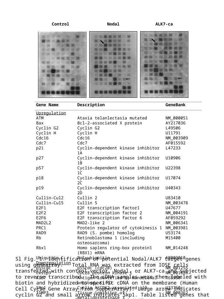
A
pcDNA3.1-CCNG2-V5
Xba IKpn I
V5 His Stop
Age I
pcDNA4-CCNG2-V5
Kpn I
V5 His Stop
Age I
pcDNA4-CCNG2-myc-His
Sac IIKpn I
myc His Stop
pCCNG2-GFP
Sac IIEcoR I
GFP Stop
p3xFLAG-CCNG2
BamH I
Stop3xFlag
EcoR I
pcDNA3-CCNG2-V5-His
Sac IIKpn I
V5 His Stop
B
CCNG2-V5
g
12h 24h 48h
0 5 10 15 0 5 10 15 0 5 10 15
CCNG2-V5
-actin
-actin
Post-transfection
21
6448
37
82
2619
116 a b c d e
212121 21C
Sl Fig. 2. Generation of cyclin G2 expression constructs and detection of cyclin G2 protein. A) Generation of human CCNG2 plasmids. These plasmids contain the fusion tags either at N-terminus or C-terminus. Arrow indicates a start site of translation. B) Detection of exogenous CG2 in a dose and time-dependent manner. IOSE397 and OV2008 cells were transiently transfected with CG2-V5 plasmid. CG2 fusion protein was detected using a V5 antibody. β-actin was used as control for the equal loading. C) Representative Western blots probed with different cyclin G2 antibodies. OV2008 cells were transiently transfected with an empty vector (1) or CCNG2-V5 (2). Proteins were extracted at 6 hours post-transfection and subjected to SDS-PAGE. Blots were probed with anti-V5 from Invitrogen (a), anti-CCNG2 from Abcam (b), anti-CCNG2 from Epitomics (c); anti-CCNG2 from Santa Cruz (d), and anti-CCNG2 from Abnova (e). Only the antibody from Santa Cruz appeared to recognize cyclin G2.
OV
20
08
IOS
E

Xu et al., Fig. Sl3
A
B
DAPI CCNG2 Merge
DAPI CCNG2 Skp2 Merge
Skp2
DAPI CCNG2 pcDNA Merge
DAPI
DAPI
CCNG2
CCNG2
Skp2
ALK7-ca
Merge
Merge
Sl Fig. 3. Effect of Skp2 on cyclin G2 expression as detected by immunofluorescent staining. A) Cells were co-transfected with cyclin G2 and control vector (pcDNA4, upper panel), Skp2 (middle panel), or ALK7-ca (lower panel). In cells expressing Skp2, cyclin G2 level was low whereas in cells expressing ALK7-ca, strong cyclin G2 signal was detected. B) Cells were transfected with cyclin G2 and control-siRNA (upper panel) or Skp2-siRNA (lower panel). Strong cyclin G2 signals were observed in the presence of Skp2-siRNA compared to control-siRNA. Scale bar: 100m.
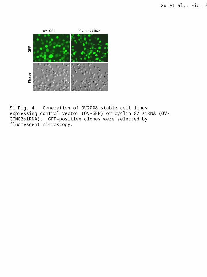
OV-GFP OV-siCCNG2G
FP
Pha
se
Sl Fig. 4. Generation of OV2008 stable cell lines expressing control vector (OV-GFP) or cyclin G2 siRNA (OV-CCNG2siRNA). GFP-positive clones were selected by fluorescent microscopy.
Xu et al., Fig. Sl4
Top Related