


![[SVY] Survey Data - Data Analysis and Statistical Software · jfor the jth observation means, roughly speaking, that the jth observation represents w j elements in the population](https://static.fdocuments.us/doc/165x107/5ac3ebc17f8b9af91c8c9113/svy-survey-data-data-analysis-and-statistical-software-the-jth-observation-means.jpg)


Languages
Pages
Legal

Clustering of GPVI dimers upon adhesion tocollagen as a mechanism to regulate GPVIsignalling in plateletsPoulter, Natalie; Pollitt, Alice; Owen, Dylan M.; Gardiner, Elizabeth E.; Andrews, Robert;Shimizu, Hiroyuki; Ishikawa, Daisuke; Bihan, Dominique; Farndale, Richard W; Moroi,Masaaki; Watson, Steve; Jung, Stephanie MDOI:10.1111/jth.13613
License:Other (please specify with Rights Statement)
Document VersionPeer reviewed version
Citation for published version (Harvard):Poulter, N, Pollitt, A, Owen, DM, Gardiner, EE, Andrews, R, Shimizu, H, Ishikawa, D, Bihan, D, Farndale, RW,Moroi, M, Watson, S & Jung, SM 2017, 'Clustering of GPVI dimers upon adhesion to collagen as a mechanismto regulate GPVI signalling in platelets: GPVI-dimer clustering on collagen' Journal of thrombosis andhaemostasis : JTH. DOI: 10.1111/jth.13613
Link to publication on Research at Birmingham portal
Publisher Rights Statement:This is the peer reviewed version of the following article: Clustering of GPVI dimers upon adhesion to collagen as a mechanism to regulateGPVI signalling in platelets, Journal of Thrombosis and Haemostatis, which has been published in final form athttp://dx.doi.org/10.1111/jth.13613. This article may be used for non-commercial purposes in accordance with Wiley Terms and Conditionsfor Self-Archiving
Checked 9/1/2017
General rightsUnless a licence is specified above, all rights (including copyright and moral rights) in this document are retained by the authors and/or thecopyright holders. The express permission of the copyright holder must be obtained for any use of this material other than for purposespermitted by law.
•Users may freely distribute the URL that is used to identify this publication.•Users may download and/or print one copy of the publication from the University of Birmingham research portal for the purpose of privatestudy or non-commercial research.•User may use extracts from the document in line with the concept of ‘fair dealing’ under the Copyright, Designs and Patents Act 1988 (?)•Users may not further distribute the material nor use it for the purposes of commercial gain.
Where a licence is displayed above, please note the terms and conditions of the licence govern your use of this document.
When citing, please reference the published version.
Take down policyWhile the University of Birmingham exercises care and attention in making items available there are rare occasions when an item has beenuploaded in error or has been deemed to be commercially or otherwise sensitive.
If you believe that this is the case for this document, please contact [email protected] providing details and we will remove access tothe work immediately and investigate.
Download date: 13. Jun. 2018

Acc
epte
d A
rtic
le
This article has been accepted for publication and undergone full peer review but has not
been through the copyediting, typesetting, pagination and proofreading process, which may
lead to differences between this version and the Version of Record. Please cite this article as
doi: 10.1111/jth.13613
This article is protected by copyright. All rights reserved.
Received Date : 25-May-2016
Revised Date : 10-Dec-2016
Accepted Date : 29-Dec-2016
Article type : Original Article - Platelets
Clustering of GPVI dimers upon adhesion to collagen as a mechanism to regulate
GPVI signalling in platelets
N. S. Poulter1,8, A. Y. Pollitt1,2, D. M. Owen3, E. E. Gardiner4, R. K. Andrews5, H. Shimizu6, D.
Ishikawa6, D. Bihan7, R. W. Farndale7, M. Moroi7, S. P. Watson1,8, and S. M. Jung7,
1Institute of Cardiovascular Sciences, College of Medical and Dental Sciences, University of
Birmingham, Birmingham, B15 2TT, United Kingdom
2Institute for Cardiovascular and Metabolic Research, School of Biological Sciences,
University of Reading, Reading, UK RG6 6AS (current address)
3 Department of Physics and Randall Division of Cell and Molecular Biophysics, King's
College London, London, United Kingdom
4 Dept Cancer Biology and Therapeutics, John Curtin School of Medical Research,
Australian National University, Canberra, ACT 2600 Australia
5 Australian Centre for Blood Diseases, Monash University, Melbourne, Australia
6 Research Department, Chemo-Sero-Therapeutic Research Institute, Kaketsuken,
Kumamoto, Japan
7 Department of Biochemistry, University of Cambridge, Cambridge, United Kingdom, CB2
1QW
8 Centre for Membrane Proteins and Receptors (COMPARE), Institute of Cardiovascular
Sciences, College of Medical and Dental Sciences, University of Birmingham, Birmingham,
B15 2TT, United Kingdom
¶Correspondence:
Stephanie M. Jung, Ph.D.
Department of Biochemistry
University of Cambridge
Tennis Court Road
Cambridge CB2 1QW

Acc
epte
d A
rtic
le
This article is protected by copyright. All rights reserved.
United Kingdom
e-mail: [email protected]
Running title: GPVI-dimer clustering on collagen
Keywords: platelet membrane glycoproteins; platelet adhesiveness; platelet activation;
receptors, collagen; glycoprotein
Summary (250 words) and Main text (3986 words)
Essentials
Dimeric high-affinity collagen receptor glycoprotein VI (GPVI) is present on resting
platelets.
Spatio-temporal organization of platelet GPVI-dimers was evaluated using advanced
microscopy.
Upon platelet adhesion to collagenous substrates, GPVI-dimers coalesce to form
clusters.
Clustering of GPVI-dimers may increase avidity and facilitate platelet activation
Summary
Background: Platelet GPVI binding to subendothelial collagen exposed upon blood vessel
injury initiates thrombus formation. Dimeric GPVI has high affinity for collagen and occurs
constitutively on resting platelets. Objective: To identify higher order oligomerisation
(clustering) of pre-existing GPVI-dimers upon interaction with collagen as a mechanism to
initiate GPVI-mediated signalling. Methods: GPVI was located using fluorophore-
conjugated GPVI-dimer-specific Fab (antigen-binding fragment). Tested substrates include
Horm collagen I fibres, soluble collagen III, GPVI-specific collagen peptides and fibrinogen.
GPVI-dimer clusters on the platelet surface interacting with these substrates were visualised
Acc
epte
d A
rtic
le
This article is protected by copyright. All rights reserved.
using complementary imaging techniques: Total Internal Reflection Fluorescence
Microscopy (TIRFM) to monitor real time interactions and direct Stochastic Optical
Reconstruction Microscopy (dSTORM), providing relative quantification of GPVI cluster size
and density. Confocal microscopy was used to locate GPVI-dimer clusters, GPIb, integrin
α2β1, and phosphotyrosine. Results: Upon platelet adhesion to all collagenous substrates,
GPVI-dimers coalesced to form clusters; notably clusters formed along the fibres of Horm
collagen. dSTORM revealed that GPVI density within clusters depended on the substrate,
collagen III being most effective. Clusters on fibrinogen-adherred platelets were much
smaller and more numerous; whether these are pre-existing oligomers of GPVI-dimers or
fibrinogen-induced is not conclusive. Some GPVI-dimer clusters colocalized with areas of
phosphotyrosine, indicative of signalling activity. Integrin α2β1 localized to collagen fibres
close to GPVI-dimer clusters. GPVI-clustering depends on a dynamic actin cytoskeleton.
Conclusions: Platelet adhesion to collagen induces GPVI-dimer clustering. GPVI-clustering
increases both avidity for collagen and proximity of GPVI-associated signalling molecules,
which may be crucial for initiation and persistence of signalling.
Introduction
Upon blood vessel injury, circulating platelets interact with exposed subendothelial collagen
through the collagen receptor glycoprotein VI (GPVI). This 65-kDa immune receptor signals
through its associated Fc-receptor γ-chain, which contains an immunoreceptor tyrosine-
based activation motif (ITAM) in its intracellular domain. Binding of GPVI to collagen
induces phosphorylation of the ITAM residues, which can then bind to Syk, itself becoming
phosphorylated and activated. This process initiates signalosome assembly [1], leading to a
series of downstream signals, resulting in platelet activation, finally culminating in thrombus
formation.

Acc
epte
d A
rtic
le
This article is protected by copyright. All rights reserved.
GPVI contains two extracellular Ig-like domains: D1 containing the collagen binding site
[2,3] and D2, connected via an O-glycosylated stem to its transmembrane domain and short
cytoplasmic tail [4]. GPVI binds to tandem GPO (glycine-proline-hydroxyproline) sequences
in collagen [5,6]. Surface plasmon resonance showed that dimerized recombinant GPVI
(D1D2-Fc)2 bound collagen fibres with high affinity but binding of its monomeric form (D1D2)
was too low to measure [7]. Monomeric and dimeric recombinant D1D2 showed similar
affinity for collagen-related peptide (CRP), a triple-helical peptide containing 10 contiguous
GPO triplets, suggesting GPVI-dimer may have a specific conformation that recognizes the
higher-order structure of fibrous collagen, beyond simply the GPO sequences. The crystal
structure of a D1D2 dimeric assembly [8] allowed docking simulations [3,5,8] which
suggested that D1 contained grooves large enough to accommodate the triple-helical CRP.
In 2009, Jung et al. provided direct evidence for the presence of dimers on the resting
platelet surface with GPVI-dimer specific, inhibitory m-Fab-F [9]. Later, they reported a non-
inhibitory Fab, 204-11, which recognized GPVI-dimers [10], and used it to show that GPVI-
dimers were constitutively present on resting platelets. These observations suggested that
the first interaction in collagen-induced activation of platelets is collagen binding to GPVI-
dimers. Several groups, however, reported that platelet activation induced formation of
GPVI-dimers. Arthur et al. (11) provided biochemical evidence for disulphide-linked dimers in
activated platelets. Loyau et al., employing GPVI-dimer–specific mab 9E18, reported that
GPVI dimerization was induced by soluble agonists or VWF, with almost no dimersdetected
on resting platelets [12], leading them to propose dimer formation as a means to control
collagen-induced platelet activation. Dimerization is an accepted mechanism for cell
activation through receptor tyrosine kinases [13], where ligand binding to the receptor
extracellular domains induces dimerization, causing a conformational change that brings
together the kinase domains in the cytoplasmic tails, facilitating autophosphorylation, thereby
initiating intracellular signals. The Src-family kinase Lyn associates with the cytoplasmic tail
of GPVI [14], which lacks intrinsic kinase activity, so that FcRγ attached to one GPVI

Acc
epte
d A
rtic
le
This article is protected by copyright. All rights reserved.
monomer might be phosphorylated by Lyn associated with a second monomer, brought into
proximity by dimerization.
An alternative activation mechanism would be higher-order receptor clustering [15], which
does not preclude the presence of constitutive GPVI-dimers in non-activated platelets.
Clustering has been demonstrated for many classes of receptor, including G protein–
coupled receptors [16]; adhesion receptors such as platelet integrin αIIbβ3 [17]; platelet
CLEC-2 [18] and notably, the discoidin domain receptor, DDR1, a constitutively-dimeric
tyrosine kinase receptor for collagen [19].
As GPVI dimers are present on resting platelets we hypothesize that a further level of control
is required. We hypothesize that clustering of GPVI-dimers is a plausible mechanism to
modulate platelet activation once the high-affinity, but low-copy-number, GPVI-dimers
engage the limited number of binding sites on collagen [2,20]. The formation of GPVI
clusters could result in increased avidity and may bring associated signalling molecules
together to facilitate platelet activation. We used complementary imaging techniques to test
this hypothesis: TIRFM (Total Internal Reflection Fluorescence Microscopy), to visualize in
real-time GPVI-dimer distribution at the interface between the platelet membrane and
immobilized collagenous substrates; dSTORM (Direct Stochastic Optical Reconstruction
Microscopy) for relative quantification of GPVI cluster size and density; and confocal
microscopy to compare localization of GPVI-dimers and two other receptors involved in the
platelet-collagen interaction, GPIb and integrin α2β1, and whether signalling was associated
with the clustered GPVI-dimers.
Materials and methods
Non-inhibitory, recombinant dimer-specific 204-11 Fab [10] was derived from clone 204-11
[21] by Kaketsuken (Kumamoto, Japan). Antibodies were fluorescently labelled by a
Microscale Protein Labelling kit (Molecular Probes); degree of fluorescent labelling =2–5 dye

Acc
epte
d A
rtic
le
This article is protected by copyright. All rights reserved.
molecules/protein molecule. 4G10 (anti-phosphotyrosine; Millipore Merck); Anti-human
CD42b/GPIb, clone 486805 (R&D Systems); 16B4 (mouse anti-human CD49b/integrin α2
chain; AbD Serotec); Alexafluor 647–conjugated AffiniPure F(ab’)2 fragment goat anti-
mouse IgG, Fcγ–specific (Jackson); Gi9 (anti-integrin α2; Abcam), Alexafluor 647–anti-
human CD62P (Bio-Rad). Other materials, reagent grade or better, were obtained from
commercial sources.
Platelet preparation
Washed platelets were prepared from ACD-anti-coagulated blood from healthy volunteers
(22) and resuspended at 3–5 x 10⁷ platelets/ml in Hepes-Tyrodes buffer (HT: 134 mM NaCl,
0.34 mM Na2HPO4, 2.9 mM KCl, 12 mM NaHCO3, 20 mM HEPES, 5 mM glucose, pH 7.3).
Preparation of collagenous substrate–coated glass dishes for imaging
35-mm glass (0.7 mm)–bottomed MatTek dishes (MatTek, USA) were coated with 10 µg/ml
CRP-XL, III-30, or collagen III, in phosphate-buffered saline (PBS: 0.01 M phosphate buffer,
0.0027 M KCl, 0.137 M NaCl,pH 7.4 ); Horm collagen, in manufacturer-supplied diluent)
overnight at 4C. The dishes were PBS-washed, 1% bovine serum albumin–blocked
(BSA/PBS; heat-denatured,filtered) for 1 h, and then PBS-washed ready for platelet
spreading.
TIRFM
Adhesion of Alexafluor 488–204-11 Fab–labelled washed platelets (3 x 10⁷ platelets/ml in
HT buffer containing 2 mM MgCl2) to immobilized collagenous substrate was imaged by
TIRFM (Nikon TIRF system mounted on a Nikon Eclipse Ti inverted microscope, with Nikon
60x NA 1.49 TIRF objective). Images were obtained at 5-s intervals for 20–30 min at 37oC,
followed by fixation in formalin and confocal imaging. Integrin α2β1–blockade was achieved
Acc
epte
d A
rtic
le
This article is protected by copyright. All rights reserved.
by preincubating platelets with 10 μg/ml Gi9 blocking antibody. Platelet morphology was
followed using differential interference contrast (DIC) microscopy
dSTORM
Alexafluor 647-204-11 Fab–labelled washed platelets were allowed to adhere to
collagenous-substrate-coated MatTek dishes. Adhered platelets were fixed, permeabilised
and stained with Phalloidin-Alexa488. Samples were imaged in switching buffer as
described previously (22) on a Nikon Eclipse Ti-E N-STORM system in dSTORM mode
using Perfect Focus, with a CFl SR Apochromat TIRF 100x Oil, 1.49NA objective lens and
an N-STORM filter cube with excitation using the Agilent Ultra High Power Dual Output
Laser bed (170-mW 647-nm laser), and image capture with an Andor IXON Ultra 897
EMCCD camera. 30,000 frames were captured using Nikon NIS Elements v4.2 and
reconstructed using STORM analysis module v3.2 with drift correction and Gaussian
rendering of data points. Detected points with a photon count of less than 500 were
discarded from reconstructed data before further processing. Points in the reconstructed
images represent individually identified fluorescent blinking events, which are referred to as
molecules.
Confocal imaging
Platelets were preincubated with Alexa 488 or 647–conjugated 204-11 Fab (4 μg/ml) alone
or with another fluorescently labelled antibody. In some experiments, platelets were also
treated with an inhibitor or inhibitory antibody. The platelets were allowed to adhere to a
collagenous substrate, fixed and imaged with an FV300 IX81 Laser-scanning confocal
microscope, 60x oil immersion objective (Olympus). Where indicated, platelets were
permeabilized (0.1% Triton/PBS) following fixation and stained for actin (Alexafluor 647–
Phalloidin) or phosphotyrosine (4G10,5 μg/ml).

Acc
epte
d A
rtic
le
This article is protected by copyright. All rights reserved.
Flow cytometry to measure GPVI-dimers
Washed platelets were pre-incubated with DMSO (0.25% final concentration) or actin
antagonist (cytochalasin D, latrunculin A, or jasplakinolide; 10 µM; 0.25% DMSO, final
concentration), and GPVI-dimer formation was measured by flow cytometry [10].
Data analyses
Data analyses,performed with Prism v7 (GraphPad, San Diego, CA), are described in the
figure legends. Differences among treatment groups in the flow cytometry experiments were
calculated by paired t-test. dSTORM cluster analysis was performed within MATLAB for
each 3 x 3 µm ROI (region of interest) as described by Owen et al. [23] with modifications
described in Pollitt et al. [18]. Platelets were identified by phalloidin staining of the actin
cytoskeleton. ROIs were positioned within the platelet ensuring that the edges of the ROI
fell within the platelet boundary. Clusters of <3 points were discounted. The numerical data,
processed using Microsoft Excel, were analysed as detailed in the figure legends. The
degree of colocalization between the GPVI and phophotyrosine confocal images was
quantified by Image-Pro Premiere 9.2 (Molecular Cybernetics).
Results
TIRFM imaging of GPVI-dimer–cluster formation in live platelets interacting with immobilized
collagenous substrates
We investigated the spatial and temporal distribution of GPVI dimers on the surface of
platelets interacting with different immobilized collagenous substrates (Table I). Washed
platelets labelled for GPVI-dimer were allowed to adhere, and cluster formation and
dynamics were visualized by TIRFM (Fig.1; supplemental movie). Discrete clustered GPVI-

Acc
epte
d A
rtic
le
This article is protected by copyright. All rights reserved.
dimers can be visualized in spreading platelets, whereas non-clustered GPVI-dimers are
poorly-resolved, appearing as diffuse fluorescence over the platelet surface.
GPVI dimers form clusters on platelets adhering to all collagenous substrates tested.
Cluster formation on fibrous Horm collagen (Horm) follows a distinct distribution,
concentrated along the collagen fibre (Fig. 1A: i, ii), initially forming at the point of contact
between the platelet and the fibre, then propagating along the fibre (Fig. 1Aii). Clusters are
not restricted to the visible collagen fibre and are also present on the platelet surface
presumably due to indirect signal-induced clustering (e.g., outside-in signalling through
released substances such as fibrinogen, ADP, VWF, etc.), through contact with smaller
(non-visible) fibres in these portions of the surface, or both. Non-fibrous collagenous
substrates CRP-XL (Fig. 1B), collagen III (Fig. 1C, Col III), and III-30 (Fig. 1D, III-30) also
support formation of GPVI-dimer clusters, each with similar cluster distributions. Small
clusters were evident upon first contact of the platelet with the coated surface, eventually
forming throughout the platelet surface; some clusters expanded and coalesced.
Different collagenous substrates induce different degrees of clustering
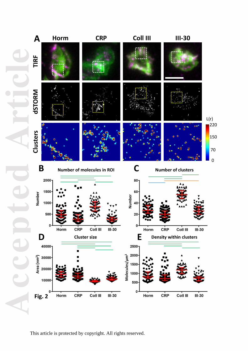
GPVI-clustering in fixed platelets spread on the different collagenous substrates was
quantitated using dSTORM, which has a typical x-y resolution of 20–30 nm (24, 25).
Widefield TIRF imaging of the platelets labelled for F-actin and GPVI dimer show the same
distribution of GPVI as that observed in the live-cell imaging (Fig. 2A). Cluster analysis of
dSTORM data is represented by the heat maps in Fig.2A. High and low levels of GPVI-
clustering are seen as red and blue, respectively. All collagenous substrates induced more
GPVI-clustering than expected to occur randomly; however, the cluster distribution
depended upon the specific substrate. Horm induced high degrees of clustering along the
fibres, whereas the other substrates induced clusters more evenly distributed throughout the
ROI. Quantification shows that there are more GPVI dimers and a greater number of clusters
found per unit area on platelets spread on Col III than on the other substrates (Fig.
2B,C).These clusters are small (Fig. 2D) but contain the highest density of GPVI dimers (Fig.

Acc
epte
d A
rtic
le
This article is protected by copyright. All rights reserved.
2E, median values). Horm induced the next highest amount of GPVI dimer per ROI, 42%
less than Col III, and Horm-induced clusters were 31% less dense than those on Col III.
CRP-XL and III-30 were least effective in dimer formation (each with ~70% fewer molecules
detected in the ROI compared to Col III) and cluster densities were also correspondingly
reduced when compared to Col III (40% and 37% reduction, respectively). The size of
GPVI-dimer clusters formed on Horm and CRP are not significantly different from each other
but were significantly larger than those formed on Col III and III-30 (Fig. 2D). In summary,
all collagenous substrates cause GPVI dimers to cluster, but different numbers of dimers
and densities of GPVI within clusters are formed, depending on the nature of the
collagenous substrate.
GPVI dimers are not restricted to the platelet membrane in direct contact with the visible
collagen fibres (Figs. 1A, 2A). Potential explanations for this are the existence of “non
visible” microfibers in those areas, to which GPVI-dimers can still bind, and/or fibrinogen or
other substances released from adhered platelets may induce cluster formation via outside-
in signalling. To determine whether GPVI clusters can form by collagen-independent platelet
activation, platelets were spread on fibrinogen and clustering of GPVI dimer quantified using
dSTORM (Fig 3). GPVI-dimer clusters seen on fibrinogen differ from those seen on Horm
collagen: relative density of dimers in platelets spread on fibrinogen is ~50% less when
compared to platelets spread on Horm (Fig 3B); there are many more small clusters on the
fibrinogen, and the small clusters are ~40% less dense than those formed on Horm (3C–E).
Although it is possible that GPVI-dimer clustering can occur by a GPVI-independent
mechanism, collagen greatly increases the formation of GPVI dimers, which coalesce into
larger and denser clusters.

Acc
epte
d A
rtic
le
This article is protected by copyright. All rights reserved.
Relationship between clustered GPVI-dimers and other receptors involved in the platelet–
collagen interaction
Localisations of other receptors involved in the platelet-collagen interaction, GPIb (Fig. 4A)
and integrin 2β1 (Fig. 4B), were compared to that of GPVI-dimer clusters on platelets
adhered to collagenous substrates. GPVI-dimer clusters form along Horm collagen fibres,
whereas they appeared throughout the lamellipodia of platelets spread on Col III and III-30.
Like GPVI-dimer clusters, α2β1 was also found to localize along Horm collagen fibres, but
was confined to the cell body of platelets adhered to Col III and III-30; no co-localisation with
GPVI-dimer clusters was observed on the platelet lamellipodia. GPIb (VWF receptor) did not
associate with GPVI-dimer clusters in the lamellipodia of any of the collagenous substrates
tested, remaining in the platelet cell body.
Contribution of collagen receptor integrin α2β1 to platelet adhesion and GPVI-dimer cluster
formation
Platelets labelled with Alexafluor 488–204-11 Fab with or without Gi9 (α2β1-blocking
antibody), were allowed to settle onto immobilized non-fibrous collagen III or Horm fibres,
and monitored by TIRFM. Gi9 treatment prevented platelet adhesion to collagen III, but
merely decreased the extent of GPVI-dimer cluster formation on Horm fibres (Fig. 4C). Gi9
had no effect on platelet adhesion/cluster formation on the GPVI-specific ligands CRP-XL
and III-30 (data not shown).
Effect of Src-family and Syk kinase inhibition on GPVI cluster formation
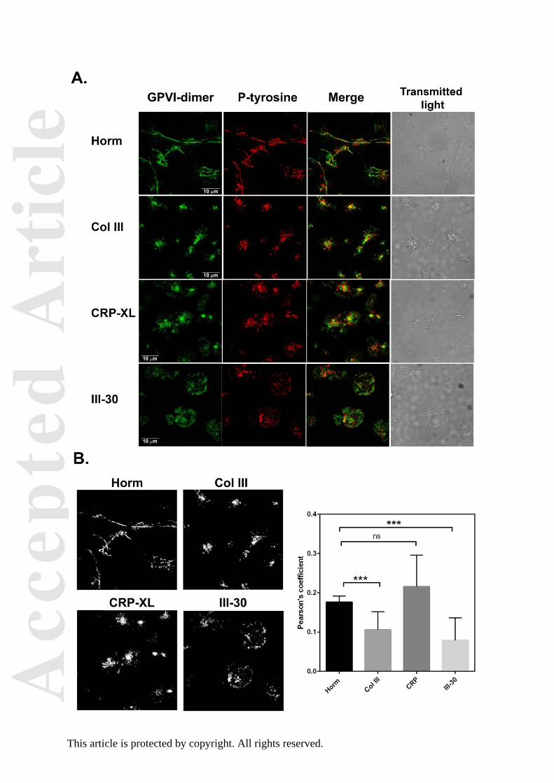
Confocal images of platelets adhered to Horm, Col III, III-30, or CRP-XL show that areas rich
in phosphotyrosine co-localize with some of the GPVI-dimer clusters (Fig. 5A), suggesting
signalling is occurring in these regions. Degree of colocalization was analysed using
Pearson’s correlation (Fig. 5B, right). To investigate whether protein tyrosine
phosphorylation is important in GPVI cluster formation, platelets were treated with the Syk

Acc
epte
d A
rtic
le
This article is protected by copyright. All rights reserved.
inhibitor PRT-060318 or Src-family kinase inhibitor PP2. Both inhibitors, at concentrations
demonstrated to inhibit spreading and phosphorylation in human platelets [26], inhibited
adhesion on Horm and Col III. Both inhibitors reduced platelet spreading on all collagenous
substrates (Supplementary Fig. 1 shows corresponding DIC images that enable visualisation
of platelet morphology in the presence and absence of the inhibitors). Despite having a
reduction in spreading, discrete GPVI-dimer clusters were visible in platelets adhered to
CRP XL and III-30 in the presence of PRT-060318 or PP2 (Fig. 6).
Effect of disrupting actin dynamics on GPVI-dimerisation and clustering
Flow cytometry was used to quantify effects of drugs that inhibit actin dynamics on GPVI-
clustering: Cytochalasin D (cyt) and Latrunculin A (lat), actin polymerization–blockers, and
Jasplakinolide (jas), F-actin stabilizer. All three inhibitors (10 μM) significantly decreased
GPVI-dimer levels in resting platelets (Fig. 7A,B). To avoid total abolition of adhesion, we
used low-dose (2 μM) actin antagonist to assess the role of the actin cytoskeleton in
clustering of GPVI-dimers on Horm, Col III, CRP-XL, and III-30 (Figs. 7C–D,respectively).
Low-dose Latrunculin A disrupted the actin cytoskeleton and severely depressed cluster
formation, with platelets that did adhere not spreading. Low-dose Jasplakinolide had similar
but more severe effects on cluster formation, with no apparent F-actin staining, and fewer
adhered, but non-spread platelets.
Discussion
GPVI-dimers constitutively present on resting platelets are the collagen-binding form of this
receptor, having over 100-fold higher affinity for collagen than the monomer [9,10]. The
constitutive presence of dimers suggests that a mechanism beyond the formation of dimers
may be necessary to initiate signalling through GPVI. One mechanism may be the formation
of higher-order oligomers (clusters) which has been demonstrated to play an important role
in the amplification, maintenance and termination of receptor signalling in many cell types

Acc
epte
d A
rtic
le
This article is protected by copyright. All rights reserved.
[27,28], including platelets [18,27]. We propose that GPVI clusters which may recruit
signalling molecules to facilitate platelet activation. To explore this hypothesis, we used
complementary imaging methods: TIRFM, dSTORM, and confocal microscopy.
TIRFM enabled the temporal visualisation of GPVI-cluster formation at the surface of
platelets spreading on collagenous substrates. After the initial contact of GPVI-dimer with
the substrate, the number of GPVI-dimer clusters increases rapidly. Notably, in platelets
adhered to fibrous Horm collagen, discrete clusters form along the fibres first, then
throughout the surface of the spread platelet (Fig.1A). This is consistent with GPVI-dimers
interacting with the surface of the collagen fibre, forming clusters at those points, followed by
signalling that induces more cluster formation in regions of the platelet not in direct contact
with the visible fibre. A different clustering pattern is observed on the immobilized non-
fibrous collagenous substrates, CRP-XL (Fig. 1B) and III-30 (Fig. 1D), which exclusively bind
GPVI, and soluble Col III (Fig. 1C), which binds GPVI and α2β1. Immobilized non-fibrous
substrates are randomly-orientated triple-helical molecules and would be expected to
support a different clustering pattern than Horm collagen fibers, which contain a highly
organised parallel assembly of triple-helical tropocollagen molecules within the microfibrils.
This structure dictates that GPVI-binding sites will be distributed on the surface of a fibre at
fixed lateral and axial intervals, and suggests that a GPVI-dimer would be able interact with
neighbouring tropocollagen molecules presenting either separate or a composite binding
sites [20].
dSTORM allows single fluorophore molecules to be detected and located with very high
spatial precision. Combined with cluster analysis, dSTORM allows quantitation of the
number of GPVI dimer molecules and cluster number, size and density in ROIs within a
spread platelet. This information permits the relative differences in GPVI clusters to be
compared in platelets spread on different substrates. Horm collagen fibres induced a high
degree of clustering by virtue of their structure, outlined above. The spacing and orientation
of the two proposed collagen-binding grooves of GPVI-dimer might allow it to bind with
increased avidity to sites on adjacent tropocollagen molecules, allowing clustering to occur

Acc
epte
d A
rtic
le
This article is protected by copyright. All rights reserved.
[20]. The ability of immobilized III-30, CRP-XL, and Col III to bind GPVI will depend on the
density and relative orientation of the immobilized substrate helices. CRP and III-30 contain
only GPVI-binding motifs, the former having more GPVI-reactive GPO triplets per molecule,
whereas immobilized soluble Col III binds both GPVI and α2β1, which may support
cooperative platelet binding, since motifs for each receptor are located close together. Such
differences may explain the variation in cluster density observed between the different
collagenous substrates. Indeed, both Horm and Col III had significantly higher GPVI cluster
density than III-30, and Col III clusters were also significantly denser than those on CRP-XL,
suggesting a measurable role for 21 in these events.
On platelets adhered to non-fibrous Col III and III-30, GPIb (Fig. 4A) and 21 (Fig. 4B)
do not co-localize with the majority of the GPVI-dimer clusters, which are located in the
lamellipodia of the spread platelet. However, α2β1 is indispensable for platelet binding to
soluble Col III, since adhesion is severely reduced by the blocking anti-integrin α2β1
antibody Gi9. Synergism between GPVI and α2β1 has also been observed using
immobilized model peptides in flowing blood thrombus deposition studies [31]. In contrast,
Gi9 did not prevent platelet adhesion/cluster formation on fibrous Horm collagen, indicating
that GPVI binding to the collagen fibre is much stronger than to non-fibrous substrates.
These observations are consistent with GPVI being sufficient to support platelet binding to
collagen fibres but not to soluble collagen [32]. Integrin 21 was also located along Horm
collagen fibres in bound platelets, and the magnified confocal images (Fig. 4B) suggest that
α2β1 binds close to some of the GPVI-dimer–clusters on the fibre. The model proposed by
Herr and Farndale [20] suggests that α2β1 and GPVI might bind ~10nm apart in collagen III,
and inspection of the collagen I sequence suggests that similar considerations would apply
to Horm collagen. Although non-activated α2β1 may bind its high affinity motif, GFOGER, in
Horm fibres [33], this is not essential for GPVI-dimer binding and clustering. However, 21
may serve to accelerate platelet adhesion to the collagen fibre [34], thus facilitating further
GPVI-clustering. Under the present static adhesion conditions, however, the distribution of

Acc
epte
d A
rtic
le
This article is protected by copyright. All rights reserved.
GPIb differed from that of the GPVI dimer–clusters on collagen fibres, although GPIb was
observed in the platelet cell body, adjacent to the fibre (Fig. 4A). A Mab against GPIb (SZ2)
was reported to block CRP-XL–induced platelet aggregation, and GPIb to co-precipitate with
GPVI in both resting and thrombin-stimulated platelets, suggesting an interaction between
the two receptors [35]. Platelet adhesion to collagen under high shear requires VWF
multimers, via GPIb, to tether the platelet to collagen, but our experiments are performed
under static conditions in the absence of plasma VWF, so GPIb is less important.
What induces GPVI clustering? One possibility is that platelet activation induced by the
initial interaction of GPVI with collagen leads to GPVI oligomerisation. However, in spite of
severe inhibition of platelet spreading by the Syk inhibitor PRT-060318, limited cluster
formation on all four immobilized collagenous substrates was still observed (Fig. 6, right).
The marked inhibition of cluster formation on Col III (which lacks the high-affinity motif,
GFOGER) would be due to blocking α2β1 activation, hence removing the contribution of its
high affinity form to stabilize platelet binding to collagen. Src-family kinase inhibitor PP2 also
inhibited platelet adhesion, but to a lesser extent than PRT-060318 (Fig. 6, middle).
Alternatively, blockade of both Src and Syk might potently inhibit secretion [36,37],
preventing release of active substances (including fibrinogen), thus reducing clustering via
secondary signaling pathways. These results suggest that, whilst platelet adhesion and
spreading is fully dependent on GPVI-mediated signalling, GPVI-cluster formation is only
partly so.
Movement of membrane proteins is controlled, in part, by the cytoskeleton in other cells
[38]. Inhibitors of actin dynamics at 10 μM inhibited GPVI-dimer formation in resting platelets
and prevented the GPVI-dimer increase in CRP-XL– or thrombin-activated platelets (Fig.
7A,B). At 2 μM, a threshold inhibitory concentration, there is severely limited adhesion of
non-spread platelets on all 4 tested substrates, but there is evidence of residual cluster
formation and disturbed actin filament distribution. These results suggest the contribution of
Acc
epte
d A
rtic
le
This article is protected by copyright. All rights reserved.
the peripheral membrane cytoskeleton to GPVI cluster formation, a topic for future
investigation.
Although the GPVI-dimers on resting platelets are competent to bind collagen, they are
not exposed to it in an uninjured vessel and the low density of GPVI-dimers in resting
platelets (1500/platelet) [10] suggests that they may be too far apart to induce efficient
signalling; thus platelets remain inactive, the GPVI-dimers requiring both receptor ligation
and proximity to activate platelets. However, upon vessel injury, subendothelial collagen
fibres are exposed to the bloodstream, and efficient platelet activation is necessary to
prevent bleeding. Once a vessel is injured, the binding sites on fibres of collagen types I and
III become accessible to the receptors involved in the platelet–collagen interaction GPVI,
integrin α2β1, and GPIb. The proximity of GPVI-dimer binding sites on the fibre surface
enable clustering of GPVI-dimers, increasing avidity and bring together necessary signalling
components to initiate signalling and lead to efficient platelet activation and thrombus
formation. We examined GPVI-dependent signalling in the vicinity of GPVI clusters, and
whether inhibitors of GPVI-mediated signalling affected clustering. The proximity of
phosphotyrosine to some of the GPVI clusters (Fig. 5), particularly on the Horm fibres,
suggests local tyrosine kinase activity. Jamasbi et al. (39) recently reported that GPVI-Fc
(Revacept) bound to collagen fibres can be clustered by the addition of anti-Fc, consistent
with dimers binding in close enough proximity in the collagen fiber to allow cluster formation.
Activation induces formation of more dimers, as evidenced by the increased dimer numbers
in CRP-XL- or thrombin-activated platelets [10], enabling formation of even more clusters.
Moreover, activation of platelets leads to activation of 21, increasing its affinity for
collagen, causing firm adhesion. Our present study suggests that platelets have two layers
of activity regulation through GPVI—conversion of monomers to high affinity dimers, and
clustering of the GPVI-dimers. Clustering would serve to increase both avidity for collagen
and signalling molecule recruitment, leading to efficient platelet activation during thrombus
formation.

Acc
epte
d A
rtic
le
This article is protected by copyright. All rights reserved.
Acknowledgments
These studies were supported by a Project Grant (PG/10/011/28199, to SMJ, MM, RWF,
and SPW) and a Special Project Grant (SP/13/7/30575, to SMJ) from the British Heart
Foundation and a Wellcome Trust Biomedical Resource Grant (09440/Z/10/Z, to RWF).
SPW and NSP are supported by the British Heart Foundation (CH/03/003). AYP was funded
by Wellcome Trust Grant 088410 (to SPW).
Authors have no conflicts of interests to disclose.
Addendum
N. S. Poulter designed experiments, performed dSTORM imaging, analysed and interpreted
data, made figures, and wrote the paper. A. Y. Pollitt designed experiments, performed
TIRFM, analysed and interpreted data, made figures, and wrote the paper. D Owen provided
the MATLAB cluster analysis algorithm and provided expert advice. E. E. Gardiner provided
antibodies and critically read the manuscript. R. K. Andrews, H. Shimizu, and D. Ishikawa
provided antibodies. D. Bihan synthesized Toolkit peptide III-30. M. Moroi designed and
performed the flow cytometry analyses, made figures, and interpreted data. R. W. Farndale
and S. P. Watson discussed and interpreted data and critically read the manuscript. S. M.
Jung, the corresponding and senior author, designed and performed experiments (TIRFM,
confocal imaging), analysed and interpreted data, coordinated and wrote the paper, and
made figures.
References
1. Watson SP, Herbert JMJ, Pollitt AY. GPVI and CLEC-2 in hemostasis and vascular
integrity. J Thromb Haemost 2010; 8: 1457–67.
2. Smethurst PA, Joutsi-Korhonen L, O’Connor MN, Wilson E, Jennings NS, Garner SF,
Zhang Y, Knight CG, Dafforn TR, Buckle A, Ijsseldijk MJW, De Groot PG, Watkins NA,

Acc
epte
d A
rtic
le
This article is protected by copyright. All rights reserved.
Farndale RW, Ouwehand WH. Identification of the primary collagen-binding surface on
human glycoprotein VI by site-directed mutagenesis and by a blocking phage antibody.
Blood 2004; 103: 903–11.
3. Brondijk THC, de Ruiter T, Ballering J, Wienk H, Lebbink RJ, van Ingen H, Boelens
R, Farndale RW, Meyaard L, Huizinga EG. Crystal structure and collagen-binding site of
immune inhibitory receptor LAIR-1: unexpected implications for collagen binding by platelet
receptor GPVI. Blood 2010; 115: 1364–73.
4. Clemetson JM, Polgar J, Magnenat E, Wells TNC, Clemetson KJ. The platelet collagen
receptor glycoprotein VI is a member of the immunoglobulin superfamily closely related to Fc
alpha R and the natural killer receptors. J Biol Chem 1999; 274: 29019–24.
5. Smethurst PA, Onley DR, Jarvis GE, O’Connor MN, Knight CG, Herr AB, Ouwehand
WH, Farndale RW. Structural basis for the platelet collagen interaction: the smallest motif
within collagen that recognizes and activates platelet Glycoprotein VI contains two glycine-
proline-hydroxyproline triplets. J Biol Chem 2007; 282: 1296–1304.
6. Jarvis GE, Raynal N, Langford JP, Onley DF, Andrews A, Smethurst PA, Farndale
RW. Identification of a major GpVI-binding locus in human type III collagen. Blood 2008;
111: 4986–96.
7. Miura Y, Takahashi T, Jung SM, Moroi M. Analysis of the interaction of platelet
collagen receptor glycoprotein (GPVI) with collagen: a dimeric form of GPVI, but not the
monomeric form, shows affinity to fibrous collagen. J Biol Chem 2002; 277: 46197–204.
8. Horii K, Kahn ML, Herr AB. Structural basis for platelet collagen responses by the
immune-type receptor glycoprotein VI. Blood 2006; 108: 936–42.

Acc
epte
d A
rtic
le
This article is protected by copyright. All rights reserved.
9. Jung SM, Tsuji K, Moroi M. Glycoprotein (GP) VI dimer as a major collagen-binding
site of native platelets. Direct evidence obtained with dimeric GPVI-specific Fabs. J Thromb
Haemost 2009; 7: 1347–55.
10. Jung SM, Moroi M, Soejima K, Nakagaki T, Miura Y, Berndt MC, Gardiner EE,
Howes JM, Pugh N, Bihan D, Watson SP, Farndale RW. Constitutive dimerization of
glycoprotein VI (GPVI) in resting platelets is essential for binding to collagen and activation
in flowing blood. J Biol Chem 2012; 287: 30000–13.
11. Arthur JF, Shen Y, Kahn, ML, Berndt MC, Andrews RK, Gardiner EE. Ligand binding
rapidly induces disulphide-dependent dimerization of glycoprotein VI on the platelet plasma
membrane. J Biol Chem 2007; 282: 30434–41.
12. Loyau S, Dumont B, Ollivier V, Boulaftali Y, Feldman L, Ajzenberg N, Jandrot-Perrus
M. Platelet glycoprotein VI dimerization, an active process inducing receptor competence, is
an indicator of platelet reactivity. Arterioscler Thromb Vasc Biol 2012; 32: 778–85.
13. Ross EM, Cobb MH. Principles of cell signaling. In: Plopper G, Sharp D, Sikorski E
eds. Lewin’s Cells (3rd edition),Burlington, MA: Jones & Barlett Learning, LLC, 2015,825–8.
14. Suzuki-Inoue K, Tulasne K, Shen Y, Bori-Sanz T, Inoue O, Jung SM, Moroi M,
Andrews RK, Berndt MC, Watson SP. Association of Fyn and Lyn with the proline-rich
domain of glycoprotein VI regulates intracellular signaling. J Biol Chem 2002; 277: 21561–6.
15. Schamel WWA, Reth M. Clustering models. In: Sigalov AB, Multichain Immune
Recognition Receptor Signaling From Spatiotemporal Organization to Human Diseases,
New York: Landes Bioscience, 2008, 64–69.
16. Palczewski K. Oligomeric forms of G protein-coupled receptors (GPCRs). Trends
Biochem Sci 2010; 36: 595–600.
17. Bunch TA. Integrin αIIbβ3 in Chinese hamster ovary cells and platelets increases
clustering rather than affinity. J Biol Chem 2010; 285: 1841–1849.
18. Pollitt AY, Poulter NS, Gitz E, Navarro-Núñez L, Wang Y-J, Hughes CE, Thomas SG,
Niewswandt B, Douglas MR, Owen DM, Jackson DG, Dustin ML, Watson SP. Syk and Src
Acc
epte
d A
rtic
le
This article is protected by copyright. All rights reserved.
family kinases regulate CLEC-1 mediated clustering of Podoplannin and platelet adhesion to
lymphatic endothelial cells. J Biol Chem 2014; 289; 35695–710.
19. Xu J, Abe T, Liu JKH, Zalivina I, Hohenester E, Leitinger B. Normal activation of
discoidin domain receptor 1 mutants with disulfide cross-links, insertions, or deletions in the
extracellular juxstamembrane region. Mechanistic Implications. J Biol Chem 2014; 289:
13565–74.
20. Herr AB, Farndale RW. Structural insights into the interactions between platelet
receptors and fibrillary collagen. J Biol Chem 2009; 284: 19981-85.
21. Moroi M, Mizuguchi J, Kawashima S, Nagamatsu M, Miura Y, Nakagaki T, Ito K,
Jung SM.. A new monoclonal antibody mAb 204-11, that influences the binding of platelet
GPVI to fibrous collagen. Thromb Haemost 2003; 89: 951–1113.
22. Pearce AC, Senis YA, Billadeau D, Turner M, Watson SP, Vigorito E. Vav1 and Vav3
have critical but redundant roles in mediating platelet activation by collagen. J Biol Chem
2004; 279: 53955– 62.
23. Owen DM, Rentero C, Rossy J, Magenau A, Williamson D, Rodriguez M, Gaus K.
PALM imaging and cluster analysis of protein heterogeneity at the cell surface. J
Biophotonics 2010; 7:446-54.
24. Metcalf DJ, Edwards R, Kumarswami N, Knight AE. Test Samples for Optimizing
STORM Super-Resolution Microscopy. J Vis Exp : JoVE. 2013;(79):50579.
doi:10.3791/50579.
25. Toomre D, Bewersdorf J. A new wave of cellular imaging. Annu Rev Cell Dev Biol
2010; 26: 285–314.
26. Zhi H, Dai J, Liu J, Zhu J, Newman DK, Gao C, Newman PJ. Platelet Activation and
Thrombus formation over IgG immune complexes requires integrin αIIbβ3 and Lyn kinase.
PLoS One 2015; 10: 1–14. http://dx.doi.org/10.1371/journal.pone.0135738
27. Berlanga O, Bori-Sanz T, James JR, Frampton J, Davis SJ, Tomlinson MG, Watson
SP. Glycoprotein VI oligomerization in cell lines and platelets. J Thromb Haemost 2007; 5:
1026–33.

Acc
epte
d A
rtic
le
This article is protected by copyright. All rights reserved.
28. Bethani I, Skånland SS, Dikic I, Acker-Palmer A. Spatial organization of
transmembrane receptor signaling. EMBO J 2010; 29: 2677–88.
29. Morton LF, Hargreaves PG, Farndale RW, Young RD, Barnes MJ. Integrin α2β1-
independent activation of platelets by simple collagen-like peptides: collagen tertiary (
structure and quaternary (polymeric) structure are sufficient alone for α2β1-independent
platelet reactivity. Biochem J 1995; 306, 337–44.
30. Raynal N, Hamaia SW, Siljander P R-M, Maddox B, Peachey AR, Fernandez R,
Foley LJ, Slatter DA, Jarvis GE, Farndale RW. Use of synthetic peptides to locate novel
integrin alpha2beta1-binding motifs in human collagen III. J Biol Chem 2006; 281:3821–
3831.
31. Pugh N, Simpson AMC, Smethurst PA, de Groot PG, Raynal N, Farndale RW.
Synergism between platelet collagen receptors defined using receptor-specific collagen
mimetic peptide substrata in flowing blood. Blood 2010;115: 5069–79.
32. Jung SM, Moroi M. Platelets interact with soluble and insoluble collagens through
characteristically different reactions. J Biol Chem 1998; 273: 14827–37.
33. Siljander P R-M, Hamaia S, Peachey AR, Slatter DA, Smethurst PA, Ouwehand WH,
Knight G, Farndale RW. Integin activation state determines selectivity for novel recognition
sites in fibrillary collagens. J Biol Chem 2004; 279: 47763–72.
34. Pugh PN, Bihan D, Perry DJ, Farndale RW. Dynamic analysis of platelet deposition to
resolve platelet adhesion receptor activity in whole blood at arterial shear rate. Platelets
2015; 26: 216–19.
35. Arthur JF, Gardiner EE, Matzaris M, Taylor SG, Wijeyewickrema L, Ozaki Y, Kahn ML,
Andrews RK, Berndt MC. Glycoprotein VI is associated with GPIb-IX-V on the membrane of
resting and activated platelets. Thromb Haemost 2005; 93: 716–28.
36. Li A, Zhang G, Liu J, Stojanovic A, Ruan C, Lowell R, Du X. An important role of the Src
family kinase Lyn in stimulating platelet granule secretion. J Biol Chem 2010; 285: 12559–
70.

Acc
epte
d A
rtic
le
This article is protected by copyright. All rights reserved.
37. Lhermusier T, van Rottem J, Garcia C, Xuereb JM, Ragab A, Martin V, Gratacap MP, Sié
P, Payrastre B. The Syk-kinase inhibitor R406 impairs platelet activation and monocyte
tissue factor expression triggered by heparin-PF4 complex directed antibodies. J Thromb
Haemost 2011; 9: 2067–76.
38. Luna EJ, Hitt AL: Cytoskeleton-plasma membrane interactions. Science 1992; 258:
955–64.
39. Jamasbi J, Megens RTA, Bianchini M, Uhland K, Münch G, Ungerer M, Sherman S,
Faussner A Brandl R, John C, Buchner J, Weber C, Lorenz R, Elia N, Siess W. Cross-linking
GPVI-Fc by anti-Fc antibodies potentiates its inhibition of atherosclerotic plaque-and
collagen-induced platelet activation. J Am Coll Cardiol Basic Trans Science 2016; 1:131-42
Figure Legends
Fig. 1. GPVI forms clusters when platelets spread on immobilized collagenous substrates.
TIRF microscopy time course of washed human platelets, labelled with Alexafluor-488–
conjugated, dimer-specific 204-11 Fab, interacting with 10 µg/ml immobilized collagenous
substrates at 37°C. (A (i)) Horm collagen, (B) CRP-XL, (C) non-fibrous collagen type III and
(D) peptide III-30. The position of Horm collagen fibres are indicated on a DIC image by the
dashed lines (A (ii)). Images are a single platelet representative of three independent
experiments. See Supplemental Movie 1. Time stamp: seconds. Scale bar: 2µm.
Figure 2: dSTORM analysis of GPVI clustering on collagenous substrates
A) Platelets spread on the collagenous substrates indicated were labelled for dimeric GPVI
using the Alexa-647 conjugated Fab 204-11 (magenta) and F-actin using Alexa-488-
Phalloidin (green) and imaged by TIRF (top row). The GPVI was also imaged by dSTORM
with the localised points (molecules) shown in the second row. The cluster heat map of the
GPVI dSTORM data in the 3µm x 3µm region of interest (ROI; dashed box in images) is
shown in the third row where red indicates high degrees of clustering. The threshold value of

Acc
epte
d A
rtic
le
This article is protected by copyright. All rights reserved.
a cluster was set to L(r)=100. Quantitative analysis of GPVI dSTORM clustering shows the
number of molecules detected in the 3µm x 3µm ROI (B), the number of clusters in the ROI
(C), the size of the clusters (in nm2; (D)) and the density of the molecules within the clusters
(E). All graphs have the median and interquartile range indicated in red. Statistical analysis:
Non-parametric Kruskal-Wallis ANOVA with Dunn’s multiple comparison. Green lines
indicate significance of P<0.001, Blue is P<0.01, Orange P<0.05, no line indicates no
significance. Scale bar: 5 µm. A total number of n≥70 ROIs taken from 3 or 4 independent
experiments were analysed for each collagenous substrate.
Fig. 3: Comparison of GPVI clustering on Horm collagen and fibrinogen
A) Platelets spread on the Horm or fibrinogen as indicated on top were labelled for dimeric
GPVI using the Alexa-647 conjugated Fab 204-11 (magenta) and F-actin using Alexa-488-
Phalloidin (green) and imaged by TIRF. The GPVI was also imaged by dSTORM with the
localised points (molecules) shown in the second row. The cluster heat map of the GPVI
dSTORM data in the 3µmx3µm region of interest (ROI; dashed box in images) is shown in
the third row where red indicates high degrees of clustering. The threshold value of a cluster
was set to L(r)=100. Quantitative analysis of GPVI dSTORM clustering shows the number of
molecules detected in the 3µm x 3µm ROI (B), the number of clusters in the ROI (C), the
size of the clusters (in nm2; (D)) and the density of the molecules within the clusters (E). All
graphs have the median and interquartile range indicated in red. Statistical analysis: If data
passed a normality test then a t-test was performed (For ‘number of clusters’). If data was
not normally distributed a non-parametric Mann-Whitney test was used. *** is where P<
0.001. Scale bar: 5 µm. A total number of n≥90 ROIs taken from 3 independent experiments
were analysed for each substrate.

Acc
epte
d A
rtic
le
This article is protected by copyright. All rights reserved.
Fig. 4. Comparison of distributions of GPVI-dimer clusters, GPIb, and integrin α2β1 on
adhered platelets and the effect of inhibiting integrin α2β1 on cluster formation and
adhesion.
A. (GPIb) and B (integrin α2β1): Confocal images of adhered platelets prelabelled with
Alexa-fluor 488–204-11 Fab (anti-GPVI-dimer; 4 μg/ml, green) and Alexafluor 647–anti-
human CD42 clone 486805 (anti-GPIb, 5 μg/ml, red) or Alexafluor 647–16B4 (anti-integrin
α2β1; 5 μg/ml, red) and then allowed to adhere on Col III, III-30, or Horm coated dishes.
Transmitted light images are included to show degree of spreading and magnified images of
the platelet(s) indicated by white arrows are included. Distributions of GPIb are different
from those of the GPVI-dimers for all three collagenous substrates and are not associated
with the Horm fibres. Integrin α2β1 bound to the Horm collagen fibres, following a similar
pattern as the GPVI-dimer clusters, coinciding with GPVI-dimer clusters at some points, but
not associated with the lammelipodia of the platelets spread on Col III or III-30. C: TIRF
images (15-min time point, upper row in each 4-image group) and DIC images (lower row in
each 4-image group). Platelets were prelabelled with Alexa-fluor 488–204-11 Fab, treated
with Gi9 (anti-integrin α2β1, 5 μg/ml; + Gi9) or an equal volume of PBS (No Gi9), and
allowed to adhere on Col III or Horm under TIRF monitoring for 30 min. Gi9 treatment
decreased but did not prevent adhesion and GPVI-dimer clustering on fibrous Horm
collagen, but little or no adhesion was seen on collagen type III even at the 30-min time
point.
Fig. 5. Phospho-tyrosine staining and GPVI-dimer clustering.
To determine if signalling reactions may be occurring in the vicinity of GPI-dimer clusters,
washed platelets were prelabelled with Alexa-fluor 488–204-11 Fab and allowed to adhere
on collagenous substrate, followed by formalin fixation, permeabilization with 0.5%
Triton/PBS, and staining with 4G10 (anti-phosphotyrosine)/Alexa-fluor-647 conjugated anti-
mouse Fc. The experiments shown are performed with platelets of one donor, on the same

Acc
epte
d A
rtic
le
This article is protected by copyright. All rights reserved.
day, and same imaging conditions. A: Confocal images show that visually, phospho-tyrosine
(P-tyr, red) was found among but not necessarily coincident, with the GPVI-dimer clusters
(green) for platelets adhered to Col III, CRP-XL, and III-30. Notably, the P-tyr staining in
platelets adhered to Horm collagen very closely followed the pattern of GPVI-dimer staining,
which was mainly confined to the fibres. The morphology of spread platelets was identified
by transmitted light.
B: Calculation of colocalization. In the left-side set of figures, colocalised pixels are
presented as a binary threshold mask, calculated by Image Pro Premier ver. 9.2 (Media
Cybernetics); they correspond to the respective merged images in 5A. The right-side graph
shows the calculated Pearson’s correlation coefficients for co-localization (mean ±SD),
calculated from 9–11 images, and are as follows: Horm collagen (0.176 ± 0.016); collagen
type III (0.106 ± 0.046), CRP-XL (0.216 ± 0.024), and toolkit peptide III-30 (0.079 ± 0.057);
the Pearson coefficients of the representative images shown in Fig. 5A are 0.178, 0.166,
0.179, 0.074, for Horm collagen, Col III, CRP-XL, and III-30, respectively. The Pearson’s
coefficient for Horm collagen is significantly different from that of Col III (***P0.001) and III-
30 (***P0.001), but not different from that of CRP (paired t-test, Prism v7).These values
suggest that only some GPVI-dimer clusters are localized with regions of high signalling
activity; it is notable that under the limitations of the resolution afforded by the confocal
microscope, both GPVI-dimer clustering and signalling can be seen along the Horm collagen
fibers.
Fig. 6. Effect of signalling inhibitors on GPVI-dimer cluster formation in live platelets
adhering to immobilized collagenous substrate.
Alexafluor-488–204-11 labelled platelets were reacted with DMSO (vehicle), 10 μM PP2, or
PRT (5 μM) and then their adhesion to immobilized collagenous substrate was followed by
TIRF. PRT markedly inhibited platelet adhesion to all substrates, but the platelets that did

Acc
epte
d A
rtic
le
This article is protected by copyright. All rights reserved.
adhere still showed GPVI-dimer cluster formation. The effect of PP2 was weaker but similar
to that of PRT.
Fig. 7. Effect of actin antagonists on GPVI dimerization and GPVI-dimer clustering.
A: Raw flow-cytometry data: Effect of the actin antagonists latrunculin A (Lat A) and
jasplakinolide (Jas) on the level of dimers in resting and activated washed platelets were
determined by flow cytometry (Accuri C6) using FITC-labeled mFab-F (dimer-specific). Panel
A provides a representative histogram and dot-plots of the control (Control, FITC-anti-human
Fab), non-treated resting platelets (No inhibitor, vehicle DMSO at 0.2% final concentration),
and Lat A (10 μM)–treated resting platelets (+ Lat A). There are no obvious differences in the
platelet region (SSC-A vs. FL1-A plot) in the Lat-A–treated and non-treated resting platelets.
However, the histogram (upper graph in panel A, FLA-1 vs. count) shows that Lat A
treatment markedly decreases the level of GPVI-dimers, as evidenced by the clear leftward
shift of the Lat A–treated platelets relative to the untreated resting platelets. B: Comparison
of effects of actin-antagonists on GPVI-dimer level in resting and activated platelets. Washed
platelets were treated with the vehicle DMSO or an actin antagonist at 10 μM: cytochalasin
D (cyt), lartrunculin A (lat), or jasplakinolide (jas) and then added with Hepes Tyrodes buffer
(resting), CRP, or thrombin. The samples were processed for flow cytometry using FITC–
mFab-F or FITC-anti-human Fab (control). Differences among treatment groups were
calculated by paired t-test, by PRISM ver. 7. All tested actin inhibitors decreased GPVI-
dimers (MFI, mean ±SEM) in resting platelets [cyt, lat, jas, each P0.05, n = 5, compared to
the vehicle alone (0.2% DMSO, final concentration, n = 8)]; CRP-XL–induced platelets [cyt,
lat, jas, each P 0.05 (n = 5), compared to 0.2% DMSO]; and thrombin-induced platelets [cyt
and jas, each P0.05 (n = 5), and lat, P<0.005 (n = 5), compared to 0.2% DMSO). C – F:
Confocal images of GPVI-dimers and F-actin in non-treated and actin-antagonist–treated
platelets adhered to Horm (C), Col III (D), CRP-XL (E), and III-30 (F). Washed platelets
labeled with Alexa-fluor 488–204-11 Fab (green), with or without treatment with Lat A or Jas,
were allowed to adhere on immobilized collagenous substrate, formalin-fixed, permeabilized,

Acc
epte
d A
rtic
le
This article is protected by copyright. All rights reserved.
and then stained for F-actin with Alexa-fluor 647–phalloidin. Lat A (2 μM) and Jas (2 μM)
were used at threshold inhibitory concentrations, so that platelet adhesion is not completely
prevented. Images were obtained on a confocal microscope; the following images are
shown: grey scale images of the GPVI-dimer and F-actin; merged images (green = GPVI-
dimer clusters and red = F-actin), and transmitted light images. Lat A severely inhibited the
formation of large GPVI-clusters, but a limited number of small clusters could still be
observed for platelet adhesion to all tested collagenous substrates. The spreading is clearly
inhibited on all the collagenous substrates. Jas produced more severe inhibition of F-actin
and no F-actin staining could be observed in the Jas-treated platelets. In the Jas-treated
platelets, some green fluorescence could still be observed in the adhered cells, but it is not
clear whether this corresponds to small GPVI-clusters or is due to the higher density of
residual GPVI-dimers due to the much more compact size of the non-spread platelets. Note
that in spite of the cytoskeleton being severely compromised in the inhibitor-treated platelets
and the total inhibition of spreading, there is residual ability for the platelets to adhere to the
collagenous substrates, possibly due to GPVI-dimers, and this is most evident in the
platelets adhered along the Horm collagen fibers in the Lat A– or Jas- treated preparations.
Table I. Collagenous substrates used in this study
Collagenous substrate Abbr Description Structure Collagen receptor specificity
aCollagen-related peptide-XL
CRP-XL Triple helical, crosslinked
*GCO-(GPO)10GCOG-NH2 GPVI
bCollagen Toolkit III peptide 30 III-30 Triple helical *GPC-(GPP)5-GAOGARGGA- GPOGPEGGKGAAGPOGPO- (GPP)5-GPC-NH2
GPVI
cHorm collagen (equine, type I) Horm Fibrous Type I
[α1(I)]2α2(I) GPVI Integrin α2β1
dBovine collagen type I Col I Non-fibrous [α1(I)]2α2(I) GPVI Integrin α2β1
dBovine collagen type III Col III Non-fibrous [α1(III)]3 GPVI Integrin α2β1
*GPVI-binding GPO triplets are shown in bold print for CRP and III-30 a Cross-linked Collagen Related Peptide (CRP-XL) was synthesized as described by Morton et al. [29].

Acc
epte
d A
rtic
le
This article is protected by copyright. All rights reserved.
b Triple-helical peptide III-30 is from the Collagen Toolkit III, a set of overlapping triple helical
peptides encompassing the entire Col domain of human collagen III; it was synthesized as described
by Raynal et al. [30]. c Nycomed Pharma GmbH (1 mg/ml; Munich, Germany). d Koken Co., Ltd. (3 mg/ml; Tokyo, Japan)
Supplementary Movie
GPVI forms clusters when platelets spread on immobilized collagenous substrates. TIRF
microscopy time course of washed human platelets, labelled with Alexa 488 labelled dimer-
specific 204-11 Fab, interacting with 10µg/ml immobilized collagenous substrates at 37oC. (A
(i)) Horm collagen, (B) CRP-XL, (C) peptide III-30 and (D) non-fibrous collagen III. Movies
are a single platelet representative of three independent experiments. Frames were taken
every 5 seconds. Time stamp: seconds. Scale bar: 2 µm.
Supplemental Fig. 1. DIC images corresponding to the fluorescence images in Figure 6.
Platelets were reacted with DMSO (vehicle), 10 μM PP2, or PRT (5 μM) and then their
adhesion to immobilized collagenous substrates was followed by DIC. PRT markedly
inhibited platelet adhesion to all substrates, but the platelets that did adhere still showed
GPVI-dimer cluster formation. The effect of PP2 was weaker but similar to that of PRT.

Acc
epte
d A
rtic
le
This article is protected by copyright. All rights reserved.
Acc
epte
d A
rtic
le
This article is protected by copyright. All rights reserved.

Acc
epte
d A
rtic
le
This article is protected by copyright. All rights reserved.

Acc
epte
d A
rtic
le
This article is protected by copyright. All rights reserved.
Acc
epte
d A
rtic
le
This article is protected by copyright. All rights reserved.

Acc
epte
d A
rtic
le
This article is protected by copyright. All rights reserved.

Acc
epte
d A
rtic
le
This article is protected by copyright. All rights reserved.

Acc
epte
d A
rtic
le
This article is protected by copyright. All rights reserved.
Top Related